Article

Characterization of a Hg2+-Selective Fluorescent Probe Based on Rhodamine B and Its Imaging in Living Cells

1
Laboratory of Environmental Monitoring, School of Tropical and Laboratory Medicine, Hainan Medical University, Haikou 571199, China
2
School of Public Health, Hainan Medical University, Haikou 571101, China
3
Laboratory of Tropical Biomedicine and Biotechnology, Hainan Medical University, Haikou 571101, China
*
Author to whom correspondence should be addressed.
These authors contributed equally to this work.
Molecules 2021, 26(11), 3385; https://doi.org/10.3390/molecules26113385
Submission received: 18 April 2021 / Revised: 31 May 2021 / Accepted: 1 June 2021 / Published: 3 June 2021
(This article belongs to the Special Issue Stimuli-Responsive Molecules for Biological Applications)

Abstract

:
A small organic molecule P was synthesized and characterized as a fluorometric and colorimetric dual-modal probe for Hg2+. The sensing characteristics of the proposed probe for Hg2+ were studied in detail. A fluorescent enhancing property at 583 nm (>30 fold) accompanied with a visible colorimetric change, from colorless to pink, was observed with the addition of Hg2+ to P in an ethanol-water solution (8:2, v/v, 20 mM HEPES, pH 7.0), which would be helpful to fabricate Hg2+-selective probes with “naked-eye” and fluorescent detection. Meanwhile, cellular experimental results demonstrated its low cytotoxicity and good biocompatibility, and the application of P for imaging of Hg2+ in living cells was satisfactory.

1. Introduction

Mercury can exist in elemental, inorganic, and organic forms in the environment, among which Hg2+ is a carcinogenic and caustic material with high biological toxicity [1]. It can form methylmercury naturally by biomethylation in aquatic environments. As is known, this form of organic mercury is much more toxic than Hg2+, which can cause brain damage and other serious diseases [2,3]. Therefore, it is of great importance to develop efficient analytical methods to detect Hg2+ in the environment and biosystems. Over the past few years, different analytical methods, including electrochemical methods [4], inductively coupled plasma-mass spectrometry [5], and UV-Vis spectrometry [6], have been applied for the detection of Hg2+, but most of these methods are complicated, costly, and especially not suitable for in vitro/vivo applications. In recent years, the design of Hg2+-selective and sensitive fluorescent probes have attracted considerable interest due to the fact of their remarkable advantages such as low cost, operational simplicity, and non-destructiveness [7]. Though examples of “turn-on” Hg2+ probes have become available that display high selectivity and sensitivity for Hg2+ in micellar media and neutral aqueous samples [8,9], even imaging in zebrafish [10], most of the reported Hg2+-selective fluorescent probes are based on fluorescence quenching (“on–off”) mechanisms due to the spin–orbit coupling effect of Hg2+ [11,12,13], which is not favored over a fluorescence enhancement signal in light of selectivity and sensitivity concerns. Therefore, the synthesis of fluorescence enhancement (“off–on”)-type Hg2+-selective probes is still a challenge.
Rhodamine spirocyclic form derivatives are non-fluorescent and colorless, whereas strong fluorescence emission and a visible color change can be displayed upon combination with the targets. This recognition progress is caused by ring-opening of the corresponding rhodamine spirolactam [14]. This structural change has been widely used as a recognition mode to construct fluorescent and colorimetric probes for many analysts [15,16,17]. As to visualizing the subcellular distribution of metal ions in physiological processes, fluorescence imaging techniques have become a powerful tool [18]. Some Hg2+-selective fluorescent probes derived from rhodamine have been reported [19,20,21,22,23,24,25,26,27]. However, these reported rhodamine-based Hg2+-selective fluorescent probes still have shortcomings that need to be overcome, such as cross-sensitivities toward other metal ions and anions [27], pH dependency [28], and non-suitability for cell imaging [22,23], which could lower the sensitivity and limit the practical application of probes in environmentally and biologically relative targets. Compared with some successful fluorescence “turn-on” probes for imaging intracellular metal ions, such as Cu2+ [29], Al3+ [30], and Mg2+ [31], the development of highly selective, sensitive, and cell membrane-penetrable Hg2+ fluorescent probes with “off–on” signals is still a bottleneck. Therefore, ”off–on” fluorescent probes for Hg2+ based on rhodamine derivatives in environmental water samples and living cells are still a very active and significant challenge now and in the future. Benzoyl hydrazide derivatives have been extensively utilized to construct fluorescent probes in view of their remarkable optical properties. Moreover, benzoyl hydrazide derivatives are efficient selective receptors for the recognition of metal ions due to the multiple N and O binding sites [32,33,34], which effectively modulates their fluorescence. Accordingly, benzoyl hydrazide derivatives could play dual roles both as receptor units and reporters in probes.
In light of the abovementioned reasons, a 2-hydroxybenzoyl hydrazide-modified rhodamine derivative, P, was synthesized in this paper, and it was successfully characterized as a highly Hg2+ selective and sensitive fluorescent probe both in aqueous media and living cells. The synthesis route of proposed P is shown in Scheme 1.

2. Results and Discussion

2.1. pH Effect on the Fluorescent Response of P and a P-Hg2+ System

The content of water in the testing system has a great effect on the response of the fluorescent probes, because the addition of water can lead to the precipitation of the probe that could cause a decrease in fluorescent intensity. In order to prevent this phenomenon, the volume ratio 8:2 of ethanol and water was adopted, which laid a foundation for the application of the probe P in the environmental samples. Real-time determination was necessary, and the time evolution of the responses of P (5 μM) in the presence of 10 equivalent of Hg2+ in the same buffer solution was also investigated (Figure S1); the recognition interaction was completed after the addition of Hg2+ within 15 min.
Subsequently, the effects of pH on the probe P and the P-Hg2+ system spectra were investigated (Figure 1). The pH had no obvious effect on the fluorescent spectra of P, which meant insensitive to acidity. For the P-Hg2+ system, the fluorescent intensity at 583 nm reached a maximum within the range 7.0–7.5, which is beneficial for use in biological systems. Thus, the followed fluorescent measurements were all conducted in the optimized conditions (ethanol-water, 8:2, v/v, 20 mM HEPES, pH 7.0).

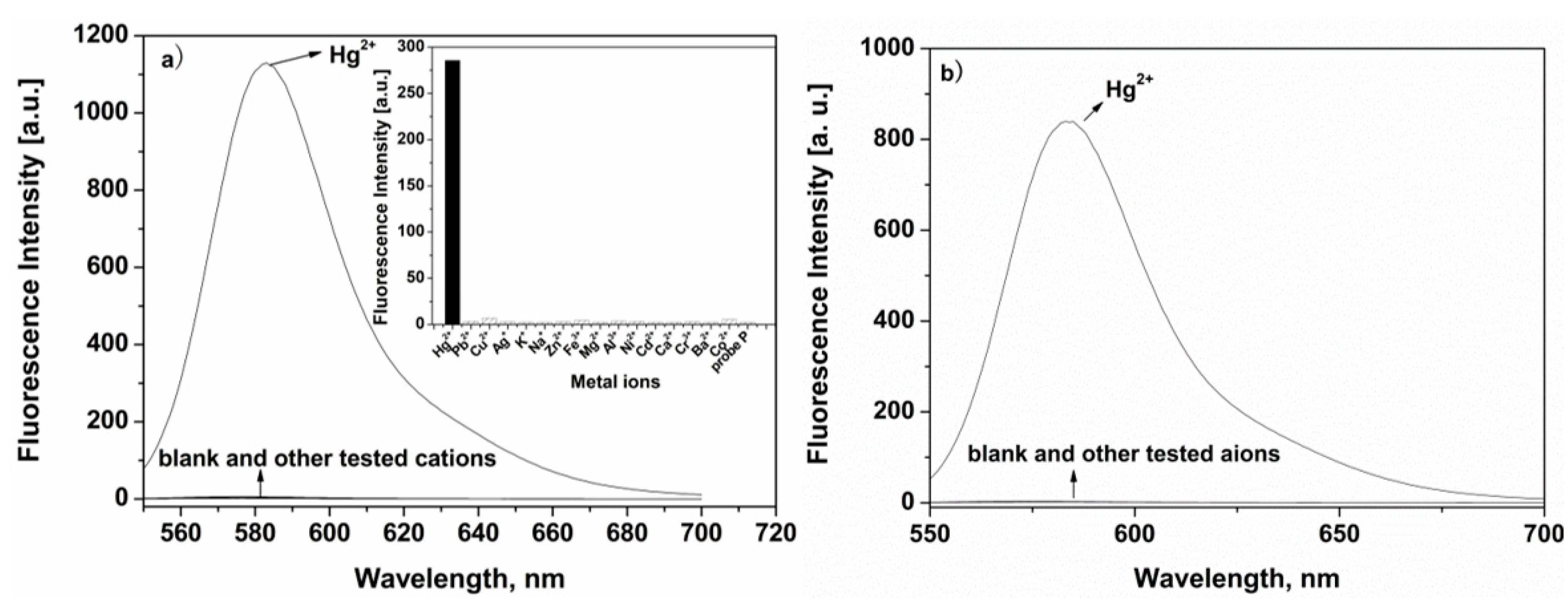
2.2. Selectivity and Sensitivity Measurement of P

A fluorescent probe with good selectivity was required for the detection of environmental or biological targets with complex backgrounds. The selectivity of this proposed probe, P, was conducted in an aqueous media (ethanol-water, 8:2, v/v, 20 mM HEPES, pH 7.0), and the tested metal ions were alkali, alkali-earth metals, divalent transition metal ions, including K+, Na+, Ag+, Ca2+, Mg2+, Zn2+, Pb2+, Cd2+, Ni2+, Co2+, Cu2+, Hg2+, Cr3+, and Fe3+, and the anions were Br, I, NO3, H2PO4, ClO4, CO32, and SO42. The results showed that like most of the spirocycle rhodamine derivatives [19,20,21], the free P displayed a very weak fluorescence, which indicates that the spirolactam form was the predominant species. Introduction of the Hg2+ to probe P elicited an obviously fluorescent enhancement at 583 nm. By contrast, other metal ions and anions had almost no influence on the fluorescent spectra of P (Figure 2). Most likely, it was the addition of Hg2+ to P that caused the opening of the spirolactam in the structure of the rhodamine part [19,20,21,22,23,24,25,26,27], inducing an enhancement in fluorescence intensity. Furthermore, for the further study of the selectivity of P, a competition experiment was also conducted (Figure S2); all the tested metal ions and anions did not show any obvious interference to the response of P with Hg2+, except I had some influence on the response of P. This revealed that this proposed probe P could work in a complicated environment and has potential application in real samples. All these results also demonstrate that P could be employed as an Hg2+-selective probe.

2.3. Fluorescent and UV-Vis Titration Experiments of P to Hg2+

In order to further study the sensing properties and mechanism between P and Hg2+, fluorescence and absorption titration experiments in aqueous media (ethanol-water, 8:2, v/v, 20 mM HEPES, pH 7.0) were recorded (Figure 3). As the sequential introduction of Hg2+ to P (5 μM), the fluorescent intensity at 583 nm enhanced gradually, and the linear fluorescent intensity was proportional to the concentrations of Hg2+ in the range 1.0–20 μM with a detection limit of 0.33 μM (Figure S3). The UV-Vis spectra also gave similar results (Figure 3b); the maximum absorption peak at 560 nm appeared with increasing intensity upon successive addition of Hg2+, and a linear dependence of absorbance at 560 nm was observed as a function of Hg2+ concentration (inset of Figure 3b) in the range 2.0–20 μM. The association constant K was determined from the slope to be 3.18 × 10⁴ M−1 [35], corresponding to a stronger binding capability toward Hg2+ (Figure S4). The results showed that P was capable of detecting Hg2+, both qualitatively and quantitatively.

2.4. Coordination Mechanism of P with Hg2+

The stoichiometry of the P-Hg2+ complex was determined by a Job’s plot experiment in aqueous media (ethanol-water, 8:2, v/v, 20 mM HEPES, pH 7.0), and the total concentrations of P and Hg2+ was kept at 10 µM. When the mole ratio P/Hg2+ was at 1:1, the fluorescent intensity at 583 nm reached the maximum (Figure 4), which indicates that P coordinated with Hg2+ in a 1:1 stoichiometric relationship. Meanwhile, an experiment with Na2S as a competitive complexing agent could serve as experimental evidence to support this semi-reversible spiro ring-opening mechanism (Figure S5). To further explore the binding mode of P with Hg2+, the 1H-NMR spectra of P and P-Hg2+ in DMSO-d6 were carried out (Figures S6 and S7). The proton peaks of –OH and O=C–NH in P alone existed in the form of hydrogen bonds and showed wide peaks and δ values that were somewhat larger than normal protons in the spectra of 1H-NMR, and the addition of Hg2+ to P in DMSO-d6 solution led to a high-field shift of the signals –OH and O=C–NH at the degrees 0.0237 and 0.0069. It may be that the coordination of Hg2+ with P destroyed the formation of hydrogen bonds, the proton peaks of –OH and O=C–NH turned sharp, and the δ values became smaller than in P. According to the abovementioned results, P was most likely to chelate with Hg2+ via its oxygen on phenol hydroxylation, oxygen on the carbonyl group as well as nitrogen on the hydrazine. The proposed reaction mechanism of P with Hg2+ is shown in Scheme 2.

2.5. Preliminary Application of P in Cell Imaging

To further explore the biological applicability of P for Hg2+ in practical samples, intracellular Hg2+ imaging in HeLa cells by fluorescence microscopy was performed (Figure 5). Obvious fluorescence was not observed upon incubation with P (1.0 µM) for 30 min at 37 °C (Figure 5a), suggesting that autofluorescence from the cells could be avoided. Under the same testing conditions, stronger fluorescent change was detected after the addition of exogenous Hg2+ (1.0 µM) to the P-loaded HeLa cells (Figure 5b) which demonstrated that P could penetrate the cell membrane and coordinate with Hg2+ inside the cells. Moreover, brightfield imaging confirmed that the cells were viable after incubation with Hg2+ and/or P (Figure 5(a2,b2)). Meanwhile, P was also applied to the subcellular locations of Hg2+ in the HeLa cells using confocal fluorescence microscopy. The cells were co-treated with P (1.0 µM) and Hoechst 33342 (0.25 µg/mL) for 30 min with the same conditions as those used in Figure 5a,b. Our work revealed that the cellular localization and distribution of P was located primarily in the cytoplasm of those living HeLa cells as shown in Figure 5c. All the results indicated that the proposed P was an effective probe for imaging changes in Hg2+ intracellularly under biological conditions.
To evaluate the cytotoxicity of the fluorescent probe P in living cells, an MTT assay on PC12 cells with P concentrations from 0 to 10 µM was taken (Figure S8). After treatment with P for 48 h, the cellular viability was estimated at approximately 92%, which exhibited the low toxicity of P to cultured cells.

3. Materials and Methods

3.1. Main Reagents and Instruments

All reagents were of commercially analytical grade and used directly.
Fluorescent spectra were recorded using a Hitachi F-4600 spectrofluorometer (Tokyo, Japan), and UV-Vis spectra were determined on a Hitachi U-2910 spectrophotometer. 1H- and 13C-NMR spectra were carried out with a Brucker AV 400 nuclear magnetic resonance instrument (Faellanden, Switzerland), and the chemical shift is given in ppm from tetramethylsilane (TMS). Mass spectra were obtained using a thermo TSQ Quantum Access Agilent 1100 mass spectrometer (Santa Clara, CA, USA). Fluorescence imaging was performed with Olympus FluoView Fv3000 laser scanning microscope (Tokyo, Japan).

3.2. Synthesis of Probe P

Compounds RBH and RBHO were obtained according to the reported method [36].
An amount of 0.1521 g of 2-hydroxybenzoyl hydrazide (1.0 mmol) and 0.4964 g RBHO (1.0 mmol) were dissolved in ethanol (40 mL) and added to a round-bottom flask (100 mL). The mixture was reacted under reflux for 6 h, and then cooled to room temperature, and the yellow precipitate so obtained was filtered off and washed with cold ethanol. Yields: 85.6%. MS m/z: 631.5 [M + H]+, 629.5 [M − H+]. 1H-NMR (DMSO-d6, δ ppm): 11.82 (s, 1H), 11.38 (s, 1H), 7.98 (d, 1H, J = 8.16), 7.92 (d, 1H, J = 7.40), 7.88 (d, 1H, J = 8.16), 7.70 (d, 1H, J = 6.80), 7.61 (t, 1H, J = 8.00), 7.57 (t, 1H, J = 6.00), 7.40 (t, 1H, J = 8.36), 7.06 (d, 1H, J = 7.52), 6.93 (t, 1H, J = 7.20), 6.89 (s, 1H), 6.45 (s, 1H), 6.44 (d, 3H, J = 7.28), 6.36 (d, 2H, J = 8.00), 3.32 (m, 8H, J = 8.70), 1.07 (t, 12H, J = 6.98). 13C-NMR (DMSO-d6, δ ppm): 164.88, 164.77, 158.77, 152.65, 152.53, 149.13, 147.56, 143.55, 135.02, 134.29, 129.40, 127.80, 127.42, 124.17, 123.79, 119.48, 117.57, 117.05, 108.67, 104.71, 97.93, 65.43, 56.50, 44.13, 19.04, 12.89. (Supplementary Materials Figure S6, Figure S9–S11)

3.3. General Spectroscopic Methods

The stock solution (1.0 mM) of P was obtained by dissolving P with DMSO. All the salts solutions (1.0 mM) were obtained by dissolving in deionized water. Before fluorescent and UV-Vis spectroscopic measurements, all the testing solutions were obtained by diluting the stock solutions to the desired concentration. The excitation and emission monochromator slit widths of the fluorescence spectrophotometer were 10 nm and 10 nm, respectively, and the excitation wavelength was fixed as 530 nm.

3.4. Cell Incubation and Imaging

HeLa cells were placed on coverslips and washed with PBS (phosphate-buffered saline). It was then incubated with 1.0 μM P (dissolved with DMSO) for 30 min at 37 °C, followed by washing three times with PBS. The cells were further cultured with 1.0 μM of HgCl2 for 30 min at 37 °C and washed with PBS three times again. The fluorescence cell imaging of intracellular Hg2+ in HeLa cells was conducted by a confocal fluorescence microscopy on an Olympus FluoView Fv1000 laser scanning microscope.

4. Conclusions

In summary, a highly selective Hg2+ probe, P, derived from rhodamine B was designed and investigated by fluorescence and UV-Vis techniques. On the basis of the change between spirolactam and open-cycle forms in the rhodamine unit, the sensing mechanism of this proposed probe was studied in detail. Furthermore, confocal fluorescence microscopy experiments demonstrated that probe P can be applied to image Hg2+ in living cells.

Supplementary Materials

The following are available online: Figure S1: Influences of time on the fluorescence spectra of P (5 μM) with Hg2+ (50 μM) in ethanol solution; Figure S2: Fluorescence response of 5 μM of P to 10 μM of Hg2+ and to the mixture of 10 μM of individual other metal ions (a) and anions (b) with 10 μM of Hg2+; Figure S3: The fluorescence intensity at 583 nm of P (5 μM) as a function of Hg2+ concentrations (1–20 μM); Figure S4: Benesi–Hildebrand plot of P, assuming a 1:1 stoichiometry for the association between P and Hg2+; Figure S5: Reversible titration response of P to Hg2+; Figure S6: 1H-NMR spectrum of P; Figure S7: 1H-NMR titration experiment of P + Hg2+; Figure S8: MTT assay of P in living cells; Figure S9: ESI (−)-MS of P; Figure S10: ESI (+)-MS of P; Figure S11: 13C-NMR spectrum of P.

Author Contributions

Conceptualization, J.Z.; methodology, C.Y.; software, W.Z. and M.Y.; formal analysis, W.Z.; resources, S.W.; writing—original draft preparation, W.Z. and J.Z.; writing—review and editing, C.Y. All authors have read and agreed to the published version of the manuscript.

Funding

This research was funded by the Natural Science Foundation of Hainan Province (grant number: 820RC626), Research Project of Education and Teaching Reform in Colleges and Universities of Hainan Province (grant number: Hnjg2019ZD-21), and the National Natural Science Foundation of China (grant numbers: 81760387 and 81860381).

Institutional Review Board Statement

The study was conducted according to the guidelines of the Declaration of Helsinki and approved by the Ethics Committee of Hainan Medical University.

Informed Consent Statement

Not applicable.

Data Availability Statement

Data supporting the reported results are available online.

Conflicts of Interest

The authors declare no conflict of interest. The funders had no role in the design of the study; in the collection, analyses, or interpretation of data; in the writing of the manuscript, or in the decision to publish the results.

Sample Availability

A sample of the compound P is available from the authors.

References

  1. Eisler, R. Health Risks of Gold Miners: A Synoptic Review. Environ. Geochem. Health 2003, 25, 325–345. [Google Scholar] [CrossRef] [PubMed]
  2. Boening, D.W. Ecological effects, transport, and fate of mercury: A general review. Chemosphere 2000, 40, 1335–1351. [Google Scholar] [CrossRef]
  3. Ikenna, O.; Albert, R.N.; Erwin, B. Biomolecule—Mercury interactions: Modalities of DNA base—Mercury binding mecha-nisms. Remediation strategies. Chem. Rev. 2005, 104, 5911–5929. [Google Scholar]
  4. Antonio, C.; Vega, L.; David, C.; Alberto, T.; Arturo, E.; Rafaela, G.; José, V.G.; Concepció, R.; Klaus, W.; Pedro, M.; et al. Electroactive thiazole derivatives capped with ferrocenyl units showing charge-transfer transition and selective ion-sensing properties: A combined experimental and theoretical study. Inorg. Chem. 2007, 46, 825–838. [Google Scholar]
  5. Vallant, B.; Kadnar, R.; Goessler, W. Development of a new HPLC method for the determination of inorganic and methyl-mercury in biological samples with ICP-MS detection. J. Anal. At. Spectrom. 2007, 22, 322–325. [Google Scholar] [CrossRef]
  6. Tan, J.; Yan, X.-P. 2,1,3-Benzoxadiazole-based selective chromogenic chemosensor for rapid naked-eye detection of Hg2+ and Cu2+. Talanta 2008, 76, 9–14. [Google Scholar] [CrossRef]
  7. Ji, H.F.; Ma, F.L.; Dai, Y.P.; Zhao, X.X.; Xue, K.; Misal, S.; Zhang, P.; Qi, Z.J.; Zhu, H.Y. A near-infrared ratiometric fluores-cent probe for selectively tracking human cytochrome P450 3A5 in living cells and tumor-bearing mice. Sens. Actuators B Chem. 2021, 331, 129372. [Google Scholar] [CrossRef]
  8. Avirah, R.R.; Jyothish, K.; Ramaiah, D. Dual-Mode Semisquaraine-Based Sensor for Selective Detection of Hg2+ in a Micellar Medium. Org. Lett. 2007, 9, 121–124. [Google Scholar] [CrossRef]
  9. Guo, X.; Qian, X.; Jia, L. A Highly Selective and Sensitive Fluorescent Chemosensor for Hg2+ in Neutral Buffer Aqueous Solution. J. Am. Chem. Soc. 2004, 126, 2272–2273. [Google Scholar] [CrossRef]
  10. Ma, X.; Wang, J.; Shan, Q.L.; Tan, Z.W.; Wei, G.H.; Wei, D.B.; Du, Y.G. A “turn-on” fluorescent Hg2+ chemosensor based on ferrier carbocyclization. Org. Lett. 2012, 14, 820–823. [Google Scholar] [CrossRef]
  11. Li, A.-L.; Wang, Z.-L.; Wang, W.-Y.; Liu, Q.-S.; Sun, Y.; Wang, S.-F.; Gu, W. A novel dehydroabietic acid-based fluorescent probe for detection of Fe3+ and Hg2+ ions and its application in live-cell imaging. Microchem. J. 2021, 160, 105682. [Google Scholar] [CrossRef]
  12. Pandey, S.; Azam, A.; Pandey, S.; Chawla, H.M. Novel dansyl-appended calix[4]arene frameworks: Fluorescence properties and mercury sensing. Org. Biomol. Chem. 2009, 7, 269–279. [Google Scholar] [CrossRef]
  13. Bhatt, S.; Vyas, G.; Paul, P. A New Molecular Probe for Colorimetric and Fluorometric Detection and Removal of Hg2+ and its Application as Agarose Film-Based Sensor for On-Site Monitoring. J. Fluoresc. 2020, 30, 1531–1542. [Google Scholar] [CrossRef]
  14. Kim, H.M.; Lee, M.H.; Kim, H.J.; Kim, J.S.; Yoon, J. A new trend in rhodamine-based chemosensors: Application of spirolac-tam ring-opening to sensing ions. Chem. Soc. Rev. 2008, 37, 1465–1472. [Google Scholar] [CrossRef] [PubMed]
  15. Ghezelsefloo, S.; Rad, J.K.; Hajiali, M.; Mahdavian, A.R. Rhodamine-based fluorescent polyacrylic nanoparticles: A highly selective and sensitive chemosensor for Fe (II) and Fe (III) cations in water. J. Environ. Chem. Eng. 2021, 9, 105082. [Google Scholar] [CrossRef]
  16. Zhao, H.; Ding, H.; Kang, H.; Fan, C.; Liu, G.; Pu, S. A solvent-dependent chemosensor for fluorimetric detection of Hg2+ and colorimetric detection of Cu2+ based on a new diarylethene with a rhodamine B unit. RSC Adv. 2019, 9, 42155–42162. [Google Scholar] [CrossRef] [Green Version]
  17. Chen, X.; Jia, J.; Ma, H.; Wang, S.; Wang, X. Characterization of rhodamine B hydroxylamide as a highly selective and sensitive fluorescence probe for copper(II). Anal. Chim. Acta 2009, 632, 9–14. [Google Scholar] [CrossRef] [PubMed]
  18. Lu, P.; Zhang, X.; Ren, T.; Yuan, L. Molecular engineering of ultra-sensitive fluorescent probe with large Stokes shift for imaging of basal HOCl in tumor cells and tissues. Chin. Chem. Lett. 2020, 31, 2980–2984. [Google Scholar] [CrossRef]
  19. Wu, J.S.; Hwang, I.-C.; Kim, K.S.; Kim, J.S. Rhodamine-based Hg2+-selective chemodosimeter in aqueous solution: Fluo-rescent off-on. Org. Lett. 2007, 9, 907–910. [Google Scholar] [CrossRef]
  20. Du, J.; Fan, J.; Peng, X.; Sun, P.; Wang, J.; Li, H.; Sun, S. A New Fluorescent Chemodosimeter for Hg2+: Selectivity, Sensitivity, and Resistance to Cys and GSH. Org. Lett. 2010, 12, 476–479. [Google Scholar] [CrossRef]
  21. Huang, W.; Zhou, P.; Yan, W.B.; He, C.; Xiong, L.Q.; Li, F.Y.; Duan, C.Y. A bright water-compatible sugar-rhodamine fluo-rescence sensor for selective detection of Hg2+ in natural water and living cells. J. Environ. Monit. 2009, 11, 330–335. [Google Scholar] [CrossRef]
  22. Ma, Q.-J.; Zhang, X.-B.; Zhao, X.-H.; Jin, Z.; Mao, G.-J.; Shen, G.-L.; Yu, R.-Q. A highly selective fluorescent probe for Hg2+ based on a rhodamine–coumarin conjugate. Anal. Chim. Acta 2010, 663, 85–90. [Google Scholar] [CrossRef]
  23. Singh, S.; Coulomb, B.; Boudenne, J.-L.; Bonne, D.; Dumur, F.; Simon, B.; Robert-Peillard, F. Sub-ppb mercury detection in real environmental samples with an improved rhodamine-based detection system. Talanta 2021, 224, 121909. [Google Scholar] [CrossRef]
  24. Wen, D.; Yu, Y.H. A novel turn-on fluorescent probe for Hg2+ detection based on rhodamine B spirolactam derivative. Int. J. Environ. Anal. Chem. 2019, 99, 1515–1527. [Google Scholar]
  25. Petdum, A.; Panchan, W.; Sirirak, J.; Promarak, V.; Sooksimuang, T.; Wanichacheva, N. Colorimetric and fluorescent sensing of a new FRET system via [5]helicene and rhodamine 6G for Hg2+ detection. New J. Chem. 2018, 42, 1396–1402. [Google Scholar] [CrossRef]
  26. Vanjare, B.D.; Mahajan, P.G.; Ryoo, H.-I.; Dige, N.C.; Choi, N.G.; Han, Y.; Kim, S.J.; Kim, C.-H.; Lee, K.H. Novel rhodamine based chemosensor for detection of Hg2+: Nanomolar detection, real water sample analysis, and intracellular cell imaging. Sens. Actuators B Chem. 2021, 330, 129308. [Google Scholar] [CrossRef]
  27. Shi, W.; Ma, H. Rhodamine B thiolactone: A simple chemosensor for Hg2+ in aqueous media. Chem. Commun. 2008, 16, 1856–1858. [Google Scholar] [CrossRef]
  28. Zheng, H.; Qian, Z.; Xu, L.; Yuan, F.; Lan, L.; Xu, J. Switching the recognition preference of rhodamine B spirolactam by re-placing one atom: Design of rhodamine B thiohydrazide for recognition of Hg(II) in aqueous solution. Org. Lett. 2006, 8, 859–861. [Google Scholar] [CrossRef]
  29. Zhou, Z.L.; Tang, H.H.; Chen, S.Y.; Huang, Y.H.; Zhu, X.H.; Li, H.T.; Zhang, Y.Y.; Yao, S.Z. A turn-on red-emitting fluores-cent probe for determination of copper(II) ions in food samples and living zebrafish. Food Chem. 2021, 343, 128513. [Google Scholar] [CrossRef]
  30. Yu, C.W.; Jian, L.; Ji, Y.X.; Zhang, J. Al(III)-Responsive “off-on” chemosensor based on rhodamine derivative and its application in cell imaging. RSC Adv. 2018, 8, 31106–31112. [Google Scholar] [CrossRef] [Green Version]
  31. Yu, C.; Cui, S.; Ji, Y.; Wen, S.; Jian, L.; Zhang, J. A pH tuning single fluorescent probe based on naphthalene for dual-analytes (Mg2+ and Al3+) and its application in cell imaging. RSC Adv. 2020, 10, 21399–21405. [Google Scholar] [CrossRef]
  32. Ji, Y.; Yu, C.; Wen, S.; Zhang, J. Characterization of an Al3+-selective fluorescent probe based on a benzoyl hydrazine derivative and its application in cell imaging. Turk. J. Chem. 2016, 40, 625–630. [Google Scholar] [CrossRef]
  33. He, H.; Cheng, Z.; Zheng, L. Aqueous Zn2+ analysis: Specific recognition and instant imaging by Schiff base fluorescent probes. J. Mol. Struct. 2021, 1227, 129522. [Google Scholar] [CrossRef]
  34. Zhu, H.; Zhu, J.; Chen, C.; Wei, Z.; Tian, L. Synthesis, Characterization and Antibacterial Activities of Manganese(II), Nickel(II), Copper(II) and Zinc(II) Complexes of the Hydrazone Compounds Derived from 1-Phenyl-3-methyl-4-acyl-pyrazole and Benzoyl Hydrazine. Asian J. Chem. 2013, 25, 8444–8446. [Google Scholar] [CrossRef]
  35. Rodríguez-Cáceres, M.I.; Agbaria, R.A.; Warner, I.M. Fluorescence of Metal–Ligand Complexes of Mono- and Di-Substituted Naphthalene Derivatives. J. Fluoresc. 2005, 15, 185–190. [Google Scholar] [CrossRef]
  36. Li, N.; Yu, C.; Ji, Y.; Zhang, J. Characterization of a Cu2+-selective fluorescent probe derived from rhodamine B with 1,2,4-triazole as subunit and its application in cell imaging. Turk. J. Chem. 2015, 39, 660–666. [Google Scholar] [CrossRef]
Scheme 1. Synthesis route of P.
Molecules 26 03385 sch001
Figure 1. The effect of pH on P (5 µM) and the P (5 µM)-Hg2+ (50 µM) system (●) in the aqueous media (ethanol-water, 8:2, v/v, 20 mM HEPES). The HEPES buffer was adjusted using 1.0 M HCl or 1.0 M NaOH.
Molecules 26 03385 g001
Figure 2. (a) Fluorescence spectra of P (5 μM) with different cations (50 µM) in the aqueous media (ethanol-water, 8:2, v/v, 20 mM HEPES, pH 7.0). Inset: The fluorescence intensity at 583 nm of P (5 μM) in the presence of different metal ions (Hg2+ is 10 μM and K+, Na+, Ag+, Ca2+, Mg2+, Zn2+, Pb2+, Cd2+, Ni2+, Co2+, Cu2+, Cr3+, and Fe3+ are 100 μM, respectively). (b) Fluorescence spectra of P (5 μM) with different anions (Br, I, NO3, H2PO4, ClO4, CO32, and SO42, 50 µM).
Molecules 26 03385 g002
Figure 3. Fluorescent titration (a) and absorption titration (b) experiments of P (5 μM) with different concentrations of Hg2+ in the aqueous media (ethanol-water, 8:2, v/v, 20 mM HEPES, pH 7.0). Inset: (a) the fluorescence intensity at 583 nm and (b) absorbance at 560 nm of P (5 μM) as a function of Hg2+ concentrations.
Molecules 26 03385 g003
Figure 4. Job’s plot experiment of P with Hg2+. Total [P + Hg2+] was kept at 10 µM.
Molecules 26 03385 g004
Scheme 2. The proposed binding mode of the P-Hg2+ complex.
Molecules 26 03385 sch002
Figure 5. Confocal fluorescence images of HeLa cells. (a) HeLa cells with P (1.0 μM) for 30 min; (b) HeLa cells with P (1.0 μM) for 30 min and then with Hg2+ (1.0 μM) for 30 min ((a1b1): dark field; (a2b2): bright field; (a3b3): (a1b1) merged with (a2b2), respectively); (c) HeLa cells with P (1.0 μM) for 30 min, then with Hg2+ (1.0 μM) for 30 min and Hoechst 33342 (0.25 μg/mL) for 15 min (c1: green channel with P; c2: blue channel with Hoechst 33342; c3: overlay of images showing fluorescence from P (c1) and Hoechst 3342 (c2)).
Molecules 26 03385 g005
Publisher’s Note: MDPI stays neutral with regard to jurisdictional claims in published maps and institutional affiliations.

