
Non-Invasive Identification of Nutrient Components in Grain

 , and
, and 
Abstract
1. Introduction
2. Results and Discussion
3. Materials and Methods
3.1. Raman Spectroscopy
3.2. Statistical Analysis
4. Conclusions
Author Contributions
Funding
Institutional Review Board Statement
Informed Consent Statement
Data Availability Statement
Conflicts of Interest
Sample Availability
References
- Lew, T.T.S.; Sarojam, R.; Jang, I.C.; Park, B.S.; Naqvi, N.I.; Wong, M.H.; Singh, G.P.; Ram, R.J.; Shoseyov, O.; Saito, K.; et al. Species-independent analytical tools for next-generation agriculture. Nat. Plants 2020, 6, 1408–1417. [Google Scholar] [CrossRef]
- Payne, W.Z.; Kurouski, D. Raman-based diagnostics of biotic and abiotic stresses in plants. A review. Front. Plant. Sci. 2021, 11, 616672. [Google Scholar] [CrossRef]
- Mantri, N.; Patade, V.; Penna, S.; Ford, R.; Pang, E. Abiotic stress responses in plants: Present and future. In Abiotic Stress Responses in Plants; Springer: New York, NY, USA, 2012; pp. 1–19. [Google Scholar]
- Waqas, M.A.; Kaya, C.; Riaz, A.; Farooq, M.; Nawaz, I.; Wilkes, A.; Li, Y. Potential Mechanisms of Abiotic Stress Tolerance in Crop Plants Induced by Thiourea. Front. Plant. Sci. 2019, 10, 1336. [Google Scholar] [CrossRef]
- Wang, K.D.; Borrego, E.J.; Kenerley, C.M.; Kolomiets, M.V. Oxylipins Other Than Jasmonic Acid Are Xylem-Resident Signals Regulating Systemic Resistance Induced by Trichoderma virens in Maize. Plant. Cell 2020, 32, 166–185. [Google Scholar] [CrossRef]
- He, Y.; Borrego, E.J.; Gorman, Z.; Huang, P.C.; Kolomiets, M.V. Relative contribution of LOX10, green leaf volatiles and JA to wound-induced local and systemic oxylipin and hormone signature in Zea mays (maize). Phytochemistry 2020, 174, 112334. [Google Scholar] [CrossRef] [PubMed]
- Farber, C.; Mahnke, M.; Sanchez, L.; Kurouski, D. Advanced Spectroscopic Techniques for Plant Disease Diagnostics. A Review. Trends Analyt. Chem. 2019, 118, 43–49. [Google Scholar] [CrossRef]
- Kurouski, D. A Spectroscopic Revolution in Agricultural World. Spectroscopy 2020, 35, 38–40. [Google Scholar]
- Qin, J.; Kim, M.S.; Chao, K.; Cho, B.-K. Raman Chemical Imaging Technology for Food and Agricultural Applications. J. Biosyst. Eng. 2017, 42, 170–189. [Google Scholar]
- Farber, C.; Kurouski, D. Detection and Identification of Plant Pathogens on Maize Kernels with a Hand-Held Raman Spectrometer. Anal. Chem. 2018, 90, 3009–3012. [Google Scholar] [CrossRef]
- Sanchez, L.; Pant, S.; Xing, Z.; Mandadi, K.; Kurouski, D. Rapid and noninvasive diagnostics of Huanglongbing and nutrient deficits on citrus trees with a handheld Raman spectrometer. Anal. Bioanal. Chem. 2019. [Google Scholar] [CrossRef] [PubMed]
- Gupta, S.; Huang, C.H.; Singh, G.P.; Park, B.S.; Chua, N.-H.; Ram, R.J. Portable Raman leaf-clip sensor for rapid detection of plant stress. Sci. Rep. 2020, 10, 20206. [Google Scholar] [CrossRef]
- Egging, V.; Nguyen, J.; Kurouski, D. Detection and Identification of Fungal Infections in Intact Wheat and Sorghum Grain Using a Hand-Held Raman Spectrometer. Anal. Chem. 2018, in press. [Google Scholar] [CrossRef]
- Farber, C.; Sanchez, L.; Kurouski, D. Confirmatory Non-Invasive and Non-Destructive Identification of Poison Ivy Using A Hand-Held Raman Spectrometer. RCS Adv. 2020, 10, 21530–21534. [Google Scholar]
- Farber, C.; Sanchez, L.; Rizevsky, S.; Ermolenkov, A.; McCutchen, B.; Cason, J.; Simpson, C.; Burrow, M.; Kurouski, D. Raman Spectroscopy Enables Non-Invasive Identification of Peanut Genotypes and Value-Added Traits. Sci. Rep. 2020, 10, 7730. [Google Scholar] [CrossRef] [PubMed]
- Farber, C.; Shires, M.; Ong, K.; Byrne, D.; Kurouski, D. Raman spectroscopy as an early detection tool for rose rosette infection. Planta 2019, 250, 1247–1254. [Google Scholar] [CrossRef] [PubMed]
- Sanchez, L.; Ermolenkov, A.; Tang, X.T.; Tamborindeguy, C.; Kurouski, D. Non-invasive diagnostics of Liberibacter disease on tomatoes using a hand-held Raman spectrometer. Planta 2020, 251, 64. [Google Scholar] [CrossRef]
- Sanchez, L.; Farber, C.; Lei, J.; Zhu-Salzman, K.; Kurouski, D. Noninvasive and Nondestructive Detection of Cowpea Bruchid within Cowpea Seeds with a Hand-Held Raman Spectrometer. Anal. Chem. 2019, 91, 1733–1737. [Google Scholar] [CrossRef]
- Sanchez, L.; Pant, S.; Irey, M.S.; Mandadi, K.; Kurouski, D. Detection and Identification of Canker and Blight on Orange Trees Using a Hand-Held Raman Spectrometer. J. Raman Spectrosc. 2019, 50, 1875–1880. [Google Scholar] [CrossRef]
- Sanchez, L.; Pant, S.; Mandadi, K.; Kurouski, D. Raman Spectroscopy vs Quantitative Polymerase Chain Reaction In Early Stage Huanglongbing Diagnostics. Sci. Rep. 2020, 10, 10101. [Google Scholar] [CrossRef] [PubMed]
- Altangerel, N.; Ariunbold, G.O.; Gorman, C.; Alkahtani, M.H.; Borrego, E.J.; Bohlmeyer, D.; Hemmer, P.; Kolomiets, M.V.; Yuan, J.S.; Scully, M.O. In vivo diagnostics of early abiotic plant stress response via Raman spectroscopy. Proc. Natl. Acad. Sci. USA 2017, 114, 3393–3396. [Google Scholar] [CrossRef]
- Mandrile, L.; Rotunno, S.; Miozzi, L.; Vaira, A.M.; Giovannozzi, A.M.; Rossi, A.M.; Noris, E. Nondestructive Raman Spectroscopy as a Tool for Early Detection and Discrimination of the Infection of Tomato Plants by Two Economically Important Viruses. Anal. Chem. 2019, 91, 9025–9031. [Google Scholar] [CrossRef]
- Yeturu, S.; Vargas Jentzsch, P.; Ciobotă, V.; Guerrero, R.; Garrido, P.; Ramos, L.A. Handheld Raman spectroscopy for the early detection of plant diseases: Abutilon mosaic virus infecting Abutilon sp. Anal. Methods 2016, 8, 3450–3457. [Google Scholar] [CrossRef]
- Huang, C.H.; Singh, G.P.; Park, S.H.; Chua, N.H.; Ram, R.J.; Park, B.S. Early Diagnosis and Management of Nitrogen Deficiency in Plants Utilizing Raman Spectroscopy. Front. Plant. Sci. 2020, 11, 663. [Google Scholar] [CrossRef]
- Farber, C.; Bryan, R.; Paetzold, L.; Rush, C.; Kurouski, D. Non-Invasive Characterization of Single-, Double- and Triple-Viral Diseases of Wheat With a Hand-Held Raman Spectrometer. Front. Plant. Sci. 2020, 11, 01300. [Google Scholar] [CrossRef]
- Sanchez, L.; Ermolenkov, A.; Biswas, S.; Septiningshih, E.M.; Kurouski, D. Raman Spectroscopy Enables Non-invasive and Confirmatory Diagnostics of Salinity Stresses, Nitrogen, Phosphorus, and Potassium Deficiencies in Rice. Front. Plant. Sci. 2020, 11, 573321. [Google Scholar] [CrossRef]
- Krimmer, M.; Farber, C.; Kurouski, D. Rapid and Noninvasive Typing and Assessment of Nutrient Content of Maize Kernels Using a Handheld Raman Spectrometer. ACS Omega 2019, 4, 16330–16335. [Google Scholar] [CrossRef]
- Sanchez, L.; Filter, C.; Baltensperger, D.; Kurouski, D. Confirmatory Non-Invasive and Non-Destructive Differentiation Between Hemp and Cannabis Using A Hand-Held Raman Spectrometer. RCS Adv. 2020, 10, 3212–3216. [Google Scholar] [CrossRef]
- Almeida, M.R.; Alves, R.S.; Nascimbem, L.B.; Stephani, R.; Poppi, R.J.; de Oliveira, L.F. Determination of amylose content in starch using Raman spectroscopy and multivariate calibration analysis. Anal. Bioanal. Chem. 2010, 397, 2693–2701. [Google Scholar] [CrossRef] [PubMed]
- Schulz, H.; Baranska, M.; Baranski, R. Potential of NIR-FT-Raman spectroscopy in natural carotenoid analysis. Biopolymers 2005, 77, 212–221. [Google Scholar] [CrossRef]
- Edwards, H.G.; Farwell, D.W.; Webster, D. FT Raman microscopy of untreated natural plant fibres. Spectrochim. Acta A 1997, 53, 2383–2392. [Google Scholar] [CrossRef]
- Cao, Y.; Shen, D.; Lu, Y.; Huang, J. A Raman-scattering study on the net orientation of biomacromolecules in the outer epidermal walls of mature wheat stems (Triticum aestivum). Ann. Bot. 2006, 97, 1091–1094. [Google Scholar] [CrossRef] [PubMed]
- Jamieson, L.E.; Li, A.; Faulds, K.; Graham, D. Ratiometric analysis using Raman spectroscopy as a powerful predictor of structural properties of fatty acids. R. Soc. Open Sci. 2018, 5, 181483. [Google Scholar] [CrossRef]
- Yu, M.M.; Schulze, H.G.; Jetter, R.; Blades, M.W.; Turner, R.F. Raman microspectroscopic analysis of triterpenoids found in plant cuticles. Appl. Spectrosc. 2007, 61, 32–37. [Google Scholar] [CrossRef]
- Kang, L.; Wang, K.; Li, X.; Zou, B. High pressure structural investigation of benzoic acid: Raman spectroscopy and X-ray diffraction. J. Phys. Chem. C 2016, 120, 14758–14766. [Google Scholar] [CrossRef]
- Agarwal, U.P. Raman imaging to investigate ultrastructure and composition of plant cell walls: Distribution of lignin and cellulose in black spruce wood (Picea mariana). Planta 2006, 224, 1141–1153. [Google Scholar] [CrossRef]
- Matousek, P.; Stone, N. Development of deep subsurface Raman spectroscopy for medical diagnosis and disease monitoring. Chem. Soc. Rev. 2016, 45, 1794–1802. [Google Scholar] [CrossRef]
- López-López, M.; García-Ruiz, C. Infrared and Raman spectroscopy techniques applied to identification of explosives. Trends Anal. Chem. 2014, 54, 36–44. [Google Scholar] [CrossRef]
- Bloomfield, M.; Andrews, D.; Loeffen, P.; Tombling, C.; York, T.; Matousek, P. Non-invasive identification of incoming raw pharmaceutical materials using Spatially Offset Raman Spectroscopy. J. Pharm. Biomed. Anal. 2013, 76, 65–69. [Google Scholar] [CrossRef] [PubMed]
- Sharma, B.; Ma, K.; Glucksberg, M.R.; Van Duyne, R. Seeing through bone with surface-enhanced spatially-offset Raman spectroscopy. J. Am. Chem. Soc. 2013, 135, 17290–17293. [Google Scholar] [CrossRef] [PubMed]
- Morey, R.; Ermolenkov, A.; Payne, W.Z.; Scheuring, D.C.; Koym, J.W.; Vales, M.I.; Kurouski, D. Non-invasive identification of potato varieties and prediction of the origin of tuber cultivation using spatially offset Raman spectroscopy. Anal. Bioanal. Chem. 2020, 412, 4585–4594. [Google Scholar] [CrossRef] [PubMed]


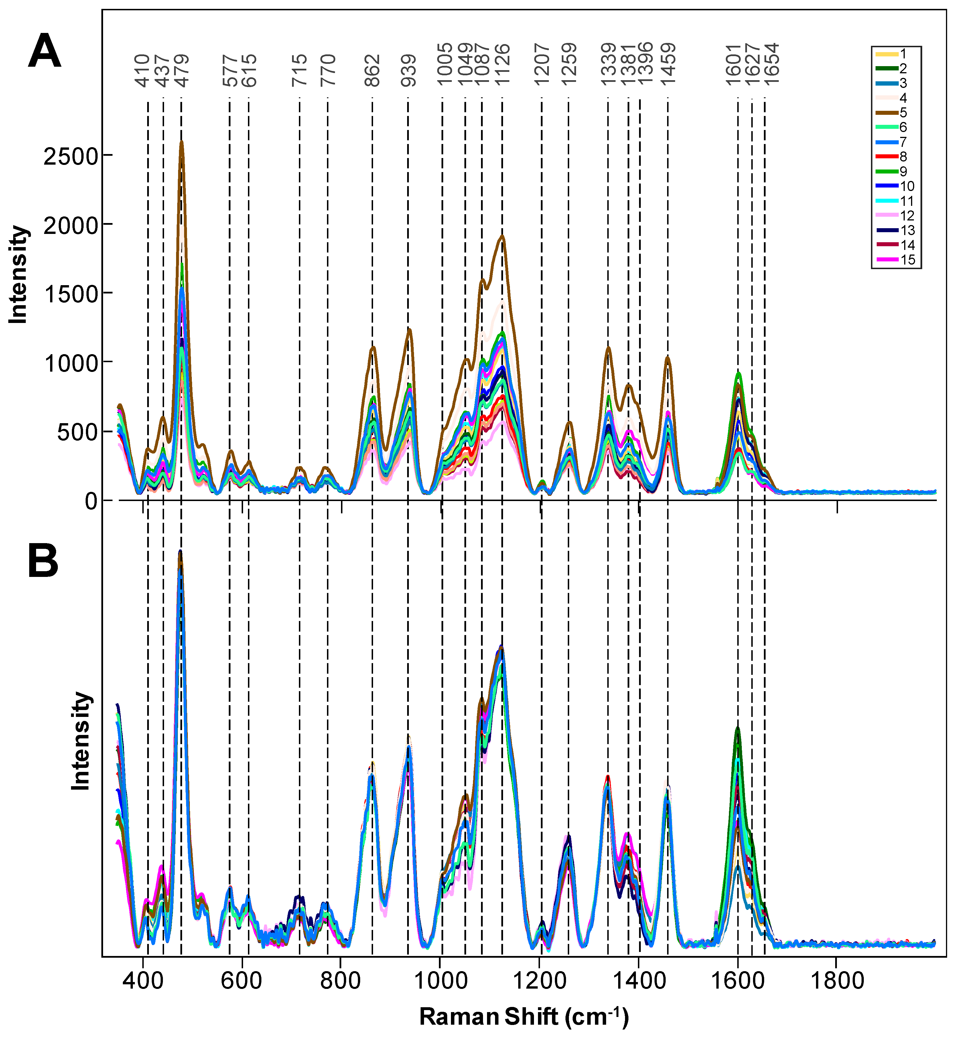
| Band | Vibrational Mode | Assignment |
|---|---|---|
| 410–479 | C-C-O and C-C-C deformations; related to glycosidic ring skeletal deformations δ(C-C-C) + τ(C-O) scissoring of C-C-C and out-of-plane bending of C-O | Carbohydrates [29] |
| 577–615 | ν(C-O-C) Glycosidic | Carbohydrates [29] |
| 715–770 | δ(C-C-O) | Carbohydrates [29] |
| 862–937 | (C6–C5–O5–C1–O1) | Carbohydrates [29] |
| 1005 | In-plane CH3 rocking of polyene aromatic ring of phenylalanine | Carotenoids [30]; protein |
| 1049 | ν(C-O) + ν(C-C) + δ(C-O-H) | Cellulose, lignin [31] |
| 1087 | ν(C-O) + ν(C-C) + δ(C-O-H) | Carbohydrates [29] |
| 1126 | ν(C-O) + ν(C-C) + δ(C-O-H) | Carbohydrates [29] |
| 1207 | δ(C-C-H) | Carbohydrates [29] |
| 1259 | Guaiacyl ring breathing, C-O stretching (aromatic); -C=C- | Lignin [32], carbohydrates, [29] unsaturated fatty acids [33] |
| 1339 | ν(C-O); δ(C-O-H) | Aliphatic, [34] carbohydrates [29] |
| 1381–1396 | δCH2 bending | Aliphatics [34] |
| 1460 | δ(CH2) + δ(CH3) | Aliphatics [34] |
| 1601–1627 | ν(C-C) aromatic ring + σ(CH) | Lignin [35,36] |
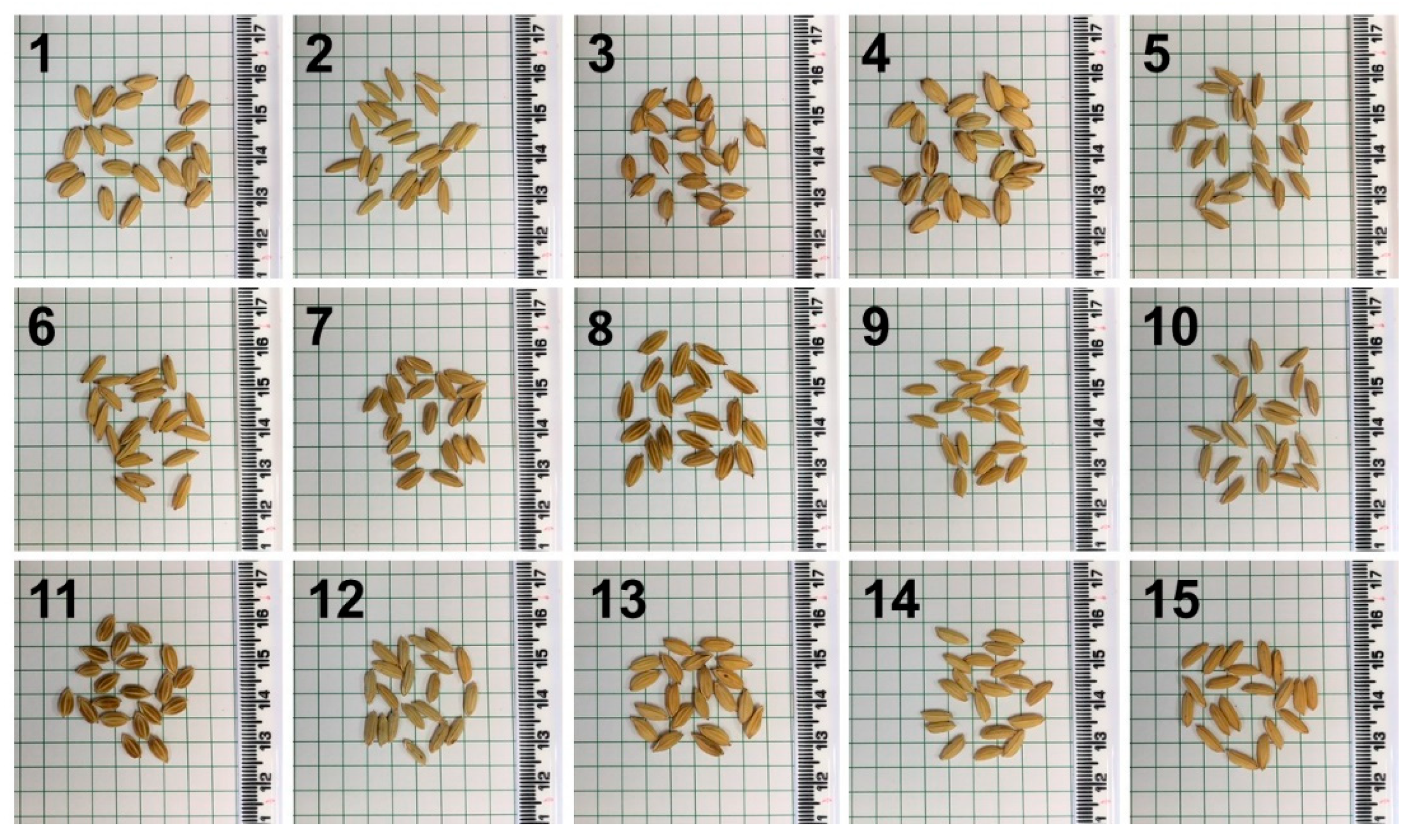
| Genotype Number | Members | True Positive Rate | 1 | 2 | 3 | 4 | 5 | 6 | 7 | 8 | 9 | 10 | 11 | 12 | 13 | 14 | 15 |
|---|---|---|---|---|---|---|---|---|---|---|---|---|---|---|---|---|---|
| 1 | 20 | 75% | 15 | 0 | 1 | 4 | 0 | 0 | 0 | 0 | 0 | 0 | 0 | 0 | 0 | 0 | 0 |
| 2 | 21 | 81% | 0 | 17 | 0 | 0 | 0 | 0 | 0 | 1 | 2 | 1 | 0 | 0 | 0 | 0 | 0 |
| 3 | 20 | 90% | 1 | 0 | 18 | 0 | 0 | 0 | 0 | 0 | 0 | 1 | 0 | 0 | 0 | 0 | 0 |
| 4 | 13 | 85% | 1 | 0 | 0 | 11 | 0 | 0 | 0 | 0 | 0 | 0 | 0 | 0 | 1 | 0 | 0 |
| 5 | 20 | 80% | 0 | 0 | 3 | 0 | 16 | 0 | 0 | 0 | 0 | 1 | 0 | 0 | 0 | 0 | 0 |
| 6 | 21 | 76% | 0 | 1 | 0 | 0 | 0 | 16 | 0 | 0 | 1 | 1 | 0 | 0 | 0 | 0 | 2 |
| 7 | 20 | 85% | 0 | 0 | 0 | 0 | 0 | 0 | 17 | 0 | 0 | 2 | 0 | 1 | 0 | 0 | 0 |
| 8 | 20 | 85% | 0 | 0 | 0 | 0 | 0 | 0 | 0 | 17 | 1 | 0 | 1 | 0 | 0 | 0 | 1 |
| 9 | 20 | 95% | 0 | 1 | 0 | 0 | 0 | 0 | 0 | 0 | 19 | 0 | 0 | 0 | 0 | 0 | 0 |
| 10 | 20 | 90% | 0 | 0 | 1 | 0 | 0 | 0 | 0 | 0 | 0 | 18 | 0 | 1 | 0 | 0 | 0 |
| 11 | 20 | 90% | 0 | 0 | 0 | 0 | 0 | 0 | 0 | 0 | 2 | 0 | 18 | 0 | 0 | 0 | 0 |
| 12 | 16 | 87% | 0 | 0 | 0 | 0 | 0 | 0 | 0 | 0 | 0 | 2 | 0 | 14 | 0 | 0 | 0 |
| 13 | 20 | 100% | 0 | 0 | 0 | 0 | 0 | 0 | 0 | 0 | 0 | 0 | 0 | 0 | 20 | 0 | 0 |
| 14 | 20 | 75% | 0 | 0 | 0 | 1 | 0 | 1 | 0 | 0 | 2 | 0 | 0 | 0 | 0 | 15 | 1 |
| 15 | 20 | 100% | 0 | 0 | 0 | 0 | 0 | 0 | 0 | 0 | 0 | 0 | 0 | 0 | 0 | 0 | 20 |
| Total | 342 | 86.2% |
Publisher’s Note: MDPI stays neutral with regard to jurisdictional claims in published maps and institutional affiliations. |
© 2021 by the authors. Licensee MDPI, Basel, Switzerland. This article is an open access article distributed under the terms and conditions of the Creative Commons Attribution (CC BY) license (https://creativecommons.org/licenses/by/4.0/).
Share and Cite
Farber, C.; Islam, A.S.M.F.; Septiningsih, E.M.; Thomson, M.J.; Kurouski, D. Non-Invasive Identification of Nutrient Components in Grain. Molecules 2021, 26, 3124. https://doi.org/10.3390/molecules26113124
Farber C, Islam ASMF, Septiningsih EM, Thomson MJ, Kurouski D. Non-Invasive Identification of Nutrient Components in Grain. Molecules. 2021; 26(11):3124. https://doi.org/10.3390/molecules26113124
Chicago/Turabian StyleFarber, Charles, A. S. M. Faridul Islam, Endang M. Septiningsih, Michael J. Thomson, and Dmitry Kurouski. 2021. "Non-Invasive Identification of Nutrient Components in Grain" Molecules 26, no. 11: 3124. https://doi.org/10.3390/molecules26113124
APA StyleFarber, C., Islam, A. S. M. F., Septiningsih, E. M., Thomson, M. J., & Kurouski, D. (2021). Non-Invasive Identification of Nutrient Components in Grain. Molecules, 26(11), 3124. https://doi.org/10.3390/molecules26113124