
Electrochemical Biosensors in Food Safety: Challenges and Perspectives


Abstract
1. Introduction
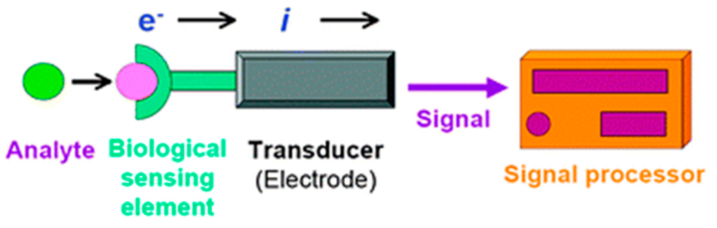
2. Electrochemical Biosensors
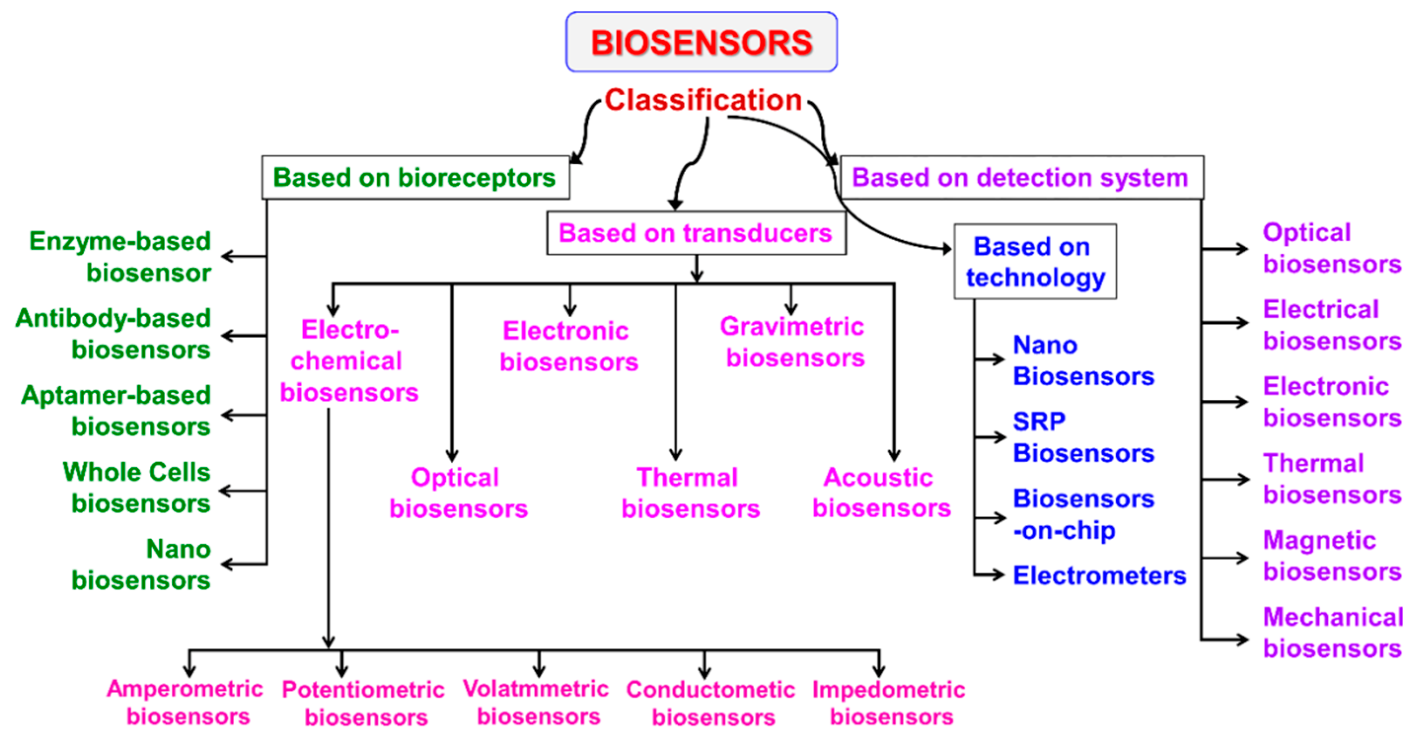
2.1. Electrochemical Biosensors Classification
2.2. Biorecognition Keys: Bioreceptors
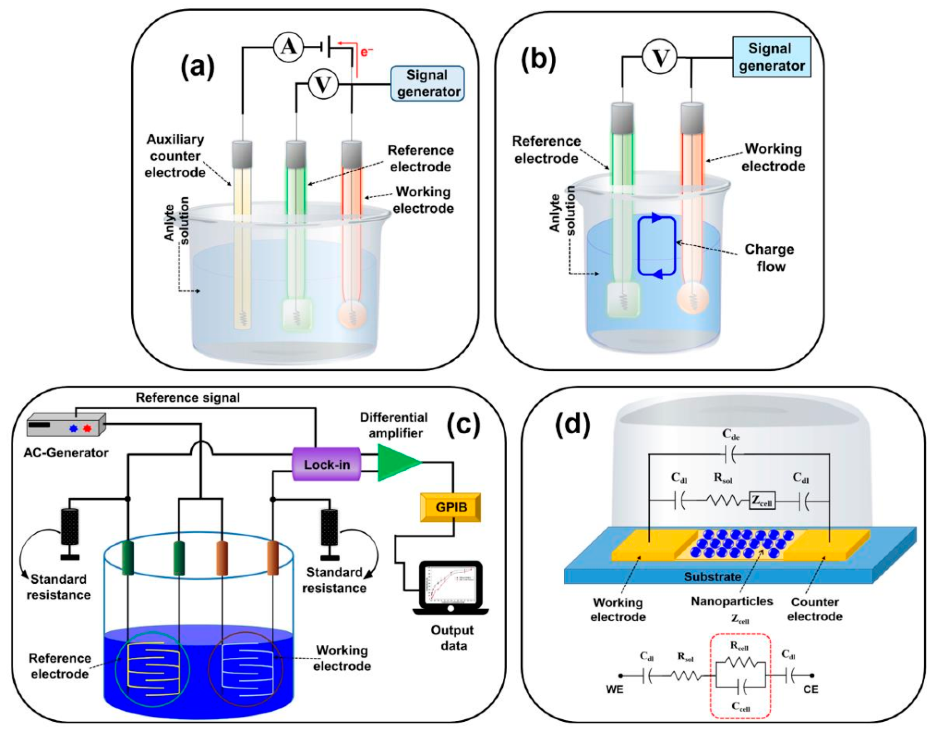
2.3. Sensing Materials and Electrodes
3. Application of Electrochemical Biosensors in Food Analysis
3.1. Toxins
3.1.1. Shellfish Toxins
3.1.2. Mycotoxins

| Electrode | (Bio)Sensor Format | Electrochemical Technique | Analyte/Sample | L.R. | LOD | References |
|---|---|---|---|---|---|---|
| SPCEs | Label-free electrochemical aptasensor based on DNA nanotetrahedron and DNA triplex | SWV | Saxitoxin/seawater | 1–400 nM | 0.92 nM | [43] |
| CB-SPCEs | Enzyme-linked immunomagnetic electrochemical (ELIME) assay | CA | DA/shellfish scallop | 5–62 ng mL−1 | 0.4 ng mL−1 | [53] |
| Phosphorene–gold–SPCE (BP–AuSPCE) | Electrochemical microfluidic biochip, including BP-SPCE with an OA aptamer | DPV | OA/mussels | 10–250 nM | 8 pM | [55] |
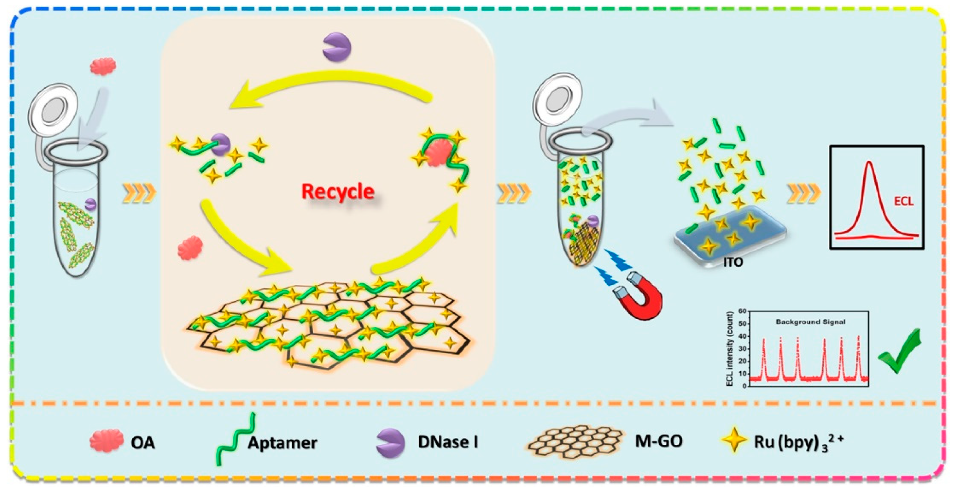
| Indium–tin-oxide electrode (ITO) | Electrochemiluminescence (ECL) aptasensor supported by magnetic graphene oxide (M-GO | ECL/CV | OA/mussels | 0.01–10 ng mL−1 | 4 pg mL−1 | [57] |
| AuSPE | Electrochemical aptasensor based on aptamer-complementary strands of aptamer complex forming a π-shape structure on the surface of the electrode and exonuclease I (Exo I) | DPV | AFB1/human serum, grape juice | 7–500 pg mL−1 | 2 pg mL−1 | [64] |
| AuSPE | Electrochemical immunosensor utilizing a competitive assay format | DPV | OTA and AFM1/red wine, milk | - | OTA 15 ng mL−1 AFM1 37 ng mL−1 | [65] |
| SPCE | Magnetically assembled aptasensor for label-free determination of AFB1 employing a disposable screen-printed carbon electrode (SPCE) covered with polydimethylsiloxane (PDMS) film as the micro electrolytic cell | EIS | AFB1/peanuts | 20–50 pg mL−1 | 15 pg mL−1 | [66] |
| GO–PAA | Aptasensor employing PAA modified with GO and an aptamer of AFB1 | Amperometry | AFB1/no real samples | 1–20 ng mL−1 | 0.13 ng mL−1 | [67] |
| SPCEs | Aptasensor using a competitive format and modified screen-printed electrode | DPV | AFB1/maize flour | Dose–response curve 0.1–10 ng mL−1 | 0.086 ng mL−1 | [68] |
| AuE | Aptasensor having methylene blue (MB) as redox tag | SWV | AFB1/white wine | 2 nM–4 μM | 2 nM | [69] |
| Screen-printed bipolar electrode (BPE) | BPE-ECL aptasensor | ECL | AFB1/rice, wheat, corn, sorghum, barley, and buckwheat grains | 0.1–100 ng mL−1 | 0.033 ng mL−1 | [70] |
| GCE | Biosensor for AFB1 and ZEN using Escherichia coli as biorecognition element | CA | AFB1 and ZEN/peanut and corn oil | AFB1 0.01–0.3 mg mL−1 ZEN 0.05–0.5 mg mL−1 | AFB1 1 ng mL−1 ZEN 6 ng mL−1 | [71] |
| AuE | Immunosensor based on DNA tetrahedron-structured probe (DTP), obtained from the conjugation between DNA tetrahedron nanostructures and HRP -labeled AFB1 monoclonal antibody | DPV | AFB1/rice, wheat, corn, sorghum, barley, and buckwheat grains | 0.05–20 ng mL−1. | 0.033 ng mL−1 | [73] |
| LbL-GCE | Aptasensor assembled via layer-by-layer deposition of differently charged layers onto GCE. The AFB1 aptamer was immobilized onto the negatively charged layer | EIS | AFB1/oil and soy sauce | 0.001–0.10 ng mL−1 | 0.002 ng mL−1 | [74] |
| AuNPs–GO–PABA–GCE | Immunosensor where AFB1 antibodies are linked to AuNPs–GO–PABA nanocomposite, deposited on GCE | EIS | AFB1/vegetable oils | 0.01–1.0 ng mL−1; 1–25 ng mL−1 | 0.001 ng mL−1 | [75] |
| AuE | Aptasensor where AFB1 aptamer is immobilized onto MCH layer self-assembled on AuE | SWV | AFB1/wine, milk, corn flour | 8 pM–25 nM; 25 nM–3 μM | 6 pM | [76] |
| MBs–SPCEs | Electrochemical magnetoimmunosensor involving magnetic beads (MBs) and disposable carbon screen-printed electrodes (SPCEs) | Amperometry | FB1/beer | Nonlinear calibration curves performed | 0.33 mg L−1 | [80] |
| AuNPs–PPy–rGO–SPCEs | Immunosensor using AuNPs–PPy–rGO nanocomposite as a platform for immobilizing anti-toxin antibody | DPV | FB1, DON/corn | FB1 0.2–4.5 ppm; DON 0.05–1 ppm | FB1 4.2 ppb; DON 8.6 ppb | [81] |
| NanoMIPs–PPY–ZnP–Pt | Chemosensor based on nano imprinted polymer nanoparticles (nano MIPs) immobilized | DPV, EIS | FB1/maize flour | 1 fM–10 pM | EIS 0.7 fM; DPV 0.03 fM | [82] |
| SPCE | Label-free electrochemical impedimetric aptasensor based on the diazonium-coupling reaction mechanism for the immobilization of anti-OTA aptamer at SPCEs | EIS | OTA/cocoa beans | 0.15–2.5 ng mL−1 | 0.15 ng mL−1 | [85] |
| AuE | Aptasensor based on the modified gold electrode with conductive polypyrrole layer covalently bound to polyamidoamine dendrimers of the fourth generation (PAMAM G4), where the OTA aptamer was immobilized | EIS | OTA/wine | - | 2 ng L−1 | [86] |
| SPCE | Competitive aptasensor where biotin-labeled and free OTA compete to bind with immobilized aptamer onto the surface of a screen-printed carbon electrode (SPCE) | DPV | OTA/cocoa beans | 0.15–5 ng mL−1 | 0.07 ng mL−1 | [87] |
| Au thin-film single electrodes | Impedimetric label-free immunosensor using two antibody immobilization methods (oriented, including protein A/G and not oriented) | EIS | OTA/cocoa beans | Oriented 0.01–5 ng mL−1 Not oriented 5 × 10⁻³–0.05 ng mL−1 | Oriented 0.01 ng mL−1 Not oriented 5 × 10⁻³ ng mL−1 | [88] |
| Bismuth-coated glassy carbon electrode (BFE) | Aptasensor assembled by combining nanocomposites of gold nanoparticles (AuNPs) functionalized silica-coated iron-oxide magnetic nanoparticles (mSiO2@Au) and cadmium telluride quantum dots (CdTe QDs) modified graphene/AuNPs nanocomposites (AuNPs/CdTe) | SWV | OTA/no real samples | 0.2–4 ng mL−1 | 0.07 pg mL−1 | [90] |
| AuE | Label-free electrochemical OTA aptasensor based on the peroxidase-like activity of g-C3N4 nanosheet (g-CNNS) and its high affinity toward single-strand DNA | CV | OTA/red wines, juices, corns | 0.2–500 nM | 0.073 nM, | [91] |
| AuE | Signal-on electrochemical aptasensor for OTA assay based on DNA controlled layer-by-layer assembly of dual gold nanoparticle (AuNP) conjugates | DPV | OTA/wine | 0.001–500 ppb | 0.001 ppb | [92] |
| TGA–AuE | Electrochemical immunosensor based on self-assembling a 2-mercaptoacetic (TGA) monolayer on the surface of Au electrode to form the Au/TGA/bovine serum albumin (BSA)-OTA/anti-OTA monoclonal antibody composite probe | DPV | OTA/malt | 0.1–1.0 ng mL−1 | 0.08 ng mL−1 | [93] |
3.2. Pathogenic Bacteria
3.2.1. Salmonella
3.2.2. Escherichia coli
3.2.3. Staphylococcus aureus
3.2.4. Listeria monocytogenes
| Electrode | (Bio)Sensor Format | Electrochemical Technique | Analyte/Sample | L.R. | LOD | References |
|---|---|---|---|---|---|---|
| GCE | Electrochemical immunosensor based on high-density gold nanoparticles (AuNPs), dispersed in chitosan (CHI) hydrogel, and modified glassy carbon electrode (GCE) | DPV | Salmonella/milk, water | 10–105 CFU mL−1 | 5 CFU mL−1 | [100] |
| SPCEs | Label-free impedimetric aptasensor assembled by grafting a diazonium-supporting layer onto screen-printed carbon electrodes (SPCEs), followed by chemical immobilization of aminated-aptamer | EIS | Salmonella/apple juice | 10–108 CFU mL−1 | 6 CFU mL−1 | [101] |
| AuE | Label-free impedimetric aptasensor based on combining poly-[pyrrole-co-3-carboxyl-pyrrole] copolymer and the Salmonella aptamer | EIS | Salmonella/apple juice | 102–108 CFU mL−1 | 3 CFU mL−1 | [102] |
| GCE | Electrochemical aptasensor developed using electrochemically reduced graphene-oxide–chitosan (rGO–CHI) composite deposited onto GCE | DPV | Salmonella/chicken | 10–106 CFU mL−1 | 10 CFU mL−1 | [103] |
| AuE | Electrochemical aptasensor developed by combining target-induced aptamer displacement on gold nanoparticles (AuNPs) deposited onto Au electrode with rolling circle amplification (RCA) | DPV | Salmonella/milk, mineral water | 20–207 CFU mL−1 | 16 CFU mL−1 | [104] |
| GF-GCE | Electrochemical immunosensor based on anti- Salmonella antibody immobilized on the surface of the graphite felt electrode | OSWV | Salmonella/no real samples | - | 105 E. coli cells mL−1 | [105] |
| BiSPCE | Immunosensor where bacterial cells were separated immunomagnetically and reacted with conjugate; labeled with an electrochemical indicator, including hyperbranched dendron molecules and heavy metal-derived quantum dots (CdTe QDs). Square-wave anodic stripping voltammetry (SWASV) employing screen-printed carbon electrodes with in situ formed Bi(III) film (BiSPCE) was used for the detection and quantification of metal ions released from the QDs and correlated with the bacterium amount | SWASV | Salmonella/milk | - | 4 CFU mL−1 | [106] |
| AuIME | Electrochemical aptasensor using aptamer-coated gold-interdigitated microelectrode (IAuE) for target capture and impedance measurement, and antibody-modified nickel nanowires (NiNWs) for target separation and impedance amplification | EIS | Salmonella/chicken | 102–106 CFU mL−1 | 80 CFU mL−1 | [107] |
| AuIME | Immunosensor using multiple magnetic nanobead (MNB) nets in a ring channel for continuous-flow separation of target bacteria from the sample volume, manganese dioxide nanoflowers (MnO2 NFs) for efficient amplification of the biological signal, and an interdigitated microelectrode to measure impedance change | EIS | Salmonella/chicken | 30–30 × 105 CFU mL−1 | 19 CFU mL−1 | [108] |
| AuIME | Impedimetric immunosensor using rotary magnetic separation and cascade reaction | EIS | Salmonella/chicken | 10–106 CFU mL−1 | 10 CFU mL−1 | [109] |
| AuE | Electrochemical genosensor based on the immobilization of complementary DNA on the gold electrode surface, which hybridizes with a pathogen-specific fragment gene to make a sandwich structure | DPV | E. coli/beef | - | 1.97 × 10⁻¹⁴ M | [113] |
| Magnetic-graphite epoxy composite (m-GEC) electrode (m-GECE) | Electrochemical magneto-genosensor based on the detection of the tagged amplified DNA obtained by single-tagging PCR with a set of pathogen-specific primers, followed by electrochemical magneto-genosensing on silica magnetic particles | Amperometry | E. coli/no real samples | 0.03–3 ng mL−1 | 0.05 ng mL−1 | [114] |
| AuE | Label-free impedimetric immunosensor using reduced graphene wrapped copper (II)-assisted cysteine hierarchical structure (rGO–CysCu) as the sensing layer | EIS | E. coli/water, fruit juice, milk | 10–108 CFU mL−1 | 3.8 CFU mL−1 | [115] |
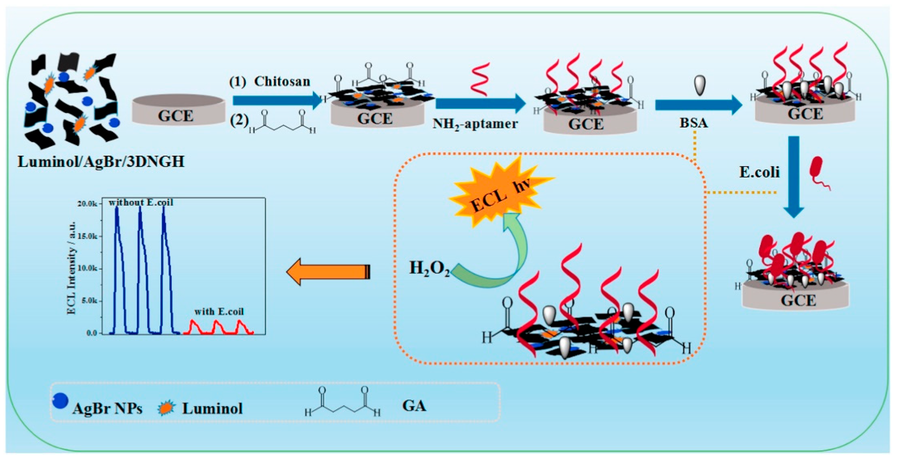
| GCE | ECL aptasensor based on AgBr nanoparticles (NPs) anchored on 3D nitrogen-doped graphene hydrogel (3DNGH) nanocomposites for immobilizing luminol and enhancing its ECL behavior | ECL | E. coli/meal samples | 0.5–500 CFU mL−1 | 0.17 CFU mL−1 | [116] |
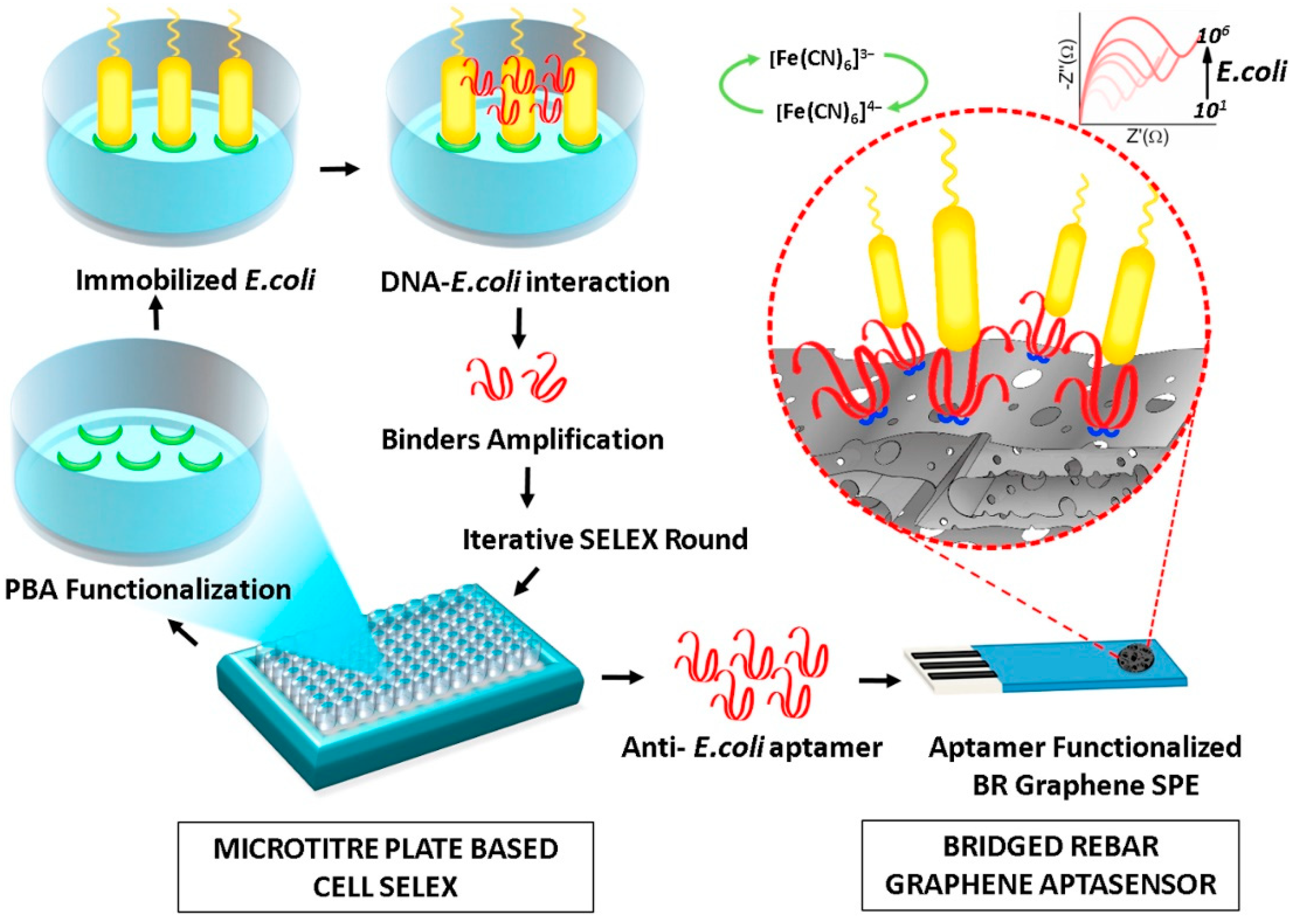
| SPCEs | Label-free impedimetric aptasensor using 3D hierarchical nanostructured bridged rebar graphene (BRG) for modifying SPCES | EIS | E. coli/water, juice, and milk. | 102–106 CFU mL−1 | 10 CFU mL−1 | [117] |
| PGE | Electrochemical immunosensor based on the PPy/AuNP/MWCNT/CHI hybrid nanocomposite modified pencil graphite electrode (PGE) | Amperometry | E. coli/no real samples | 30–306 CFU mL−1 | 30 CFU mL−1 | [118] |
| SPCEs | Electrochemical immunoassay using silica-coated Fe3O4 magnetic nanoparticles (Fe3O4@SiO2) and Au@Pt nanoparticles loaded on neutral red (NR) functionalized graphene to form composite complex rGO–NR–Au@Pt | CV | E. coli/pork and milk | 4.0 × 10³–4.0 × 10⁸ CFU mL−1 | 4.0 × 10² CFU mL−1 | [119] |
| GF-GCE | Electrochemical immunosensor based on anti- Escherichia coli antibody immobilized on the surface of the graphite felt electrode | OSWV | E. coli/beef | - | 400 cells mL−1 | [120] |
| AuIME | Electrochemical biosensor based on hybridization chain reaction (HCR) | MSPQC | S. aureus/milk and human serum | 50–107 CFU mL−1 | 50 CFU mL−1 | [122] |
| AuE | Electrochemical biosensor based on a triple-helix molecular switch, which can control the switching of electrochemical signals | DPV | S. aureus/water and honey | 30–30 × 108 CFU mL−1 | 8 CFU mL−1 | [123] |
| AuE | Label-free impedimetric immunosensor based on bacteria-imprinted conductive poly(3-thiopheneacetic acid) (BICP) film | EIS | S. aureus/milk | 10–10 × 108 CFU mL−1 | 2 CFU mL−1 | [125] |
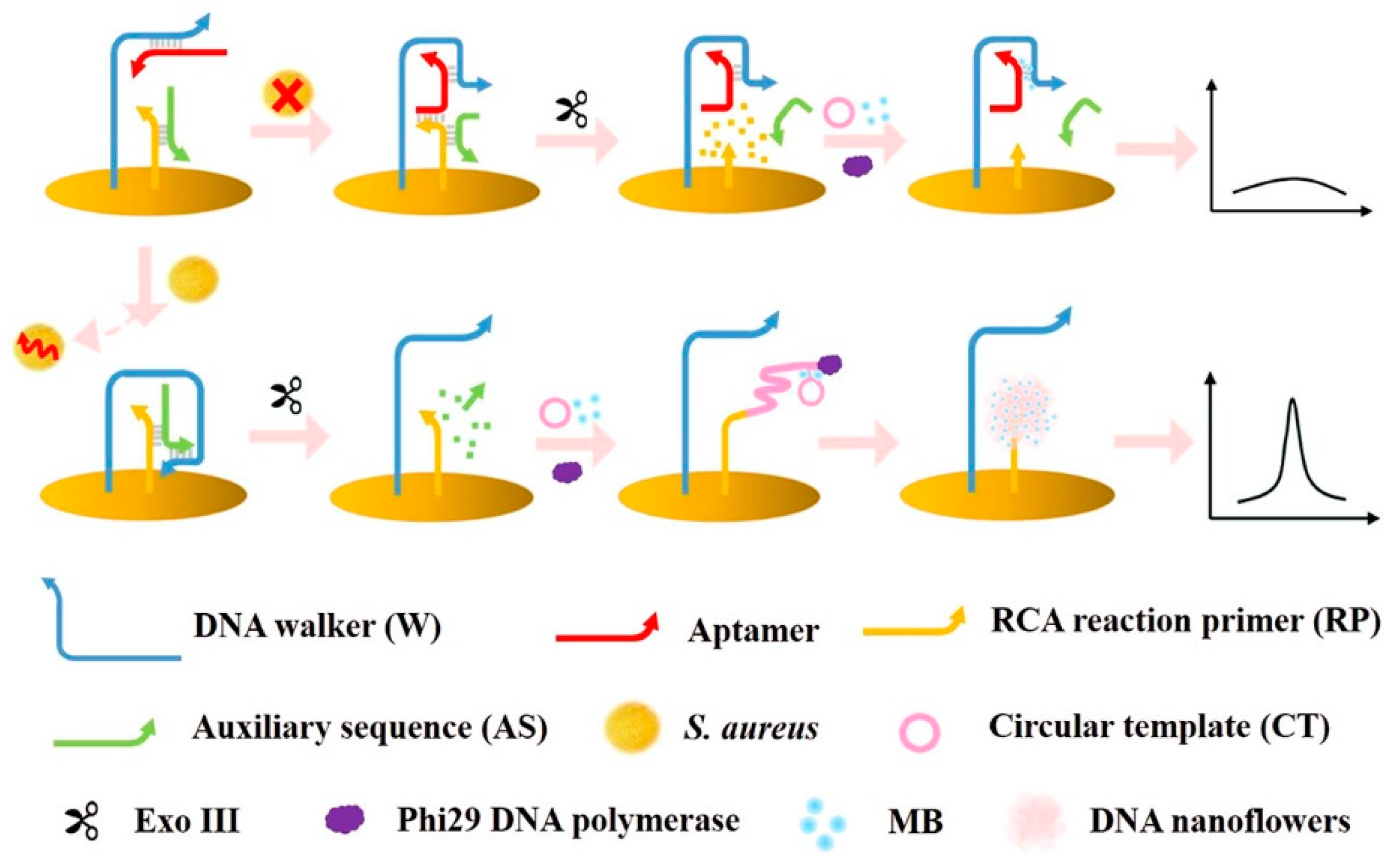
| AuE | Dual signal amplification electrochemical biosensor based on a DNA walker and DNA nanoflowers | DPV | S. aureus/water and honey | 60–60 × 107 CFU mL−1 | 9 CFU mL−1 | [126] |
| SPCNF/AuNPsE | plcA-based electrochemical DNA biosensor using screen-printed CNF/AuNPs electrode | CV | L. monocytogenes/milk | 0–0.234 ng/6 μL | 82 fg/6 μL | [136] |
| Pt-IME | Aptasensor using platinum interdigitated microelectrodes (Pt-IME) biofunctionalized with Listeria-specific aptamer and a smartphone-based signal acquisition system | EIS | L. monocytogenes/vegetable broth, hydroponic media | 102–106 CFU mL−1 | 23 CFU mL−1 | [137] |
| SPCEs | Electrochemical immunosensor using a disposable screen-printed electrode as transducer surface and monoclonal and polyclonal antibodies specifically recognizing Listeria monocytogenes p60 protein used as the sandwich immuno-pair | CV | L. monocytogenes/milk | 5–150 ng mL−1 | 1.5 ng mL−1 | [138] |
| Disposable electrical printed (DEP) microarray electrode s | Electrochemical biosensor assembled by selectively functionalizing the array electrodes with bacteria-specific peptides | SWV | L. monocytogenes/no real samples | 10–107 CFU mL−1 | 9 CFU mL−1 | [139] |
3.3. Pesticides
3.3.1. Insecticides
3.3.2. Herbicides
3.3.3. Fungicides
| Electrode | (Bio)Sensor Format | Electrochemical Technique | Analyte/Sample | L.R. | LOD | References |
|---|---|---|---|---|---|---|
| GCE | Electrochemical sensor using GCE modified with MCNHs and zein (SWCNH-ZE/GCE) | DPACSV | Fenitrothion/orange juice | 9.9 × 10⁻⁷–1.2 × 10⁻⁵ M | 1.2 × 10⁻⁸ M | [146] |
| SPCEs | Electrochemical sensor based on SCPCEs modified with cellulose microfibers supported reduced graphene-oxide composite | DPV | Fenitrothion/water | 0.03–1333.8 μM | 8 nM | [147] |
| GCE | Electrochemical biosensor using glutaraldehyde (Glu) crosslinked with acetylcholinesterase (AChE) immobilized on s-SWCNTs wrapped with bovine serum albumin (BSA) | DPV | Parathion/strawberry and apple juice | 1 × 10⁻¹⁰–5 × 10⁻⁶ M | 3.75 × 10⁻¹¹ M | [148] |
| ITO | Electrochemical sensor using ITO electrode modified with poly-3,4-ethylenedioxythiophene (PEDOT) membrane and zirconia nanoparticles (ZrO2 NPs) | CV | Parathion/water | 5–2000 ng·mL−1 | 2.8 ng·mL−1 | [149] |
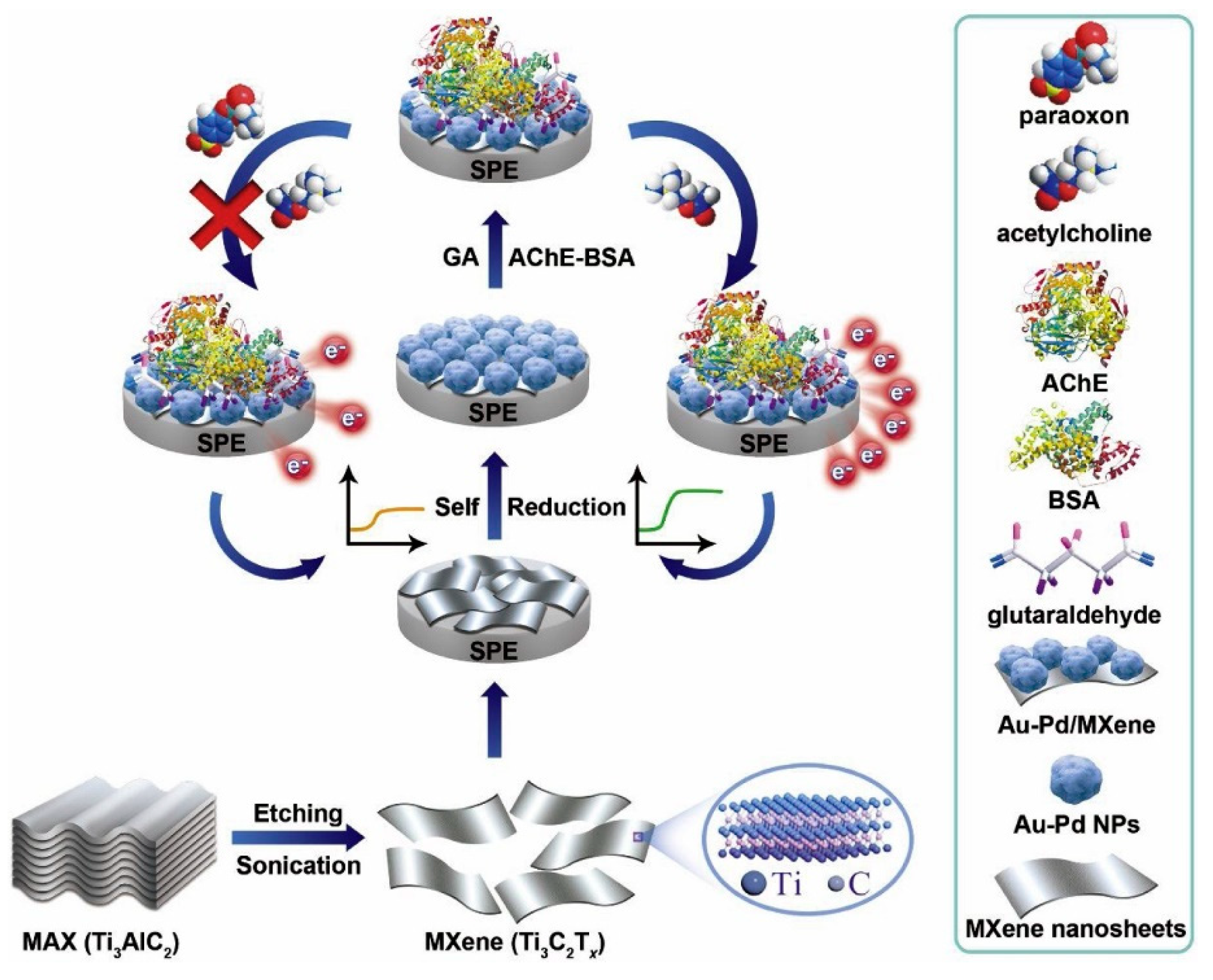
| SPCEs | Electrochemical biosensor using the multidimensional nanocomposite (MXene/Au–Pd) as the functional platform for immobilizing AChE | Amperometry | Paraoxon/pear and cucumber | 0.1–1000 μg L−1, | 1.75 ngL−1 | [150] |
| Fluorine–tin-oxide glass electrodes (FTO) | Electrochemical sensor developed by immobilizing hemoglobin (Hb), redox-active proteins on electrochemically reduced graphene-oxide–chitosan (ERGO–CHI/Hb/FTO) | SWV | Parathion/onion, lettuce | 0.076–0.988 mM | 79.77 nM | [151] |
| GCE | Electrochemical sensor using reduced graphene oxide (RGO) decorated fumed silica (FS) to modify glassy carbon (FS@RGO–GCE) | DPV | Fenitrothion/orange juice and tomato | 0.005–1.0 μM | 0.00019 μM | [152] |
| GCE | Electrochemical sensor using silver nanoparticles/dodecane modified glassy carbon electrode | DPV | Fenitrothion/paddy grains and potato | 0.1–7 nM | 0.60 nM | [153] |
| PGE | Electrochemical biosensor using WO3/g-C3N4 nanocomposite modified pencil graphite electrode as an immobilizing platform for Tribolium castaneum (red flour beetle) acetylcholinesterase (Tc-AChE) | Amperometry | Phosmet/wheat flour | 5–125 nM | 3.6 nM | [154] |
| SPCEs | Electrochemical sensor based on strontium hexaferrite (nanorods) decorated on porous graphitic carbon nitride (SrFe12O19/g-C3N4) to modify SPCEs | DPV | Fenitrothion/grapes, apricots, orange, cranberry, guava, mango | 0.005–378.15 mM | 1.4 nM | [155] |
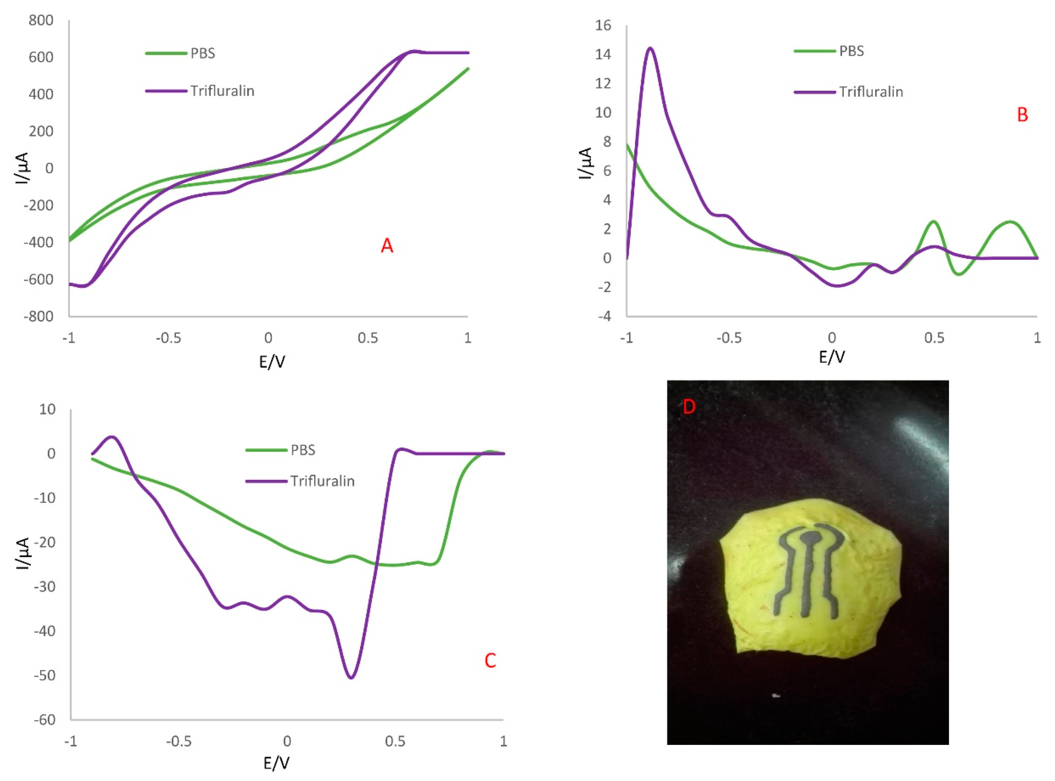
| Ag–citrate/GQDs nano-ink/leaf or skin | Electrochemical sensor prepared by direct writing on the surface of the samples, using Ag-citrate/graphene quantum dots (GQDs) nano-ink | DPV, SWV | Trifluralin/apple skin | 0.005–0.04 mM | 0.005 mM | [158] |
| PGE | Electrochemical biosensor platform developed to detect the pesticide–DNA interaction by using disposable pencil graphite electrodes (PGEs) where DNA was immobilized via passive absorption | DPV | Monitoring glyphosate and 2,4-dichlorophenoxyacetic acid DNA interactions | - | - | [160] |
| CPE | Electrochemical sensor using a carbon paste electrode modified with recrystallized zeolite | SWV | Thiram and carbendazim/honey and grape juice | 0.36–4.99 × 10⁻⁷ M Thiram 0.10–2.35 × 10⁻⁶ M Carbendazim | 6.74 × 10⁻⁹ M Thiram 3.51 × 10⁻⁹ M Carbendazim | [162] |
| NPG–GCe | Electrochemical sensor based on modified GCE with nanoporous gold film | DPV | Carbendazim and methyl parathion/water | 0.5–150 mM Methyl parathion 3.0–120 mM Carbendazim | 0.02 mM methyl parathion 0.24 mM Carbendazim | [163] |
| GCE | Electrochemical sensor based on gadolinium-oxide nanorods embedded on the graphene aerogel (GdO NRs/GA) | CV | Carbendazim/water | 0.01–75 μM | 3.0 nM | [165] |
| SPCEs | Electrochemical sensor based on SPCEs modified with carbon spherical shells (CSS) or Printex carbon nanoballs (PCNB) | DPV | Carbendazim and diuron/cabbages, apples, and orange juice | 0.1–1.0 μM carbendazim 1–10 mM diuron | 4.7 × 10⁻⁸ M carbendazim 9.2 × 10⁻⁷ M diuron | [166] |
| SPCEs | Electrochemical sensor based on SPCEs modified with carbon spherical shells (CSS) or Printex carbon nanoballs (PCNB) | SWV | Paraquat and fenitrothion/cabbages, apples, and orange juice | 0.1–1.0 μM paraquat 1–10 mM fenitrothion | 2.4 × 10⁻⁸ M paraquat 6.4 × 10⁻⁷ M fenitrothion | [166] |
3.4. Antibiotics
3.5. Endocrine-Disrupting Chemicals
3.5.1. Bisphenol A
3.5.2. Estrogens
| Electrode | (Bio)Sensor Format | Electrochemical Technique | Sample/Analyte | L.R. | LOD | References |
|---|---|---|---|---|---|---|
| Interdigitated electrode (IDE) | Label-free impedimetric aptasensor printed circuit board (PCB) technique | EIS | BPA/canned food | 1 fM–10 pM | 152.93 aM | [184] |
| BDDE | Impedimetric aptasensor based on Au nanoparticles (Au-NPs) coated boron-doped diamond (BDD) modified with aptamers, and 6-mercapto-1-hexanol (MCH) | EIS | BPA/milk | 1 × 10⁻¹⁴–1 × 10⁻⁹ M | 7.2 × 10⁻¹⁵ M | [185] |
| AuE | Label-free electrochemical aptasensor based on functionalized multiwall carbon nanotubes/gold nanoparticles (f-MWCNTs/AuNPs) nanocomposite film modified gold electrode | SWV | BPA/mineral water, orange juice, milk | 0.1–10 nM | 0.05 nM | [186] |
| GCE | Electrochemical sensor using hierarchical Ce-metal–organic framework (Ce-MOF) modified with cetyltrimethylammonium bromide (CTAB) as a sensing platform | DPV | BPA/milk | 0.005–50 mM | 2 nM | [187] |
| GCE | Electrochemical sensor based on the AuPd nanoparticles incorporated carboxylic multi-walled carbon nanotubes (MWCNT) | DPV | BPA/milk | 0.18–18 μM | 60 nM | [188] |
| GCE | Electrochemical aptasensor based on MWCNT/SiO2@Au nanocomposite | SWV | BPA/water, orange juice, milk | 0.1–100 mM | 10 pM | [189] |
| GCE | Electrochemical sensor using as sensing platform multi-walled carbon nanotubes and chitosan (MWCNTs–CH) self-assembled on graphene nanoplatelets GNPs (GNPs–MWCNTs–CH) | DPV | BPA/milk | 0.1–100 μM | 0.05 | [190] |
| GCE | Electrochemical sensor based on three-dimensional hierarchical cylinder-like nickel nanoparticle/nitrogen-doped carbon nanosheet/chitosan nanocomposite (NiNP/NCN/CHI) | DPV | BPA/milk | 0.1–2.5 mM and 2.5–15.0 mM | 45 nM | [191] |
| BDDE | Electrochemical sensor, using a cathodically pretreated boron-doped diamond (Cpt-BDD) electrode combined with QuEChERS extraction method | SWV | E,E-dienestrol/fish tissue | 2.30 × 10⁻⁷–9.69 × 10⁻⁶ M | 5.43 × 10⁻⁸ M | [197] |
| SPCE | Impedimetric aptasensor based on carbon nanodots modified SPC electrode | EIS | 17b-estradiol/water | 1.0 × 10⁻⁷–1.0 × 10⁻¹² M, | 0.5 × 10⁻¹² M. | [198] |
| AuE | Electrochemical biosensor based on graphene quantum dots (GQDs)/conducting polymer and laccase modified gold electrodes | CV | 17b-estradiol/no real samples | 0.1–120 × 10−6 M | 1 mM | [199] |
3.6. Allergens
3.6.1. Gliadin
3.6.2. Milk Allergens
| Electrode | (Bio)Sensor Format | Electrochemical Technique | Analyte/Sample | L.R. | LOD | References |
|---|---|---|---|---|---|---|
| GCE | Electrochemical immunosensor based on zein polymer coupled with carbon nanotubes as a sensing platform to immobilize capture antibody | SWV | Gliadin/wheat flour | 0.5–100 ppm | 0.5 ppm | [208] |
| SPCE | Microfluidic electrochemical aptasensing system based on a combination of 2D nanomaterial molybdenum disulfide (MoS2) and graphene with the addition of gold nanoparticles | DPV | Gliadin/wheat flour | 4–250 nM | 7 pM | [209] |
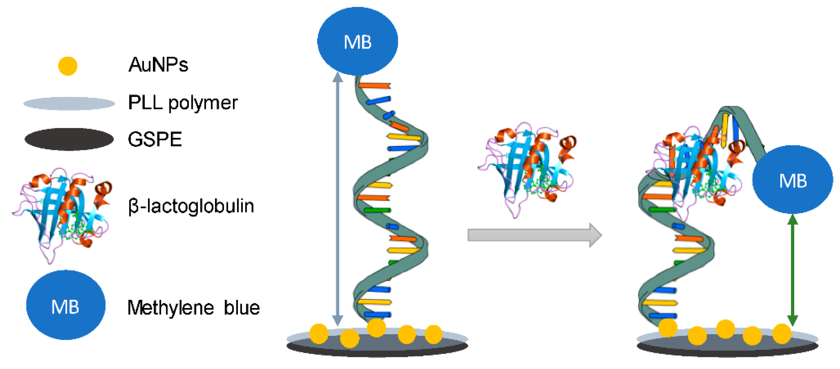
| GSPE | Electrochemical aptasensor based on poly(aniline-co-anthranilic acid) (PANI/PAA) composite polymer coupled with a specific aptamer | DPV | β-lactoglobulin/milk | 0.01–1.0 μg mL−1 | 0.053 μg mL−1 | [211] |
| GSPE | Electrochemical aptasensor based on poly-L-lysine modified graphite electrodes | DPV | β-lactoglobulin/milk, yogurt | 0.1–10 ng mL−1 | 0.09 ng mL−1. | [212] |
| SPAuE | Electrochemical label-free immunosensor using polypyrrole (PPY) electropolymerized as immobilizing platform for the capture antibody | DPV | a-lactoglobulin/meal | 355–2840 pg mL−1 | 0.192 fg mL−1 | [213] |
| ITO | Electrochemical aptasensor based on a highly selective DNA aptamer and flower-like Au@BiVO4 microspheres | Amperometry | β-lactoglobulin/infant food formula | 0.01–1000 ng mL−1 | 0.007 ng mL−1 | [214] |
| GSPE | Electrochemical immunosensor based on gold-nanocluster-modified graphene screen-printed electrodes | DPV | β-lactoglobulin/milk | 0.01–100 ng mL−1 | 0.08 ng mL−1 | [215] |
| PGE | Electrochemical sensor based on graphene-oxide modified pencil graphite electrode | CV | β-lactoglobulin/milk | 530–11.160 Mg L−1 | 270 mg L−1 | [216] |
| SPCE | Disposable amperometric magnetoimmunosensor using a sandwich configuration involving selective capture and detector antibodies and carboxylic acid-modified magnetic beads (HOOC-MBs) | Amperometry | Ara h 2/flour | 87–10.000 pg mL−1 | 26 pg mL−1 | [217] |
| GCE | DNA biosensor based on gold–palladium nanowaxberries (AuPd NWs)/dodecylamine functionalized graphene quantum dots (D-GQDs)-graphene micro-aerogel (GMA) composite | DPV | Ara h 1/peanut milk | 1.0 × 10⁻²²–1.0 × 10⁻¹⁷ M | 4.7 × 10⁻²³ M | [218] |
| SPCE | Paper-based capacitance mast cell sensor based on 3D paper chip printed with carbon electrodes as a noncontact capacitance sensing platform, using a polyvinyl alcohol (PVA)-gelatin methacryloyl (GelMA)-nano-hydroxyapatite (nHAP) composite hydrogel (PGHAP gel) to improve the conductivity and biocompatibility of the cellulose paper | Capacitance measurement | Ara h 2/raw and fried peanut | 0.1–100 ng mL−1 | 0.028 ng mL−1 | [219] |
| Magnetic glassy carbon electrode (MGCE) | Cell sensor, based on fluorescent magnetic beads | EIS | Tropomyosin and parvalbumin/crucian carp and brown shrimp | - | Tropomyosin 0.03 μg mL−1 Parvalbumin 0.16 ng mL−1 | [220] |
| SPCE | Label-free electrochemical immunosensor assembled by electrochemically reducing 4-carboxyphenyl diazonium salt, which was electrochemically generated in situ, to a stable 4-carboxyphenyl layer on carbon nanofiber-modified screen-printed electrode | DPV | Porcine serum albumin/pork fresh meat | 0.5–500 pg mL−1 | 0.5 pg mL−1 | [221] |
| CE | Electrochemical sensor using molecularly imprinted polymers (MIPs) for detecting genistein, an allergenic soy marker | DPV | Genistein/no real samples | 100 ppb-10 ppm | 100 ppb | [222] |
3.6.3. Peanut Allergens
4. Conclusions, Challenges, and Future Perspectives
Author Contributions
Funding
Institutional Review Board Statement
Informed Consent Statement
Data Availability Statement
Acknowledgments
Conflicts of Interest
References
- Rodriguez, R.S.; O’Keefe, T.L.; Froehlich, C.; Lewis, R.E.; Sheldon, T.R.; Haynes, C.L. Sensing Food Contaminants: Advances in Analytical Methods and Techniques. Anal. Chem. 2021, 93, 23–40. [Google Scholar] [CrossRef]
- Campuzano, S.; Yáñez-Sedeño, P.; Pingarrón, J.M. Electrochemical Affinity Biosensors in Food Safety. Chemosensors 2017, 5, 8. [Google Scholar] [CrossRef]
- Scognamiglio, V.; Arduini, F.; Palleschi, G.; Rea, G. Biosensing technology for sustainable food safety. TrAC Trends Anal. Chem. 2014, 62, 1–10. [Google Scholar] [CrossRef]
- Caballero, B.; Trugo, L.; Finglas, P. Encyclopedia of Food Sciences and Nutrition: Volumes 1–10; Elsevier: New York, NY, USA, 2003. [Google Scholar]
- Pividori, M.I.; Alegret, S. Electrochemical biosensors for food safety. Contrib. Sci. 2010, 6, 173–191. [Google Scholar]
- Ricci, F.; Volpe, G.; Micheli, L.; Palleschi, G. A review on novel developments and applications of immunosensors in food analysis. Anal. Chim. Acta 2007, 605, 111–129. [Google Scholar] [CrossRef]
- Mishra Kumar, G.; Barfidokht, A.; Tehrani, F.; Kumar Mishra, R. Food Safety Analysis Using Electrochemical Biosensors. Foods 2018, 7, 141. [Google Scholar] [CrossRef]
- Thakur, M.S.; Ragavan, K.V. Biosensors in food processing. J. Food Sci. Technol. 2013, 50, 625–641. [Google Scholar] [CrossRef]
- Barfidokht, A.; Gooding, J.J. Approaches toward allowing electroanalytical devices to be used in biological fluids. Electroanalysis 2014, 26, 1182–1196. [Google Scholar] [CrossRef]
- Ronkainen, N.J.; Halsall, H.B.; Heineman, W.R. Electrochemical biosensors. Chem. Soc. Rev. 2010, 39, 1747–1763. [Google Scholar] [CrossRef]
- Chen, A.; Chatterjee, S. Nanomaterials based electrochemical sensors for biomedical applications. Chem. Soc. Rev. 2013, 42, 5425–5438. [Google Scholar] [CrossRef]
- Curulli, A. Nanomaterials in Electrochemical Sensing Area: Applications and Challenges in Food Analysis. Molecules 2020, 25, 5759. [Google Scholar] [CrossRef]
- Ahmed, M.U.; Hossain, M.M.; Tamiya, E. Electrochemical Biosensors for Medical and Food Applications. Electroanalysis 2008, 20, 616–626. [Google Scholar] [CrossRef]
- Neethirajan, S.; Raghavan, K.V.; Weng, X. Agro-defense: Biosensors for food from healthy crops and animals. Trends Food Sci. Technol. 2018, 73, 25–44. [Google Scholar] [CrossRef]
- Moro, G.; De Wae, K.; Moretto, L.M. Challenges in the electrochemical (bio)sensing of nonelectroactive food and environmental contaminants. Curr. Opin. Electrochem. 2019, 16, 57–65. [Google Scholar] [CrossRef]
- Naresh, V.; Lee, N. Review on Biosensors and Recent Development of Nanostructured Materials-Enabled Biosensors. Sensors 2021, 21, 1109. [Google Scholar] [CrossRef]
- Griesche, C.; Baeumner, A.J. Biosensors to support sustainable agriculture and food safety. TrAC Trends Anal. Chem. 2020, 128, 115906. [Google Scholar]
- Ru Choi, J.; Wey Yong, K.; Yu Choi, J.; Cowie, A.C. Emerging Point-of-care Technologies for Food Safety Analysis. Sensors 2019, 19, 817. [Google Scholar] [CrossRef]
- Zeng, Y.; Zhu, Z.; Dua, D.; Lin, Y. Nanomaterial-based electrochemical biosensors for food safety. J. Electroanal. Chem. 2016, 781, 147–154. [Google Scholar] [CrossRef]
- Yoo, E.-H.; Lee, S.-Y. Glucose Biosensors: An Overview of Use in Clinical Practice. Sensors 2010, 10, 4558–4576. [Google Scholar] [CrossRef]
- Patris, S.; Vandeput, M.; Kauffmann, J.M. Antibodies as target for affinity biosensors. TrAC Trends Anal. Chem. 2016, 79, 239–246. [Google Scholar] [CrossRef]
- Bard, A.J.; Faulkner, L.R. Electrochemical Methods: Fundamentals and Applications, 2nd ed.; John Wiley & Sons: New York, NY, USA, 2001. [Google Scholar]
- Mishra, G.K.; Sharma, V.; Mishra, R.K. Electrochemical aptasensors for food and environmental safeguarding: A review. Biosensors 2018, 8, 28. [Google Scholar] [CrossRef]
- Bartlett, P.N. Bioelectrochemistry 45: Fundamentals, Experimental Techniques, and Applications; John Wiley & Sons: Hoboken, NJ, USA, 2008. [Google Scholar]
- Bonizzoni, M.; Anslyn, E.V. Combinatorial Methods for Chemical and Biological Sensors. J. Am. Chem. Soc. 2009, 131, 14597–14598. [Google Scholar] [CrossRef]
- Suni, I.I. Impedance methods for electrochemical sensors using nanomaterials. TrAC Trends Anal. Chem. 2008, 27, 604–611. [Google Scholar] [CrossRef]
- Katz, E.; Willner, I. Probing Biomolecular Interactions at Conductive and Semiconductive Surfaces by Impedance Spectroscopy: Routes to Impedimetric Immunosensors, DNA-Sensors, and Enzyme Biosensors. Electroanalysis 2003, 15, 913–947. [Google Scholar] [CrossRef]
- Wang, J. Analytical Electrochemistry, 2nd ed.; Wiley/VCH: New York, NY, USA, 2000. [Google Scholar]
- Bockris, J.O.M.; Reddy, A.K.N.; Gamboa-Aldeco, M. Modern Electrochemistry 2A: Fundamentals of Electrodics, 2nd ed.; Kluwer Academic/Plenum Publishers: New York, NY, USA, 2000; Volume 2. [Google Scholar]
- Mustafa, F.; Andreescu, S. Nanotechnology-based approaches for food sensing and packaging applications. RSC Adv. 2020, 10, 19309. [Google Scholar] [CrossRef]
- Xiao, T.; Huang, J.; Wang, D.; Meng, T.; Yang, X. Au and Au-Based nanomaterials: Synthesis and recent progress in electrochemical sensor applications. Talanta 2020, 206, 120210. [Google Scholar] [CrossRef]
- Teradal, N.L.; Jelinek, R. Carbon Nanomaterials: Carbon Nanomaterials in Biological Studies and Biomedicine. Adv. Healthc. Mater. 2017, 6, 1700574. [Google Scholar] [CrossRef]
- Pumera, M. Graphene-based nanomaterials and their electrochemistry. Chem. Soc. Rev. 2010, 39, 4146–4157. [Google Scholar] [CrossRef]
- Smart, A.; Crew, A.; Pemberton, R.; Hughes, G.; Doran, O.; Hart, J.P. Screen-printed carbon-based biosensors and their applications in agri-food safety. TrAC Trends Anal. Chem. 2020, 127, 115898. [Google Scholar] [CrossRef]
- Hughes, G.; Westmacott, K.; Honeychurch, K.C.; Crew, A.; Pemberton, R.M.; Hart, J.P. Recent Advances in the Fabrication and Application of Screen-Printed Electrochemical (Bio)Sensors Based on Carbon Materials for Biomedical, Agri-Food and Environmental Analyses. Biosensors 2016, 6, 50. [Google Scholar] [CrossRef]
- Trojanowicz, M. Impact of nanotechnology on design of advanced screen-printed electrodes for different analytical applications. TrAC Trends Anal. Chem. 2016, 84, 22–47. [Google Scholar] [CrossRef]
- Janik, E.; Ceremuga, M.; Saluk-Bijak, J.; Bijak, M. Biological toxins as the potential Tools for bioterrorism. Int. J. Mol. Sci. 2019, 20, 1181. [Google Scholar] [CrossRef]
- World Health Organization (WHO). Food Safety 2021. Available online: https://www.who.int/health-topics/food-safety/ (accessed on 22 February 2021).
- World Health Organization. Foodborne Disease Outbreaks: Guidelines for Investigation and Control; World Health Organization Publisher: Geneva, Switzerland, 2008. [Google Scholar]
- Vale, P.; Botelho, M.J.; Rodrigues, S.M.; Gomes, S.S.; Sampayo, M.A.D.M. Two decades of marine biotoxin monitoring in bivalves from Portugal (1986–2006): A review of exposure assessment. Harmful Algae 2008, 7, 11–25. [Google Scholar] [CrossRef]
- Tian, Y.; Du, L.; Zhu, P.; Chen, Y.; Chen, W.; Wu, C.; Wa, P. Recent progress in micro/nano biosensors for shellfish toxin detection. Biosens. Bioelectron. 2021, 176, 112899. [Google Scholar] [CrossRef] [PubMed]
- Scientific Opinion of the Panel on Contaminants in the Food Chain, Marine biotoxins in shellfish—Saxitoxin group. EFSA J. 2009, 1019, 1–76.
- Qi, X.; Yan, X.; Zhao, L.; Huang, Y.; Wang, S.; Liang, X. A facile label-free electrochemical aptasensor constructed with nanotetrahedron and aptamer-triplex for sensitive detection of small molecule: Saxitoxin. J. Electroanal. Chem. 2020, 858, 113805. [Google Scholar] [CrossRef]
- Wiese, M.; D’Agostino, P.M.; Mihali, T.K.; Moffitt, M.C.; Neilan, B.A. Neurotoxic Alkaloids: Saxitoxin and Its Analogs. Mar. Drugs 2010, 8, 2185–2211. [Google Scholar] [CrossRef] [PubMed]
- Tolle, F.; Brändle, G.M.; Matzner, D.; Mayer, G. A versatile approach towards nucleobase modified aptamers. Angew. Chem. Int. Ed. 2015, 54, 10971–10974. [Google Scholar] [CrossRef]
- Zhao, L.H.; Huang, Y.F.; Dong, Y.Y.; Han, X.T.; Wang, S.; Liang, X.G. Aptamers and aptasensors for highly specific recognition and sensitive detection of marine biotoxins: Recent advances and perspectives. Toxins 2018, 10, 427. [Google Scholar] [CrossRef]
- Ma, C.; Wu, K.; Han, Z.; Liu, H.; Wang, K.; Xia, K. Fluorometric aptamer-based determination of ochratoxin a based on the use of graphene oxide and RNase H-aided amplification. Microchim. Acta 2018, 185, 347–354. [Google Scholar] [CrossRef]
- Sekhon, S.S.; Park, G.Y.; Park, D.Y.; Kim, S.Y.; Wee, J.H.; Ahn, J.Y. Aptasensors for pesticide detection. Toxicol. Environ. Health 2018, 10, 229–236. [Google Scholar] [CrossRef]
- Hao, L.; Duan, N.; Wu, S.; Dai, S.; Gu, H.; Ye, H.; Wang, Z. Advances in aptasensors for the detection of food contaminants. Analyst 2016, 141, 3942–3961. [Google Scholar]
- Hesieh, K.; Patterson, A.S.; Ferguson, B.S.; Plaxco, K.W.; Soh, H.T. Rapid, sensitive, and quantitative detection of pathogenic DNA at the point of care through microfluidic electrochemical quantitative loop-mediated isothermal amplification. Angew. Chem. Int. Ed. 2012, 124, 4980–4984. [Google Scholar] [CrossRef]
- Wang, S.; Zhang, L.; Wan, S.; Cansiz, S.; Cui, C.; Liu, Y.; Cai, R.; Hong, C.; Teng, I.T.; Shi, M.; et al. Aptasensor with expanded nucleotide using DNA nanotetrahedral for electrochemical detection of cancerous exosomes. ACS Nano 2017, 11, 3943–3949. [Google Scholar] [CrossRef] [PubMed]
- Nguyen, V.T.; Kwon, Y.S.; Gu, M.B. Aptamer-based environmental biosensor for small molecule contaminants. Curr. Opin. Biotech. 2017, 45, 15–23. [Google Scholar] [CrossRef]
- Nelis, J.L.D.; Migliorelli, D.; Jafari, S.; Generelli, S.; Franco, J.L.J.; Salvador, P.; Marco, M.P.; Cao, C.; Elliott, C.T.; Campbell, K. The benefits of carbon black, gold, and magnetic nanomaterials for point-of-harvest electrochemical quantification of domoic acid. Microchim. Acta 2020, 187, 164–175. [Google Scholar] [CrossRef]
- Volpe, G.; Palleschi, G. New Biosensors for Microbiological Analysis of Food. In Detecting Pathogens in Food, Woodhead Publishing Series in Food Science, Technology and Nutrition; Elsevier: Amsterdam, The Netherlands, 2003; pp. 294–331. [Google Scholar]
- Ramalingam, S.; Chand, R.; Singh, C.B.; Singh, A. Phosphorene-gold nanocomposite based microfluidic aptasensor for the detection of okadaic acid. Biosens. Bioelectron. 2019, 135, 14–21. [Google Scholar] [CrossRef]
- Verma, M.; Chaudhary, M.; Singh, A.; Kaur, N.; Singh, N. Naphthalimide-gold-based nanocomposite for the ratiometric detection of okadaic acid in shellfish. J. Mater. Chem. B 2020, 8, 8405–8413. [Google Scholar] [CrossRef] [PubMed]
- Yang, W.; Zhang, G.; Ni, J.; Wang, Q.; Lin, Z. From signal amplification to restrained background: Magnetic graphene oxide assisted homogeneous electrochemiluminescence aptasensor for highly sensitive detection of okadaic acid. Sens. Actuators B Chem. 2021, 32, 128872. [Google Scholar] [CrossRef]
- Pitt, J.I.; Miller, J.D. A Concise History of Mycotoxin Research. J. Agric. Food Chem. 2017, 65, 7021–7033. [Google Scholar] [CrossRef]
- Commission Regulation (EC) No 1881/2006 of 19 December 2006 Setting Maximum Levels for Certain Contaminants in Foodstuffs, Official Journal of the European Union, 2006, L364/5–L364/24. Available online: https://www.fsai.ie/uploadedFiles/Regulation-EC-1881-2006.pdf (accessed on 22 February 2021).
- Zhang, N.; Liu, B.; Cui, X.; Li, Y.; Tang, J.; Wang, H.; Zhang, D.; Li, Z. Recent advances in aptasensors for mycotoxin detection: On the surface and in the colloid. Talanta 2021, 223, 121729. [Google Scholar] [CrossRef]
- Jia, M.; Liao, X.; Fang, L.; Jia, B.; Liu, M.; Li, D.; Zhou, L.; Kong, W. Recent advances on immunosensors for mycotoxins in foods and other commodities. TrAC Trends Anal. Chem. 2021, 136, 116193. [Google Scholar] [CrossRef]
- Liu, D.; Li, W.; Zhu, C.; Li, Y.; Shen, X.; Li, L.; Yan, X.; You, T. Recent progress on electrochemical biosensing of aflatoxins: A review. TrAC Trends Anal. Chem. 2020, 133, 115966. [Google Scholar] [CrossRef]
- IARC Working Group on the Evaluation of Carcinogenic Risks to Humans. International Agency for Research on Cancer, Some Traditional Herbal Medicines, Some Mycotoxins, Naphthalene and Styrene; IARC Publisher: Lyon, France, 2002; Volume 82. [Google Scholar]
- Abnousa, K.; Daneshc, N.M.; Alibolandia, M.; Ramezanic, M.; Sarreshtehdar Emranie, A.; Zolfagharif, R.; Taghdisig, S.M. A new amplified π-shape electrochemical aptasensor for ultrasensitive detection of aflatoxin B1. Biosens. Bioelectron. 2017, 94, 374–379. [Google Scholar] [CrossRef]
- Karczmarczyka, A.; Baeumner, A.J.; Fellera, K.-H. Rapid and sensitive inhibition-based assay for the electrochemical detection of Ochratoxin A and Aflatoxin M1 in red wine and milk. Electrochim. Acta 2017, 243, 82–89. [Google Scholar] [CrossRef]
- Wang, C.; Qian, J.; An, K.; Ren, C.; Lu, X.; Hao, N.; Liu, Q.; Li, H.; Huang, X.; Wang, K. Fabrication of magnetically assembled aptasensing device for label-free determination of aflatoxin B1 based on EIS. Biosens. Bioelectron. 2018, 108, 69–75. [Google Scholar] [CrossRef]
- Mo, R.; He, L.; Yan, X.; Su, T.; Zhou, C.; Wang, Z.; Hong, P.; Sun, S.; Li, C. A novel aflatoxin B1 biosensor based on a porous anodized alumina membrane modified with graphene oxide and an aflatoxin B1 aptamer. Electrochem. Commun. 2018, 95, 9–13. [Google Scholar] [CrossRef]
- Selvolini, G.; Lettieri, M.; Tassoni, L.; Gastaldello, S.; Grillo, M.; Maran, C.; Marrazza, G. Electrochemical enzyme-linked oligonucleotide array for aflatoxin B1 detection. Talanta 2019, 203, 49–57. [Google Scholar] [CrossRef]
- Wang, C.; Lia, Y.; Zhao, Q. A signal-on electrochemical aptasensor for rapid detection of aflatoxin B1 based on competition with complementary DNA. Biosens. Bioelectron. 2019, 144, 111641. [Google Scholar] [CrossRef]
- Xiong, X.; Li, Y.; Yuan, W.; Lu, Y.; Xiong, X.; Li, Y.; Chen, X.; Liu, Y. Screen printed bipolar electrode for sensitive electrochemiluminescence detection of aflatoxin B1 in agricultural products. Biosens. Bioelectron. 2020, 150, 111873. [Google Scholar] [CrossRef]
- Chen, Y.; Yang, Y.; Wang, Y.; Peng, Y.; Nie, J.; Gao, G.; Zhi, J. Development of an Escherichia coli-based electrochemical biosensor for mycotoxin toxicity detection. Bioelectrochemistry 2020, 133, 107453. [Google Scholar] [CrossRef]
- EFSA Panel on Contaminants in the Food Chain (CONTAM). Risks for animal health related to the presence of zearalenone and its modified forms in feed. EFSA J. 2017, 15, 4851–4974. [Google Scholar]
- Xiong, X.; Yuan, W.; Li, Y.; Lu, Y.; Xiong, X.; Li, Y.; Liu, Y.; Lu, L. Sensitive electrochemical detection of aflatoxin B1 using DNA tetrahedron nanostructure as substrate of antibody ordered assembly and template of aniline polymerization. Food Chem. 2020, 331, 127368. [Google Scholar] [CrossRef]
- Lin, T.; Shen, Y. Fabricating electrochemical aptasensors for detecting aflatoxin B1 via layer-by-layer self-assembly. J. Electroanal. Chem. 2020, 870, 114247. [Google Scholar] [CrossRef]
- Shi, L.; Wang, Z.; Yang, G.; Yang, H.; Zhao, F. A novel electrochemical immunosensor for aflatoxin B1 based on Au nanoparticles-poly 4-aminobenzoic acid supported graphene. Appl. Surf. Sci. 2020, 527, 146934. [Google Scholar] [CrossRef]
- Wang, C.; Zhao, Q. A reagentless electrochemical sensor for aflatoxin B1 with sensitive signal-on responses using aptamer with methylene blue label at specific internal thymine. Biosens. Bioelectron. 2020, 167, 112478. [Google Scholar] [CrossRef] [PubMed]
- Stockmann-Juvala, H.; Savolainen, K. A review of the toxic effects and mechanisms of action of fumonisin B1. Hum. Exp. Toxicol. 2008, 27, 799–809. [Google Scholar] [CrossRef]
- EFSA Panel on Contaminants in the Food Chain (CONTAM). Appropriateness to set a group health-based guidance value for fumonisins and their modified forms. EFSA J. 2018, 16, 5172–5247. [Google Scholar]
- Lin, X.; Guo, X. Advances in Biosensors, Chemosensors and Assays for the Determination of Fusarium Mycotoxins. Toxins 2016, 8, 161. [Google Scholar] [CrossRef]
- Jodra, A.; López, M.Á.; Escarpa, A. Disposable and reliable electrochemical magnetoimmunosensor for Fumonisins simplified determination in maize-based foodstuffs. Biosens. Bioelectron. 2015, 64, 633–638. [Google Scholar] [CrossRef]
- Lu, L.; Seenivasan, R.; Wang, Y.-C.; Yu, J.-H.; Gunasekaran, S. An Electrochemical Immunosensor for Rapid and Sensitive Detection of Mycotoxins Fumonisin B1 and Deoxynivalenol. Electrochim. Acta 2016, 213, 89–97. [Google Scholar] [CrossRef]
- Munawara, H.; Garcia-Cruz, A.; Majewska, M.; Karim, K.; Kutner, W.; Piletsky, S.A. Electrochemical determination of fumonisin B1 using a chemosensor with a recognition unit comprising molecularly imprinted polymer nanoparticles. Sens. Actuators B Chem. 2020, 321, 128552. [Google Scholar] [CrossRef]
- EFSA Panel on Contaminants in the Food Chain (CONTAM). Risk assessment of ochratoxin A in food. EFSA J. 2020, 18, 6113–6263. [Google Scholar]
- Lv, L.; Wang, X. Recent Advances in Ochratoxin A Electrochemical Biosensors: Recognition Elements, Sensitization Technologies, and Their Applications. J. Agric. Food Chem. 2020, 68, 4769–4787. [Google Scholar] [CrossRef]
- Mishra, R.K.; Hayat, A.; Catanante, G.; Ocana, C.; Marty, J.-L. A label free aptasensor for Ochratoxin A detection in cocoa beans: An application to chocolate industries. Anal. Chim. Acta 2015, 889, 106–112. [Google Scholar] [CrossRef]
- Mejri-Omrani, N.; Miodek, A.; Zribi, B.; Marrakchi, M.; Hamdi, M.; Marty, J.-L.; Korri-Youssoufi, H. Direct detection of OTA by impedimetric aptasensor based on modified polypyrrole-dendrimers. Anal. Chim. Acta 2016, 920, 37–46. [Google Scholar] [CrossRef]
- Mishra, R.K.; Hayat, A.; Catanante, G.; Istamboulie, G.; Marty, J.-L. Sensitive quantitation of Ochratoxin A in cocoa beans using differential pulse voltammetry based aptasensor. Food Chem. 2016, 192, 799–804. [Google Scholar] [CrossRef]
- Malvano, F.; Albanese, D.; Pilloton, R.; Di Matteo, M. A highly sensitive impedimetric label free immunosensor for Ochratoxin measurement in cocoa beans. Food Chem. 2016, 212, 688–694. [Google Scholar] [CrossRef]
- Liu, Y.; Yu, J. Oriented immobilization of proteins on solid supports for use in biosensors and biochips: A review. Microchim. Acta 2016, 183, 1–19. [Google Scholar] [CrossRef]
- Hao, N.; Jiang, L.; Qian, J.; Wang, K. Ultrasensitive electrochemical Ochratoxin A aptasensor based on CdTe quantum dots functionalized graphene/Au nanocomposites and magnetic separation. J. Electroanal. Chem. 2016, 781, 332–338. [Google Scholar] [CrossRef]
- Zhu, X.; Kou, F.; Xu, H.; Han, Y.; Yang, G.; Huang, X.; Chen, W.; Chi, Y.; Line, Z. Label-free ochratoxin A electrochemical aptasensor based on target-induced noncovalent assembly of peroxidase-like graphitic carbon nitride nanosheet. Sens. Actuators B Chem. 2018, 270, 263–269. [Google Scholar] [CrossRef]
- Chen, W.; Yan, C.; Cheng, L.; Yao, L.; Xue, F.; Xu, J. An ultrasensitive signal-on electrochemical aptasensor for ochratoxin A determination based on DNA controlled layer-by-layer assembly of dual gold nanoparticle conjugates. Biosens. Bioelectron. 2018, 117, 845–851. [Google Scholar] [CrossRef]
- Sun, C.; Liao, X.; Huang, P.; Shan, G.; Ma, X.; Fu, L.; Zhou, L.; Kong, W. A self-assembled electrochemical immunosensor for ultra-sensitive detection of ochratoxin A in medicinal and edible malt. Food Chem. 2020, 315, 126289. [Google Scholar] [CrossRef]
- Riu, J.; Giussani, B. Electrochemical biosensors for the detection of pathogenic bacteria in food. TrAC Trends Anal. Chem. 2020, 126, 115863. [Google Scholar] [CrossRef]
- The Commission of the European Communities. Commission Regulation (EC) No 2073/2005 of 15th November 2005 on Microbiological Criteria for Foodstuffs; The Commission of the European Communities: Luxembourg, 2005. [Google Scholar]
- Zhang, Z.; Zhou, J.; Du, X. Electrochemical Biosensors for Detection of Foodborne Pathogens. Micromachines 2019, 10, 222. [Google Scholar] [CrossRef]
- US Food and Drug Administration. Bad Bug Book, Foodborne Pathogenic Microorganisms and Natural Toxins, 2nd ed.; FDA: Silver Spring, MD, USA, 2012.
- Silva, N.F.D.; Magalhães, J.M.C.S.; Freire, C.; Delerue-Matosa, C. Electrochemical biosensors for Salmonella: State of the art and challenges in food safety assessment. Biosens. Bioelectron. 2018, 99, 667–682. [Google Scholar] [CrossRef]
- Shen, Y.; Xu, L.; Li, Y. Biosensors for rapid detection of Salmonella in food: A review. Compr. Rev. Food Sci. Food Saf. 2021, 20, 149–197. [Google Scholar] [CrossRef]
- Xiang, C.; Li, R.; Adhikari, B.; She, Z.; Li, Y.; Kraatz, H.-B. Sensitive electrochemical detection of Salmonella with chitosan-gold nanoparticles composite film. Talanta 2015, 140, 122–127. [Google Scholar] [CrossRef]
- Bagheryan, Z.; Raoof, J.-B.; Golabi, M.; Turner, A.P.F.; Beni, V. Diazonium-based impedimetric aptasensor for the rapid label-free detection of Salmonella typhimurium in food sample. Biosens. Bioelectron. 2016, 80, 566–573. [Google Scholar] [CrossRef]
- Sheikhzadeh, E.; Chamsaz, M.; Turner, A.P.F.; Jager, E.W.H.; Beni, V. Label-free impedimetric biosensor for Salmonella Typhimurium detection based on poly [pyrrole-co-3-carboxyl-pyrrole] copolymer supported aptamer. Biosens. Bioelectron. 2016, 80, 194–200. [Google Scholar] [CrossRef]
- Dinshaw, I.J.; Muniandy, S.; Jyan Teh, S.; Ibrahim, F.; Fen Leo, B.; Lin Thong, K. Development of an aptasensor using reduced graphene oxide chitosan complex to detect Salmonella. J. Electroanal. Chem. 2017, 806, 88–96. [Google Scholar] [CrossRef]
- Ge, C.; Yuan, R.; Yi, L.; Yang, J.; Zhang, H.; Li, L.; Nian, W.; Yi, G. Target-induced aptamer displacement on gold nano-particles and rolling circle amplification for ultrasensitive live Salmonella typhimurium electrochemical biosensing. J. Electroanal. Chem. 2018, 826, 174–180. [Google Scholar] [CrossRef]
- Capobianco, J.A.; Lee, J.; Armstrong, C.M.; Gehring, A.G. Rapid detection of Salmonella enterica serotype Typhimurium in large volume samples using porous electrodes in a flow-through, enzyme-amplified immunoelectrochemical sensor. Anal. Bioanal. Chem. 2019, 411, 5233–5242. [Google Scholar] [CrossRef]
- Murasova, P.; Kovarova, A.; Kasparova, J.; Brozkova, I.; Hamiot, A.; Pekarkova, J.; Dupuy, B.; Drbohlavova, J.; Bilkova, Z.; Korecka, L. Direct culture-free electrochemical detection of Salmonella cells in milk based on quantum dots-modified nanostructured dendrons. J. Electroanal. Chem. 2020, 863, 114051. [Google Scholar] [CrossRef]
- Wang, L.; Huo, X.; Qi, W.; Xia, Z.; Li, Y.; Lin, J. Rapid and sensitive detection of Salmonella Typhimurium using nickel nanowire bridge for electrochemical impedance amplification. Talanta 2020, 211, 120715. [Google Scholar] [CrossRef]
- Xue, L.; Guo, R.; Huang, F.; Qi, W.; Liu, Y.; Cai, G.; Lin, J. An impedance biosensor based on magnetic nanobead net and MnO2 nanoflowers for rapid and sensitive detection of foodborne bacteria. Biosens. Bioelectron. 2021, 173, 112800. [Google Scholar] [CrossRef]
- Huang, F.; Xue, L.; Qi, W.; Cai, G.; Liu, Y.; Lin, J. An ultrasensitive impedance biosensor for Salmonella detection based on rotating high gradient magnetic separation and cascade reaction signal amplification. Biosens. Bioelectron. 2021, 176, 112921. [Google Scholar] [CrossRef]
- Kim, Y.S.; Song, M.Y.; Jurng, J.; Kim, B.C. Isolation, and characterization of DNA aptamers against Escherichia coli using a bacterial cell-systematic evolution of ligands by exponential enrichment approach. Anal. Biochem. 2013, 436, 22–28. [Google Scholar] [CrossRef]
- Kaper, J.B.; Nataro, J.P.; Mobley, H.L.T. Pathogenic Escherichia coli. Nat. Rev. Microbiol. 2004, 2, 123–140. [Google Scholar] [CrossRef]
- Xu, M.; Wang, R.; Li, Y. Electrochemical biosensors for rapid detection of Escherichia coli O157:H7. Talanta 2017, 162, 511–522. [Google Scholar] [CrossRef]
- Abdalhai, M.H.; Fernandes, A.M.; Xia, X.; Musa, A.; Ji, J.; Sun, X. Electrochemical Genosensor To Detect Pathogenic Bacteria (Escherichia coli O157:H7) As Applied in Real Food Samples (Fresh Beef) To Improve Food Safety and Quality Control. J. Agric. Food Chem. 2015, 63, 5017–5025. [Google Scholar] [CrossRef]
- Liébana, S.; Brandao, D.; Cortes, P.; Campoy, S.; Alegret, S.; Pividori, M.I. Electrochemical genosensing of Salmonella, Listeria and Escherichia coli on silica magnetic particles. Anal. Chim. Acta 2016, 904, 1–9. [Google Scholar] [CrossRef]
- Mouli Pandey, C.; Tiwari, I.; Nand Singh, V.; Sood, K.N.; Sumana, G.; Dhar Malhotra, B. Highly sensitive electrochemical immunosensor based on graphene-wrapped copper oxide-cysteine hierarchical structure for detection of pathogenic bacteria. Sens. Actuators B Chem. 2017, 238, 1060–1069. [Google Scholar] [CrossRef]
- Hao, N.; Zhang, X.; Zhou, Z.; Hua, R.; Zhang, Y.; Liu, Q.; Qian, J.; Li, H.; Wang, K. AgBr nanoparticles/3D nitrogen-doped graphene hydrogel for fabricating all-solid-state luminol-electrochemiluminescence Escherichia coli aptasensors. Biosens. Bioelectron. 2017, 97, 377–383. [Google Scholar] [CrossRef] [PubMed]
- Kaur, H.; Shorie, M.; Sharma, M.; Ganguli, A.K.; Sabherwal, P. Bridged Rebar Graphene functionalized aptasensor for pathogenic E. coli O78:K80:H11 detection. Biosens. Bioelectron. 2017, 98, 486–493. [Google Scholar] [CrossRef]
- Güner, A.; Çevik, E.; Senel, M.; Alpsoy, L. An electrochemical immunosensor for sensitive detection of Escherichia coli O157:H7 by using chitosan, MWCNT, polypyrrole with gold nanoparticles hybrid sensing platform. Food Chem. 2017, 229, 358–365. [Google Scholar] [CrossRef]
- Zhu, F.; Zhao, G.; Dou, W. A non-enzymatic electrochemical immunoassay for quantitative detection of Escherichia coli O157:H7 using Au@Pt and graphene. Anal. Biochem. 2018, 559, 34–43. [Google Scholar] [CrossRef]
- Capobianco, J.A.; Lee, J.; Armstrong, C.M.; Gehring, A.G. Detection of pathogenic bacteria in large volume food samples using an enzyme-linked immunoelectrochemical biosensor. Food Control. 2021, 119, 107456. [Google Scholar] [CrossRef]
- Rubab, M.; Shahbaz, H.M.; Olaimat, A.N.; Oh, D.-H. Biosensors for rapid and sensitive detection of Staphylococcus aureus in food. Biosens. Bioelectron. 2018, 105, 49–57. [Google Scholar] [CrossRef]
- Feng, Y.; Zhou, D.; Gao, L.; He, F. Electrochemical biosensor for rapid detection of bacteria based on facile synthesis of silver wire across electrodes. Biosens. Bioelectron. 2020, 168, 112527. [Google Scholar] [CrossRef]
- Cai, R.; Zhang, Z.; Chen, H.; Tian, Y.; Zhou, N. A versatile signal-on electrochemical biosensor for Staphylococcus aureus based on triple-helix molecular switch. Sens. Actuators B Chem. 2021, 326, 128842. [Google Scholar] [CrossRef]
- Tang, P.; Zheng, J.; Tang, J.; Ma, D.; Xu, W.; Li, J.; Caob, Z.; Yang, R. Programmable DNA triple-helix molecular switch in biosensing applications: From in homogenous solutions to in living cells. Chem. Commun. 2017, 53, 2507–2510. [Google Scholar] [CrossRef]
- Wang, R.; Wang, L.; Yan, J.; Luan, D.; Sun, T.; Wu, J.; Bian, X. Rapid, sensitive, and label-free detection of pathogenic bacteria using a bacteria-imprinted conducting polymer film-based electrochemical sensor. Talanta 2021, 226, 122135. [Google Scholar] [CrossRef]
- Cai, R.; Zhang, S.; Chen, L.; Li, M.; Zhang, Y.; Zhou, N. Self-Assembled DNA Nanoflowers Triggered by a DNA Walker for Highly Sensitive Electrochemical Detection of Staphylococcus aureus. ACS Appl. Mater. Interfaces 2021, 13, 4905–4914. [Google Scholar] [CrossRef]
- Chai, H.; Miao, P. Bipedal DNA Walker based Electrochemical Genosensing Strategy. Anal. Chem. 2019, 91, 4953–4957. [Google Scholar] [CrossRef]
- Chen, J.; Luo, Z.; Sun, C.; Huang, Z.; Zhou, C.; Yin, S.; Duan, Y.; Li, Y. Research Progress of DNA Walker, and Its Recent Applications in Biosensor. TrAC Trends Anal. Chem. 2019, 120, 115626. [Google Scholar] [CrossRef]
- Huang, J.; Zhu, L.; Ju, H.; Lei, J. Telomerase Triggered DNA Walker with A Superhairpin Structure for Human Telomerase Activity Sensing. Anal. Chem. 2019, 91, 6981–6985. [Google Scholar] [CrossRef]
- Mason, S.D.; Tang, Y.; Li, Y.; Xie, X.; Li, F. Emerging Bioanalytical Applications of DNA Walkers. TrAC Trends Anal. Chem. 2018, 107, 212–221. [Google Scholar] [CrossRef]
- Liu, A.; Shen, L.; Zeng, Z.; Sun, M.; Liu, Y.; Liu, S.; Li, C.; Wang, X. A minireview of the methods for Listeria monocytogenes detection. Food Anal. Methods 2018, 11, 215–223. [Google Scholar] [CrossRef]
- Välimaa, A.-L.; Tilsala-Timisjärvi, A.; Virtanen, E. Rapid detection, and identification methods for Listeria monocytogenes in the food chain—A review. Food Control. 2015, 55, 103–114. [Google Scholar] [CrossRef]
- Soni, D.K.; Ahmad, R.; Dubey, S.K. Biosensor for the detection of Listeria monocytogenes: Emerging trends. Crit. Rev. Microbiol. 2018, 44, 590–608. [Google Scholar] [CrossRef]
- Mishra, A.; Tyagi, M.; Pilloton, R.; Jain, S.; Narang, J. Evolving techniques for the detection of Listeria monocytogenes: Underlining the electrochemical approach. Int. J. Environ. Anal. Chem. 2020, 100, 507–523. [Google Scholar] [CrossRef]
- Silva, N.F.D.; Neves, M.M.P.S.; Magalhães, J.M.C.S.; Freire, C.; Delerue-Matos, C. Emerging electrochemical bio-sensing approaches for detection of Listeria monocytogenes in food samples: An overview. Trends Food Sci. Technol. 2020, 99, 621–633. [Google Scholar] [CrossRef]
- Saini, K.; Kausha, A.; Gupta, S.; Kumar, D. PlcA-based nanofabricated electrochemical DNA biosensor for the detection of Listeria monocytogenes in raw milk samples. 3 Biotech 2020, 10, 327–337. [Google Scholar] [CrossRef] [PubMed]
- Sidhu, R.K.; Cavallaro, N.D.; Pola, C.C.; Danyluk, M.D.; McLamore, E.S.; Gomes, C.L. Planar Interdigitated Aptasensor for Flow-Through Detection of Listeria spp. in Hydroponic Lettuce Growth Media. Sensors 2020, 20, 5773. [Google Scholar] [CrossRef]
- Silva, N.F.D.; Neves, M.M.P.S.; Magalhães, J.M.C.S.; Freire, C.; Delerue-Matos, C. Electrochemical immunosensor towards invasion-associated protein p60: An alternative strategy for Listeria monocytogenes screening in food. Talanta 2020, 216, 120976. [Google Scholar] [CrossRef] [PubMed]
- Eissa, S.; Zourob, M. Ultrasensitive peptide-based multiplexed electrochemical biosensor for the simultaneous detection of Listeria monocytogenes and Staphylococcus aureus. Microchim. Acta 2020, 187, 486. [Google Scholar] [CrossRef]
- Qi, X.; Wang, Z.; Lu, R.; Liu, J.; Li, Y.; Chen, Y. One-step and DNA amplification-free detection of Listeria monocytogenes in ham samples: Combining magnetic relaxation switching and DNA hybridization reaction. Food Chem. 2021, 338, 127837. [Google Scholar] [CrossRef]
- World Health Organization. Human Biomonitoring: Facts and Figures; WHO Regional Office for Europe: Copenhagen, Denmark, 2015. [Google Scholar]
- EU Pesticides Database. Available online: https://ec.europa.eu/food/plant/pesticides/eu-pesticides-db_en (accessed on 2 March 2021).
- EU Legislation on MRLs. Regulation EC 396/2005 and Amendments. Available online: https://ec.europa.eu/food/plant/pesticides/max_residue_levels/eu_rules_en (accessed on 2 March 2021).
- Vinotha Alex, A.; Mukherjee, A. Review of recent developments (2018–2020) on acetylcholinesterase inhibition-based biosensors for organophosphorus pesticides detection. Microchem. J. 2021, 161, 105779. [Google Scholar] [CrossRef]
- Mishra, A.; Kumar, J.; Melo, J.S.; Prakash Sandaka, B. Progressive development in biosensors for detection of dichlorvos pesticide: A review. J. Environ. Chem. Eng. 2021, 9, 105067. [Google Scholar] [CrossRef]
- Itkes, M.P.M.; de Oliveira, G.G.; Almeida Silva, T.; Fatibello-Filho, O.; Janegitz, B.C. Voltammetric sensing of fenitrothion in natural water and orange juice samples using a single-walled carbon nanohorns and zein modified sensor. J. Electroanal. Chem. 2019, 840, 21–26. [Google Scholar] [CrossRef]
- Velusamy, V.; Palanisamy, S.; Chen, S.-W.; Balu, S.; Yang, T.C.K.; Banks, C.E. Novel electrochemical synthesis of cellulose microfiber entrapped reduced graphene oxide: A sensitive electrochemical assay for detection of fenitrothion organophosphorus pesticide. Talanta 2019, 192, 471–477. [Google Scholar] [CrossRef]
- Kumar, T.H.V.; Sundramoorthy, A.K. Electrochemical biosensor for methyl parathion based on single-walled carbon nanotube/glutaraldehyde crosslinked acetylcholinesterase-wrapped bovine serum albumin nanocomposites. Anal. Chim. Acta 2019, 1074, 131–141. [Google Scholar] [CrossRef] [PubMed]
- Zhou, H.; Wei, C.; Fang, J.; Fu, L.; Liu, G.; Liu, Q. Poly 3,4-ethylenedioxythiophene and zirconia nanoparticles composite modified sensor for methyl parathion determination. J. Electroanal. Chem. 2019, 848, 113282. [Google Scholar] [CrossRef]
- Zhao, F.; Yao, Y.; Jiang, C.; Shao, Y.; Barceló, D.; Ying, Y.; Ping, J. Self-reduction bimetallic nanoparticles on ultrathin MXene nanosheets as functional platform for pesticide sensing. J. Hazard. Mater. 2020, 384, 121358. [Google Scholar] [CrossRef] [PubMed]
- Kaur, R.; Rana, S.; Lalit, K.; Singh, P.; Kaur, K. Electrochemical detection of methyl parathion via a novel biosensor tailored on highly biocompatible electrochemically reduced graphene oxide-chitosan-hemoglobin coatings. Biosens. Bioelectron. 2020, 167, 112486. [Google Scholar] [CrossRef]
- Özcan, A.; Topçuoğulları, D.; Atılır Özcan, A. Fenitrothion sensing with reduced graphene oxide decorated fumed silica nanocomposite modified glassy carbon electrode. Sens. Actuators B Chem. 2019, 284, 179–185. [Google Scholar] [CrossRef]
- Kumaravel, A.; Murugananthan, M. Electrochemical detection of fenitrothion using nanosilver/dodecane modified glassy carbon electrode. Sens. Actuators B Chem. 2021, 331, 129467. [Google Scholar] [CrossRef]
- Bilal, S.; Hassan, M.M.; Fayyaz ur Rehman, M.; Nasir, M.; Sami, A.J.; Hayat, A. An insect acetylcholinesterase biosensor utilizing WO3/g-C3N4 nanocomposite modified pencil graphite electrode for phosmet detection in stored grains. Food Chem. 2021, 346, 128894. [Google Scholar] [CrossRef]
- Rajaji, U.; Chinnapaiyan, S.; Chen, T.-W.; Chen, S.-M.; Mani, G.; Mani, V.; Ajmal Ali, M.; Al-Hemaid, F.M.A.; El-Shikh, M.S. Rational construction of novel strontium hexaferrite decorated graphitic carbon nitrides for highly sensitive detection of neurotoxic organophosphate pesticide in fruits. Electrochim. Acta 2021, 371, 137756. [Google Scholar] [CrossRef]
- Kumar, V.; Vaid, K.; Anil Bansal, S.; Kim, K.-H. Nanomaterial-based immunosensors for ultrasensitive detection of pesticides/herbicides: Current status and perspectives. Biosens. Bioelectron. 2020, 165, 112382. [Google Scholar] [CrossRef] [PubMed]
- Laghrib, F.; Bakasse, M.; Lahrich, S.; El Mhammedi, M.A. Electrochemical sensors for improved detection of paraquat in food samples: A review. Mater. Sci. Eng. C 2020, 107, 110349. [Google Scholar] [CrossRef]
- Saadati, A.; Hassanpour, S.; Hasanzadeh, M. Lab-on-fruit skin and lab-on-leaf towards recognition of trifluralin using Ag-citrate/GQDs nanocomposite stabilized on the flexible substrate: A new platform for the electroanalysis of herbicides using direct writing of nano-inks and pen-on paper technology. Heliyon 2020, 6, e05779. [Google Scholar] [PubMed]
- Neuhold, C. The ‘Legislative Backbone’ Keeping the Institution Upright? The Role of European Parliament Committees in the EU Policy-Making Process. Eur. Integr. Online Pap. 2001, 5, 10. [Google Scholar] [CrossRef]
- Congur, G. Monitoring of glyphosate-DNA interaction and synergistic genotoxic effect of glyphosate and 2,4-dichlorophenoxyacetic acid using an electrochemical biosensor. Environ. Pollut. 2021, 271, 116360. [Google Scholar] [CrossRef]
- Fanjul-Bolado, P.; Fogel, R.; Limson, J.; Purcarea, C.; Vasilescu, A. Advances in the Detection of Dithiocarbamate Fungicides: Opportunities for Biosensors. Biosensors 2021, 11, 12. [Google Scholar] [CrossRef] [PubMed]
- Maximiano, E.M.; de Lima, F.; Cardoso, C.A.L.; Arruda, G.J. Modification of carbon paste electrodes with recrystallized zeolite for simultaneous quantification of thiram and carbendazim in food samples and an agricultural formulation. Electrochim. Acta 2018, 259, 66–76. [Google Scholar] [CrossRef]
- Gao, X.; Gao, Y.; Bian, C.; Ma, H.; Liu, H. Electroactive nanoporous gold driven electrochemical sensor for the simultaneous detection of carbendazim and methyl parathion. Electrochim. Acta 2019, 310, 78–85. [Google Scholar] [CrossRef]
- Kim, S.H. Nanoporous gold: Preparation and applications to catalysis and sensors. Curr. Appl. Phys. 2018, 18, 810–818. [Google Scholar] [CrossRef]
- Kokulnathan, T.; Chen, S.-M. Design and Construction of the Gadolinium Oxide Nanorod-Embedded Graphene Aerogel: A Potential Application for Electrochemical Detection of Postharvest Fungicide. ACS Appl. Mater. Interfaces 2020, 12, 16216–16226. [Google Scholar] [CrossRef]
- Raymundo-Pereira, P.A.; Gomes, N.O.; Shimizu, F.M.; Machado, S.A.S.; Oliveira, O.N., Jr. Selective and sensitive multiplexed detection of pesticides in food samples using wearable, flexible glove-embedded non-enzymatic sensors. Chem. Eng. J. 2021, 408, 127279. [Google Scholar] [CrossRef]
- Ben, Y.; Fu, C.; Hu, M.; Liu, L.; Wong, M.H.; Zheng, C. Human health risk assessment of antibiotic resistance associated with antibiotic residues in the environment: A review. Environ. Res. 2018, 169, 483–493. [Google Scholar] [CrossRef]
- Bacanli, M.; Basaran, N. Importance of antibiotic residues in animal food. Food Chem. Toxicol. 2019, 125, 462–466. [Google Scholar] [CrossRef]
- Leibovici, L.; Paul, M.; Garner, P.; Sinclair, D.J.; Afshari, A.; Pace, N.L.; Cullum, N.; Williams, H.C.; Smyth, A.; Skoetz, N. Addressing resistance to antibiotics in systematic reviews of antibiotic interventions. J. Antimicrob. Chemother. 2016, 71, 2367–2369. [Google Scholar] [CrossRef]
- Commission Regulation no 37/2010 of 22 December 2009 on Pharmacologically Active Substances and Their Classification Regarding Maximum Residue Limits in Foodstuff of the Animal Origin Official Journal of the European Union20.01.2010 L51/1-72; FAO: Rome, Italy, 2009.
- Majdinasab, M.; Kumar Mishra, R.; Tang, X.; Marty, J.-L. Detection of antibiotics in food: New achievements in the development of biosensors. TrAC Trends Anal. Chem. 2020, 127, 115883. [Google Scholar] [CrossRef]
- Sun, Y.; Zhao, J.; Liang, L. Recent development of antibiotic detection in food and environment: The combination of sensors and nanomaterials. Microchim. Acta 2021, 188, 21–43. [Google Scholar] [CrossRef] [PubMed]
- Shen, Z.; He, L.; Cao, Y.; Hong, F.; Zhang, K.; Hu, F.; Lin, J.; Wu, D.; Gan, N. Multiplexed electrochemical aptasensor for antibiotics detection using metallic-encoded apoferritin probes and double stirring bars-assisted target recycling for signal amplification. Talanta 2019, 197, 491–499. [Google Scholar] [CrossRef]
- Taghdisi, S.M.; Danesh, N.M.; Alinezhad Nameghi, M.; Ramezani, M.; Alibolandi, M.; Abnous, K. An electrochemical sensing platform based on ladder-shaped DNA structure and label-free aptamer for ultrasensitive detection of ampicillin. Biosens. Bioelectron. 2019, 133, 230–235. [Google Scholar] [CrossRef]
- He, B.; Wang, L.; Dong, X.; Yan, X.; Li, M.; Yan, S.; Yan, D. Aptamer-based thin film gold electrode modified with gold nanoparticles and carboxylated multi-walled carbon nanotubes for detecting oxytetracycline in chicken samples. Food Chem. 2019, 300, 125179. [Google Scholar] [CrossRef]
- Wang, T.; Yin, H.; Zhang, Y.; Wang, L.; Dub, Y.; Zhuge, Y.; Ai, S. Electrochemical aptasensor for ampicillin detection based on the protective effect of aptamer-antibiotic conjugate towards DpnII and Exo III digestion. Talanta 2019, 197, 42–48. [Google Scholar] [CrossRef]
- Wang, Y.; Yao, L.; Ning, G.; Wu, Y.; Wu, S.; Mao, S.; Liu, G.-Q. An electrochemical strategy for tetracycline detection coupled triple helix aptamer probe with catalyzed hairpin assembly signal amplification. Biosens. Bioelectron. 2019, 143, 111613. [Google Scholar] [CrossRef]
- Huang, Y.; Yan, X.; Zhao, L.; Qi, X.; Wang, S.; Liang, X. An aptamer cocktail-based electrochemical aptasensor for direct capture and rapid detection of tetracycline in honey. Microchem. J. 2019, 150, 104179. [Google Scholar] [CrossRef]
- Han, X.; Yu, Z.; Li, F.; Shi, W.; Fu, C.; Yan, H.; Zhang, G. Two kanamycin electrochemical aptamer-based sensors using different signal transduction mechanisms: A comparison of electrochemical behaviour and sensing performance. Bioelectrochemistry 2019, 129, 270–277. [Google Scholar] [CrossRef]
- Yu, J.; Tang, W.; Wang, F.; Zhang, F.; Wang, Q.; He, P. Simultaneous detection of streptomycin and kanamycin based on an all solid-state potentiometric aptasensor array with a dual-internal calibration system. Sens. Actuators B Chem. 2020, 311, 127857. [Google Scholar] [CrossRef]
- Diamanti-Kandarakis, E.; Bourguignon, J.-P.; Giudice, L.C.; Hauser, R.; Prins, G.S.; Soto, A.M.; Zoeller, R.T.; Gore, A.C. Endocrine-Disrupting Chemicals: An Endocrine Society Scientific Statement. Endocr. Rev. 2009, 30, 293–342. [Google Scholar] [CrossRef] [PubMed]
- World Health Organization (WHO). State of the Science of Endocrine Disrupting Chemicals-2012; Bergman, Å., Heindel, J.J., Jobling, S., Kidd, K.A., Zoeller, R.T., Eds.; United Nations Environment Programme: Nairobi, Kenya; World Health Organization: Geneva, Switzerland, 2013. [Google Scholar]
- Rajabnejad, S.-H.; Badibostan, H.; Verdian, A.; Karimi, G.R.; Fooladi, E.; Feizy, J. Aptasensors as promising new tools in bisphenol A detection—An invisible pollution in food and environment. Microchem. J. 2020, 155, 104722. [Google Scholar] [CrossRef]
- Mirzajani, H.; Cheng, C.; Wu, J.; Chen, J.; Eda, S.; Najafi Aghdam, E.; Badri Ghavifekr, H. A highly sensitive and specific capacitive aptasensor for rapid and label-free trace analysis of Bisphenol A (BPA) in canned foods. Biosens. Bioelectron. 2017, 89, 1059–1067. [Google Scholar] [CrossRef]
- Ma, Y.; Liu, J.; Li, H. Diamond-based electrochemical aptasensor realizing a femtomolar detection limit of bisphenol A. Biosens. Bioelectron. 2017, 92, 21–25. [Google Scholar] [CrossRef]
- Deiminiat, B.; Rounaghi, G.H.; Arbab-Zavar, M.H.; Razavipanah, I. A novel electrochemical aptasensor based on f-MWCNTs/AuNPs nanocomposite for label-free detection of bisphenol A. Sens. Actuators B Chem. 2017, 242, 158–166. [Google Scholar] [CrossRef]
- Zhang, J.; Xu, X.; Chen, L. An ultrasensitive electrochemical bisphenol A sensor based on hierarchical Ce-metal-organic framework modified with cetyltrimethylammonium bromide. Sens. Actuators B Chem. 2018, 261, 425–433. [Google Scholar] [CrossRef]
- Mo, F.; Xie, J.; Wu, T.; Liu, M.; Zhang, Y.; Yao, S. A sensitive electrochemical sensor for bisphenol A on the basis of the AuPd incorporated carboxylic multi-walled carbon nanotubes. Food Chem. 2019, 292, 253–259. [Google Scholar] [CrossRef]
- Razavipanah, I.; Rounaghi, G.H.; Deiminiat, B.; Damirchi, S.; Abnous, K.; Izadyar, M.; Khavani, M. A new electrochemical aptasensor based on MWCNT-SiO2@Au core-shell nanocomposite for ultrasensitive detection of bisphenol A. Microchem. J. 2019, 146, 1054–1063. [Google Scholar] [CrossRef]
- Zou, J.; Yuan, M.-M.; Huang, Z.-N.; Chen, X.-Q.; Jiang, X.-Y.; Jiao, F.-P.; Zhou, N.; Zhou, Z.; Yu, J.-G. Highly sensitive and selective determination of bisphenol A in milk samples based on self-assembled graphene nanoplatelets-multiwalled carbon nanotube-chitosan nanostructure. Mater. Sci. Eng. C 2019, 103, 109848. [Google Scholar] [CrossRef] [PubMed]
- Wang, Y.; Yin, C.; Zhuang, Q. An electrochemical sensor modified with nickel nanoparticle/nitrogen-doped carbon nanosheet nanocomposite for bisphenol A detection. J. Alloys Compd. 2020, 827, 154335. [Google Scholar] [CrossRef]
- Fuentes, N.; Silveyra, P. Estrogen receptor signaling mechanisms. Adv. Protein Chem. Struct. Biol. 2019, 116, 135–170. [Google Scholar]
- Tran, T.K.A.; Yu, R.M.K.; Islam, R.; Nguyen, T.H.T.; Bui, T.L.H.; Kong, R.Y.C.; O’Connor, W.A.; Leusch, F.D.L.; Andrew-Priestley, M.; MacFarlane, G.R. The utility of vitellogenin as a biomarker of estrogenic endocrine disrupting chemicals in molluscs. Environ. Pollut. 2019, 248, 1067–1078. [Google Scholar] [CrossRef]
- Wathudura, P.D.; Kavinda, T.; Gunatilake, S.R. Determination of steroidal estrogens in food matrices: Current status and future perspectives. Curr. Opin. Food Sci. 2019, 28, 104–113. [Google Scholar] [CrossRef]
- Zamfir, L.-G.; Puiu, M.; Bala, C. Advances in Electrochemical Impedance Spectroscopy Detection of Endocrine Disruptors. Sensors 2020, 20, 6443. [Google Scholar] [CrossRef]
- Lu, X.; Sun, J.; Sun, X. Recent advances in biosensors for the detection of estrogens in the environment and food. TrAC Trends Anal. Chem. 2020, 127, 115882. [Google Scholar] [CrossRef]
- Medeiros, A.S.; Silva, D.B.; Santos, A.O.; Castro, S.S.L.; Oliveira, T.M.B.F. Voltammetric sensing of E,E-dienestrol in fish tissue by combining a cathodically pretreated boron-doped diamond electrode and QuEChERS extraction method. Microchem. J. 2020, 155, 104718. [Google Scholar] [CrossRef]
- Hazani Mat Zaid, M.; Abdullah, J.; Rozi, N.; Rozlan, A.A.M.; Hanifah, S.A. A Sensitive Impedimetric Aptasensor Based on Carbon Nanodots Modified Electrode for Detection of 17ß-Estradiol. Nanomaterials 2020, 10, 1346. [Google Scholar] [CrossRef]
- Spychalska, K.; Baluta, S.; Świst, A.; Cabaj, J. Biosensors for β17-estradiol detection based on graphene quantum dots (GQDs)/conducting polymer and laccase modified platinum/gold electrodes. Int. J. Electrochem. Sci. 2020, 15, 3127–3142. [Google Scholar] [CrossRef]
- Bruijnzeel-Koomen, C.; Ortolani, C.; Bindslev-Jensen, K.A.C.; Bjorksten, B.; Moneret-Vautrin, D.; Wüthrich, B. Adverse reactions to food. Eur. Acad. Allergol. Clin. Immunol. Subcomm. Allergy 1995, 50, 623–635. [Google Scholar] [CrossRef]
- Montalto, M.; Santoro, L.; Onofrio, F.D.; Curigliano, V. Adverse reactions to Food: Allergies and intolerances. Dig. Dis. 2008, 26, 96–103. [Google Scholar] [CrossRef]
- Ortolani, C.; Pastorello, E.A. Food allergies and food intolerances. Best Pract. Res. Clin. Gastroenterol. 2006, 20, 467–483. [Google Scholar] [CrossRef] [PubMed]
- Regulation (EU) No 1169/2011 of the European Parliament and of the Council of 25 October 2011 on the Provision of Food Information to Consumers, Amending Regulations (EC) No 1924/2006 and (EC) No 1925/2006 of the European Parliament and of the Council. Available online: https://eur-lex.europa.eu/eli/reg/2011/1169/oj/eng (accessed on 2 March 2021).
- Vasilescu, A.; Nunes, G.; Hayat, A.; Latif, U.; Marty, J.-L. Electrochemical Affinity Biosensors Based on Disposable Screen-Printed Electrodes for Detection of Food Allergens. Sensors 2016, 16, 1863. [Google Scholar] [CrossRef]
- Campuzano, S.; Ruiz-Valdepeñas Montiel, V.; Serafín, V.; Yáñez-Sedeño, P.; Pingarrón, J.M. Cutting-Edge Advances in Electrochemical Affinity Biosensing at Different Molecular Level of Emerging Food Allergens and Adulterants. Biosensors 2020, 10, 10. [Google Scholar] [CrossRef]
- Aquino, A.; Conte-Junior, C.A. A Systematic Review of Food Allergy: Nanobiosensor and Food Allergen Detection. Biosensors 2020, 10, 194. [Google Scholar] [CrossRef] [PubMed]
- Sena-Torralba, A.; Pallas-Tamarit, Y.; Morais, S.; Maquieira, A. Recent advances and challenges in food-borne allergen detection. TrAC Trends Anal. Chem. 2020, 132, 116050. [Google Scholar] [CrossRef]
- Ramalingam, S.; Elsayed, A.; Singh, A. An electrochemical microfluidic biochip for the detection of gliadin using MoS2/graphene/gold nanocomposite. Microchim. Acta 2020, 187, 645–656. [Google Scholar] [CrossRef] [PubMed]
- Binte Rouf, T.; Díaz-Amaya, S.; Stanciu, L.; Kokini, J. Application of corn zein as an anchoring molecule in a carbon nanotube enhanced electrochemical sensor for the detection of gliadin. Food Control. 2020, 117, 107350. [Google Scholar] [CrossRef]
- Nehra, M.; Lettieri, M.; Dilbaghi, N.; Kumar, S.; Marrazza, G. Nano-Biosensing Platforms for Detection of Cow’s Milk Allergens: An Overview. Sensors 2020, 20, 32. [Google Scholar] [CrossRef]
- Lettieri, M.; Hosu, O.; Adumitrachioaie, A.; Cristea, C.; Marrazza, G. Beta-lactoglobulin Electrochemical Detection Based with an Innovative Platform Based on Composite Polymer. Electroanalysis 2020, 32, 217–225. [Google Scholar] [CrossRef]
- Amor-Gutiérrez, O.; Selvolini, G.; Fernández-Abedul, M.T.; de la Escosura-Muñiz, A.; Marrazza, G. Folding-Based Electrochemical Aptasensor for the Determination of b-Lactoglobulin on Poly-L-Lysine Modified Graphite Electrodes. Sensors 2020, 20, 2349. [Google Scholar] [CrossRef] [PubMed]
- Kilic, T.; Philipp, P.J.; Giavedoni, P.; Carrara, S. Milk Allergen Detection: Sensitive Label-Free Voltammetric Immunosensor Based on Electropolymerization. BioNanoScience 2020, 10, 512–522. [Google Scholar] [CrossRef]
- Xu, S.; Dai, B.; Zhao, W.; Jiang, L.; Huang, H. Electrochemical detection of β-lactoglobulin based on a highly selective DNA aptamer and flower-like Au@BiVO4 microspheres. Anal. Chim. Acta 2020, 1120, 1–10. [Google Scholar] [CrossRef]
- Hong, J.; Wang, Y.; Zhu, L.; Jian, L. An Electrochemical Sensor Based on Gold-Nanocluster-Modified Graphene Screen-Printed Electrodes for the Detection of b-Lactoglobulin in Milk. Sensors 2020, 20, 3956. [Google Scholar] [CrossRef]
- Surucu, O.; Abaci, S. Electrochemical determination of β-lactoglobulin in whey proteins. J. Food Meas. Charact. 2020, 14, 11–19. [Google Scholar] [CrossRef]
- Ruiz-Valdepenas Montiela, V.; Pellicanò, A.; Campuzano, S.; Magnolia Torrente-Rodríguez, R.; Reviejo, Á.J.; Cosio, M.S.; Pingarrón, J.M. Electrochemical detection of peanuts at trace levels in foods using a magnetoimmunosensor for the allergenic protein Ara h 2. Sens. Actuators Chem. 2016, 236, 825–833. [Google Scholar] [CrossRef]
- Li, R.; Liu, L.; Zhu, H.; Li, Z. Synthesis of gold-palladium nanowaxberries/dodecylamine functionalized graphene quantum dots-graphene micro-aerogel for voltammetric determination of peanut allergen Ara h 1. Anal. Chim. Acta 2018, 1008, 38–47. [Google Scholar] [CrossRef]
- Jiang, D.; Jiang, H.; Wang, L. A Novel Paper-Based Capacitance Mast Cell Sensor for Evaluating Peanut Allergen Protein Ara h 2. Food Anal. Methods 2020, 13, 1993–2001. [Google Scholar] [CrossRef]
- Jiang, D.; Zhu, P.; Jiang, H.; Ji, J.; Sun, X.; Gu, W.; Zhang, G. Fluorescent magnetic bead-based mast cell biosensor for electrochemical detection of allergens in foodstuffs. Biosens. Bioelectron. 2015, 70, 482–490. [Google Scholar] [CrossRef] [PubMed]
- Lim, S.A.; Ahmed, M.U. A label free electrochemical immunosensor for sensitive detection of porcine serum albumin as a marker for pork adulteration in raw meat. Food Chem. 2016, 206, 197–203. [Google Scholar] [CrossRef] [PubMed]
- Sundhoro, M.; Agnihotra, S.R.; Amberger, B.; Augustus, K.; Khan, N.D.; Barnes, A.; Bel Bruno, J.; Mendecki, L. An electrochemical molecularly imprinted polymer sensor for rapid and selective food allergen detection. Food Chem. 2021, 344, 128648. [Google Scholar] [CrossRef] [PubMed]


| Electrode | (Bio)Sensor Format | Electrochemical Technique | Sample/Analyte | L.R. | LOD | References |
|---|---|---|---|---|---|---|
| GCE | Multiplexed electrochemical aptasensor using metal ions encoded apoferritin probes and double stirring bars-assisted target recycling for signal amplification | SWV | Kanamycin and ampicillin/milk and fish | 0.05 pM–50 nM | Kanamycin 18 fM Ampicillin 15 fM | [173] |
| AuE | Electrochemical aptasensor based on applying a ladder-shaped DNA structure as a multilayer physical block on the surface of a gold electrode | DPV | Ampicillin/milk | 7 pM–100 nM | 1 pM | [174] |
| Thin-film gold electrode (TFGE) | Disposable and portable aptasensor using gold nanoparticles (AuNPs)/carboxylated multi-walled carbon nanotubes (cMWCNTs)@thionine connecting complementary strand of aptamer (cDNA) as signal tags | DPV | Oxytetracycline/chicken | 1 × 10⁻¹³–1 × 10⁻⁵ g mL−1 | 3.1 × 10⁻¹⁴ g mL−1 | [175] |
| GCE | Electrochemical aptasensor based on the protective effect of aptamer-antibiotic conjugate towards endonuclease DpnII activity | DPV | Ampicillin/milk and water | 0.1–100 nM | 32 pM | [176] |
| AuE | Electrochemical aptasensor incorporating elements of triple-helix aptamer probes (TAP), catalyzed hairpin assembly (CHA) signal amplification and host–guest recognition | DPV | Tetracycline/milk | 0.2–100 nM | 0.13 nM | [177] |
| SPAuE | Electrochemical aptasensor based on aptamer cocktail on the surface of gold screen-printed electrodes | DPV | Tetracycline/honey | 0.01–1000 ng mL−1 | 0.0073 ng mL−1 | [178] |
| AuE | Electrochemical aptasensor based on the classical probe conformation changing mode (PCCM) with a methylene blue (MB) label used as an electrochemical tag | SWV | Kanamycin/milk and water | 10.0 nM–10.0 μM | 3 nM | [179] |
| AuE | Electrochemical aptasensor based on the target-induced signal probe shifting (TISPS) method with a free MB label in the assay solution | SWV | Kanamycin/milk and water | 200.0 pM–1.0 μM | 60 pM | [179] |
| SPCEs | Potentiometric aptasensor array based on a 4-channel screen-printed carbon electrode | Open-circuit potential (OCP) measurement | Streptomycin and kanamycin/milk | Streptomycin 10 pM–10 μM Kanamicin 10 pM−1 μM | streptomycin 9.66 pM Kanamycin 5.24 pM | [180] |
Publisher’s Note: MDPI stays neutral with regard to jurisdictional claims in published maps and institutional affiliations. |
© 2021 by the author. Licensee MDPI, Basel, Switzerland. This article is an open access article distributed under the terms and conditions of the Creative Commons Attribution (CC BY) license (https://creativecommons.org/licenses/by/4.0/).
Share and Cite
Curulli, A. Electrochemical Biosensors in Food Safety: Challenges and Perspectives. Molecules 2021, 26, 2940. https://doi.org/10.3390/molecules26102940
Curulli A. Electrochemical Biosensors in Food Safety: Challenges and Perspectives. Molecules. 2021; 26(10):2940. https://doi.org/10.3390/molecules26102940
Chicago/Turabian StyleCurulli, Antonella. 2021. "Electrochemical Biosensors in Food Safety: Challenges and Perspectives" Molecules 26, no. 10: 2940. https://doi.org/10.3390/molecules26102940
APA StyleCurulli, A. (2021). Electrochemical Biosensors in Food Safety: Challenges and Perspectives. Molecules, 26(10), 2940. https://doi.org/10.3390/molecules26102940