Article

Design, Synthesis of Novel Tetrandrine-14-l-Amino Acid and Tetrandrine-14-l-Amino Acid-Urea Derivatives as Potential Anti-Cancer Agents

1
College of Pharmacy, Zunyi Medical University, Zunyi 563000, China
2
State Key Laboratory of Functions and Applications of Medicinal Plants, Guizhou Medical University, Guiyang 550014, China
3
The Key Laboratory of Chemistry for Natural Products of Guizhou Province and Chinese Academy of Sciences, Guiyang 550014, China
*
Authors to whom correspondence should be addressed.
These authors contributed equally to this work.
Molecules 2020, 25(7), 1738; https://doi.org/10.3390/molecules25071738
Submission received: 6 March 2020 / Revised: 26 March 2020 / Accepted: 31 March 2020 / Published: 9 April 2020
(This article belongs to the Special Issue Design, Synthesis and Applications of New Anti-Cancer Agents)

Abstract

:
Tetrandrine, a dibenzyltetrahydroisoquinoline alkaloid isolated from the root of the traditional Chinese medicinal plant Stephania tetrandra S. Moore, a member of the Menispermaceae, showed anti-cancer activity by inhibiting cell proliferation, preventing cell cycle progress and induction of cell death and autophagy. In this study, twelve tetrandrine-l-amino acid derivatives and twelve tetrandrine-14-l-amino acid-urea derivatives were designed and synthesized, using C14-aminotetrandrine as raw material. Then the preliminary in vitro anti-cancer activities of these derivatives against human breast cancer cell line MDA-MB-231, human leukemia cell lines HEL and K562 were evaluated. The in vitro cytotoxicity results showed that these derivatives exhibited potent inhibitory effects on cancer cell growth, and the primary structure-activity relationships were evaluated. Notably, compound 3f exhibited satisfactory anticancer activity against all three cancer cell lines, especially the HEL cell line, with the IC50 value of 0.23 µM. Further research showed that 3f could induce G1/S cycle arrest and apoptosis in a dose- and time- dependent manner on the leukemia cell line HEL. The results suggested that 3f may be used as a potential anti-cancer agent for human leukemia.

1. Introduction

Cancer is one of the most serious disease threats to human health worldwide. Based on the report of the International Agency for Research on Cancer (IARC), it was estimated that there were 18.1 million new cancer cases and 9.6 million cancer deaths in 2018 [1]. Cancer is the first or second leading cause of death for people under 70 years old across 91 countries at the global level [2]. Chemotherapy has one of the most important ways to fight back against cancer since the 1940s when nitrogen mustard and antifolates were introduced to treat non-Hodgkin’s lymphoma and pediatric acute leukemia [3,4,5]. More than 200 chemotherapeutic drugs have been approved by the FDA for treating cancers, and 75% of them are derived from natural products [6]. Over the past decades, natural products isolated from microorganisms and plants such as doxorubicin, mitomycin C, camptothecin, vincristine, taxol and podophyllotoxin as well as their structurally modified derivatives have been used as approved chemotherapeutic drugs [7,8,9,10].
Tetrandrine (Figure 1), a bisbenzylisoquinoline (BBI) alkaloid isolated from the dried roots of the traditional Chinese medicinal herb Stephania tetrandra S. Moore [11], has been used as a antiphlogistic, antalgic, calcium channel antagonistic, anti-radical and anticancer agent [12,13,14]. Recent research indicated that the anticancer mechanism of tetrandrine was multifarious. Tetrandrine is used as a potential CDKs inhibitor that directly inhibits CDK4, CDK2-CycE to arrest the cell cycle in the G1/S phase [15,16,17], and then the effects of tetrandrine on controlling the cancer-associated gene (GAGE) expression are able to activate the apoptosis and autophagy pathway in cancer cells [18,19,20]. Aside from the aforesaid anticancer effects, tetrandrine increases the sensibility to other chemotherapeutic drugs and reverses the MDR [21] by regulating ABC transporter activity and reversal of P-g expression [22] and inhibiting the functions of P-gp [23].
As a potential anticancer agent with multiple mechanisms of action, the structural modification of tetrandrine is an attractive subject for many research groups. Since the 21st century, structural modifications have mainly focused on introducing halogens and alkyl groups at the C5 and C14 positions of tetrandrine [24,25,26], or quaternary ammonium salts at the N2 and N1 positions [27,28]. Recently, our group prepared a serious of C14-amino substituted tetrandrine derivatives which exhibited satisfactory inhibitory effects on human hepatocellular carcinoma (HCC), human leukemia (HEL and K562), human breast carcinoma (MDA-MD-231), human PCa (PC3), and human melanoma (WM9) cell lines [29,30,31]. Even though these derivatives are reported as potential anticancer agents, their poor water solubility and low bioavailability limits their application for developing lead anticancer compounds [32,33].
Amino acid functional groups often used for development of antiviral, antiparasitic, antibacterial and anticancer drugs [34,35], in order to improve the oral absorption, sensitivity, physiochemical property and pharmacology of drugs [36]. Further studies showed that certain cancer cells were rich in oligopeptide transporters on their cytomembrane [37,38], so the amino acid fragment was promising for the improvement of the selectivity of anticancer drugs [39], such as floxuridine and brivanib (Figure 2) [40,41]. In addition to amino acid fragments, the aryl urea moiety was also proved to be good fragment for anticancer agents [42]. Based on this background, we have now designed and synthesized a series of tetrandrine derivatives with amino acid and urea groups at the C14-position and evaluated their in vitro anticancer bioactivity. Primary SAR and mechanistic studies were also performed in this study.

2. Results and Discussion

2.1. Chemistry

The synthetic route of tetrandrine derivatives 1a3k is shown in Scheme 1. The mixture of concentrated nitric acid and acetic anhydride at low temperature was used as nitration reagent to obtain C14-nitro-tetrandrine selectively. The nitrotetrandrine could be restored to amino-substituted tetrandrine by using hydrazine hydrate as reducing agent in a methanol reaction medium containing palladium on carbon [43]. The C14-amino-tetrandrine was then reacted with Boc-l-amino acids in the presence of EDCI and HOBT to give tetrandrine-l-amino acid derivatives 1 in good yield. The tert-butyl carbonate groups of 1a and 1b were removed in a mixed solvent of CH2Cl2 and TFA at room temperature to obtain compounds 1k and 1l, which were then reacted with isocyanate to give tetrandrine-l-amino acid-urea derivatives 2a3k in satisfactory yield.

2.2. Biological Evaluation

2.2.1. In Vitro Cytotoxicity Assay

Twenty-seven tetrandrine derivatives were tested for their cytotoxicity against a human leukemia cell line (HEL), K562 and a breast cancer cell line (MDA-MB-231). The IC50 values of the tetrandrine derivatives, positive control vinblastine, the parent compounds tetrandrine and fangchinoline for 48 h were determined by the MTT assay [44], as presented in Table 1.
Compared with vinblastine, tetrandrine and fangchinoline, most of the tetrandrine derivatives showed better in vitro anti-cancer activities on all the three human cancer cell lines and the IC50 values were as follows: 0.230-13.856 µM for HEL, 0.392-15.025 µM for K562, 0.812-9.088 µM for MDA-MB-231, respectively. Among the derivatives, six of them (1c, 1i, 3f-3i) showed better inhibitory effects on HEL cell line with IC50 values of 0.631, 0.821, 0.230, 0.261, 0.386 and 0.940 µM, respectively. The compound 3f showed the strongest cytotoxic activity, so it was chosen for further mechanistic studies.

2.2.2. Structure-Activity Relationship Study

Based on the MTT results, a preliminary Structure-Activity Relationship (SAR study could be performed. The substitution of L-amino acid and L-amino acid-urea, which are supposed to introduce a pivotal pharmacophore at the C14-position of tetrandrine, could enhance the anti-cancer activities of the derivatives.
Compared with the cytotoxicity of the tetrandrine-l-amino acid derivatives on all three cell lines, tetrandrine-l-amino acid-urea derivatives showed better anti-cancer activities. For compounds 1a1l, when the R1 substituents are electron-withdrawing side chains (i.e., compounds 1i, 1j), these compounds showed worse in vitro anti-cancer activities than those compounds whose R1 substituents contained electron-donating side chains (1a, 1c, 1e). Longer branched aliphatic side chain substituents at R1 were able to improve the inhibitory effects of the compounds (1d, 1g, 1h), as these compounds showed better activities than compound 1b, whose R1 substituent was a methyl. The anti-cancer activities of compounds 1a and 1k didn’t display prominent differences on the three cancer cell lines and the same situation happened between compounds 1b and 1l, so it followed that the tert-butyl carbonate group on the L-amino acid substituent was not essential for anti-cancer activity.
Compounds 2a3k showed better inhibitory effects on HEL and MDA-MB-231 cell lines than K562 cell line. The change of R1 substituent in the tetrandrine-l-amino acid-urea derivatives could influence their inhibitory effects, on account of the different R1 substituents, compounds 2a and 3a showed prominent differences in anti-cancer activity. Compound 2a, whose R1 substituent was benzyl, showed better activities on HEL and K562 cell line with IC50 values of 1.171 µM and 1.616 µM, which were 2-fold and 9-fold higher than compound 3a, whose R1 substituent was a methyl. The probable cause of the different activities between compounds 2a and 3a was the electronic effect of the R1 substituent. The electron accepting effect of the R2 substituent could also affect the anti-cancer activities of tetrandrine-l-amino acid-urea derivatives. When the R2 substituent was a phenyl with electron-withdrawing groups (-F, -CF3, -OCF3, -Cl) in the para-position, the products showed increased antiproliferative activities (3f3i).

2.2.3. The Effect of Compound 3f on Cell Proliferation

Microscopic examination was used to evaluate morphological changes within HEL cells. Cell growth curves were observed by measuring the OD value at 12, 24, 48 and 72 h using the MTT method, where the OD value is proportional to the cell viability. Compared with the control group, the microscopy examination (Figure 3A) showed that the number of HEL cells was significantly reduced and cells had obviously died and dispersed, with the appearance of apoptotic bodies. The cell growth curve (Figure 3B) showed that compound 3f exerted inhibitory activity on the proliferation of the HEL cell line in a time and dose dependent manner (Figure 3).

2.2.4. Compound 3f Induced Cell Apoptosis on HEL Cell Line

Depending on the effects of 3f on cell cycle progression, it was shown that the treatment of compound 3f led to the cell cycle arrest of the HEL cell line in the G1/S phase (Figure 4A). Because the 3f treatment led to cellular morphological transformation and cell death, the effects of compound 3f on cell apoptosis were tested as well. Flow cytometry analysis showed that 3f treatment significantly increased the proportion of early apoptotic cells from 0.29% to 8.13%, 12.91% and 31.84%, and the proportion of late apoptotic cells was also increased from 0.09% to 1.62%, 5.98% and 15.63% after 3f treatment (Figure 4B) in a dose dependent manner. From these results, it could be suggested that compound 3f might induce cancer cell apoptosis in a dose dependent manner.

3. Materials and Methods

3.1. Instruments and Materials

Tetrandrine was obtained with purity ≥ 98%. The reagents and solvents were purchased from Adamas (Shanghai, China), J&K Chemical (Chengdu, China), Energy Chemical (Shanghai, China) and other local commercial dealers. All the reagents and solvents were commercially analytical or guaranteed purity products and used without further purification. Column chromatography was performed on silica gel (Qingdao Haiyang Chemical, Qingdao, China 200-300 mesh) using the indicated eluents. Thin-layer (0.25 mm, GF254) chromatography was carried out on silica gel plates (Qingdao Haiyang Chemical, Qingdao, China). 1H-NMR spectra were recorded on 600 MHz (Bruker, Boston, MA, USA) and 400 MHz (Varian, Palo Alto, CA, USA) spectrometers in appropriate solvents using TMS as internal standard or the solvent signals as secondary standards and the chemical shifts are shown in δ scales. Multiplicities of NMR signals are designated as s (singlet), d (doublet), t (triplet), br (broad), and m (multiplet, for unresolved lines). 13C-NMR spectra were recorded at 150 and 100 MHz. High-resolution mass spectra were obtained by using an ESI-QTOF mass spectrometer (Bruker, Beijing, China). All the NMR spectra can be found in Supplementary Materials (Figures S1–S52). Melting points (uncorrected) were determined on a WRX-4 micro melting point apparatus (Tansoole, Shanghai, China).

3.2. Methods of Synthesis

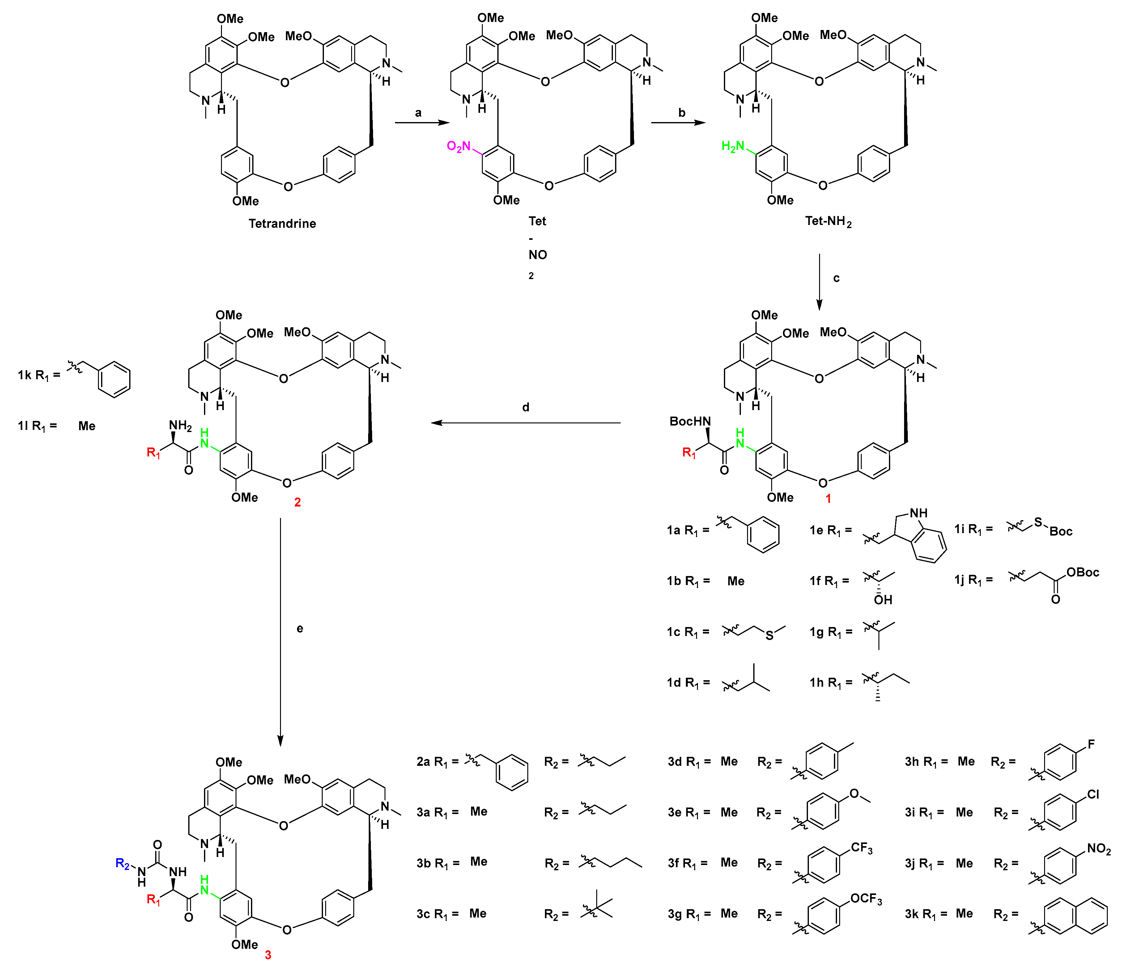
3.2.1. General Procedure for the Preparation of 14-Nitrotetrandrine (Tet-NO2)

Under the protection of an argon atmosphere, concentrated HNO3 (69%, 1.4 mL, 22.4 mmol) was slowly added dropwise into (CH3CO)2O (2.0 mL, 21.3 mmol) in an ice-salt bath and stirred for 10 min. Then, the tetrandrine (0.7 g, 1.12 mmol) dissolved in dry DCM (4 mL) was added dropwise into the reaction mixture and stirred in an ice-salt bath. TLC was used to monitor reaction. Upon completion, the reaction mixture was quenched with saturated aqueous solution of sodium bicarbonate, extracted with DCM (3 × 15 mL), dried over anhydrous sodium sulfate and filtered. The solvent was removed under reduced pressure. The residue was purified by silica gel chromatography from DCM/MeOH (30/1 v/v, 0.5% TEA) to afford the compound Tet-NO2. Light yellow amorphous solid, yield: 93%. Mp: 176–177 °C. 1H-NMR (400 MHz, CDCl3) δ 7.42 (1H, s), 7.37 (1H, dd, J = 2.0, 8.0 Hz), 7.12 (1H, dd, J = 2.4, 8.0 Hz), 6.77 (1H, dd, J = 2.8, 8.4 Hz), 6.54 (1H, s), 6.52 (1H, s), 6.30 (1H, s), 6.28 (1H, d, J = 2.0 Hz), 5.98 (1H, s), 3.98 (3H, s), 3.91 (1H, dd, J = 6.0, 10.8 Hz), 3.75 (3H, s), 3.69–3.63 (1H, m), 3.52–3.49 (2H, m), 3.38 (3H, s), 3.30–3.25 (1H, m), 3.18 (3H, s), 2.96–2.73 (7H, m), 2.63 (3H, s), 2.53 (1H, d, J = 12.8 Hz), 2.35 (1H, m), 2.21 (3H, s). 13C-NMR (CDCl3, 100 MHz) δ 152.3, 152.1, 151.5, 148.7, 148.2, 146.5, 144.2, 143.5, 137.5, 136.4, 133.1, 130.5, 130.4, 128.9, 128.1, 127.6, 121.6, 121.4, 121.3, 119.9, 117.2, 112.5, 108.2, 105.8, 63.6, 61.7, 60.3, 56.3, 55.8, 55.7, 45.3, 43.2, 42.8, 41.5, 37.9, 36.8, 25.3, 21.6. HRMS (ESI) calcd. for C38H42N3O8: 668.2972 [M + H]+, found: 668.2965.

3.2.2. General Procedure for the Preparation of 14-Aminotetrandrine (Tet-NH2)

To a mixture of Tet-NO2 (400.0 mg, 0.60 mmol) and palladium on carbon (5%, 40 mg) were added analytical methanol (20 mL) and hydrazine hydrate (85%, 0.18 mL, 4.80 mmol). The mixture was stirred at 65 °C for about 4 h before it was filtered by celite under reduced pressure. The filter was quenched with saturated sodium chloride solution, extracted with DCM (5 × 20 mL), dried over anhydrous sodium sulfate and filtered. The solvent was removed under reduced pressure. The crude product was recrystallized from cyclohexane and acetone (2/7, v/v) to give Tet-NH2. White amorphous solid, yield: 84%. Mp: 164–166 °C. 1H-NMR (400 MHz, CDCl3) δ 7.28 (1H, d, J = 9.6 Hz), 7.18 (1H, dd, J = 2.0, 8.0 Hz), 6.60 (1H, dd, J = 2.0, 8.4 Hz), 6.50 (1H, s), 6.46 (1H, s), 6.31 (1H, s), 6.29 (1H, s), 6.12 (1H, dd, J = 1.6, 8.0 Hz), 5.87 (1H, s), 3.94 (1H, d, J = 9.2 Hz), 3.87 (3H, s), 3.80 (1H, dd, J = 5.2, 11.2 Hz), 3.73 (3H, s), 3.64 (1H, m), 3.42 (1H, m), 3.35 (3H, s), 3.26 (1H, dd, J = 5.2, 12.4 Hz), 3.11 (3H, s), 2.88 (7H, m), 2.61 (3H, s), 2.42 (3H, s), 2.35 (2H, m). 13C-NMR (CDCl3, 100 MHz) δ 156.6, 151.6, 149.4, 148.7, 148.5, 144.2, 142.0, 140.8, 138.0, 133.2, 132.6, 129.3, 128.0, 127.6, 127.4, 122.6, 122.1, 121.3, 120.9, 120.5, 120.2, 112.3, 105.8, 100.6, 64.2, 61.5, 59.9, 56.1, 55.6, 55.5, 44.9, 43.2, 42.3, 40.8, 40.0, 38.7, 24.6, 20.6. HRMS (ESI) calcd. for C38H44N3O6: 638.3230 [M + H]+, found: 638.3233.

3.2.3. General Procedure for the Preparation of Compounds 1a1k

To a mixture of Tet-NH2 (100 mg, 0.16 mmol), HOBT (8.47 mg, 0.63 mmol), EDCI (27.3 mg, 0.17 mmol) and Boc-l-amino acid (0.17 mmol, 1.1 eq) was added DCM (2.0 mL) under the protection of argon atmosphere, and stirred at room temperature for 1.5 to 3 h. The reaction mixture was quenched with saturated aqueous solution of sodium bicarbonate, extracted with DCM (3 × 10 mL), dried over anhydrous sodium sulfate and filtered. The solvent was removed under reduced pressure, and the residue was purified by silica gel chromatography eluated with DCM/MeOH (40/1 v/v, 0.5% TEA) to afford compounds 1a1k.
14-((R)-2-(N-(tert-butoxycarbonyl)amino)-3-phenylpropanamido)tetrandrine (1a). White to light yellow amorphous solid, yield: 85%. Mp: 136–137 °C. 1H-NMR (600 MHz, CDCl3) δ 12.20 (s, 1H), 7.58 (s, 1H), 7.36–7.29 (m, 5H), 7.26 (t, J = 7.2 Hz, 1H), 7.23 (dd, J = 7.8, 2.4 Hz, 1H), 6.63 (dd, J = 8.4, 2.4 Hz, 1H), 6.57 (s, 1H), 6.49 (s, 1H), 6.32 (s, 1H), 6.16 (dd, J = 8.4, 1.8 Hz, 1H), 5.91 (s, 1H), 5.44 (d, J = 12.0 Hz, 1H), 4.47 (m, 1H), 3.94 (d, J = 9.0 Hz, 4H), 3.83 (dd, J = 10.8, 5.4 Hz, 1H), 3.76 (s, 3H), 3.58 (m, 1H), 3.47 (m, 1H), 3.36 (s, 3H), 3.27 (m, 2H), 3.16–3.08 (m, 5H), 3.01–2.88 (m, 4H), 2.79 (t, J = 12.0 Hz, 1H), 2.70 (dd, J = 16.2, 5.4 Hz, 1H), 2.62 (s, 3H), 2.49 (dd, J = 17.4, 4.2 Hz, 1H), 2.40 (d, J = 17.4 Hz, 4H), 1.46 (s, 9H). 13C-NMR (150 MHz, CDCl3) δ 169.2, 155.8, 155.2, 152.2, 149.4, 148.6, 148.1, 145.6, 144.2, 138.2, 136.6, 134.2, 132.9, 131.5, 129.7, 129.6, 128.6, 128.5, 127.8, 127.2, 127.0, 125.8, 121.4, 121.1, 121.1, 120.8, 120.6, 112.3, 106.9, 105.8, 79.6, 77.3, 77.1, 76.8, 64.2, 61.4, 60.1, 56.3, 56.2, 55.8, 55.6, 53.4, 45.1, 43.2, 42.5, 40.7, 40.0, 39.6, 38.9, 29.7, 28.4, 24.8, 20.7. HRMS (ESI) calcd. for C52H61N4O9: 885.4429 [M + H]+, found 885.4433.
14-((R)-2-(N-(tert-butoxycarbonyl)amino)-propanamido)tetrandrine (1b). White to light yellow amorphous solid, yield: 78%. Mp: 149–150 °C. 1H-NMR (600 MHz, CDCl3) δ 12.35 (s, 1H), 7.84 (s, 1H), 7.32 (dd, J = 7.8, 1.8 Hz, 1H), 7.23 (dd, J = 7.8, 2.4 Hz, 1H), 6.61–6.56 (m, 2H), 6.48 (s, 1H), 6.33 (s, 1H), 6.15 (dd, J = 8.4, 1.8 Hz, 1H), 5.91 (s, 1H), 5.57 (d, J = 7.8 Hz, 1H), 4.31 (m, 1H), 4.02 (d, J = 9.0 Hz, 1H), 3.97 (s, 3H), 3.82 (dd, J = 11.4, 5.4 Hz, 1H), 3.76 (s, 3H), 3.68 (m, 1H), 3.46 (m, 1H), 3.37 (s, 3H), 3.25 (dd, J = 12.0, 5.4 Hz, 1H), 3.16 (dd, J = 13.8, 5.4 Hz, 1H), 3.12 (s, 3H), 3.06 (dd, J = 15.0, 9.6 Hz, 1H), 3.02–2.86 (m, 3H), 2.78 (t, J = 11.8 Hz, 1H), 2.69 (dd, J = 16.2, 4.8 Hz, 1H), 2.62 (s, 3H), 2.55–2.49 (m, 4H), 2.45 (d, J = 15.0 Hz, 1H), 1.51 (d, J = 7.2 Hz, 12H). 13C-NMR (150 MHz, CDCl3) δ 170.8, 156.1, 155.2, 152.2, 149.5, 148.6, 148.4, 145.1, 144.2, 138.3, 134.0, 132.9, 132.2, 129.6, 128.6, 127.8, 127.1, 125.2, 121.5, 121.2, 121.1, 121.0, 120.6, 112.3, 106.2, 105.9, 79.5, 64.2, 61.2, 60.1, 56.2, 55.8, 55.5, 53.4, 50.6, 45.1, 43.1, 42.5, 40.7, 39.9, 38.9, 28.4, 24.8, 20.4. HRMS (ESI) calcd. for C46H57N4O9: 809.4117 [M + H]+, found 809.4120.
14-((R)-2-(N-(tert-butoxycarbonyl)amino)-4-methylthio-butylamido)tetrandrine (1c). White to light yellow amorphous solid, yield: 83%. Mp: 133-134 °C. 1H-NMR (600 MHz, CDCl3) δ 12.41 (s, 1H), 7.75 (s, 1H), 7.32 (dd, J = 8.1, 2.0 Hz, 1H), 7.23 (dd, J = 8.1, 2.5 Hz, 1H), 6.59 (d, J = 8.7 Hz, 2H), 6.48 (s, 1H), 6.33 (s, 1H), 6.15 (dd, J = 8.4, 2.0 Hz, 1H), 5.91 (s, 1H), 5.50 (d, J = 8.2 Hz, 1H), 4.38 (m, 1H), 4.03–3.99 (m, 1H), 3.96 (s, 3H), 3.84 (dd, J = 11.1, 5.6 Hz, 1H), 3.76 (s, 3H), 3.67 (m, 1H), 3.52–3.44 (m, 1H), 3.37 (s, 3H), 3.25 (m, 2H), 3.11 (s, 3H), 3.06 (dd, J = 14.8, 9.5 Hz, 1H), 3.03–2.88 (m, 3H), 2.78 (t, J = 11.8 Hz, 1H), 2.73–2.63 (m, 3H), 2.62 (s, 3H), 2.55–2.50 (m, 4H), 2.44 (d, J = 14.8 Hz, 1H), 2.3–2.16 (m, 1H), 2.15 (s, 3H), 2.05–2.00 (m, 1H), 1.47 (s, 9H). 13C-NMR (150 MHz, CDCl3) δ 169.6, 156.0, 155.5, 152.2, 149.4, 148.7, 148.31, 145., 4144.3, 138.3, 134.0, 132.9, 131.9, 131.9, 129.7, 128.5, 127.2, 125.7, 121.5, 121.1, 121.0, 120.7, 112.3, 106.6, 105.9, 79.7, 77.3, 77.1, 76.9, 64.2, 61.1, 60.1, 56.3, 55.8, 55.6, 54.3, 45.0, 43.1, 42.4, 40.8, 39.8, 38.8, 34.1, 30.2, 29.7, 28.4, 24.7, 20.6, 15.8. HRMS (ESI) calcd. for C48H61N4O9S: 869.4152 [M + H]+, found 869.4154.
14-((R)-3-methyl-2-(N-(tert-butoxycarbonyl)amino)-amylamido)tetrandrine (1d). White to light yellow amorphous solid, yield: 81%. Mp: 145–146 °C. 1H-NMR (600 MHz, CDCl3) δ 12.16 (s, 1H), 7.72 (s, 1H), 7.33 (dd, J = 8.4, 2.4 Hz, 1H), 7.24 (dd, J = 7.8, 2.4 Hz, 1H), 6.60 (d, J = 7.2 Hz, 2H), 6.49 (s, 1H), 6.34 (s, 1H), 6.15 (dd, J = 8.4, 1.8 Hz, 1H), 5.91 (s, 1H), 5.29 (d, J = 9.0 Hz, 1H), 4.29 (m, 1H), 4.01 (d, J = 9.6 Hz, 1H), 3.96 (s, 3H), 3.85 (dd, J = 11.4, 5.4 Hz, 1H), 3.77 (s, 3H), 3.70 (td, J = 13.8, 13.2, 4.8 Hz, 1H), 3.53–3.46 (m, 1H), 3.38 (s, 3H), 3.29 (dd, J = 12.0, 5.4 Hz, 1H), 3.22 (dd, J = 14.4, 6.0 Hz, 1H), 3.12 (s, 3H), 3.08 (dd, J = 15.0, 9.6 Hz, 1H), 3.08–2.88 (m, 3H), 2.79 (t, J = 12.0 Hz, 1H), 2.72 (dd, J = 15.6, 5.4 Hz, 1H), 2.63 (s, 3H), 2.53 (s, 4H), 2.43 (d, J = 15.0 Hz, 1H), 1.84 (m, 1H), 1.70 (t, J = 7.2 Hz, 2H), 1.46 (s, 9H), 1.07 (d, J = 6.6 Hz, 3H), 1.02 (d, J = 6.6 Hz, 3H). 13C-NMR (150 MHz, CDCl3) δ 171.1, 156.0, 155.4, 152.2, 149.4, 148.7, 148.3, 145.3, 144.3, 138.3, 133.9, 132.9, 131.9, 129.7, 128.3, 127.3, 125.8, 121.5, 121.3, 121.1, 121.0, 120.6, 112.3, 106.8, 105.9, 79.4, 64.2, 61.4, 60.1, 56.3, 55.8, 55.6, 53.8, 45.0, 43.4, 43.2, 42.3, 40.9, 39.8, 38.9, 29.7, 28.4, 24.8, 24.7, 23.3, 22.7, 20.7. HRMS (ESI) calcd. for C49H63N4O9: 851.4590 [M + H]+, found 851.4590.
14-((R)-3-(indolyl-3)-2-(N-(tert-butoxycarbonyl)amino)-propanamido)tetrandrine (1e). White to light yellow amorphous solid, yield: 84%. Mp: 152–153 °C. 1H-NMR (600 MHz, CDCl3) δ 12.06 (s, 1H), 8.16 (s, 1H), 7.72 (d, J = 7.8 Hz, 1H), 7.50 (s, 1H), 7.35 (d, J = 8.4 Hz, 1H), 7.32 (dd, J = 8.4, 2.1 Hz, 1H), 7.22 (dd, J = 8.4, 2.4 Hz, 1H), 7.21–7.18 (m, 1H), 7.17–7.11 (m, 2H), 6.64–6.61 (m, 1H), 6.55 (s, 1H), 6.48 (s, 1H), 6.30 (s, 1H), 6.16 (dd, J = 8.4, 2.4 Hz, 1H), 5.90 (s, 1H), 5.51 (d, J = 8.4 Hz, 1H), 4.59–4.52 (m, 1H), 3.92–3.81 (m, 5H), 3.75 (s, 3H), 3.46 (m, 3H), 3.33 (d, J = 18.6 Hz, 4H), 3.28 (dd, J = 12.0, 5.4 Hz, 1H), 3.11 (s, 3H), 3.02 (dd, J = 13.8, 6.0 Hz, 1H), 2.97–2.83 (m, 4H), 2.79 (t, J = 11.4 Hz, 1H), 2.71 (dd, J = 15.6, 5.4 Hz, 1H), 2.62 (s, 3H), 2.44–2.34 (m, 2H), 2.14 (s, 3H), 1.48 (s, 9H). 13C-NMR (150 MHz, CDCl3) δ 169.8, 155.8, 155.3, 155.3, 152.1, 149.3, 148.0, 145.5, 144.3, 138.1, 136.2, 132.9, 131.5, 129.7, 127.9, 127.3, 125.9, 125.9, 122.8, 122.1, 121.4, 121.2, 120.8, 120.5, 119.6, 119.1, 112.3, 111.1, 110.9, 110.8, 107.2, 105.8, 79.5, 64.2, 61.3, 60.1, 56.3, 55.8, 55.7, 55.6, 45.0, 43.0, 42.3, 40.3, 39.6, 38.9, 29.7, 29.5, 29.5, 28.4, 24.7, 20.7. HRMS (ESI) calcd. for C54H62N5O9: 924.4537 [M + H]+, found 924.4542.
14-((R)-3-hydroxy-2-(N-(tert-butoxycarbonyl)amino)-butylamido)tetrandrine (1f). White to light yellow amorphous solid, yield: 79%. Mp: 163–165 °C. 1H-NMR (600 MHz, CDCl3) δ 12.46 (s, 1H), 7.81 (s, 1H), 7.32 (dd, J = 8.4, 2.4 Hz, 1H), 7.23 (dd, J = 7.8, 2.4 Hz, 1H), 6.61 (s, 1H), 6.59 (dd, J = 8.4, 2.4 Hz, 1H), 6.49 (s, 1H), 6.33 (s, 1H), 6.15 (dd, J = 8.4, 2.4 Hz, 1H), 5.90 (s, 1H), 5.60 (d, J = 9.0 Hz, 1H), 4.25–4.19 (m, 1H), 4.15–4.11 (m, 1H), 4.01 (d, J = 9.0 Hz, 1H), 3.96 (s, 3H), 3.83 (dd, J = 11.4, 5.4 Hz, 1H), 3.76 (s, 3H), 3.66 (m, 1H), 3.50–3.45 (m, 1H), 3.38 (s, 3H), 3.26 (dd, J = 12.6, 5.4 Hz, 1H), 3.21 (dd, J = 12.6, 6.0 Hz, 1H), 3.11 (s, 4H), 3.03–2.86 (m, 4H), 2.79 (t, J = 12.0 Hz, 1H), 2.71 (dd, J = 16.2, 5.4 Hz, 1H), 2.62 (s, 3H), 2.51 (s, 4H), 2.45 (d, J = 14.8 Hz, 1H), 1.48 (s, 9H), 1.34 (d, J = 6.0 Hz, 3H). 13C-NMR (150 MHz, CDCl3) δ 169.5, 156.2, 156.1, 152.2, 149.4, 148.6, 148.3, 145.5, 144.3, 138.3, 134.0, 132.9, 131.7, 129.7, 128.5, 127.7, 127.3, 125.8, 121.6, 121.2, 121.0, 120.6, 112.3, 106.7, 105.9, 79.9, 77.3, 77.0, 76.8, 69.3, 64.2, 61.3, 60.1, 59.8, 56.3, 55.8, 55.6, 45.1, 43.3, 42.4, 40.8, 40.0, 38.8, 29.7, 28.4, 24.8, 20.8, 19.7. HRMS (ESI) calcd. for C47H59N4O10: 839.4230 [M + H]+, found 839.4226.
14-((R)-3-methyl-2-(N-(tert-butoxycarbonyl)amino)-butylamido)tetrandrine (1g). White to light yellow amorphous solid, yield: 84%. Mp: 143–144 °C. 1H-NMR (600 MHz, CDCl3) δ 12.28 (s, 1H), 7.78 (s, 1H), 7.32 (dd, J = 8.1, 1.9 Hz, 1H), 7.24 (dd, J = 7.8, 2.4 Hz, 1H), 6.60 (d, J = 9.8 Hz, 2H), 6.48 (s, 1H), 6.34 (s, 1H), 6.17–6.13 (m, 1H), 5.91 (s, 1H), 5.42 (d, J = 9.0 Hz, 1H), 4.09 (dd, J = 9.0, 6.0 Hz, 1H), 4.03 (d, J = 9.3 Hz, 1H), 3.96 (s, 3H), 3.82 (dd, J = 11.1, 5.5 Hz, 1H), 3.77 (s, 3H), 3.69 (m, 1H), 3.51–3.44 (m, 1H), 3.38 (s, 3H), 3.26 (dd, J = 12.3, 5.5 Hz, 1H), 3.21 (dd, J = 14.0, 5.9 Hz, 1H), 3.12 (s, 3H), 3.07 (dd, J = 14.8, 9.5 Hz, 1H), 3.03–2.85 (m, 3H), 2.79 (t, J = 11.8 Hz, 1H), 2.72–2.67 (m, 1H), 2.62 (s, 3H), 2.57–2.49 (m, 4H), 2.44 (d, J = 14.8 Hz, 1H), 2.12 (m, 1H), 1.47 (s, 9H), 1.11 (d, J = 6.8 Hz, 3H), 1.03 (d, J = 6.7 Hz, 3H). 13C-NMR (150 MHz, CDCl3) δ 170.1, 156.1, 156.0, 152.2, 149.5, 148.6, 148.3, 145.2, 144.2, 138.3, 134.1, 132.9, 132.0, 129.7, 128.6, 127.2, 125.6, 121.6, 121.2, 121.2, 121.0, 120.6, 112.3, 106.4, 105.9, 79.3, 64.2, 61.3, 60.3, 60.1, 56.3, 55.8, 55.5, 53.4, 45.1, 43.2, 42.5, 40.9, 39.8, 38.9, 32.9, 28.4, 24.8, 20.6, 19.9, 17.8. HRMS (ESI) calcd. for C48H61N4O9: 837.4421 [M + H]+, found 837.4433.
14-((2R,3R)-3-methyl-2-(N-(tert-butoxycarbonyl)amino)-amylamido)tetrandrine (1h). White to light yellow amorphous solid, yield: 79%. Mp: 158–159 °C. 1H-NMR (600 MHz, CDCl3) δ 12.25 (s, 1H), 7.79 (s, 1H), 7.33 (dd, J = 7.8, 1.8 Hz, 1H), 7.24 (dd, J = 7.8, 2.4 Hz, 1H), 6.60 (d, J = 8.4 Hz, 2H), 6.49 (s, 1H), 6.34 (s, 1H), 6.14 (dd, J = 8.4, 1.8 Hz, 1H), 5.91 (s, 1H), 5.38 (d, J = 9.0 Hz, 1H), 4.12–4.07 (m, 1H), 4.03 (d, J = 9.6 Hz, 1H), 3.96 (s, 3H), 3.85 (dd, J = 11.4, 5.4 Hz, 1H), 3.77 (s, 3H), 3.69 (m, 1H), 3.52–3.46 (m, 1H), 3.38 (s, 3H), 3.28 (dd, J = 12.0, 5.4 Hz, 1H), 3.21 (dd, J = 13.8, 6.0 Hz, 1H), 3.12 (s, 3H), 3.08 (dd, J = 15.0, 9.6 Hz, 1H), 3.03–2.89 (m, 3H), 2.78 (t, J = 12.0 Hz, 1H), 2.72 (dd, J = 15.6, 5.4 Hz, 1H), 2.63 (s, 3H), 2.55–2.50 (m, 4H), 2.44 (d, J = 15.0 Hz, 1H), 1.87 (m, 1H), 1.66 (m, 1H), 1.46 (s, 9H), 1.26–1.20 (m, 1H), 1.09 (d, J = 6.6 Hz, 3H), 0.97 (t, J = 7.2 Hz, 3H). 13C-NMR (150 MHz, CDCl3) δ 170.3, 156.1, 155.9, 152.2, 149.4, 148.7, 148.3, 145.2, 144.3, 138.3, 133.9, 132.9, 132.0, 129.7, 128.4, 127.4, 127.3, 125.6, 121.6, 121.3, 121.2, 121.0, 120.6, 112.3, 106.5, 105.9, 79.3, 77.3, 77.1, 76.8, 64.1, 61.3, 60.1, 59.9, 56.3, 55.8, 55.5, 45.0, 43.2, 42.3, 40.9, 39.9, 39.3, 38.9, 29.7, 28.4, 24.5, 20.6, 16.0, 11.6. HRMS (ESI) calcd. for C49H63N4O9 [M + H]+: 851.4583, found 851.4590.
14-((R)-3-(S-(tert-butoxycarbonyl)sulfydryl)-propanamido)tetrandrine (1i). White to light yellow amorphous solid, yield: 82%. Mp: 147–149 °C. 1H-NMR (600 MHz, CDCl3) δ 12.48 (s, 1H), 7.75 (s, 1H), 7.32 (dd, J = 8.4, 2.4 Hz, 1H), 7.23 (dd, J = 7.8, 2.4 Hz, 1H), 6.61–6.57 (m, 2H), 6.49 (s, 1H), 6.33 (s, 1H), 6.16 (dd, J = 8.4, 2.4 Hz, 1H), 5.91 (s, 1H), 5.58 (d, J = 8.4 Hz, 1H), 4.44 (m, 1H), 4.01 (d, J = 9.6 Hz, 1H), 3.96 (s, 3H), 3.82 (dd, J = 10.8, 5.4 Hz, 1H), 3.76 (s, 3H), 3.64 (m, 1H), 3.51–3.44 (m, 1H), 3.38 (s, 4H), 3.27–3.19 (m, 3H), 3.12 (s, 3H), 3.05 (dd, J = 15.0, 9.6 Hz, 1H), 3.02–2.86 (m, 4H), 2.79 (t, J = 12.0 Hz, 1H), 2.70 (dd, J = 16.2, 5.4 Hz, 1H), 2.62 (s, 3H), 2.55 (s, 3H), 2.51 (dd, J = 16.8, 4.8 Hz, 1H), 2.44 (d, J = 14.4 Hz, 1H), 1.50 (s, 9H), 1.47 (s, 9H). 13C-NMR (150 MHz, CDCl3) δ 168.5, 168.1, 156.0, 155.1, 152.18, 149.44, 148.61, 148.24, 145.45, 144.20, 138.25, 134.11, 132.92, 131.73, 129.66, 128.58, 127.77, 127.3, 125.7, 121.5, 121.2, 121.0, 120.9, 120.7, 112.3, 106.9, 105.8, 85.1, 79.8, 64.2, 61.3, 60.1, 56.3, 55.8, 55.6, 54.9, 53.5, 43.1, 42.5, 40.8, 39.9, 38.9, 34.3, 28.4, 28.2, 24.9, 20.7. HRMS (ESI) calcd. for C51H65N4O11S: 941.4366 [M + H]+, found 941.4365.
14-((R)-4-(C-(tert-butoxycarbonyl)carboxyl)-2-(N-(tert-butoxycarbonyl)amino)-butylamido)tetrandrine (1j) White to light yellow amorphous solid, yield: 87%. Mp: 131–132 °C. 1H-NMR (600 MHz, CDCl3) δ 12.19 (s, 1H), 7.98 (s, 1H), 7.31 (dd, J = 7.8, 1.8 Hz, 1H), 7.23 (dd, J = 7.8, 2.4 Hz, 1H), 6.59 (s, 1H), 6.58–6.55 (m, 1H), 6.49 (s, 1H), 6.33 (s, 1H), 6.13 (dd, J = 8.4, 1.8 Hz, 1H), 5.90 (s, 1H), 5.29 (d, J = 8.4 Hz, 1H), 4.25 (m, 1H), 3.99 (d, J = 9.0 Hz, 1H), 3.96 (s, 3H), 3.83 (dd, J = 11.2, 5.4 Hz, 1H), 3.77 (s, 3H), 3.62 (td, J = 12.6, 3.6 Hz, 1H), 3.53–3.47 (m, 1H), 3.38 (s, 3H), 3.29 (dd, J = 12.0, 4.8 Hz, 1H), 3.11 (s, 3H), 3.09–2.88 (m, 5H), 2.78 (t, J = 12.0 Hz, 1H), 2.72 (dd, J = 15.0, 4.8 Hz, 1H), 2.63 (s, 3H), 2.51 (s, 5H), 2.42 (dd, J = 23.6, 12.0 Hz, 2H), 2.32 (m, 1H), 2.07 (m, 1H), 1.51 (s, 9H), 1.45 (s, 9H). 13C-NMR (150 MHz, CDCl3) δ 171.6, 170.4, 156.5, 155.7, 152.2, 149.5, 148.7, 148.4, 144.6, 144.3, 138.3, 133.6, 132.9, 132.9, 129.6, 128.4, 127.1, 124.7, 121.6, 121.4, 120.9, 120.5, 112.3, 106.0, 105.9, 82.1, 79.6, 77.3, 77.1, 76.8, 64.3, 61.1, 60.1, 56.2, 55.8, 55.6, 54.2, 53.4, 45.1, 43.1, 42.4, 40.6, 40.2, 38.9, 34.0, 28.9, 28.4, 28.0, 24.7, 20.7. HRMS (ESI) calcd. for C53H67N4O13: 967.4699 [M + H]+, found 967.4671.

3.2.4. General Procedure for the Preparation of Compounds 1k and 1l

Trifluoroacetic acid (0.1 mL) was slowly added dropwise to a solution of 1k (160 mg, 0.16 mmol) or 1l (130 mg, 0.16 mmol) in DCM (2 mL) at 0 °C. After 10 min, the reaction mixture was warmed up to room temperature, and stirred for 0.5 to 1.5 h before the reaction finished. The reaction mixture was quenched with saturated aqueous solution of sodium bicarbonate, extracted with DCM (3 × 10 mL), dried over anhydrous sodium sulfate and filtered. The solvent was removed under reduced pressure. The residue was purified by silica gel chromatography from DCM/MeOH (30/1 v/v, 0.5 % TEA) to afford the pure compounds 1k and 1l.
14-((R)-2-amino-3-phenyl-propanamido)tetrandrine (1k). White amorphous solid, yield: 76%. Mp: 140–141 °C. 1H-NMR (600 MHz, CDCl3) δ 11.83 (s, 1H), 7.74 (s, 1H), 7.37–7.29 (m, 5H), 7.28–7.25 (m, 1H), 7.23 (dd, J = 8.4, 2.4 Hz, 1H), 6.63 (dd, J = 8.4, 2.4 Hz, 1H), 6.58 (s, 1H), 6.49 (s, 1H), 6.32 (s, 1H), 6.16 (dd, J = 8.4, 2.4 Hz, 1H), 5.92 (s, 1H), 4.24 (m, 1H), 4.14 (m, 1H), 3.98 (s, 3H), 3.93 (d, J = 9.6 Hz, 1H), 3.84 (dd, J = 11.2, 5.4 Hz, 1H), 3.76 (s, 3H), 3.65 (dd, J = 7.8, 6.0 Hz, 1H), 3.56–3.42 (m, 2H), 3.37 (s, 3H), 3.27 (dd, J = 12.6, 6.0 Hz, 1H), 3.21 (dd, J = 13.8, 6.0 Hz, 1H), 3.12 (s, 3H), 3.01–2.87 (m, 6H), 2.79 (t, J = 12.0 Hz, 1H), 2.71 (dd, J = 16.2, 5.4 Hz, 1H), 2.63 (s, 3H), 2.51–2.45 (m, 1H), 2.41 (d, J = 14.4 Hz, 1H), 2.37 (s, 3H). 13C-NMR (150 MHz, CDCl3) δ 173.4, 155.7, 152.1, 149.4, 148.6, 148.0, 145.4, 144.2, 138.3, 137.9, 134.1, 132.9, 131.73, 129.7, 129.4, 128.7, 128.5, 127.7, 127.2, 126.8, 125.8, 121.5, 121.4, 121.2, 120.8, 120.4, 112.4, 107.1, 105.8, 64.2, 61.44, 60.2, 58.2, 56.3, 55.8, 55.6, 45.1, 43.9, 42.5, 42.3, 41.0, 39.9, 38.8, 24.9, 21.0. HRMS (ESI) calcd. for C47H53N4O7: 785.3904 [M + H]+, found 785.3909.
14-((R)-2-amino-propanamido)tetrandrine. (1l). White to light yellow amorphous solid, yield: 83%. Mp: 137–139 °C. 1H-NMR (600 MHz, CDCl3) δ 11.96 (s, 1H), 7.88 (s, 1H), 7.32 (dd, J = 8.2, 2.2 Hz, 1H), 7.23 (dd, J = 7.8, 2.4 Hz, 1H), 6.60 (d, J = 6.0 Hz, 2H), 6.49 (s, 1H), 6.33 (s, 1H), 6.15 (dd, J = 8.4, 2.4 Hz, 1H), 5.91 (s, 1H), 4.02 (s, 1H), 3.97 (s, 3H), 3.83 (dd, J = 10.8, 5.4 Hz, 1H), 3.76 (s, 3H), 3.63 (m, 1H), 3.53 (m, 1H), 3.49–3.43 (m, 1H), 3.38 (s, 3H), 3.26 (dd, J = 12.6, 5.4 Hz, 1H), 3.11 (s, 3H), 3.06–2.87 (m, 7H), 2.78 (t, J = 11.8 Hz, 1H), 2.73–2.68 (m, 1H), 2.62 (s, 3H), 2.51 (s, 4H), 2.45 (d, J = 14.8 Hz, 1H), 1.46 (d, J = 7.2 Hz, 3H). 13C-NMR (150 MHz, CDCl3) δ 174.9, 156.0, 152.2, 149.5, 148.6, 148.3, 145.0, 144.2, 138.4, 133.9, 132.9, 132.3, 129.7, 128.5, 127.7, 127.1, 125.2, 121.6, 121.4, 121.0, 120.6, 112.3, 106.5, 105.9, 64.2, 61.2, 60.1, 56.2, 55.8, 55.6, 52.0, 45.0, 43.6, 42.4, 40.9, 40.0, 38.8, 29.7, 24.8, 22.0, 20.8. HRMS (ESI) calcd. for C41H49N4O7: 704.3589 [M + H]+, found 704.3596.

3.2.5. General Procedure for the Preparation of Compounds 2a3k

Compound 1l (100 mg, 0.14 mmol) was dissolved in DCM (2.0 mL), triethylamine (98 %, 4.0 µL, 0.03 mmol) and isocyanate (98%, 0.16 mmol) were added into the solution in turn, and the reaction mixture was stirred for 0.5 to 1.5 h. Upon completion, the solvent was removed under reduced pressure. The residue was purified by silica gel chromatography from DCM/MeOH (50/1 v/v, 0.5 % TEA) to afford the pure compounds 2a, 2b and 2c. The compounds 3a-3k were obtained using the same method.
14-((R)-2-(3-propylureido)-3-phenylpropanamido)tetrandrine (2a). White amorphous solid, yield: 91%. Mp: 156–157 °C. 1H-NMR (600 MHz, CDCl3) δ 12.39 (s, 1H), 7.52 (s, 1H), 7.33 (d, J = 4.8 Hz, 5H), 7.28–7.19 (m, 2H), 6.63 (dd, J = 8.4, 2.4 Hz, 1H), 6.56 (s, 1H), 6.49 (s, 1H), 6.32 (s, 1H), 6.17 (dd, J = 8.4, 1.8 Hz, 1H), 5.91 (s, 1H), 5.83 (d, J = 7.4 Hz, 1H), 4.92 (s, 1H), 4.71 (m, 1H), 3.94 (s, 3H), 3.92 (d, J = 9.6 Hz, 1H), 3.84 (dd, J = 11.4, 5.4 Hz, 1H), 3.76 (s, 3H), 3.67–3.62 (m, 1H), 3.48 (dd, J = 13.8, 8.4 Hz, 1H), 3.37 (s, 3H), 3.26 (dd, J = 13.2, 7.2 Hz, 2H), 3.20 (dd, J = 13.8, 6.0 Hz, 1H), 3.11 (s, 3H), 3.11–2.87 (m, 7H), 2.80 (t, J = 12.0 Hz, 1H), 2.71 (dd, J = 15.6, 5.4 Hz, 1H), 2.63 (s, 3H), 2.51 (dd, J = 17.4, 4.8 Hz, 1H), 2.42–2.35 (m, 4H), 1.35 (m, 2H), 0.83 (t, J = 7.2 Hz, 3H). 13C-NMR (150 MHz, CDCl3) δ 170.5, 157.5, 155.8, 152.1, 149.3 148.64=, 148.1, 145.7, 144.2, 138.1, 133.0, 131.4, 129.7, 129.7, 128.4, 127.4, 126.8, 126.3, 121.4, 121.2, 120.9, 120.8, 120.7, 112.3, 106.9, 105.9, 64.2, 61.4, 60.1, 56.3, 55.7, 55.7, 55.6, 45.1, 43.1, 42.4, 42.1, 40.7, 40.7, 39.5, 39.0, 24.7, 23.4, 20.6, 11.3. HRMS (ESI) calcd. for C51H60N5O8: 870.4429 [M + H]+, found 870.4436.
14-((R)-2-(3-propylureido)propanamido)tetrandrine (3a). White amorphous solid, yield: 94%. Mp: 151–152 °C. 1H-NMR (600 MHz, CDCl3) δ 12.62 (s, 1H), 7.76 (s, 1H), 7.32 (dd, J = 8.4, 2.4 Hz, 1H), 7.23 (dd, J = 7.8, 2.4 Hz, 1H), 6.60 (s, 1H), 6.58 (dd, J = 8.4, 2.4 Hz, 1H), 6.48 (s, 1H), 6.33 (s, 1H), 6.15 (dd, J = 8.4, 2.4 Hz, 1H), 6.06–5.99 (m, 1H), 5.90 (s, 1H), 5.15–5.06 (m, 1H), 4.55 (m, 1H), 4.01 (d, J = 9.6 Hz, 1H), 3.96 (s, 3H), 3.81 (dd, J = 11.4, 5.4 Hz, 1H), 3.76 (s, 3H), 3.68 (m, 1H), 3.45 (m, 1H), 3.37 (s, 3H), 3.23 (m, 2H), 3.11 (s, 3H), 3.05 (dd, J = 15.0, 9.6 Hz, 2H), 3.01–2.86 (m, 4H), 2.78 (t, J = 12.0 Hz, 1H), 2.72–2.65 (m, 3H), 2.61 (s, 3H), 2.53 (s, 3H), 2.53–2.49 (m, 1H), 2.44 (d, J = 15.0 Hz, 1H), 1.55 (d, J = 7.2 Hz, 3H), 1.13 (t, J = 7.2 Hz, 3H). 13C-NMR (150 MHz, CDCl3) δ 172.3, 157.8, 157.7, 156.0, 152.2, 149.4, 148.6, 148.4, 145.4, 144.2, 138.2, 134.2, 133.0, 132.0, 129.7, 128.6, 127.9, 127.3, 125.8, 125.8, 121.5, 121.3, 121.0, 121.0, 120.6, 112.3, 106.3, 105.9, 77.3, 77.1, 76.8, 64.2, 61.2, 60.1, 56.3, 55.7, 55.6, 50.2, 46.1, 45.1, 43.0, 42.5, 42.1, 40.6, 39.8, 38.8, 24.9, 23.4, 22.7, 21.1, 20.5. HRMS (ESI) calcd. for C45H56N5O8: 794.4123 [M + H]+, found 794.4117.
14-((R)-2-(3-butylureido)propanamido)tetrandrine (3b). Light yellow amorphous solid, yield: 92%. Mp: 160–161 °C. 1H-NMR (600 MHz, CDCl3) δ 12.60 (s, 1H), 7.78 (s, 1H), 7.32 (dd, J = 8.4, 2.4 Hz, 1H), 7.23 (dd, J = 7.8, 2.4 Hz, 1H), 6.61 (s, 1H), 6.59 (dd, J = 8.4, 2.4 Hz, 1H), 6.49 (s, 1H), 6.33 (s, 1H), 6.15 (dd, J = 8.4, 2.4 Hz, 1H), 5.91 (s, 1H), 5.89 (d, J = 7.2 Hz, 1H), 4.89 (s, 1H), 4.55 (m, 1H), 4.02 (d, J = 9.0 Hz, 1H), 3.96 (s, 3H), 3.82 (dd, J = 11.4, 5.4 Hz, 1H), 3.76 (s, 3H), 3.71–3.66 (m, 1H), 3.47 (m, 1H), 3.38 (s, 3H), 3.27–3.20 (m, 2H), 3.12 (s, 4H), 3.09–2.86 (m, 5H), 2.79 (t, J = 12.0 Hz, 1H), 2.72–2.67 (m, 1H), 2.62 (s, 3H), 2.54 (s, 4H), 2.45 (d, J = 15.0 Hz, 1H), 1.55 (d, J = 7.2 Hz, 3H), 1.34–1.26 (m, 4H), 0.87 (t, J = 7.2 Hz, 3H). 13C-NMR (150 MHz, CDCl3) δ 172.1, 157.6, 156.1, 152.2, 149.4, 148.6, 148.4, 145.3, 144.2, 138.2, 134.2, 133.0, 132.03, 129.7, 128.6, 127.8, 127.3, 125.7, 121.5, 121.4, 121.0, 121.0, 120.6, 112.3, 106.3, 105.9, 64.2, 61.2, 60.1, 56.3, 55.7, 55.6, 50.2, 45.1, 43.1, 42.5, 40.7, 40.2, 39.8, 38.9, 32.3, 24.8, 21.1, 20.6, 20.1, 13.8. HRMS (ESI) calcd. for C46H58N5O8: 808.4280 [M + H]+, found 808.4272.
14-((R)-2-(3-tert-butylureido)propanamido)tetrandrine (3c). White to light yellow amorphous solid, yield: 90%. Mp: 215–216 °C. 1H-NMR (600 MHz, CDCl3) δ 12.40 (s, 1H), 7.82 (s, 1H), 7.33 (dd, J = 8.4, 2.4 Hz, 1H), 7.24 (dd, J = 7.8, 2.4 Hz, 1H), 6.60 (s, 1H), 6.60–6.58 (m, 1H), 6.49 (s, 1H), 6.34 (s, 1H), 6.15 (dd, J = 8.4, 2.4 Hz, 1H), 5.91 (s, 1H), 5.47 (d, J = 7.4 Hz, 1H), 4.51 (m, 1H), 4.45 (s, 1H), 4.01 (d, J = 9.0 Hz, 1H), 3.97 (s, 3H), 3.83 (dd, J = 10.8, 5.4 Hz, 1H), 3.77 (s, 3H), 3.69 (m, 1H), 3.51–3.45 (m, 1H), 3.37 (s, 3H), 3.30–3.25 (m, 1H), 3.21 (dd, J = 13.8, 6.0 Hz, 1H), 3.12 (s, 3H), 3.06 (dd, J = 15.0, 9.6 Hz, 1H), 2.95 (m, 3H), 2.79 (t, J = 12.0 Hz, 1H), 2.71 (dd, J = 15.6, 6.0 Hz, 1H), 2.63 (s, 3H), 2.53 (s, 4H), 2.45 (d, J = 15.0Hz, 1H), 1.52 (d, J = 7.2 Hz, 3H), 1.33 (s, 9H). 13C-NMR (150 MHz, CDCl3) δ 171.9, 156.7, 156.1, 152.2, 149.4, 148.6, 148.3, 145.2, 144.2, 138.2, 134.1, 132.9, 132.2, 129.7, 128.6, 127.8, 127.3, 125.5, 121.5, 121.2, 121.1, 121.0, 120.6, 112.3, 106.4, 105.9, 64.2, 61.2, 60.1, 56.2, 55.7, 55.5, 50.3, 49.9, 45.1, 43.1, 42.5, 40.7, 39.8, 38.9, 29.5, 24.8, 21.0, 20.5. HRMS (ESI) calcd. for C46H58N5O8: 808.4280 [M + H]+, found 808.4272.
14-((R)-2-(3-(p-tolyl)ureido)propanamido)tetrandrine (3d). White amorphous solid, yield: 90%. Mp: 165–167 °C. 1H-NMR (600 MHz, CDCl3) δ 12.71 (s, 1H), 7.73 (s, 1H), 7.33 (dd, J = 8.4 2.4 Hz, 1H), 7.27 (s, 1H), 7.23 (dd, J = 7.8, 2.4 Hz, 1H), 7.10–7.00 (m, 4H), 6.61 (s, 1H), 6.60–6.53 (m, 2H), 6.49 (s, 1H), 6.33 (s, 1H), 6.15 (dd, J = 8.4, 2.4 Hz, 1H), 5.92 (s, 1H), 4.66 (m, 1H), 4.03 (d, J = 9.6 Hz, 1H), 3.84 (s, 4H), 3.77 (s, 3H), 3.71 (m, 1H), 3.48 (m, 1H), 3.38 (s, 3H), 3.25 (m, 2H), 3.12 (s, 4H), 3.02–2.87 (m, 3H), 2.79 (t, J = 11.4 Hz, 1H), 2.73–2.68 (m, 1H), 2.62 (s, 3H), 2.55 (s, 3H), 2.52 (dd, J = 16.8, 4.8 Hz, 1H), 2.46 (d, J = 15.0 Hz, 1H), 2.30 (s, 3H), 1.61 (d, J = 7.2 Hz, 3H). 13C-NMR (150 MHz, CDCl3) δ 172.0, 156.0, 155.4, 152.2, 149.4, 148.6, 148.5, 145.6, 144.2, 138.2, 136.2, 134.2, 133.0, 131.8, 129.7, 129.6, 128.7, 127.9, 127.3, 125.9, 121.5, 121.3, 121.0, 120.6, 120.5, 112.3, 106.5, 105.9, 64.2, 61.3, 60.1, 56.2, 55.7, 55.6, 50.2, 46.1, 45.1, 43.1, 42.5, 40.7, 39.8, 38.9, 24.9, 21.0, 20.8, 20.5. HRMS (ESI) calcd. for C49H56N5O8: 842.4116 [M + H]+, found 842.4123.
14-((R)-2-(3-(4-methoxyphenyl)ureido)propanamido)tetrandrine (3e). White amorphous solid, yield: 90%. Mp: 183–184 °C. 1H-NMR (600 MHz, CDCl3) δ 12.69 (s, 1H), 7.71 (s, 1H), 7.32 (dd, J = 8.4, 2.4 Hz, 1H), 7.23 (dd, J = 7.8, 2.4 Hz, 1H), 7.10 (t, J = 6.0 Hz, 3H), 6.82–6.78 (m, 2H), 6.60 (s, 1H), 6.57 (dd, J = 8.4, 2.4 Hz, 1H), 6.49 (s, 1H), 6.41 (d, J = 7.8 Hz, 1H), 6.33 (s, 1H), 6.15 (dd, J = 8.4, 2.0 Hz, 1H), 5.91 (s, 1H), 4.64 (m, 1H), 4.02 (d, J = 9.6 Hz, 1H), 3.85 (s, 3H), 3.84–3.81 (m, 1H), 3.80 (s, 3H), 3.76 (s, 3H), 3.73–3.67 (m, 1H), 3.46 (m, 1H), 3.38 (s, 3H), 3.24 (m, 2H), 3.12 (s, 3H), 3.08 (dd, J = 15.0, 9.6 Hz, 1H), 3.01–2.86 (m, 3H), 2.79 (t, J = 11.4 Hz, 1H), 2.72–2.66 (m, 1H), 2.62 (s, 3H), 2.54 (s, 3H), 2.53–2.48 (m, 1H), 2.45 (d, J = 15.0 Hz, 1H), 1.60 (d, J = 6.6 Hz, 3H). 13C-NMR (150 MHz, CDCl3) δ 171.9, 156.0, 155.8, 152.3, 149.4, 148.6, 148.5, 145.5, 144.2, 138.2, 134.2, 133.0, 131.8, 131.4, 129.7, 128.7, 127.9, 127.2, 125.8, 123.3, 121.5, 121.3, 121.0, 120.6, 114.5, 114.4, 112.3, 106.5, 105.9, 64.2, 61.3, 60.1, 56.2, 55.74, 55.6, 55.5, 50.2, 45.1, 43.1, 42.5, 40.7, 39.8, 38.9, 24.9, 21.0, 20.5. HRMS (ESI) calcd. for C49H56N5O9: 858.4067 [M + H]+, found 858.4073.
14-((R)-2-(3-(4-(trifluoromethyl)phenyl)ureido)propanamido)tetrandrine (3f). Light yellow amorphous solid, yield: 95%. Mp: 156–157 °C. 1H-NMR (600 MHz, CDCl3) δ 12.93 (s, 1H), 8.09 (s, 1H), 7.62 (s, 1H), 7.36 (dd, J = 8.4, 2.4 Hz, 1H), 7.30 (s, 1H), 7.26–7.22 (m, 2H), 6.98 (d, J = 8.4 Hz, 2H), 6.66 (s, 1H), 6.59 (dd, J = 8.4, 2.4 Hz, 1H), 6.51 (s, 1H), 6.34 (s, 1H), 6.16 (dd, J = 8.4, 2.0 Hz, 1H), 5.95 (s, 1H), 4.74 (m, 1H), 4.05 (d, J = 9.6 Hz, 1H), 3.85 (dd, J = 10.8, 5.4 Hz, 1H), 3.79 (s, 3H), 3.77 (s, 3H), 3.75–3.70 (m, 1H), 3.48 (m, 1H), 3.40 (s, 3H), 3.27 (m, 2H), 3.14 (s, 4H), 3.06–2.87 (m, 4H), 2.80 (t, J = 11.4 Hz, 1H), 2.71 (dd, J = 16.6, 5.4 Hz, 1H), 2.63 (s, 3H), 2.60–2.57 (m, 3H), 2.56 (d, J = 18.0 Hz, 1H), 2.52 (d, J = 15.0 Hz, 1H), 1.70 (d, J = 7.2 Hz, 3H). 13C-NMR (150 MHz, CDCl3) δ 172.7, 155.6, 154.8, 152.3, 149.3, 148.6, 148.5, 146.5, 144.2, 142.6, 138.2, 134.5, 133.0, 131.1, 129.9, 128.8, 127.9, 127.2, 126.8, 125.8, 125.8, 121.4, 121.1, 121.0, 120.7, 120.6, 117.8, 112.3, 107.4, 105.9, 64.2, 61.6, 60.2, 56.3, 55.8, 55.6, 50.0, 46.1, 45.1, 43.3, 42.5, 40.7, 39.6, 38.9, 24.9, 21.1, 20.6. HRMS (ESI) calcd. for C49H53F3N5O8: 896.3833 [M + H]+, found 896.3841.
14-((R)-2-(3-(4-(trifluoromethoxy)phenyl)ureido)propanamido)tetrandrine (3g). Light yellow amorphous solid, yield: 91%. Mp: 173–174 °C. 1H-NMR (600 MHz, CDCl3) δ 12.90 (s, 1H), 7.86 (s, 1H), 7.65 (s, 1H), 7.35 (dd, J = 8.2, 2.2 Hz, 1H), 7.23 (dd, J = 8.1, 2.6 Hz, 1H), 7.04–6.94 (m, 5H), 6.63 (s, 1H), 6.58 (dd, J = 8.4, 2.6 Hz, 1H), 6.50 (s, 1H), 6.33 (s, 1H), 6.15 (dd, J = 8.4, 2.2 Hz, 1H), 5.93 (s, 1H), 4.71 (m, 1H), 4.04 (d, J = 9.4 Hz, 1H), 3.85 (dd, J = 11.2, 5.6 Hz, 1H), 3.80 (s, 3H), 3.77 (s, 3H), 3.72 (m, 1H), 3.48 (m, 1H), 3.39 (s, 3H), 3.29–3.21 (m, 2H), 3.13 (s, 4H), 3.03–2.88 (m, 3H), 2.80 (t, J = 11.8 Hz, 1H), 2.71 (dd, J = 16.1, 5.4 Hz, 1H), 2.63 (s, 3H), 2.57 (s, 3H), 2.54 (dd, J = 17.3, 4.7 Hz, 1H), 2.50 (s, 1H), 1.67 (d, J = 7.0 Hz, 3H). 13C-NMR (150 MHz, CDCl3) δ 172.6, 155.8, 155.1, 152.3, 149.3, 148.6, 148.5, 146.2, 144.2, 143.8, 138.2, 138.0, 134.3, 133.0, 131.3, 129.8, 128.7, 127.8, 127.2, 126.6, 121.5, 121.4, 121.2, 121.0, 120.7, 120.6, 119.9, 119.7, 112.3, 107.2, 105.9, 64.1, 61.5, 60.1, 56.3, 55.7, 55.6, 50.0, 45.8, 45.0, 43.2, 42.4, 40.7, 39.6, 38.8, 29.7, 24.8, 21.1, 20.6. HRMS (ESI) calcd. for C49H53F3N5O9: 912.3783 [M + H]+, found 912.3790.
14-((R)-2-(3-(4-fluorophenyl)ureido)propanamido)tetrandrine (3h). Light yellow amorphous solid, yield: 91%. Mp: 139–140 °C. 1H-NMR (600 MHz, CDCl3) δ 12.79 (s, 1H), 7.67 (s, 1H), 7.38 (s, 1H), 7.36–7.33 (m, 1H), 7.25 (dd, J = 8.4, 3.0 Hz, 1H), 7.03 (dd, J = 9.0, 4.8 Hz, 2H), 6.87 (t, J = 8.4 Hz, 2H), 6.68 (d, J = 7.2 Hz, 1H), 6.61 (s, 1H), 6.58 (dd, J = 8.4, 2.4 Hz, 1H), 6.50 (s, 1H), 6.33 (s, 1H), 6.15 (dd, J = 8.4, 2.4 Hz, 1H), 5.93 (s, 1H), 4.67 (m, 1H), 4.03 (d, J = 9.6 Hz, 1H), 3.89–3.85 (m, 1H), 3.84 (s, 3H), 3.77 (s, 3H), 3.69 (d, J = 15.0 Hz, 1H), 3.53–3.47 (m, 1H), 3.39 (s, 3H), 3.29 (dd, J = 7.8, 1.8 Hz, 1H), 3.24 (dd, J = 14.4, 6.0 Hz, 1H), 3.13 (s, 3H), 3.09 (dd, J = 15.0, 9.6 Hz, 1H), 3.03–2.92 (m, 3H), 2.79 (t, J = 11.4 Hz, 1H), 2.73 (d, J = 16.8 Hz, 1H), 2.64 (s, 3H), 2.56 (s, 3H), 2.53 (dd, J = 18.0, 4.2 Hz, 1H), 2.47 (d, J = 14.4 Hz, 1H), 1.64 (d, J = 7.2 Hz, 3H). 13C-NMR (150 MHz, CDCl3) δ 179.7, 170.6, 160.5, 156.1, 152.3, 149.4, 148.6, 148.4, 145.5, 144.2, 138.3, 134.2, 132.9, 131.6, 129.7, 128.7, 127.8, 127.4, 127.3, 127.2, 125.7, 121.5, 121.4, 121.0, 1209, 120.6, 117.0, 116.9, 112.3, 106.1, 105.9, 64.2, 61.2, 60.1, 56.3, 55.8, 55.6, 55.1, 46.0, 45.0, 43.3, 42.4, 40.7, 39.9, 38.9, 24.8, 20.6, 20.1. HRMS (ESI) calcd. for C48H53FN5O8: 846.3873 [M + H]+, found 846.3877.
14-((R)-2-(3-(4-chlorophenyl)ureido)propanamido)tetrandrine (3i). Light yellow amorphous solid, yield: 91%. Mp: 186–187 °C. 1H-NMR (600 MHz, CDCl3) δ 12.83 (s, 1H), 7.79 (s, 1H), 7.63 (d, J = 1.9 Hz, 1H), 7.36 (dd, J = 7.8, 2.4 Hz, 1H), 7.28–7.26 (m, 1H), 7.06 (dd, J = 9.0, 1.8 Hz, 2H), 7.00 (d, J = 7.8 Hz, 1H), 6.92 (dd, J = 9.0, 2.4 Hz, 2H), 6.63 (d, J = 1.8 Hz, 1H), 6.59 (dd, J = 8.4, 2.4 Hz, 1H), 6.50 (s, 1H), 6.34 (s, 1H), 6.16 (dd, J = 8.4, 2.4 Hz, 1H), 5.94 (d, J = 1.8 Hz, 1H), 4.70 (m, 1H), 4.03 (d, J = 9.6 Hz, 1H), 3.85–3.81 (m, 4H), 3.77 (d, J = 1.8 Hz, 3H), 3.70 (d, J = 16.8 Hz, 1H), 3.50 – 3.45 (m, 1H), 3.39 (s, 3H), 3.25 (m, 2H), 3.16–3.13 (m, 3H), 3.10 (dd, J = 13.8, 9.0 Hz, 1H), 2.96 (m, 3H), 2.80 (t, J = 12.0 Hz, 1H), 2.70 (dd, J = 16.2, 4.8 Hz, 1H), 2.62 (s, 3H), 2.57 (s, 3H), 2.55–2.52 (m, 1H), 2.49 (d, J = 15.0 Hz, 1H), 1.66 (d, J = 6.6 Hz, 3H). 13C-NMR (150 MHz, CDCl3) δ 172.7, 155.7, 155.1, 152.3, 149.3, 148.7, 148.5, 146.2, 144.2, 138.2, 137.97, 134.3, 133.0, 131.2, 129.8, 128.6, 128.6, 127.7, 127.3, 126.9, 126.6, 121.4, 121.2, 121.0, 120.7, 120.6, 120.0, 112.3, 107.2, 105.9, 64.1, 61.5, 60.1, 56.3, 55.7, 55.6, 50.0, 45.7, 45.0, 43.2, 42.4, 40.7, 39.6, 38.8, 24.8, 21.1, 20.6. HRMS (ESI) calcd. for C48H53Cl3N5O8: 852.3568 [M + H]+, found 852.3577.
14-((R)-2-(3-(4-nitrophenyl)ureido)propanamido)tetrandrine (3j). Yellow amorphous solid, yield: 94%. Mp: 175–176 °C. 1H-NMR (600 MHz, CDCl3) δ 13.03 (s, 1H), 8.43 (s, 1H), 7.93 (d, J = 9.0 Hz, 2H), 7.52 (s, 1H), 7.48 (d, J = 7.8 Hz, 1H), 7.40 (d, J = 7.4 Hz, 1H), 7.32 (dd, J = 8.4, 2.4 Hz, 1H), 6.89 (d, J = 8.4 Hz, 2H), 6.71 (s, 1H), 6.60 (dd, J = 8.4, 2.4 Hz, 1H), 6.52 (s, 1H), 6.35 (s, 1H), 6.19–6.15 (m, 1H), 5.98 (s, 1H), 4.77 (p, J = 7.2, 6.6 Hz, 1H), 4.05 (d, J = 9.6 Hz, 1H), 3.88 (dd, J = 10.4, 5.4 Hz, 1H), 3.79 (s, 3H), 3.77 (s, 3H), 3.73 (dd, J = 12.6, 3.6 Hz, 1H), 3.50 (m, 1H), 3.41 (s, 3H), 3.28 (m, 2H), 3.18–3.11 (m, 4H), 3.08–3.01 (m, 1H), 2.95 (m, 2H), 2.81 (t, J = 11.4 Hz, 1H), 2.73 (dd, J = 16.2, 5.4 Hz, 1H), 2.65 (s, 3H), 2.60 (s, 3H), 2.56 (d, J = 14.4 Hz, 2H), 1.72 (d, J = 7.2 Hz, 3H). 13C-NMR (150 MHz, CDCl3) δ 172.8, 155.2, 154.2, 152.4, 149.3, 148.6, 148.5, 147.0, 145.8, 144.1, 141.7, 138.2, 134.8, 133.1, 130.6, 130.1, 128.8, 127.9, 127.3, 127.1, 124.9, 121.4, 121.3, 120.7, 120.7, 120.2, 117.1, 112.3, 107.7, 105.8, 77.3, 77.1, 76.8, 64.1, 61.7, 60.3, 56.3, 55.8, 55.6, 49.9, 46.0, 45.1, 43.3, 42.5, 40.8, 39.4, 38.8, 24.9, 21.2, 20.6. HRMS (ESI) calcd. for C48H53N6O10: 873.3811[M + H]+, found 873.3818.
14-((R)-2-(3-(1-naphthalenyl)ureido)propanamido)tetrandrine (3k). Light yellow amorphous solid, yield: 90%. Mp: 164–166 °C. 1H-NMR (600 MHz, CDCl3) δ 12.63 (s, 1H), 8.09 (d, J = 8.4 Hz, 1H), 7.89–7.84 (m, 2H), 7.76–7.71 (m, 2H), 7.59 (s, 1H), 7.50 (t, J = 7.8 Hz, 1H), 7.48–7.45 (m, 1H), 7.40 (t, J = 7.8 Hz, 1H), 7.31 (dd, J = 8.4, 2.4 Hz, 1H), 7.20 (dd, J = 8.4, 2.4 Hz, 1H), 6.81–6.69 (m, 1H), 6.51 (dd, J = 8.4, 2.4 Hz, 1H), 6.47 (d, J = 5.4 Hz, 2H), 6.28 (s, 1H), 6.11 (dd, J = 8.4, 2.4 Hz, 1H), 5.88 (s, 1H), 4.67 (m, 1H), 3.95 (d, J = 9.6 Hz, 1H), 3.81 (dd, J = 11.4, 5.4 Hz, 1H), 3.76 (s, 3H), 3.64 (s, 3H), 3.60 (m, 1H), 3.46 (m, 1H), 3.36 (s, 3H), 3.24 (dd, J = 12.5, 5.4 Hz, 1H), 3.10 (s, 3H), 3.07 (dd, J = 14.4, 6.0 Hz, 1H), 2.99 (dd, J = 14.4, 9.6 Hz, 1H), 2.95–2.85 (m, 2H), 2.81–2.74 (m, 2H), 2.68 (dd, J = 15.6, 5.4 Hz, 1H), 2.61 (s, 3H), 2.41 (s, 3H), 2.38–2.32 (m, 2H), 1.61 (d, J = 7.2 Hz, 3H). 13C-NMR (150 MHz, CDCl3) δ 171.7, 156.4, 156.4, 156.1, 152.2, 149.4, 148.6, 148.2, 145.2, 144.2, 138.2, 134.4, 134.0, 133.7, 132.9, 131.8, 129.6, 128.6, 128.3, 127.,8 127.2, 126.1, 126.1, 125.9, 125.6, 125.4, 122.0, 121.6, 121.2, 121.0, 120.9, 120.6, 112.3, 106.1, 105.9, 64.2, 61.2, 60.0, 55.9, 55.7, 55.5, 50.4, 46.0, 45.1, 43.0, 42.5, 40.5, 39.8, 38.9, 24.8, 21.0, 20.4. HRMS (ESI) calcd. for C52H56N5O8: 878.4115 [M + H]+, found 878.4123.

3.3. Cell Lines and Cell Culture

Human leukemic cell lines (HEL and K562) and breast cell line MDA-MB-231 were obtained from the University of Toronto (Toronto, ON, Canada). Cells cultured in RPMI (HEL and K562) or DMEM (MDA-MB-231) medium (high glucose) supplemented with 5% fetal bovine serum FBS (HyClone, GE Healthcare, Sydney, Australia) and maintained in a humidified incubator of 5% CO2 at 37 °C. When the growing cells reached approximately 70-90% confluence, they were treated with 3f.

3.4. In Vitro Cytotoxicity Assay

The cells were cultured in 96-wells plates as density of 1 × 10⁴/well. The plates were incubated for 12 h to allow cell to adapt growing circumstance before the test compounds were added. After the adding of compounds in different doses, the cells were incubated for another two days. Then, each well was added with 20 μL diphenyltetrazolium bromide (MTT) and incubated for 4 h, medium removed and 200 μL of dimethyl sulfoxide (DMSO) was added. The IC50 was detected by measuring the absorbance at 490 nm on a plate reader (BioTek, Winooski, VT, USA). All experiments were in triplicates and repeated at least three times.

3.5. Cell Growth Curve Assay

The compound 3f was prepared to original solution (20 µM) by DMSO and stored at −20 °C. The human leukemic cell line HEL was cultured in 96-wells plates as density of 1 × 10⁴/well. The plates were incubated for 12 h to allow cells to adapt growing circumstance. Cells then treated with 3f for 12 h, 24 h, 48 h and 72 h. The cell viability was measured by the MTT method.

3.6. Apoptosis Analysis by Annexin V and Propidium Iodide STAINING

HEL cells (3 × 10⁵/well) were cultured in 6 well-plates and treated with 3f or DMSO as a vehicle control for 24 h. The treated cells were gathered and washed with cold PBS for three times, then redistributed in binding buffer and stained with annexin V and PI, according to manufacturer instruction (BD Biosciences, Franklin Lakes, NJ, USA). Apoptotic cells were analyzed by flow cytometer (ACEA Biosciences Inc, San Diego, CA, USA).

3.7. Cell Cycle Analysis by Flow Cytometry

HEL cells (3 × 10⁵/well) were cultured in 6 well-plates and treated with 3f or DMSO. The treated cells were collected and washed with cold PBS, then dealt with iced 70% ethanol and stored at 4 °C overnight. After that, the cells were centrifuged and washed with PBS for three times, then redistributed in PBS (0.5 mL) containing 100 μg/mL RNase and 50 μg/mL PI. After it was let sit for 1 h in the dark at 37 °C, the cellular DNA content was analyzed by flow cytometry.

3.8. Statistical Analysis

The experimental data for all in vitro anticancer experiments were repeated in triplicates at least in three independent times. The t-test was used to determine statistical differences between treated groups and controls, and P < 0.05** was considered statistically significant. The values were presented as mean ± SD of the number of experiments. The significance level was calculated using one-way analysis of variance to assess the differences between experimental groups.

4. Conclusions

In conclusion, twenty-four tetrandrine derivatives were designed and synthesized. All the derivatives were obtained efficiently under mild reaction conditions. The anti-cancer activity tests of these derivatives against the HEL, K562 and MDA-MB-231 cell lines showed that they exhibited better inhibitory effects than the original compound tetrandrine and the positive control vinblastine. Among these derivatives, compounds 3f and 3g showed the strongest cytotoxic effect against the HEL cell line, with IC50 values of 0.23 µM and 0.26 µM, which were 85-fold and 24-fold lower than those of tetrandrine, and 65-fold and 36-fold lower than those of vinblastine. Meanwhile, the preliminary mechanistic study results exhibited that compound 3f could induce cell cycle arrest in the G1/S phase of the HEL cell line. 3f could also induced HEL cell death through apoptosis. The results thus showed that 3f could be a potential agent for the treatment of leukemia, but further mechanistic and toxicologic researches should be performed to confirm this.

Supplementary Materials

The following are available online, Figure S1. 1H-NMR Spectra of Tet-NO2 in CDCl3, Figure S2. 1H-NMR Spectra of Tet-NH2 in CDCl3, Figure S3. 1H-NMR Spectra of 1a in CDCl3, Figure S4. 1H-NMR Spectra of 1b in CDCl3, Figure S5. 1H-NMR Spectra of 1c in CDCl3, Figure S6. 1H-NMR Spectra of 1d in CDCl3, Figure S7. 1H-NMR Spectra of 1e in CDCl3, Figure S8. 1H-NMR Spectra of 1f in CDCl3, Figure S9. 1H-NMR Spectra of 1g in CDCl3, Figure S10. 1H-NMR Spectra of 1h in CDCl3, Figure S11. 1H-NMR Spectra of 1i in CDCl3, Figure S12. 1H-NMR Spectra of 1j in CDCl3, Figure S13. 1H-NMR Spectra of 1k in CDCl3, Figure S14. 1H-NMR Spectra of 1l in CDCl3, Figure S15. 1H-NMR Spectra of 2a in CDCl3, Figure S16. 1H-NMR Spectra of 3a in CDCl3, Figure S17. 1H-NMR Spectra of 3b in CDCl3, Figure S18. 1H-NMR Spectra of 3c in CDCl3, Figure S19. 1H-NMR Spectra of 3d in CDCl3, Figure S20. 1H-NMR Spectra of 3e in CDCl3, Figure S21. 1H-NMR Spectra of 3f in CDCl3, Figure S22. 1H-NMR Spectra of 3g in CDCl3, Figure S23. 1H-NMR Spectra of 3h in CDCl3, Figure S24. 1H-NMR Spectra of 3i in CDCl3, Figure S25. 1H-NMR Spectra of 3j in CDCl3, Figure S26. 1H-NMR Spectra of 3k in CDCl3, Figure S27. 13C-NMR Spectra of Tet-NO2 in CDCl3, Figure S28. 13C-NMR Spectra of Tet-NH2 in CDCl3, Figure S29. 13C-NMR Spectra of 1a in CDCl3, Figure S30. 13C-NMR Spectra of 1b in CDCl3, Figure S31. 13C-NMR Spectra of 1c in CDCl3, Figure S32. 13C-NMR Spectra of 1d in CDCl3, Figure S33. 13C-NMR Spectra of 1e in CDCl3, Figure S34. 13C-NMR Spectra of 1f in CDCl3, Figure S35. 13C-NMR Spectra of 1g in CDCl3, Figure S36. 13C-NMR Spectra of 1h in CDCl3, Figure S37. 13C-NMR Spectra of 1i in CDCl3, Figure S38. 13C-NMR Spectra of 1j in CDCl3, Figure S39. 13C-NMR Spectra of 1k in CDCl3, Figure S40. 13C-NMR Spectra of 1l in CDCl3, Figure S41. 13C-NMR Spectra of 2a in CDCl3, Figure S42. 13C-NMR Spectra of 3a in CDCl3, Figure S43. 13C-NMR Spectra of 3b in CDCl3, Figure S44. 13C-NMR Spectra of 3c in CDCl3, Figure S45. 13C-NMR Spectra of 3d in CDCl3, Figure S46. 13C-NMR Spectra of 3e in CDCl3, Figure S47. 13C-NMR Spectra of 3f in CDCl3, Figure S48. 13C-NMR Spectra of 3g in CDCl3, Figure S49. 13C-NMR Spectra of 3h in CDCl3, Figure S50. 13C-NMR Spectra of 3i in CDCl3, Figure S51. 13C-NMR Spectra of 3j in CDCl3, Figure S52. 13C-NMR Spectra of 3k in CDCl3.

Author Contributions

Conceptualization, W.-D.P.; Data curation, S.-C.H. and J.Y.; Formal analysis, S.-C.H. and J.Y.; Funding acquisition, W.-D.P.; Methodology, S.-C.H. and J.Y.; Project administration, C.C., J.-R.S. and W.-D.P.; Resources, C.C., J.-R.S. and W.-D.P.; Writing – original draft, S.-C.H.; Writing – review & editing, S.-C.H., C.C., J.-R.S. and W.-D.P. All authors have read and agreed to the published version of the manuscript.

Funding

This work was financially supported by the National Natural Science Foundation of China (No. 81960635, 81360479 and U1812403), and the Science and Technology Department of Guizhou Province (QKHRC [2016] 4037 and QKHPTRC [2017] 5737), and Guizhou Provincial Engineering Research Center for Natural Drugs.

Conflicts of Interest

The authors declare no conflict of interest.

References

  1. Bray, F.; Ferlay, J.; Soerjomataram, I.; Siegel, R.L.; Torre, L.A.; Jemal, A. Global cancer statistics 2018: GLOBOCAN estimates of incidence and mortality worldwide for 36 cancers in 185 countries. CA Cancer J. Clin. 2018, 68, 394–424. [Google Scholar] [CrossRef] [Green Version]
  2. Miller, K.D.; Siegel, R.L.; Lin, C.C.; Mariotto, A.B.; Kramer, J.L.; Rowland, J.H.; Stein, K.D.; Alteri, R.; Jemal, A. Cancer treatment and survivorship statistics, 2016. CA Cancer J. Clin. 2016, 66, 271–289. [Google Scholar] [CrossRef] [PubMed] [Green Version]
  3. Chabner, B.A.; Roberts, T.G. Timeline: Chemotherapy and the war on cancer. Nat. Rev. Cancer 2005, 5, 65–72. [Google Scholar] [CrossRef]
  4. Sternberg, C.N.; Donat, S.M.; Bellmunt, J.; Millikan, R.E.; Stadler, W.; De Mulder, P.; Sherif, A.; von der Maase, H.; Tsukamoto, T.; Soloway, M.S. Chemotherapy for bladder cancer: Treatment guidelines for neoadjuvant chemotherapy, bladder preservation, adjuvant chemotherapy, and metastatic cancer. Urology 2007, 69, 62–79. [Google Scholar] [CrossRef] [PubMed]
  5. Zagouri, F.; Peroukidis, S.; Tzannis, K.; Kouloulias, V.; Bamias, A. Hellenic Genito-Urinary Cancer, G., Current clinical practice guidelines on chemotherapy and radiotherapy for the treatment of non-metastatic muscle-invasive urothelial cancer: A systematic review and critical evaluation by the Hellenic Genito-Urinary Cancer Group (HGUCG). Crit. Rev. Oncol. Haematol. 2015, 93, 36–49. [Google Scholar]
  6. Newman, D.J.; Cragg, G.M.; Snader, K.M. Natural products as sources of new drugs over the period 1981-2002. J. Nat. Prod. 2003, 66, 1022–1037. [Google Scholar] [CrossRef]
  7. Mann, J. Natural products in cancer chemotherapy: Past, present and future. Nat. Rev. Cancer 2002, 2, 143–148. [Google Scholar] [CrossRef]
  8. Cragg, G.M.; Newman, D.J. Plants as a source of anti-cancer agents. J. Ethnopharmacol. 2005, 100, 72–79. [Google Scholar] [CrossRef] [Green Version]
  9. Surh, Y.J. Cancer chemoprevention with dietary phytochemicals. Nat. Rev. Cancer 2003, 3, 768–780. [Google Scholar] [CrossRef]
  10. Chikara, S.; Nagaprashantha, L.D.; Singhal, J.; Horne, D.; Awasthi, S.; Singhal, S.S. Oxidative stress and dietary phytochemicals: Role in cancer chemoprevention and treatment. Cancer lett. 2018, 413, 122–134. [Google Scholar] [CrossRef]
  11. Fabricant, D.S.; Farnsworth, N.R. The value of plants used in traditional medicine for drug discovery. Environ. Health perspect. 2001, 109, 69–75. [Google Scholar] [PubMed]
  12. Bhagya, N.; Chandrashekar, K.R. Tetrandrine--A molecule of wide bioactivity. Phytochemistry 2016, 125, 5–13. [Google Scholar] [CrossRef] [PubMed]
  13. Liu, T.; Zhang, Z.; Yu, C.; Zeng, C.; Xu, X.; Wu, G.; Huang, Z.; Li, W. Tetrandrine antagonizes acute megakaryoblastic leukaemia growth by forcing autophagy-mediated differentiation. Br. J. Pharmacol. 2017, 174, 4308–4328. [Google Scholar] [CrossRef] [PubMed]
  14. Wong, V.K.W.; Zeng, W.; Chen, J.; Yao, X.J.; Leung, E.L.H.; Wang, Q.Q.; Chiu, P.; Ko, B.C.B.; Law, B.Y.K. Tetrandrine, an activator of autophagy, induces autophagic cell death via PKC-alpha inhibition and mTOR-dependent mechanisms. Front. Pharmacol. 2017. [Google Scholar] [CrossRef] [Green Version]
  15. Meng, L.H.; Zhang, H.; Hayward, L.; Takemura, H.; Shao, R.G.; Pommier, Y. Tetrandrine induces early G1 arrest in human colon carcinoma cells by down-regulating the activity and inducing the degradation of G1-S-specific cyclin-dependent kinases and by inducing p53 and p21Cip1. Cancer Res. 2004, 64, 9086–9092. [Google Scholar] [CrossRef] [PubMed] [Green Version]
  16. Xiao, W.; Jiang, Y.; Men, Q.; Yuan, L.; Huang, Z.; Liu, T.; Li, W.; Liu, X. Tetrandrine induces G1/S cell cycle arrest through the ROS/Akt pathway in EOMA cells and inhibits angiogenesis in vivo. Int. J. Oncol. 2015, 46, 360–368. [Google Scholar] [CrossRef] [Green Version]
  17. Lee, J.H.; Kang, G.H.; Kim, K.C.; Kim, K.M.; Park, D.I.; Choi, B.T.; Kang, H.S.; Lee, Y.T.; Choi, Y.H. Tetrandrine-induced cell cycle arrest and apoptosis in A549 human lung carcinoma cells. Int. J. Oncol. 2002, 21, 1239–1244. [Google Scholar] [CrossRef]
  18. Li, X.; Su, B.; Liu, R.; Wu, D.; He, D. Tetrandrine induces apoptosis and triggers caspase cascade in human bladder cancer cells. J. Surg. Res. 2011, 166, 45–51. [Google Scholar] [CrossRef]
  19. Wang, H.; Liu, T.; Li, L.; Wang, Q.; Yu, C.; Liu, X.; Li, W. Tetrandrine is a potent cell autophagy agonist via activated intracellular reactive oxygen species. Cell Biosci. 2015, 4, 4–12. [Google Scholar] [CrossRef] [Green Version]
  20. Liu, W.; Kou, B.; Ma, Z.K.; Tang, X.S.; Lv, C.; Ye, M.; Chen, J.Q.; Li, L.; Wang, X.Y.; He, D.L. Tetrandrine suppresses proliferation, induces apoptosis, and inhibits migration and invasion in human prostate cancer cells. Asian J. Androl. 2015, 17, 850–853. [Google Scholar]
  21. Xu, W.L.; Shen, H.L.; Ao, Z.F.; Chen, B.A.; Xia, W.; Gao, F.; Zhang, Y.N. Combination of tetrandrine as a potential-reversing agent with daunorubicin, etoposide and cytarabine for the treatment of refractory and relapsed acute myelogenous leukemia. Leuk. Res. 2006, 30, 407–413. [Google Scholar] [CrossRef] [PubMed]
  22. Fu, L.W.; Zhang, Y.M.; Liang, Y.J.; Yang, X.P.; Pan, Q.C. The multidrug resistance of tumour cells was reversed by tetrandrine in vitro and in xenografts derived from human breast adenocarcinoma MCF-7/adr cells. Eur. J. Cancer 2002, 38, 418–426. [Google Scholar] [CrossRef]
  23. Sun, Y.F.; Wink, M. Tetrandrine and fangchinoline, bisbenzylisoquinoline alkaloids from Stephania tetrandra can reverse multidrug resistance by inhibiting P-glycoprotein activity in multidrug resistant human cancer cells. Phytomedicine 2014, 21, 1110–1119. [Google Scholar] [CrossRef]
  24. Wu, C.Z.; Lai, L.; Hu, X.; Lei, R.R.; Yang, Y.F. Synthesis and antitumor activity of tetrandrine derivatives. J. Asian Nat. Prod. Res. 2013, 15, 993–1002. [Google Scholar] [CrossRef] [PubMed]
  25. Niu, N.; Qu, T.; Xu, J.; Lu, X.; Bodwell, G.J.; Zhao, Z. Synthesis of 5-alkynyltetrandrine derivatives and evaluation of their anticancer activity on A549 cell lines. Anticancer Agents Med. Chem. 2019, 19, 1454–1462. [Google Scholar] [CrossRef] [PubMed]
  26. Wei, X.; Qu, T.L.; Yang, Y.F.; Xu, J.F.; Li, X.W.; Zhao, Z.B.; Guo, Y.W. Design and synthesis of new tetrandrine derivatives and their antitumor activities. J. Asian Nat. Prod. Res. 2016, 18, 966–975. [Google Scholar] [CrossRef]
  27. Shi, C.; Ahmad Khan, S.; Wang, K.; Schneider, M. Improved delivery of the natural anticancer drug tetrandrine. Int. J. Pharm. 2015, 479, 41–51. [Google Scholar] [CrossRef]
  28. Hagiwara, M.; Adachi-Akahane, S.; Nagao, T. High-affinity binding of [3H] DTZ323 to the diltiazem-binding site of L-type Ca2+ channels. Eur. J. Pharm. 2003, 466, 63–71. [Google Scholar] [CrossRef]
  29. Lan, J.J.; Wang, N.; Huang, L.; Liu, Y.Z.; Ma, X.P.; Lou, H.Y.; Chen, C.; Feng, Y.B.; Pan, W.D. Design and synthesis of novel tetrandrine derivatives as potential anti-tumor agents against human hepatocellular carcinoma. Eur. J. Med. Chem. 2017, 127, 554–566. [Google Scholar] [CrossRef]
  30. Lan, J.J.; Huang, L.; Lou, H.Y.; Chen, C.; Liu, T.J.J.; Hu, S.C.; Yao, Y.; Song, J.R.; Luo, J.; Liu, Y.Z.; et al. Design and synthesis of novel C14-urea-tetrandrine derivatives with potent anti-cancer activity. Eur. J. Med. Chem. 2018, 143, 1968–1980. [Google Scholar] [CrossRef]
  31. Song, J.R.; Lan, J.J.; Chen, C.; Hu, S.C.; Song, J.R.; Liu, W.; Zeng, X.Y.; Lou, H.Y.; Ben-David, Y.; Pan, W.D. Design, synthesis and bioactivity investigation of tetrandrine derivatives as potential anti-cancer agents. Med. Chem. Comm. 2018, 9, 1131–1141. [Google Scholar] [CrossRef] [PubMed]
  32. Liu, R.; Wang, S.; Fang, S.; Wang, J.; Chen, J.; Huang, X.; He, X.; Liu, C. Liquid Crystalline Nanoparticles as an Ophthalmic Delivery System for Tetrandrine: Development, Characterization, and In Vitro and In Vivo Evaluation. Nanoscale Res. Lett. 2016. [Google Scholar] [CrossRef] [PubMed] [Green Version]
  33. Tian, Y.; Yin, H.; Xu, H. Enhanced Pro-Apoptotic Effect of Tetrandrine Loaded Nanoparticles Against Osteosarcoma Cells. Curr. Drug Deliv. 2016, 13, 946–952. [Google Scholar] [CrossRef] [PubMed]
  34. Vale, N.; Ferreira, A.; Matos, J.; Fresco, P.; Gouveia, M.J. Amino acids in the development of prodrugs. Molecules 2018, 23, 2318. [Google Scholar] [CrossRef] [PubMed] [Green Version]
  35. Gonzalez, D.E.; Covitz, K.M.; Sadee, W.; Mrsny, R.J. An oligopeptide transporter is expressed at high levels in the pancreatic carcinoma cell lines AsPc-1 and Capan-2. Cancer Res. 1998, 58, 519–525. [Google Scholar] [PubMed]
  36. Buckley, S.T.; Fischer, S.M.; Fricker, G.; Brandl, M. In vitro models to evaluate the permeability of poorly soluble drug entities: Challenges and perspectives. Eur. J. Pharm. Sci. 2012, 45, 235–250. [Google Scholar] [CrossRef]
  37. Nakanishi, T.; Tamai, I.; Takaki, A.; Tsuji, A. Cancer cell-targeted drug delivery utilizing oligopeptide transport activity. Int. J. Cancer 2000, 88, 274–280. [Google Scholar] [CrossRef]
  38. Vig, B.S.; Lorenzi, P.J.; Mittal, S.; Landowski, C.P.; Shin, H.C.; Mosberg, H.I.; Hilfinger, J.M.; Amidon, G.L. Amino acid ester prodrugs of floxuridine: Synthesis and effects of structure, stereochemistry, and site of esterification on the rate of hydrolysis. Pharm. Res. 2003, 20, 1381–1388. [Google Scholar] [CrossRef]
  39. Landowski, C.P.; Lorenzi, P.L.; Song, X.; Amidon, G.L. Nucleoside ester prodrug substrate specificity of liver carboxylesterase. J. Pharm. Exp. Ther. 2006, 316, 572–580. [Google Scholar] [CrossRef] [Green Version]
  40. Vig, B.S.; Huttunen, K.M.; Laine, K.; Rautio, J. Amino acids as promoieties in prodrug design and development. Adv. Drug Deliv. Rev. 2013, 65, 1370–1385. [Google Scholar] [CrossRef]
  41. Diaz-Padilla, I.; Siu, L.L. Brivanib alaninate for cancer. Expert Opin. Investig. Drugs. 2011, 20, 577–586. [Google Scholar] [CrossRef] [PubMed]
  42. Li, H.Q.; Lv, P.C.; Yan, T.; Zhu, H.L. Urea derivatives as anticancer agents. Anticancer Agents Med. Chem. 2009, 9, 471–480. [Google Scholar] [CrossRef] [PubMed]
  43. Liu, Y.Z.; Xia, B.; Lan, J.J.; Hu, S.C.; Huang, L.; Chen, C.; Zeng, X.Y.; Lou, H.Y.; Lin, C.H.; Pan, W.D. Design, synthesis and anticancer evaluation of fangchinoline derivatives. Molecules 2017, 22, 1923. [Google Scholar] [CrossRef] [PubMed] [Green Version]
  44. Yang, J.; Hu, S.C.; Wang, C.L.; Song, J.R.; Chen, C.; Fan, Y.H.; Ben-David, Y.; Pan, W.D. Fangchinoline derivatives induce cell cycle arrest and apoptosis in human leukemia cell lines via suppression of the PI3K/AKT and MAPK signaling pathway. Eur. J. Med. Chem. 2020, 186, 1898–1910. [Google Scholar] [CrossRef] [PubMed]
Sample Availability: Samples of the compounds are available from the authors.
Figure 1. The structure of tetrandrine.
Molecules 25 01738 g001
Figure 2. The structures of floxuridine prodrug (a) and brivanib (b).
Molecules 25 01738 g002
Scheme 1. The synthetic routes of tetrandrine derivatives. Reagents and Conditions: (a) mixed acid (20 eq, HNO3: acetic anhydride = 7:10 v/v), DCM, 0 °C to r.t., 4 h (93%); (b) Pd/C (5%), hydrazine hydrate (80 eq), MeOH, 65 °C, 3.5 h (84%); (c) Boc-l-amino acid (1.1 eq), EDCI (1.1 eq), HOBT (0.4 eq), DCM, r.t., 1.5–3 h (78-88%); (d) TFA (1.0 eq), DCM, 0 °C to r.t., 4 h (97%); (e) isocyanate (1.1 eq), triethylamine (0.2 eq), DCM, r.t., 0.5–1.5 h (90–95%).
Molecules 25 01738 sch001
Figure 3. The inhibitory activity on proliferation of human leukemia HEL cell of 3f. (A) Cellular morphological alteration of HEL cell at different concentrations of 3f after 24 h of drug treatment. (B) The inhibition of 3f on HEL cell growth after 72 h.
Molecules 25 01738 g003
Figure 4. Apoptosis induced by compound 3f in HEL cell line. (A) Compound 3f had effect in retardation of cell cycle progression in HEL cell line. The cell cycle progression was retarded in the G1/S phase. HEL cell line was treated with compound 3f for 24 h. (B) Compound 3f induced apoptosis in HEL cell line. The HEL cell line was treated with compound 3f for 24 h and analyzed by flow cytometry, using Annexin V/PI staining.
Molecules 25 01738 g004
Table 1. The yields and IC50 values of 1a1m, 2a2c, 3a3k against MDA-MB-231, HEL and K562 cell lines.
CompoundsYield (%)IC50 (µM)
MDA-MB-231HELK562
1a852.867 ± 0.2371.941 ± 0.0941.87 ± 0.061
1b785.182 ± 0.4494.383 ± 0.3064.900 ± 0.301
1c832.206 ± 0.0810.631 ± 0.0590.392 ± 0.337
1d812.374 ± 0.1921.864 ± 0.1770.793 ± 0.032
1e842.921 ± 0.2212.453 ± 0.1192.590 ± 0.201
1f794.758 ± 0.2572.969 ± 0.2554.677 ± 0.442
1g844.514 ± 0.3802.410 ± 0.1892.263 ± 0.019
1h792.137 ± 0.1690.821 ± 0.0302.421 ± 0.107
1i823.934 ± 0.2292.288 ± 0.1762.749 ± 0.209
1j875.652 ± 0.4055.386 ± 0.4773.494 ± 0.253
1k764.949 ± 0.3982.233 ± 0.1162.081 ± 0.117
1l835.747 ± 0.5484.716 ± 0.2315.183 ± 0.227
2a911.118 ± 0.0491.171 ± 0.0681.616 ± 0.108
3a940.812 ± 0.0903.369 ± 0.22815.025 ± 1.036
3b921.088 ± 0.0371.467 ± 0.1368.726 ± 0.802
3c905.606 ± 0.5003.273 ± 0.3076.734 ± 0.638
3d904.499 ± 0.4431.507 ± 0.1184.214 ± 0.366
3e909.091 ± 0.8401.878 ± 0.1746.822 ± 0.674
3f951.119 ± 0.0490.230 ± 0.0192.887 ± 0.260
3g911.066 ± 0.1050.261 ± 0.0702.943 ± 0.020
3h911.725 ± 0.1370.386 ± 0.0585.037 ± 0.402
3i911.271 ± 0.1060.940 ± 0.2703.095 ± 0.291
3j941.401 ± 0.1061.362 ± 0.1343.560 ± 0.126
3k902.256 ± 0.2041.762 ± 0.1464.136 ± 0.327
vinblastine 17.744 ± 0.65315.980 ± 1.0239.494 ± 0.750
tetrandrine 18.452 ± 1.27119.742 ± 1.3016.433 ± 0.806
fangchinoline 58.607 ± 1.76522.709 ± 1.3535.935 ± 0.771
Note: Result of MTT assays after 48 h of drug treatment; the values are averaged for at least three independent experiments; variation ± 10%.

