Review

Glucocorticoids Influencing Wnt/β-Catenin Pathway; Multiple Sites, Heterogeneous Effects

by
Katalin Meszaros
1 and
Attila Patocs
1,2,3,*
1
Hereditary Tumours Research Group, 1089 Budapest, Hungary
2
Department of Laboratory Medicine, Semmelweis University, 1089 Budapest, Hungary
3
Department of Molecular Genetics, National Institute of Oncology, 1122 Budapest, Hungary
*
Author to whom correspondence should be addressed.
Molecules 2020, 25(7), 1489; https://doi.org/10.3390/molecules25071489
Submission received: 19 February 2020 / Revised: 17 March 2020 / Accepted: 23 March 2020 / Published: 25 March 2020
(This article belongs to the Special Issue Steroids-II)

Abstract

:
Glucocorticoid hormones are vital; their accurate operation is a necessity at all ages and in all life situations. Glucocorticoids regulate diverse physiological processes and they use many signaling pathways to fulfill their effect. As the operation of these hormones affects many organs, the excess of glucocorticoids is actually detrimental to the whole human body. The endogenous glucocorticoid excess is a relatively rare condition, but a significant proportion of adult people uses glucocorticoid medication for the treatment of chronic illnesses, therefore they are exposed to the side effects of long-term glucocorticoid treatment. Our review summarizes the adverse effects of glucocorticoid excess affecting bones, adipose tissue, brain and skin, focusing on those effects which involve the Wnt/β-catenin pathway.

1. Introduction

Glucocorticoids (GCs) are pleiotropic hormones with an overall impact on different processes: cell proliferation and survival, growth, reproduction, metabolism and cardiovascular function essential for life. They are also responsible for the proper operation of the immune system and central nervous system [1,2,3]. Glucocorticoid excess at the same time leads to the appearance of severe comorbidities and adverse events [4].
Endogenous glucocorticoid excess, Cushing’s syndrome (CS), is a relatively rare disease, with an annual incidence of 0.7–2.4 cases per million population [5]. Glucocorticoid excess from an exogenous source is much more common, considering that about 1% of the adult population use oral glucocorticoids [6] and the proportion of GC-users in aged persons is even higher [7]. Cushing’s syndrome can be characterized by broad symptomatology, and the clinical appearance of CS often overlaps with features of metabolic, rheumatologic, gynecologic, cardiovascular and neuropsychiatric disorders.
Glucocorticoid signaling engages many signaling pathways to exert its pleiotropic effect in the human body. The most studied pathways include the GH/IGF-1 axis, TGFβ-SMAD signaling, MAPK signaling, PI3/Akt signaling, Wnt-signaling and BMP signaling. All these at various levels can be responsible for signs observed in patients suffering from GC excess.

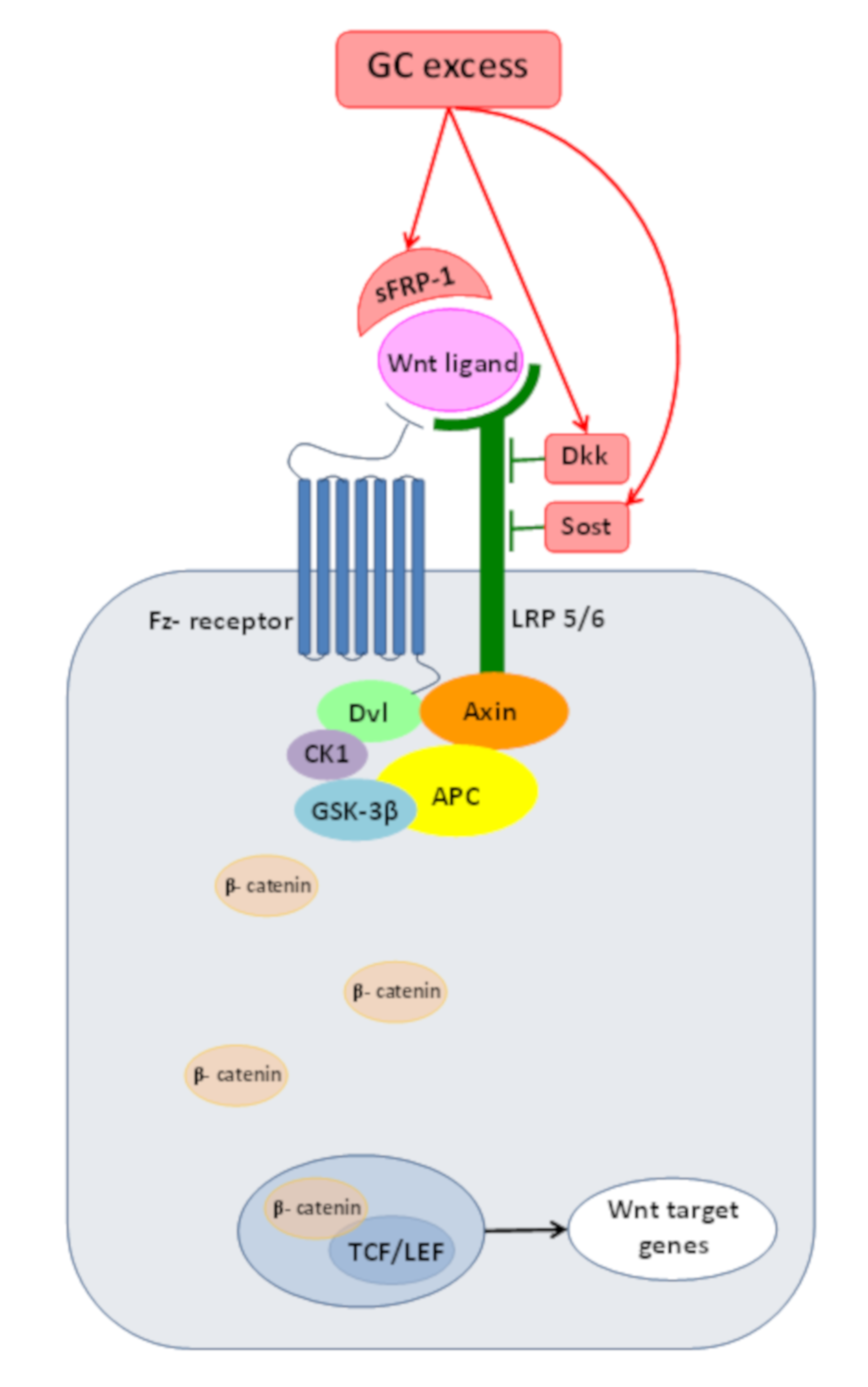
2. Wnt/β-Catenin Pathway or Canonical Wnt-Signaling

The Wnt/β-catenin pathway is an ancient signaling cascade that controls numerous cell processes (stem cell renewal, cell fate determination, differentiation and proliferation) during embryonic development and adult homeostasis [8]. Its deregulation has been detected in a number of human pathologies, including birth defect disorders, skeletal disorders and cancer [9].
Wnt proteins are a family of small signaling glycoproteins, which bind to a member of the Frizzled (Fz) receptor family. In the absence of Wnt-ligand, β-catenin is constantly degraded by a destruction complex composed of Axin and Adenomatous polyposis coli (APC) which admit the phosphorylation of β-catenin by glycogen synthase kinase 3β (GSK-3β) and casein kinase 1 (CK1). Phosphorylated β-catenin is subjected to proteosomal degradation, resulting in low free cytoplasmic and nucleic β-catenin levels in the absence of Wnt-ligand binding. The binding of Wnt to the Fz receptor requires low-density lipoprotein receptor-related-protein 5 or 6 (LRP 5/6) as coreceptors. The formation of the Wnt-Fz-LRP 5/6 complex leads to the membrane recruitment of the Axin complex by Dishevelled proteins (Dvl), Axin being bound to a cytoplasmic tail of LRP 5/6. In the absence of the degrading complex, β-catenin escapes from phosphorylation and consequential ubiquitination. This leads to an increase in free β-catenin levels which form complexes in the nucleus with the DNA-bounded T cell factor/lymphoid enhancer factor (TCF/LEF) and activate Wnt target gene expression.
Up to now,10 families of Wnt-pathway agonists were identified [10], from which the role of the Dickkopf family (Dkks), sclerostin (Sost) and secreted Frizzled-related proteins (sFRPs) were studied in depth in relation with glucocorticoid excess (Figure 1).

3. Wnt/β-catenin Pathway and Cushing’s Syndrome

The common occurrence of adrenal adenomas in patients with familial adenomatous polyposis highlighted the role of the Wnt/β-catenin pathway in adrenal tumorigenesis [14,15]. With APC being part of the degradation complex of β-catenin, abnormal Wnt/β-catenin activation was observed in both benign and malignant adrenocortical tumors: in 10 of 26 adrenocortical adenomas (38%) and in 11 of 13 adrenocortical carcinomas (77%) [16]. In these specimens, immunohistochemistry revealed atypical nuclear and/or cytoplasmic β-catenin accumulation. In addition, 27% of benign and 31% of malign tumors represented activating somatic mutations of the β-catenin gene (CTNNB1), altering the Ser45 of exon 3 [16], involved in targeted degradation of β-catenin [17].
In another study, genetic alterations were found in 5 of 33 adrenal adenomas (15%). Of these, three were cortisol-secreting, one aldosterone-producing and one nonfunctioning adenoma [18]. In two samples, point mutations were demonstrated at serine residues of codons 37 and 45, another three samples contained deletions [18]. There are also results suggesting that adrenocortical tumors with molecular alterations of CTNNB1 are primarily nonsecreting adenomas (61.1%), but the role of the Wnt/β-catenin pathway in adrenocortical tumor size and cortisol production seems plausible [19].

4. Effects of GC Excess on Bone

4.1. Bone Homeostasis and Main Cytokines

Homeostasis of the adult skeleton depends on the balance between bone-forming and bone-resorbing processes and relies on the complex communication between osteoblasts, osteocytes and osteoclasts.
Bone-forming osteoblasts are derived from mesenchymal stem cells (MSCs) located in the bone marrow. Master transcriptional regulator Runx controls the commitment of MSCs toward osteogenesis and the following differentiation processes [20]. Preosteoblasts are subjected to a maturation process resulting in matrix-synthesizing osteoblasts. Osteoblasts trapped in the mineralized matrix become osteocytes, the most substantial and long-lived cell types of the bone which communicate with each other through their cytoplasmic extensions. Osteocytes are involved in the bone turnover process through their mechanosensory activity, controlling the operation of both osteoblasts and osteoclasts [21]. Osteoclasts originate from hematopoietic stem cells and beside their “classic” function of bone resorption, the findings of recent years revealed their complex role in bone homeostasis, hematopoiesis and angiogenesis [22].
The communication between bone cells is conducted partly through the production of cytokines. Osteoblasts release M-CSF, which binding to its cognate receptor on preosteoclasts, provides signals essential for the survival and proliferation of osteoclast precursors [23]. The receptor activator of nuclear factor kappa-B°(RANK) is expressed in osteoclast precursors and osteoclasts; intracellular RANK signaling results in the induction of transcription factors required for osteoclast differentiation. RANK ligand (RANKL) is expressed mainly in osteoblasts, osteocytes and bone stroma [24]. RANKL–RANK interaction is critical for the differentiation of osteoclast precursors to mature osteoclasts [25]. Osteoprotegerin (OPG) is produced by the cells of osteoblast lineage, mainly by osteocytes [26]. Osteoprotegerin is a decoy receptor for RANKL, thereby inhibiting the differentiation and activation of osteoclasts.

4.2. Detrimental Effects of GC Excess

Glucocorticoid-induced osteoporosis (GIO), the most common cause of secondary osteoporosis [27,28], is characterized by the decrease of bone mass and microarchitectural deterioration which results in increased bone fragility [28]. As it was summarized in detail by Komori, GC excess exerts its detrimental effect on bone formation by inducing apoptosis of osteoblast and osteocytes, increasing production of reactive oxygen species and inhibiting the Wnt-signaling pathway [29]. Related to bone resorption, excess of GCs influences RANKL and OPG production besides prolonging the life span of osteoclasts [29]. The Wnt/β-catenin pathway is involved in all of these processes.

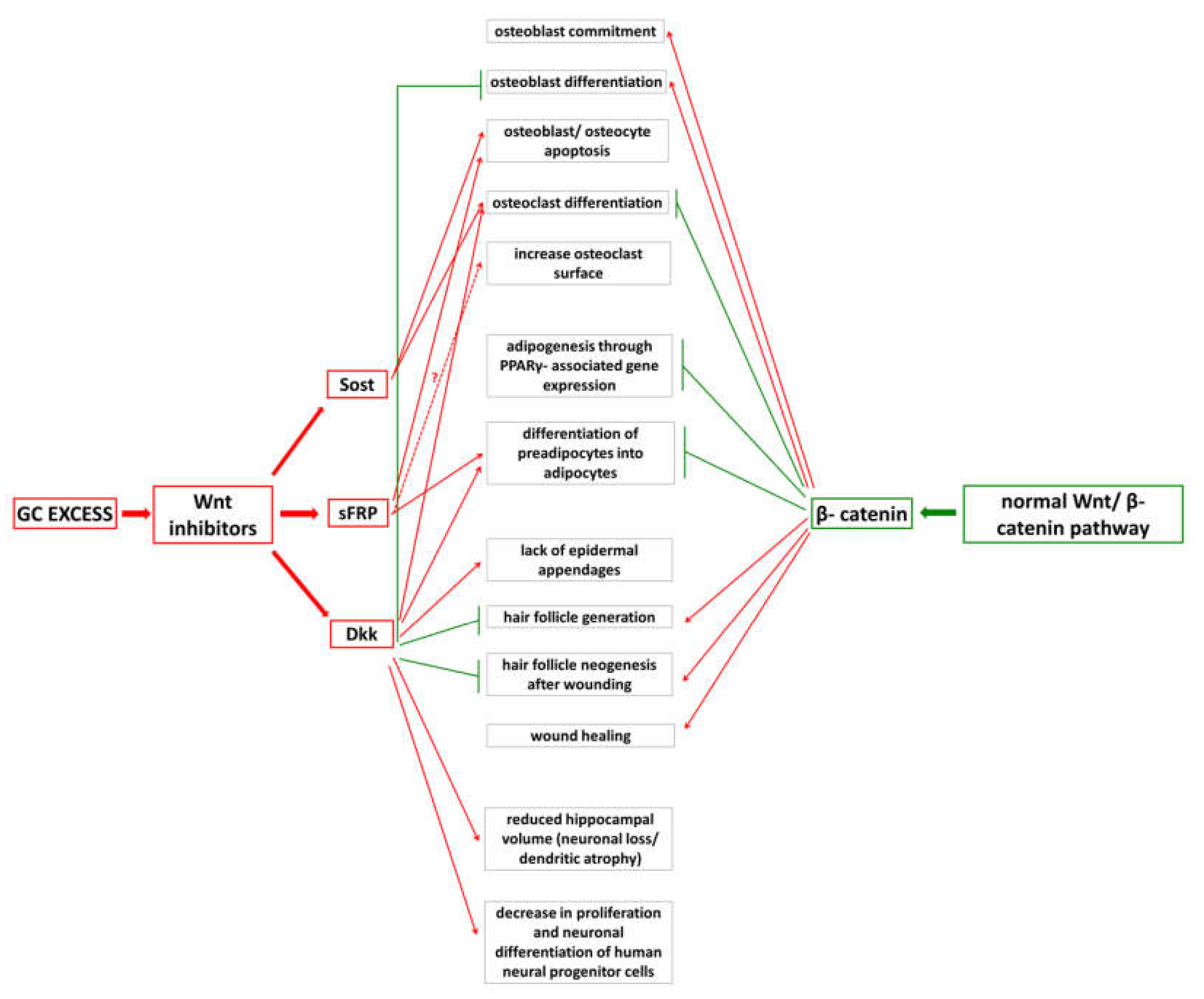
4.3. Inadequate Wnt/β-Catenin PathwayOperationdue to GC Excess

GC excess rapidly induces the apoptosis of osteoblastic cells as it was demonstrated on different cell lines and in patients with CS or receiving GC treatment. Sclerostin treatment of human osteoblastic cells initiates apoptosis through a caspase-activated mechanism [30]. sFRP-1 overexpression in human osteoblasts accelerates apoptotic cell death in osteoblasts and osteocytes [31]. These findings were also confirmed in humans: in adult patients on steroid therapy, biochemical markers were assessed at baseline and at 24, 48 and 96 h during the treatment. Osteocyte-related biochemical markers OPG and sclerostin showed a significant decrease at 48 h (after which their level was practically unchanged during the rest of the experiment); it can be explained by a rapidly induced apoptotic cell death of osteocytes [32]. Low serum sclerostin level was also found in patients with Cushing’s syndrome, but during treatment of GC excess, this level increased [33].
Glucocorticoids are detrimental to osteo-anabolic processes through their effects on oxidative stress levels [34,35]. The administration of prednisolone for 28 days to mice resulted in increased reactive oxygen species (ROS) levels in the bone marrow [34]. In human osteoblasts, dexamethasone significantly increased the number of mRNA transcripts related to oxidative stress [35]. The increase of ROS leads to enhanced activity of Forkhead box O (FoxO) transcription factors [34]. The interaction between FoxOs and β-catenin prevents β-catenin from forming complexes with TCF/LEF and activates Wnt target gene expression [36]. Diverting the limited pool of β-catenin from β-catenin-TCF/LEF mediated transcription to β-catenin-FoxO transcription itself is a significant proof for the repression of bone-anabolic processes in GC excess. In line with this, Iyer et al. demonstrated that FoxOs attenuated Wnt-signaling in lineage-committed murine osteoblast progenitor cells, hence decreasing the number of matrix-synthesizing osteoblasts [37].
Glucocorticoid excess enhances the expression of Wnt-inhibitors. Supraphysiological levels of glucocorticoids enhance sFRP-1 expression in primary mesenchymal cell cultures in vitro and in osteoblasts and bone marrow cells in vivo [38]. The expression of another Wnt-inhibitor, Dkk-1, increased after dexamethasone treatment in primary human osteoblasts [39]. Sato et al. demonstrated that glucocorticoids increase the expression of Sost mRNA and the number of sclerostin-positive osteocytes in wild-type mice while, in sclerostin deficient mice, the negative effects of GCs on bone (decreased mass, deteriorated microarchitecture, reduced structural and material strength) were prevented [40].
Wnt-pathway inhibition influences cell proliferation and maturation of cells from osteoblastic lineage. Dkk-1 expression in rat calvaria cells was associated with inhibited osteoblast differentiation and with the lack of mineralized nodules; parallelly, increased adipocyte differentiation could be detected in vitro [41]. In Yao’s experiment, expression levels of Lrp5 and Lrp6 (coreceptors of Wnt/β-catenin signaling), Runx, Osterix and osteocalcin (osteoblast differentiation and maturation markers) and unphosphorylated β-catenin were assessed in sFRP-1 transgenic and wild-type male and female mice. Lrp5 and Lrp6, β-catenin and osteocalcin levels were significantly lower in both sexes of sFRP-1 Tg mice, while Osterix and Runx (osteoblast differentiation markers) were significantly lower only in sFRP-1 male mice compared to controls [13]. These results suggest an important role for Wnt/β-catenin pathway in osteoblast differentiation and maturation, and also highlight the gender differences in GC-induced bone lesions.
The loss of osteocytes by GC-induced apoptosis damages the osteocyte-canalicular network and the mechanosensory ability of bone, which, in turn, decreases the capacity of osteocytes to regulate osteoclasts’ function. In mechanosensory osteocytes, the capacity to produce RANKL is much higher than that in osteoblasts [42]. Mice lacking RANKL in their osteocytes were protected from the adverse effects of glucocorticoid treatment on bone resorption [43].
Intact Wnt-signaling exerts an indirect negative role on osteoclastogenesis: in pluripotent mesenchymal cells, and stromal cells Wnt3a upregulates OPG expression [44], thereby inhibiting RANKL–RANK interaction and consequently the differentiation of octeoclasts. Wnt-pathway inhibitor sclerostin upregulates the expression of RANKL and downregulates OPG expression in human primary preosteocytes and murine osteocyte-like MLO-Y4 cells [45]. Dkk-1 enhances osteoclastogenesis by increasing the expression of RANKL and M-CSF and downregulating OPG in mesenchymal progenitor cells undergoing osteoblastic differentiation [25]. In another study, GC treatment of cortical bone organ cultures and primary osteoblasts did not increase significantly RANKL, but suppressed OPG production, suggesting that cortical bone loss caused by glucocorticoid excess can be attributed more likely to reduced OPG rather than elevated RANKL expression [43].
The role of sFRP-1 in the regulation of osteoclasts now seems to be bidirectional: first, sFRP-1 inhibits osteoclast formation by preventing the fusion of osteoblast precursors [46], secondly, sFRP-1 increases osteoclast surface in sFRP-1 transgenic female mice by 38% compared to WT controls [13].
Recently, Albers et al. demonstrated a direct effect of the Wnt-signaling pathway on osteoclastogenesis and bone resorption beside the indirect effects mediated by osteocytes [47]. They found elevated Frizzled8 (Fzd8) expression in osteoblasts and osteoclasts. The skeletal phenotype of Fzd8-deficient mice showed osteopenia with unaffected bone formation and enhanced osteoclastogenesis. To assess whether this is an indirect effect mediated by osteoblastic cytokines or it is the direct effect of the canonical Wnt-pathway on osteoclasts, they examined mice lacking ß-catenin in their osteoclast lineage. It was found that these animals displayed increased bone resorption despite their normal OPG level produced by osteoblasts [47].
In summary, we can conclude that the Wnt/β-catenin pathway is essential in normal bone homeostasis, and the harmful effects of GC excess are closely related to the inadequate operation of this pathway.

5. Effects of GC Excess on Adipose Tissue

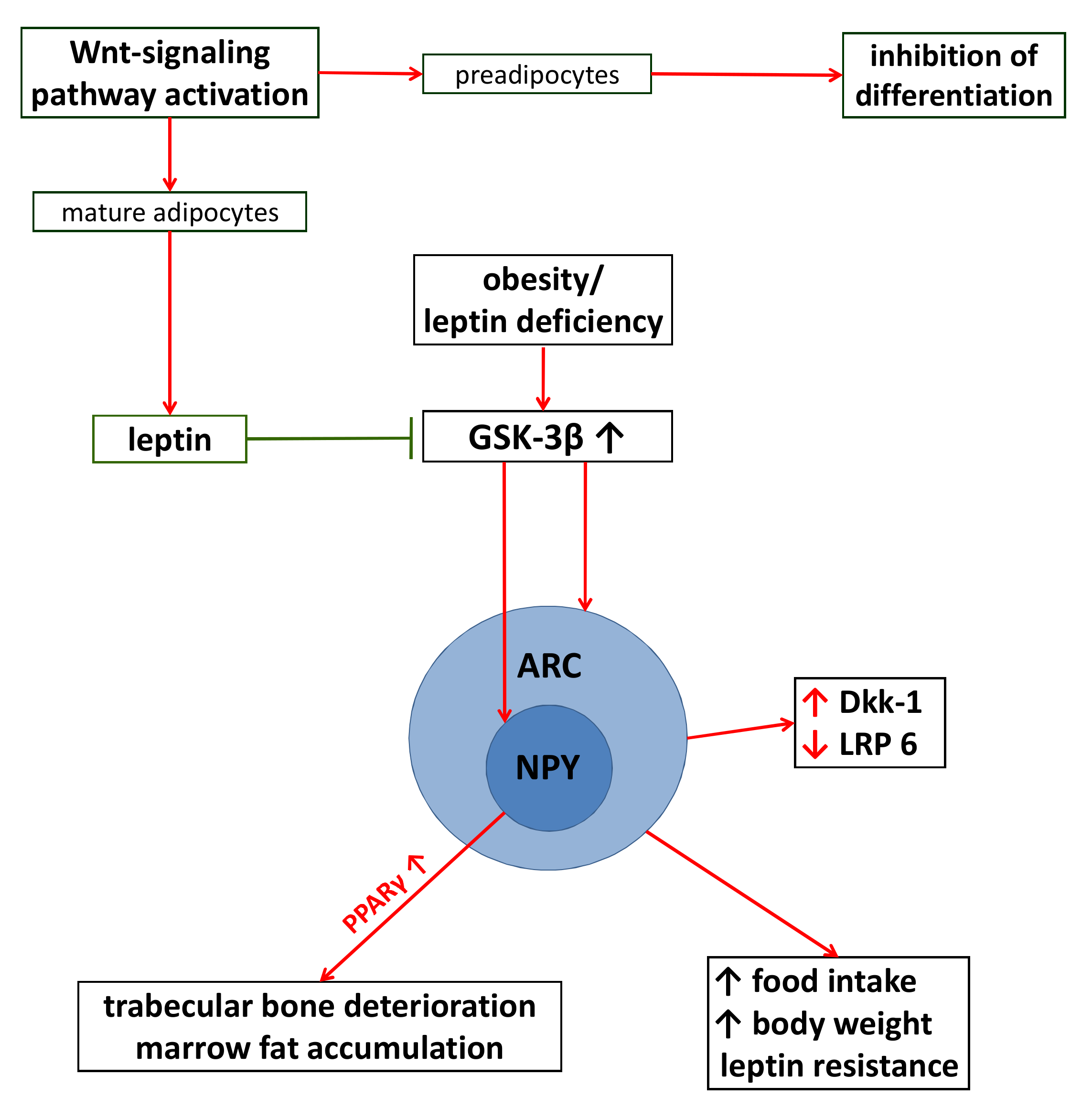
5.1. Adipogenesis and Wnt-Signaling

Adipogenesis is a well-orchestrated process during which MSC-derived preadipocytes develop into mature adipocytes. This process involves the interplay of several transcription factors, from which PPARγ functions as a master regulator [48].
There is plenty of evidence for strong communication between adipose tissue and bones, based (partly) on the interaction between PPAR- and Wnt-signaling pathways. Sustained activation of the Wnt/β-catenin pathway by expression of Wnt10b and inhibiting expression of PPARγ-associated adipogenic genes, in murine preadipocytes, maintains preadipocytes in an undifferentiated state [49]. Inversely, PPARγ induces the decrease of β-catenin expression during the differentiation process of preadipocytes into adipocytes [50]. A β-catenin binding domain within PPARγ ensures the close cooperation between the two pathways [51] and activation of PPARγ induces the proteasomal degradation of β-catenin which is preceded by its phosphorylation by GSK-3β [49]. LiCl, an inhibitor of GSK-3β, prevents PPARγ-induced decrease of β-catenin [49]. Missense mutations of the Wnt10b gene detected in families with early-onset obesity represent additional evidence for the strong relation between PPAR- and Wnt-signaling [52].

5.2. Detrimental Effects of GC Excess

Clinically, the strong but opposite-sign communication between adipogenesis and osteoblastogenesis appears most expressively in patients with Cushing’s syndrome presenting with osteoporosis, osteopenia and increased bone fragility associated with central obesity [27,53]. GC excess leads to abdominal, especially visceral, fat accumulation accompanied by the depletion of peripheral subcutaneous fat depots. GC-induced fat distribution can be partly explained by the different adipogenic and lipolytic effects of glucocorticoids on visceral adipose tissue (VAT) and subcutaneous adipose tissue (SAT). Analyzing the up- and downregulated genes during dexamethasone treatment of omental and abdominal subcutaneous tissues of obese women revealed that the PPAR signaling pathway was significantly upregulated in both types of adipose tissue but expression levels were higher in VAT than in SAT [54].
GCs change the characteristics of adipose tissue differently in VAT and SAT, as it was demonstrated in methylprednisolone-treated rats [55]. An increased number of adipocytes was experienced both in SAT and VAT, but the size of adipocytes was unchanged in VAT and decreased in SAT. Adipose mass of GC-treated rats increased only in VAT and was practically unchanged in SAT compared to controls. Importantly, GCs inhibit Wnt/β-catenin pathway both in SAT and VAT: Wnt10b and β-catenin levels were significantly decreased, while the Dkk-1 level was significantly increased in methylprednisolone-treated rats. GCs were also proved to exert a different lipolytic effect on VAT and SAT: in SAT, methylprednisolone induced a higher expression of lipolytic enzymes compared to untreated controls, in VAT a GC-induced, suppressive effect on lipolysis was demonstrated [55]. Analyzing abdominal and femoral adipose tissues in patients with Cushing’s syndrome leads to similar results: abdominal adipose tissue was characterized by enlarged fat cells, increased adipose tissue lipoprotein lipase activity and low lipolytic capacity [56].

5.3. The Consequences of Adiposity on Wnt-Signaling

Adipose tissue produces Wnt-inhibitors belonging to the sFRP family. Gene expression of all sFRPs (sFRP1-5) was analyzed in subcutaneous and visceral adipose tissues of obese and non-obese women [57]. In SAT, sFRP-1 was decreased and sFRP-2 increased in obesity and accordingly, sFRP-1 negatively, while sFRP-2 positively associated with insulin resistance [57]. Partly contradictory, another study demonstrated a biphasic regulation of sFRP-1 in mice (confirmed also in human tissues) fed with a high-fat diet. A higher expression of sFRP-1 in mild obesity and with a gradual fall of sFRP-1 in morbidly obese individuals was reported [58]. sFRP-1 overexpression promoted adipogenesis in preadipocytes through the inhibition of the Wnt/β-catenin pathway, suggesting its role in the paracrine regulation of adipogenesis [58].
Adipocytes secrete a variety of signaling proteins (called adipokines) and proinflammatory cytokines which are responsible for the auto-, para- and endocrine effects of adipose tissue. Adipose tissue-derived cytokines (leptin, chemerin, adiponectin, omentin, resistin, IL-6 and TNF-α) were proved to affect the function of osteoblasts and/or osteoclasts [26,59] and not surprisingly, many of these signaling molecules engage with the Wnt/β-catenin pathway.
Leptin is one of the first discovered adipokines and nowadays it is considered as the prototype of all signaling molecules produced by the adipose tissue. It is responsive to extensive communication between adipose tissue and brain, pancreas and reproductive organs [60]. The relation between obesity, leptin levels and Wnt-signaling was studied in depth. Chen et al. showed that activation of the Wnt-pathway, besides repressing preadipocytes’ differentiation, stimulates leptin production in mature murine adipocytes [61]. Benzler et al. revealed that GSK-3β (responsible for the continuous degradation of β catenin) activity was increased in mediobasal hypothalamic cells in obese male mice; the consequence of this status was hyperphagia with further aggravation of glucose intolerance and obesity [62]. Furthermore, they found that genes involved in the Wnt-pathway were mostly expressed in mediobasal hypothalamic cells with particular prominence in the arcuate nucleus. In brain sections of obese, leptin-deficient mice Wnt antagonist Dkk-1 level was significantly higher than in wild-type mice [62]. Leptin deficiency was proved to lead to decreased activation of LRP 6 (co-receptor of Wnt activation); the lower level of activated LRP 6 was fully restored after leptin administration. Leptin treatment, at the same time, caused inhibition of GSK-3β in orexigenic neuropeptide Y (NPY) cells [63]. Very recently, Wang et al. demonstrated the mediator role of neuropeptide Y in glucocorticoid-induced osteoporosis and bone marrow adiposity. In mice, NPY deletion was protective against glucocorticoid-induced loss of bone mass and fatty marrow development [64]. These findings altogether suggest that adipose tissue through leptin expression communicates bilaterally with mediobasal hypothalamic cells and provides evidence for the role of the Wnt/β-catenin pathway in the neuroendocrine control of metabolism (Figure 2).

6. Effects of GC Excess on Brain

6.1. Neurogenesis and Wnt/β-Catenin Pathway

Data from the last two decades have provided sufficient evidence for the role of Wnt-signaling in different processes of neurogenesis. Recently, Guo summarized these results and concluded that the Wnt/β-catenin signaling pathway plays a context-dependent role in oligodendrogenesis, oligodendrocyte differentiation and myelination. In addition, the effects associated with the Wnt/β-catenin pathway depend on the developmental stage of CNS, oligodendrocyte and CNS microenvironment [65].

6.2. Detrimental Effects of GC Excess

Detrimental effects of increased GC levels on brain structure, neurological function and emotional life have been demonstrated in numerous studies. GC excess negatively affects memory retrieval, cognitive functions and behavior and they have significant effects on brain structure [66,67].
Of brain regions, the hippocampus seems to be the most vulnerable to the neurotoxic effect of GCs [67,68,69]. Mice exposed to chronic stress showed reduced hippocampal volume with reduced neurogenesis in the dentate gyrus and neuronal loss and dendritic atrophy in the cornu ammonis 1 (CA1) region [68]. Besides, these animals showed increased hippocampal levels of Dkk-1 and reduced expression of β-catenin.
Clinically, brain development and affective problems of preadolescent children with or without fetal exposure to synthetic GCs were evaluated by Davis et al. [70]. A significant bilateral cortical thinning, particularly in the rostral anterior cingulate cortex was detected in children exposed to prenatal GC treatment. Besides this, the left rostral anterior cingulate cortex was thinner in children with affective problems, underlining the effect of GCs on behavioral disorders occurring even at later ages [70]. MRI results of the data of 339 CS patients lead to the conclusion that these patients have smaller hippocampal volumes, enlarged ventricles and cerebral atrophy; importantly, a part of structural deviations persisted even after a long-term remission [67].
Accumulating evidence suggests that long-lasting effects of GCs on the brain are regulated through epigenetic mechanisms. Rat embryonic neural stem cells exposed to dexamethasone underwent heritable alterations; GC effects induced in parent cells were detectable in daughter cells which were never exposed directly to GCs [71]. Methylation studies showed a significant decrease in global DNA methylation both in parent and daughter cells. Furthermore, dexamethasone caused a long-lasting, heritable receptivity of neural stem cells to oxidative stress manifested in increased apoptotic cell death of dexamethasone-treated daughter cells compared to control cells [71].
Extensive genome-wide alterations in promoter methylation, typically the hypomethylation of the affected promoters, were detected in the hippocampi of fetal guinea pigs treated prenatally with GCs [72]. The samples were collected 24 h and 14 days after two courses of maternal GC treatment. Twenty-four hours after the treatment, hypomethylation and hyperacetylation of the affected genes could be detected. After 14 days of GC administration, the changes previously observed did not persist, but the hypo- or hypermethylation of other promoters was noticed instead. These findings suggest a dynamic, spillover effect of GCs on the prenatal brain [72].

6.3. GC Excess and Wnt-Signaling

Inhibitory effects of dexamethasone on the neural precursor cells and the role of the Wnt/β-catenin pathway in this process were described by Boku et al. [73]. The negative effect of GCs has turned aside through the negative regulation of GSK-3β (by the administration of lithium or valproic acid, both inhibiting GSK-3β activity) and the consequential upregulation of Wnt/β-catenin signaling pathway [73,74]. Contrary to that, transgenic mice expressing a constant β-catenin level in neural precursor cells had enlarged brains with increased cerebral cortical surface and enlarged lateral ventricles [75].
The negative effects of GCs were demonstrated on human neural progenitor cells (hNPCs) responsible for the major cell types (neurons, astrocytes and oligodendrocytes) of the brain. Treating hNPCs with dexamethasone caused a marked decrease in proliferation and neuronal differentiation of hNPCs while increased glial cell formation. Dexamethasone induced the upregulation of the Wnt-signaling inhibitor Dkk-1 which effect could be inhibited by adding a GR antagonist, mifepristone. The final conclusions were that Dkk-1 was the primary target for GR in hNPCs and the regulation of hNPCs by dexamethasone and Dkk-1 was mediated through the canonical Wnt-signaling pathway [76].
High levels of GCs induced the generation of reactive oxygen species (ROS) in primary hippocampal and cortical cell cultures [77]. Accordingly, GC excess increased the level of oxidized components and decreased the antioxidant enzymes including SOD, catalase and glutathione peroxidase in the hippocampus of young rats [78]. Oxidative stress in rats caused hippocampal oxidative damage and apoptosis of pyramidal cells with a significant decrease in GRs in the hippocampal CA1 region and a consequential decline in cognitive functions [69]. In neonate rats, postnatal glucocorticoid treatment caused a significant decrease in total brain volume and neuronal loss in the dentate gyrus and CA1 region, which effects were largely prevented when dexamethasone therapy was combined with antioxidant vitamins [79]. Besides, sharing β-catenin as a common co-activator, the crosstalk between FoxO-regulated pathways and Wnt-signaling in neurogenesis was also confirmed by the identification of Wnt-inhibitors as direct targets of FoxOs. In FoxO-null neural stem cells, a significant downregulation of sFRP-1, sFRP-2 and sclerostin was demonstrated [80].

7. Effects of GC Excess on Skin

7.1. Skin and Steroidogenesis

Skin, besides its primary function of protecting the body, is an important part of immunological processes and extraadrenal steroidogenesis [81]. Recently, it was found that skin has its own HPA axis analog system with all major components present in the HPA axis and enzymes required for glucocorticoid synthesis [81,82].
Keratinocytes, one of the main cell types of the epidermis, synthesize cholesterol, the precursor of all steroids [83]. Epidermal and follicular keratinocytes, melanocytes, hair follicles and dermal fibroblasts have been proven to synthesize cortisol [82,84]. Glucocorticoids act through glucocorticoid receptors which are present in most, if not all of the skin compartments [84]. Skin expresses both types of 11-beta-hydroxysteroid-dehydrogenases (11β-HSDs); type 1 enzyme activates cortisone into cortisol, while type 2 converts cortisol into inactive cortisone [85]. The balanced activity of these enzymes is essential in the regulation of locally produced or exogenously administered glucocorticoids.

7.2. Detrimental Effects of GC Excess

Related to Cushing’s syndrome, a part of the signs and symptoms are relatively common (fatigue, weight gain, insomnia, irritability, depression, etc.) and makes CS difficult to diagnose. Some of the skin-related manifestations (easy bruising, reddish-purple striae) are between those signs which best discriminate Cushing’s syndrome from other diseases, while other skin-related signs (thin skin, poor skin healing) are very common in patients with CS (70%, respectively 80%) [86].
GCs have profound effects on the different layers of the skin. In the epidermis, the decreased size of keratinocytes, the thinning of the epidermis and the diminution of the lamellar lipid bilayer in the stratum corneum are well-known histopathological features of GC excess [87]. The molecular mechanism behind the “thinning of the epidermis” is that GCs inhibit the proliferation and migration of keratinocytes and have a dual effect on keratinocytes’ differentiation: at early stages of differentiation, inhibitory, while in late stages, stimulatory [88]. Transcriptional profiles of human keratinocytes untreated and treated with dexamethasone revealed that (unlike in many tissues affected by GCexcess) GCs exert anti-apoptotic effects on keratinocytes by inducing anti-apoptotic genes and suppressing proapoptotic genes [88].
In the dermis, GCs lead to a decreased number of fibroblasts with the rearrangement of the dermal fibrosis network [87]. The antiproliferative effect of GCs on fibroblast results in an altered collagen and elastin synthesis and in the thinning of the dermis, which causes a decrease of mechanical strength and elasticity of the skin.
There are only little data about the effect of GC excess on hypodermis, but we have to note the confusion in the literature related to the definition of skin-associated adipocyte layers. Traditionally, the thick layer of adipocytes underlining the reticular dermis has been called hypodermis or subcutis and it was considered that all skin-associated adipose tissues represent one depot [89]. The presence of a common precursor for dermal fibroblasts and intradermal adipocytes, besides the independent development of skin-related adipocytes and subcutaneous adipose tissue [90,91] justifies Driskell et al.’s proposal for a refined nomenclature. They recommend the term of intradermal adipocytes and dermal white adipose tissue for cells and adipose tissue underlying the reticular dermis [89].
Related to the effects of GC excess on skin compartments, it seems plausible that skin atrophy involves all skin layers, including dermal white adipose tissue. An indirect proof for this assumption was presented very recently by Baida et al. [92]. They studied the expression of FK506-binding protein 51 (FKBP51), a co-chaperone and regulator of the glucocorticoid receptor, in mice and human skin. They found that all skin compartments: epidermis, dermis, dermal adipose and CD34+ stem cells of FKBP51 KO mice were much more resistant to GC-induced hypoplasia than of their wild-type littermates [92].
Epidermal appendages are also influenced by GC excess. Based on the data of 481 patients published by the European Registry on Cushing’s syndrome, hair loss was present in 31% of patients [93]. Female balding occurs five times more often in patients with CD than in age-and sex-matched controls; the combination of female balding with the symptom of weakness/fatigue was 15 times more common in CD patientsthan in controls [94]. Rarely, female baldness is the main complaint of patients with Cushing’s syndrome [95].

7.3. Wnt-Pathway Alterations and Their Consequences

Ectopic expression of Wnt-inhibitor Dkk1 in epidermis drives to the lack of epidermal appendages: hair, whiskers, teeth and mammary gland [96]. Epithelial β-catenin deletion or ectopic Dkk-1 expression, induced during the growth phase of the hair cycle, causes premature hair follicle regression [97]. Another Wnt-inhibitor, sFRP-1, also plays a role in hair loss: WAY-316606, an sFRP-1 inhibitor, enhanced hair shaft production, hair shaft keratin expression and inhibited spontaneous hair follicle regression in organ-cultured human scalp ex vivo [98]. In line, a conditional gain-of-function mutation of β-catenin resulted in accelerated and excessive hair generation with densely packed hair follicles and little or no interfollicular epidermis [99].
As the canonical Wnt-signaling plays an important role in skin cells’ proliferation and differentiation, its impact on wound healing is self-evident. Wnt10a-deficient mice, besides their phenotype including alopecia and follicle growth failure, showed delayed wound healing compared to their wild-type littermates [100]. De novo development of hair follicles after injuries is also driven by Wnt-signaling: inducing expression of Dkk-1 after wounding leads to the inhibition of hair follicle neogenesis in mice [101]. Stabilization of nuclear β-catenin caused delayed wound healing in human skin organ cultures: LiCl-treated wounds (LiCl inhibits GSK-3β, an inactivator of β-catenin) showed a significantly larger wound size on day four after wounding compared to untreated wounds (12% healing rate for LiCl-treated wounds vs. 70% healing rate for untreated wounds) as LiCl treatment inhibited the migration of primary human keratinocytes in wounds [102].
In non-healing edges of venous ulcers, components of the Wnt-pathway were found to be deregulated: phosphorylated β-catenin was found mainly in the cytoplasm and nuclear; while in healthy skin specimens, β-catenin was present mostly at the membrane of the basal layer [103]. Jozic et al. demonstrated that GCs promote nuclear localization of β-catenin: dexamethasone treatment leads to a robust nuclearization of β-catenin in primary human keratinocytes, of which the effect could be blocked by pretreatment with mifepristone (RU486), a glucocorticoid receptor antagonist [104].
We have to conclude that, related to GC excess-induced skin alterations, the sole role of the Wnt/β-catenin pathway has not yet proven in many processes, but its function obviously contributes to these phenotypes. Future research can clarify the bidirectional link between the Wnt-pathway and GC-induced skin lesions.

8. Future Perspectives

Balanced operation of the Wnt/β-catenin pathway is indispensable for homeostatic processes in many tissues, including bones, brain, adipose tissue and skin (Figure 3.). However, some missing details related to its extensive impact on cell differentiation and proliferation and to its crosstalk with other pathways suggest that regulating its operation may be hazardous and it can be only done with the utmost care. In bone, treating osteoporotic and osteolytic bone lesions and treating myeloma multiplex with compound regulating Wnt-inhibitors showed some encouraging results. In myeloma cells, Dkk-1 is produced in large amounts and it is overexpressed in patients having extensive osteolytic lesions [105]. Bortezomib, a proteasome inhibitor approved for the treatment of relapsed multiple myeloma patients was proved to inhibit Dkk-1 expression in neonatal mouse calvarial cells and bone marrow stromal cells [106]. In another study, Wnt-pathway independent β-catenin activation and consequential osteoblast differentiation were demonstrated, suggesting a role for bortezomib in the partial restoration of tumor-induced suppression of Wnt-signaling [107].
Selective inhibition of sFRP-1 was also proposed as a potential tool to stimulate Wnt-signaling, thereby osteo-anabolic processes in bone [13,108]. In certain types of cancer, epigenetic inactivation of different sFRP genes was identified, thus sFRPs were hypothesized to function as tumor suppressors [109,110]. These data underline the necessity of strict controls and a careful evaluation of results obtained during experiments related to Wnt-inhibitors.
Wnt/β-catenin signaling can be stimulated by physical exercise. In a study conducted among women affected by abdominal obesity, osteoblast cell cultures were exposed to sera of these women at an initial timepoint and 4, 6 and 12 months after they attended a physical training program combined with a hypocaloric diet [111]. Proteins of the Wnt/β-catenin pathway were quantified at the specified timepoints. Results showed a time-dependent restoration of the Wnt/β-catenin pathway evident already after the fourth month of training. At the same time, a consequential decline in the inhibition of the pathway was demonstrated by decreased sclerostin levels. These results suggest that the decrease of the abdominal fat mass due to physical training and hypocaloric diet created a hormonal environment where osteoblasts restored their osteo-anabolic function [111].
A similar study was carried out among breast cancer survivors, where serum levels of Dkk-1 and sFRP-1 were followed before and after a 12-week long physical exercise training program. The conception of this study was based on the results of former studies showing that elevated serum Dkk-1 levels correlated with poor prognosis in certain types of cancer [112]. Significant decrease in serum levels of Wnt-inhibitors: Dkk-1 and sFRP-1 were demonstrated after the exercise training program, accompanied by the significant decrease of serum insulin and leptin levels and significant reduction of total body fat and visceral fat area [113].
Another evidence for the beneficial effects of physical exercises on Wnt-pathway was the mechanosensitive expression of sclerostin (specific product of osteocytes). Mechanical unloading of wild-type mice caused downregulation of Wnt/ß-catenin signaling with the upregulation of the sclerostin levels [114]. In Sost KO mice, unable to produce sclerostin, bone loss caused by mechanical unload could not be detected [114]. This finding was also confirmed in humans: serum sclerostin level was assayed in healthy adult men before and after longer bed rest and increased sclerostin level wasdetected. Lower serum sclerostin level was accompanied by significantly increased bone resorption markers (bone formation markers were unchanged), with significantly higher urinary calcium excretion and significantly declined bone mineral density at weight-bearing skeletal sites: lumbar spine, hip, femoral neck and calcaneus [115]. These results suggest that the simultaneous presence of glucocorticoid excess and mechanical unload (e.g., sedentary lifestyle) exert a cumulative negative effect on bone homeostasis.
The beneficial effects of physical exercise were also highlighted by Chen, who recently reviewed the paradox of chronic stress and chronic exercise, both known to increase basal cortisol levels. Their metanalysis underlined that solely increased basal cortisol levels are not necessarily detrimental to the human body and their work suggested that other factors are also needed for the harmful effects of glucocorticoid excess [116]. Related to GIO, Tóth et al. highlighted that one of the most peculiar features was the large interindividual variability in clinical presentation and severity [27]. In this regard, the role of hypoxia and hypoxia-induced factors seem unavoidable. In bone, despite the detailed investigations, the effect of hypoxia, whether it is osteo-anabolic or osteo-catabolic is not entirely clear. A proof for the role of hypoxia in GC excess induced detrimental effects was shown recently by Ueda. In MLO-Y4 murine osteocytic cells, dexamethasone and hypoxia separately caused apoptosis; but their simultaneous effects lead to a significant increase of necrotic celldeath [117].
Related to this, Noguchi et al. proved that the detrimental effects of the glucocorticoid excess on bone could be prevented by oxygen ultra-fine bubbles saline injection [118]. In prednisolone-treated mice, regularly administered injections diluted with oxygen ultra-fine bubbles prevented severe bone loss caused by GCs. Simple saline injections and injections diluted with nitrogen ultra-fine bubbles were not able to counteract the detrimental effects of GCs. In vitro experiments revealed that appropriate oxygen supply (through injection with oxygen ultra-fine bubbles) had not affected differentiation of osteoblasts, but inhibited osteoclastogenesis, resulting in an osteo-anabolic effect [118].
Summarizing these results, it can be assumed that the restorative effects of physical exercise on Wnt/ß-catenin pathway can be linked partly to an enhanced oxygen supply, and inversely, deleterious effects of GC excess on Wnt-pathway are more pronounced when hypoxic conditions are present and probably the severity of the damaging effects is related to the degree of hypoxia.
The relationship between GC excess and severe comorbidities is well-known: fractures occur in 30%–50% of the patients subjected to long-term hypercortisolism [28]; the rate of glucocorticoid-induced hyperglycemia or diabetes among chronic GC-users is more than 32% [119]. Besides this, glucocorticoids are responsible for cardiovascular (15%), infectious (15%), gastrointestinal (10%), psychological/behavioral (9%) and endocrine/metabolic (7%) adverse effects [4]. Therefore, the earlier treatment of endogenous GC excess is imperious and the periodic evaluation of chronic GC-users’ state is indispensable. This is also of paramount importance because a part of the adverse effects of GC excess seems to be long-lasting, definitive or even heritable. Besides the complex medical treatment of endogenous GC excess and the adverse effects of GCtreatment, the role of comprehensive lifestyle changes are also essential: physical activity and the management of obesity create a hormonal environment in which Wnt/β-catenin pathway can fulfill its role in the homeostatic processes of the adult organism.

Author Contributions

Conceptualization: K.M. and A.P., writing—original draft preparation: K.M., writing—review and editing: K.M. and A.P. All authors have read and agreed to the published version of the manuscript.

Funding

This research received no external funding

Acknowledgments

The authors acknowledge the financial support of Hungarian Academy of Sciences and Hungarian National Bionics Program (Medical bionics subtheme: AP).

Conflicts of Interest

The authors declare no conflict of interest.

References

  1. Nicolaides, N.C.; Charmandari, E.; George, P.; Chrousos, G.P.; Kino, T. Recent advances in the molecular mechanisms determining tissue sensitivity to glucocorticoids: Novel mutations, circadian rhythm and ligand- induced repression of the human glucocorticoid receptor. BMC Endocr. Disord. 2014, 14, 71. [Google Scholar] [CrossRef] [PubMed] [Green Version]
  2. Quax, R.A.; Manenschijn, L.; Koper, J.W.; Hazes, J.M.; Lamberts, S.W.J.; van Rossum, E.F.C.; Feelders, R.A. Glucocorticoid sensitivity in health and disease. Nat. Rev. Endocrinol. 2013, 9, 670–686. [Google Scholar] [CrossRef] [PubMed]
  3. Spies, C.M.; Strehl, C.; van der Goes, M.C.; Bijlsma, J.W.J.; Buttgereit, F. Glucocorticoids. Best Pract. Res. Clin. Rheumatol. 2011, 25, 891–900. [Google Scholar] [CrossRef] [PubMed]
  4. Bijlsma, J.W.J.; Buttgereit, F. Adverse events of glucocorticoids during treatment of rheumatoid arthritis: Lessons from cohort and registry studies. Rheumatology 2016, 55, ii3–ii5. [Google Scholar] [CrossRef] [PubMed]
  5. Guaraldi, F.; Salvatori, R. Cushing syndrome: Maybe not so uncommon of an endocrine disease. J. Am. Board Fam. Med. 2012, 2, 199–208. [Google Scholar] [CrossRef] [PubMed] [Green Version]
  6. Reynolds, R.M.; Labad, J.; Sears, A.V.; Williamson, R.M.; Strachan, M.W.J.; Deary, I.J.; Lowe, G.D.O.; Price, J.F.; Walker, B.R. Glucocorticoid treatment and impaired mood, memory and metabolism in people with diabetes: The Edinburgh Type 2 Diabetes Study. Eur. J. Endocrinol. 2012, 166, 861–868. [Google Scholar] [CrossRef] [Green Version]
  7. Overman, R.A.; Toliver, J.C.; Yeh, J.Y.; Gourlay, M.L.; Deal, C.L. United States adults meeting 2010 American College of Rheumatology criteria for treatment and prevention of glucocorticoid-induced osteoporosis. Arthritis Care Res. 2014, 66, 1644–1652. [Google Scholar] [CrossRef]
  8. Clevers, H.; Nusse, R. Wnt/b-Catenin Signaling and Disease. Cell 2012, 149, 1192–1205. [Google Scholar] [CrossRef] [Green Version]
  9. Komiya, Y.; Habas, R. Wnt signal transduction pathways. Organogenesis 2008, 4, 68–75. [Google Scholar] [CrossRef] [Green Version]
  10. Cruciat, C.M.; Niehrs, C. Secreted and Transmembrane Wnt Inhibitors and Activators. Cold Spring Harb. Perspect. Biol. 2013, 5, a015081. [Google Scholar] [CrossRef] [Green Version]
  11. Schulze, J.; Seitz, S.; Saito, H.; Schneebauer, M.; Marshall, R.P.; Baranowsky, A.; Busse, B.; Schilling, A.F.; Friedrich, F.W.; Albers, J.; et al. Negative Regulation of Bone Formation by the Transmembrane Wnt Antagonist Kremen-2. PLoS ONE 2010, 5, e10309. [Google Scholar] [CrossRef] [PubMed] [Green Version]
  12. Li, X.; Zhang, Y.; Kang, H.; Liu, W.; Liu, P.; Zhang, J.; Harris, S.E.; Wu, D. Sclerostin Binds to LRP5/6 and Antagonizes Canonical Wnt Signaling. J. Biol. Chem. 2005, 280, 19883–19887. [Google Scholar] [CrossRef] [PubMed] [Green Version]
  13. Yao, W.; Cheng, Z.; Shahnazari, M.; Dai, W.; Johnson, M.L.; Lane, N.E. Overexpression of secreted frizzled-related protein 1 inhibits bone formation and attenuates parathyroid hormone bone anabolic effects. J. Bone Miner. Res. 2010, 25, 190–199. [Google Scholar] [CrossRef] [PubMed] [Green Version]
  14. Marchesa, P.; Fazio, V.W.; Church, J.M.; McGannon, E. Adrenal masses in patients with familial adenomatous polyposis. Dis. Colon Rectum 1997, 40, 1023–1028. [Google Scholar] [CrossRef] [PubMed]
  15. Smith, T.G.; Clark, S.K.; Katz, D.E.; Reznek, R.H.; Phillips, R.K. Adrenal masses are associated with familial adenomatous polyposis. Dis. Colon Rectum 2000, 43, 1739–1742. [Google Scholar] [CrossRef]
  16. Tissier, F.; Cavard, C.; Groussin, L.; Perlemoine, K.; Fumey, G.; Hagneré, A.M.; René-Corail, F.; Jullian, E.; Gicquel, C.; Bertagna, X.; et al. Mutations of beta-catenin in adrenocortical tumors: Activation of the Wnt signaling pathway is a frequent event in both benign and malignant adrenocortical tumors. Cancer Res. 2005, 65, 7622–7627. [Google Scholar] [CrossRef] [Green Version]
  17. Polakis, P. Wnt signaling and cancer. Genes Dev. 2000, 14, 1837–1851. [Google Scholar] [CrossRef] [Green Version]
  18. Tadjine, M.; Lampron, A.; Ouadi, L.; Bourdeau, I. Frequent mutations of beta-catenin gene in sporadic secreting adrenocortical adenomas. Clin. Endocrinol. 2008, 68, 264–270. [Google Scholar] [CrossRef]
  19. Bonnet, S.; Gaujoux, S.; Launay, P.; Baudry, C.; Chokri, I.; Ragazzon, B.; Libé, R.; René-Corail, F.; Audebourg, A.; Vacher-Lavenu, M.C.; et al. Wnt/β-Catenin Pathway Activation in Adrenocortical Adenomas Is Frequently due to Somatic CTNNB1-Activating Mutations, Which Are Associated with Larger and Nonsecreting Tumors: A Study in Cortisol-Secreting and -Nonsecreting Tumors. J. Clin. Endocrinol. Metab. 2011, 96, E419–E426. [Google Scholar] [CrossRef] [Green Version]
  20. Rutkovskiy, A.; Stensløkken, K.O.; Vaage, I.J. Osteoblast Differentiation at a Glance. Med. Sci. Monit. Basic Res. 2016, 22, 95–106. [Google Scholar] [CrossRef] [Green Version]
  21. Sapir-Koren, R.; Livshits, G. Osteocyte control of bone remodeling: Is sclerostin a key molecular coordinator of the balanced bone resorption– formation cycles? Osteoporos. Int. 2014, 25, 2685–2700. [Google Scholar] [CrossRef] [PubMed]
  22. Cappariello, A.; Maurizi, A.; Veeriah, V.; Teti, A. The Great Beauty of the osteoclast. Arch. Biochem. Biophys. 2014, 558, 70–78. [Google Scholar] [CrossRef] [PubMed]
  23. Faccio, R.; Takeshita, S.; Zallone, A.; Ross, F.P.; Teitelbaum, S.L. c-Fms and the alphavbeta3 integrin collaborate during osteoclast differentiation. J. Clin. Investig. 2003, 111, 749–758. [Google Scholar] [CrossRef] [PubMed] [Green Version]
  24. Walsh, M.C.; Choi, Y. Biology of the RANKL–RANK-OPG System in Immunity, Bone, and Beyond. Front. Immunol. 2014, 5, 511. [Google Scholar] [CrossRef] [Green Version]
  25. Fujita, K.; Janz, S. Attenuation of WNT signaling by DKK-1 and -2 regulates BMP2-induced osteoblast differentiation and expression of OPG, RANKL and M-CSF. Mol. Cancer 2007, 6, 71. [Google Scholar] [CrossRef] [Green Version]
  26. Muruganandan, S.; Sinal, C.J. The impact of bone marrow adipocytes on osteoblast and osteoclast differentiation. IUBMB Life 2014, 66, 147–155. [Google Scholar] [CrossRef]
  27. Tóth, M.; Grossman, A. Glucocorticoid- induced osteoporosis: Lessons from Cushing’s syndrome. Clin. Endocrinol. 2013, 79, 1–11. [Google Scholar] [CrossRef]
  28. Buehring, B.; Viswanathan, R.; Binkley, N.; Busse, W. Glucocorticoid-induced osteoporosis: An update on effects and management. J. Allergy Clin. Immunol. 2013, 132, 1019–1030. [Google Scholar] [CrossRef]
  29. Komori, T. Glucocorticoid Signaling and Bone Biology. Horm. Metab. Res. 2016, 48, 755–763. [Google Scholar] [CrossRef] [Green Version]
  30. Sutherland, M.K.; Geoghegan, J.C.; Yu, C.; Turcott, E.; Skonier, J.E.; Winkler, D.G.; Latham, J.A. Sclerostin promotes the apoptosis of human osteoblastic cells: A novel regulation of bone formation. Bone 2004, 35, 828–835. [Google Scholar] [CrossRef]
  31. Bodine, P.V.; Billiard, J.; Moran, R.A.; Ponce-de-Leon, H.; McLarney, S.; Mangine, A.; Scrimo, M.J.; Bhat, R.A.; Stauffer, B.; Green, J.; et al. The Wnt antagonist secreted frizzled-related protein-1 controls osteoblast and osteocyte apoptosis. J. Cell. Biochem. 2005, 96, 1212–1230. [Google Scholar] [CrossRef] [PubMed]
  32. Brabnikova Maresova, K.; Pavelka, K.; Stepan, J.J. Acute effects of glucocorticoids on serum markers of osteoclasts, osteoblast, and osteocytes. Calcif. Tissue Int. 2013, 92, 354–361. [Google Scholar] [CrossRef] [PubMed]
  33. Van Lierop, A.H.; van der Eerden, A.W.; Hamdy, N.A.; Hermus, A.R.; den Heijer, M.; Papapoulos, S.E. Circulating sclerostin levels are decreased in patients with endogenous hypercortisolism and increase after treatment. J. Clin. Endocrinol. Metab. 2012, 97, E1953–E1957. [Google Scholar] [CrossRef] [PubMed] [Green Version]
  34. Almeida, M.; Han, L.; Ambrogini, E.; Weinstein, R.S.; Manolagas, S.C. Glucocorticoids and tumor necrosis factor α increase oxidative stress and suppress Wnt protein signaling in osteoblasts. J. Biol. Chem. 2011, 286, 44326–44335. [Google Scholar] [CrossRef] [Green Version]
  35. Hurson, C.J.; Butler, J.S.; Keating, D.T.; Murray, D.W.; Sadlier, D.M.; O’Byrne, J.M.; Doran, P.P. Gene expression analysis in human osteoblasts exposed to dexamethasone identifies altered developmental pathways as putative drivers of osteoporosis. BMC Musculoskelet. Disord. 2007, 8, 1–12. [Google Scholar] [CrossRef] [Green Version]
  36. Hoogeboom, D.; Essers, M.A.; Polderman, P.E.; Voets, E.; Smits, L.M.; Burgering, B.M. Interaction of FOXO with beta-catenin inhibits beta-catenin/T cell factor activity. J. Biol. Chem. 2008, 283, 9224–9230. [Google Scholar] [CrossRef] [Green Version]
  37. Iyer, S.; Ambrogini, E.; Bartell, S.M.; Han, L.; Roberson, P.K.; de Cabo, R.; Jilka, R.L.; Weinstein, R.S.; O’Brien, C.A.; Manolagas, S.C.; et al. FOXOs attenuate bone formation by Wnt signaling. J. Clin. Investig. 2013, 123, 3409–3419. [Google Scholar] [CrossRef]
  38. Wang, F.S.; Lin, C.L.; Chen, Y.J.; Wang, C.J.; Yang, K.D.; Huang, Y.T.; Sun, Y.C.; Huang, H.C. Secreted frizzled- related protein 1 modulates glucocorticoid attenuation of osteogenic activities and bone mass. Endocrinology 2005, 146, 2415–2423. [Google Scholar] [CrossRef] [Green Version]
  39. Ohnaka, K.; Tanabe, M.; Kawate, H.; Nawata, H.; Takayanagi, R. Glucocorticoid suppresses the canonical Wnt signal in cultured human osteoblasts. Biochem. Biophys. Res. Commun. 2005, 329, 177–181. [Google Scholar] [CrossRef]
  40. Sato, A.Y.; Cregor, M.; Delgado-Calle, J.; Condon, K.W.; Allen, M.R.; Peacock, M.; Plotkin, L.I.; Bellido, T. Protection From Glucocorticoid-Induced Osteoporosis by Anti-Catabolic Signaling in the Absence of Sost/Sclerostin. J. Bone Miner. Res. 2016, 31, 1791–1802. [Google Scholar] [CrossRef] [Green Version]
  41. Morvan, F.; Boulukos, K.; Clément-Lacroix, P.; Roman Roman, S.; Suc-Royer, I.; Vayssière, B.; Ammann, P.; Martin, P.; Pinho, S.; Pognonec, P.; et al. Deletion of a single allele of the Dkk1 gene leads to an increase in bone formation and bone mass. J. Bone Miner. Res. 2006, 21, 934–945. [Google Scholar] [CrossRef] [PubMed]
  42. Nakashima, T.; Hayashi, M.; Fukunaga, T.; Kurata, K.; Oh-Hora, M.; Feng, J.Q.; Bonewald, L.F.; Kodama, T.; Wutz, A.; Wagner, E.F.; et al. Evidence for osteocyte regulation of bone homeostasis through RANKL expression. Nat. Med. 2011, 17, 1231–1234. [Google Scholar] [CrossRef] [PubMed]
  43. Piemontese, M.; Xiong, J.; Fujiwara, Y.; Thostenson, J.D.; O’Brien, C.A. Cortical bone loss caused by glucocorticoid excess requires RANKL production by osteocytes and isassociated with reduced OPG expression in mice. Am. J. Physiol. Endocrinol. Metab. 2016, 311, E587–E593. [Google Scholar] [CrossRef] [Green Version]
  44. Jackson, A.; Vayssière, B.; Garcia, T.; Newell, W.; Baron, R.; Roman-Roman, S.; Rawadi, G. Gene array analysis of Wnt-regulated genes in C3H10T1/2 cells. Bone 2005, 36, 585–598. [Google Scholar] [CrossRef] [PubMed]
  45. Wijenayaka, A.R.; Kogawa, M.; Lim, H.P.; Bonewald, L.F.; Findlay, D.M.; Atkins, G.J. Sclerostin stimulates osteocyte support of osteoclastactivity by a RANKL- dependent pathway. PLoS ONE 2011, 6, e25900. [Google Scholar] [CrossRef] [PubMed] [Green Version]
  46. Liu, D.; Wise, G.E. A DNA microarray analysis of chemokine and receptor genes in the rat dental follicle -role of secreted frizzled-related protein-1 in osteoclastogenesis. Bone 2007, 41, 266–272. [Google Scholar] [CrossRef] [PubMed] [Green Version]
  47. Albers, J.; Keller, J.; Baranowsky, A.; Beil, F.T.; Catala-Lehnen, P.; Schulze, J.; Amling, M.; Schinke, T. Canonical Wnt signaling inhibits oteoclastogenesis independent of osteoprotegerin. J. Cell Biol. 2013, 200, 537–549. [Google Scholar] [CrossRef] [Green Version]
  48. Rosen, E.D.; Sarraf, P.; Troy, A.E.; Bradwin, G.; Moore, K.; Milstone, D.S.; Spiegelman, B.M.; Mortensen, R.M. PPAR gamma is required for the differentiation of adipose tissue in vivo and in vitro. Mol. Cell 1999, 4, 611–617. [Google Scholar] [CrossRef]
  49. Liu, J.; Farmer, S.R. Regulating the balance between peroxisome proliferator-activated receptor gamma and beta-catenin signaling during adipogenesis. A glycogen synthase kinase 3beta phosphorylation-defective mutant of beta-catenin inhibits expression of a subset of adipogenic genes. J. Biol. Chem. 2004, 279, 45020–45027. [Google Scholar] [CrossRef] [Green Version]
  50. Moldes, M.; Zuo, Y.; Morrison, R.F.; Silva, D.; Park, B.H.; Liu, J.; Farmer, S.R. Peroxisome-proliferator-activated receptor gamma suppresses Wnt/beta- catenin signalling during adipogenesis. Biochem. J. 2003, 376, 607–613. [Google Scholar] [CrossRef] [Green Version]
  51. Liu, J.; Wang, H.; Zuo, Y.; Farmer, S.R. Functional interaction between peroxisome proliferator-activated receptor gamma and beta-catenin. Mol. Cell. Biol. 2006, 26, 5827–5837. [Google Scholar] [CrossRef] [PubMed] [Green Version]
  52. Christodoulides, C.; Scarda, A.; Granzotto, M.; Milan, G.; Dalla Nora, E.; Keogh, J.; De Pergola, G.; Stirling, H.; Pannacciulli, N.; Sethi, J.K.; et al. WNT10B mutations in human obesity. Diabetologia 2006, 49, 678–684. [Google Scholar] [CrossRef] [PubMed] [Green Version]
  53. Nieman, L.K. Cushing’s syndrome: Update on signs, symptoms and biochemical screening. Eur. J. Endocrinol. 2015, 173, M33–M38. [Google Scholar] [CrossRef] [PubMed] [Green Version]
  54. Pickering, R.T.; Lee, M.J.; Karastergiou, K.; Gower, A.; Fried, S.K. Depot dependent effects of dexamethasone on gene expression in human omental and abdominal subcutaneous adipose tissues from obese women. PLoS ONE 2016, 11, e0167337. [Google Scholar] [CrossRef]
  55. Xiao, X.; Li, H.; Yang, J.; Qi, X.; Zu, X.; Yang, J.; Zhong, J.; Cao, R.; Liu, J.; Wen, G. Wnt/β-catenin signaling pathway and lipolysis enzymes participate in methylprednisolone induced fat differential distribution between subcutaneous and visceral adipose tissue. Steroids 2014, 84, 30–35. [Google Scholar] [CrossRef]
  56. Rebuffé-Scrive, M.; Krotkiewski, M.; Elfverson, J.; Björntorp, P. Muscle and adipose tissue morphology and metabolism in Cushing’s syndrome. J. Clin. Endocrinol. Metab. 1988, 67, 1122–1128. [Google Scholar] [CrossRef]
  57. Ehrlund, A.; Mejhert, N.; Lorente-Cebrián, S.; Aström, G.; Dahlman, I.; Laurencikiene, J.; Rydén, M. Characterization of the Wnt inhibitors secreted frizzled- related proteins (SFRPs) in human adipose tissue. J. Clin. Endocrinol. Metab. 2013, 98, E503–E508. [Google Scholar] [CrossRef]
  58. Lagathu, C.; Christodoulides, C.; Tan, C.Y.; Virtue, S.; Laudes, M.; Campbell, M.; Ishikawa, K.; Ortega, F.; Tinahones, F.J.; Fernández-Real, J.M.; et al. Secreted frizzled-related protein 1 regulates adipose tissue expansion and is dysregulated in severe obesity. Int. J. Obes. 2010, 34, 1695–1705. [Google Scholar] [CrossRef] [Green Version]
  59. Abdallah, B.M. Marrow adipocytes inhibit the differentiation of mesenchymal stem cells into osteoblasts via suppressing BMP- signaling. J. Biomed. Sci. 2017, 24, 1–10. [Google Scholar] [CrossRef] [Green Version]
  60. Blüher, M.; Mantzoros, C.S. From leptin to other adipokines in health and disease: Facts and expectations at the beginning of the 21st century. Metabolism 2015, 64, 131–145. [Google Scholar] [CrossRef]
  61. Chen, Z.L.; Shao, W.J.; Xu, F.; Liu, L.; Lin, B.S.; Wei, X.H.; Song, Z.L.; Lu, H.G.; Fantus, I.G.; Weng, J.P.; et al. Acute Wnt pathway activation positively regulates leptin gene expression in mature adipocytes. Cell Signal. 2015, 27, 587–597. [Google Scholar] [CrossRef] [PubMed]
  62. Benzler, J.; Ganjam, G.K.; Krüger, M.; Pinkenburg, O.; Kutschke, M.; Stöhr, S.; Steger, J.; Koch, C.E.; Ölkrug, R.; Schwartz, M.W.; et al. Hypothalamic glycogen synthase kinase 3β has a central role in the regulation of food intake and glucose metabolism. Biochem. J. 2012, 447, 175–184. [Google Scholar] [CrossRef] [PubMed] [Green Version]
  63. Benzler, J.; Andrews, Z.B.; Pracht, C.; Stöhr, S.; Shepherd, P.R.; Grattan, D.R.; Tups, A. Hypothalamic WNT signalling is impaired during obesity and reinstated by leptin treatment in male mice. Endocrinology 2013, 154, 4737–4745. [Google Scholar] [CrossRef] [PubMed] [Green Version]
  64. Wang, F.S.; Lian, W.S.; Weng, W.T.; Sun, Y.C.; Ke, H.J.; Chen, Y.S.; Ko, J.Y. Neuropeptide Y mediates glucocorticoid-induced osteoporosis and marrow adiposity in mice. Osteoporos. Int. 2016, 27, 2777–2789. [Google Scholar] [CrossRef] [PubMed]
  65. Guo, F.; Lang, J.; Sohn, J.; Hammond, E.; Chang, M.; Pleasure, D. Canonical Wnt signaling in the oligodendroglial lineage-puzzles remain. Glia 2015, 63, 1671–1693. [Google Scholar] [CrossRef] [Green Version]
  66. Tatomir, A.; Micu, C.; Crivii, C. The impact of stress and glucocorticoids on memory. Clujul Med. 2014, 87, 3–6. [Google Scholar] [CrossRef] [Green Version]
  67. Andela, C.D.; van Haalen, F.M.; Ragnarsson, O.; Papakokkinou, E.; Johannsson, G.; Santos, A.; Webb, S.M.; Biermasz, N.R.; van der Wee, N.J.; Pereira, A.M. Cushing’s syndrome causes irreversible effects on the human brain: A systematic review of structural and functional magnetic resonance imaging studies. Eur. J. Endocrinol. 2015, 173, R1–R14. [Google Scholar] [CrossRef] [Green Version]
  68. Matrisciano, F.; Busceti, C.L.; Bucci, D.; Orlando, R.; Caruso, A.; Molinaro, G.; Cappuccio, I.; Riozzi, B.; Gradini, R.; Motolese, M.; et al. Induction of the Wnt antagonist Dickkopf-1 is involved in stress-induced hippocampal damage. PLoS ONE 2011, 1, e16447. [Google Scholar] [CrossRef] [Green Version]
  69. Kobayashi, N.; Machida, T.; Takahashi, T.; Takatsu, H.; Shinkai, T.; Abe, K.; Urano, S. Elevation by oxidative stress and aging of hypothalamic-pituitary-adrenal activity in rats and its prevention by vitamin E. J. Clin. Biochem. Nutr. 2009, 45, 207–213. [Google Scholar] [CrossRef] [Green Version]
  70. Davis, E.P.; Sandman, C.A.; Buss, C.; Wing, D.A.; Head, K. Fetal glucocorticoid exposure is associated with preadolescent brain development. Biol. Psychiatry 2013, 74, 647–655. [Google Scholar] [CrossRef] [Green Version]
  71. Bose, R.; Moors, M.; Tofighi, R.; Cascante, A.; Hermanson, O.; Ceccatelli, S. Glucocorticoids induce long-lasting effects in neural stem cells resulting in senescence-related alterations. Cell Death Dis. 2010, 1, e92. [Google Scholar] [CrossRef] [PubMed] [Green Version]
  72. Crudo, A.; Suderman, M.; Moisiadis, V.G.; Petropoulos, S.; Kostaki, A.; Hallett, M.; Szyf, M.; Matthews, S.G. Glucocorticoid programming of the fetal male hippocampal epigenome. Endocrinology 2013, 154, 1168–1180. [Google Scholar] [CrossRef] [PubMed] [Green Version]
  73. Boku, S.; Nakagawa, S.; Masuda, T.; Nishikawa, H.; Kato, A.; Takamura, N.; Omiya, Y.; Kitaichi, Y.; Inoue, T.; Kusumi, I. Valproate recovers the inhibitory effect of dexamethasone on the proliferation of the adult dentate gyrus-derived neural precursor cells via GSK-3β and β-catenin pathway. Eur. J. Pharmacol. 2014, 723, 425–430. [Google Scholar] [CrossRef] [PubMed]
  74. Boku, S.; Nakagawa, S.; Masuda, T.; Nishikawa, H.; Kato, A.; Kitaichi, Y.; Inoue, T.; Koyama, T. Glucocorticoids and lithium reciprocally regulate the proliferation of adult dentate gyrus-derived neural precursor cells through GSK-3b and b-catenin/TCF pathway. Neuropsychopharmacology 2009, 34, 805–815. [Google Scholar] [CrossRef] [Green Version]
  75. Chenn, A.; Walsh, C.A. Regulation of cerebral cortical size by control of cell cycle exit in neural precursors. Science 2002, 297, 365–369. [Google Scholar] [CrossRef] [Green Version]
  76. Moors, M.; Bose, R.; Johansson-Haque, K.; Edoff, K.; Okret, S.; Ceccatelli, S. Dickkopf 1 mediates glucocorticoid-induced changes in human neural progenitor cell proliferation and differentiation. Toxicol. Sci. 2012, 125, 488–495. [Google Scholar] [CrossRef] [Green Version]
  77. McIntosh, L.J.; Sapolsky, R.M. Glucocorticoids increase the accumulation of reactive oxygen species and enhance adriamycin-induced toxicity in neuronal culture. Exp. Neurol. 1996, 141, 201–206. [Google Scholar] [CrossRef]
  78. Sato, H.; Takahashi, T.; Sumitani, K.; Takatsu, H.; Urano, S. Glucocorticoidcgenerates ROS to induce oxidative injury in the hippocampus, leading to impairment of cognitive function of rats. J. Clin. Biochem. Nutr. 2010, 47, 224–232. [Google Scholar] [CrossRef] [Green Version]
  79. Camm, E.J.; Tijsseling, D.; Richter, H.G.; Adler, A.; Hansell, J.A.; Derks, J.B.; Cross, C.M.; Giussani, D.A. Oxidative stress in the developing brain: Effects of postnatal glucocorticoid therapy and antioxidants in the rat. PLoS ONE 2011, 6, e21142. [Google Scholar] [CrossRef]
  80. Paik, J.H.; Ding, Z.; Narurkar, R.; Ramkissoon, S.; Muller, F.; Kamoun, W.S.; Chae, S.S.; Zheng, H.; Ying, H.; Mahoney, J.; et al. FoxOs cooperatively regulate diverse pathways governing neural stem cell homeostasis. Cell Stem Cell 2009, 5, 540–553. [Google Scholar] [CrossRef] [Green Version]
  81. Slominski, A. A nervous breakdown in the skin: Stress and the epidermal barrier. J. Clin. Investig. 2007, 117, 3166–3169. [Google Scholar] [CrossRef] [PubMed] [Green Version]
  82. Vukelic, S.; Stojadinovic, O.; Pastar, I.; Rabach, M.; Krzyzanowska, A.; Lebrun, E.; Davis, S.C.; Resnik, S.; Brem, H.; Tomic-Canic, M. Cortisol synthesis in epidermis is induced by IL-1 and tissue injury. J. Biol. Chem. 2011, 286, 10265–10275. [Google Scholar] [CrossRef] [PubMed] [Green Version]
  83. Jozic, I.; Stojadinovic, O.; Kirsner, R.S.; Tomic-Canic, M. Stressing the steroids in skin: Paradox or fine-tuning? J. Investig. Dermatol. 2014, 134, 2869–2872. [Google Scholar] [CrossRef] [PubMed] [Green Version]
  84. Slominski, A.; Zbytek, B.; Nikolakis, G.; Manna, P.R.; Skobowiat, C.; Zmijewski, M.; Li, W.; Janjetovic, Z.; Postlethwaite, A.; Zouboulis, C.C.; et al. Steroidogenesis in the skin: Implications for local immune functions. J. Steroid Biochem. Mol. Biol. 2013, 137, 107–123. [Google Scholar] [CrossRef] [PubMed] [Green Version]
  85. Miller, W.L.; Auchus, R.J. The molecular biology, biochemistry, and physiology of human steroidogenesis and its disorders. Endocr. Rev. 2011, 32, 81–151. [Google Scholar] [CrossRef] [PubMed] [Green Version]
  86. Ceccato, F.; Boscaro, M. Cushing’s syndrome: Screening and diagnosis. High Blood Press. Cardiovasc. Prev. 2016, 23, 209–215. [Google Scholar] [CrossRef]
  87. Schoepe, S.; Schäcke, H.; May, E.; Asadullah, K. Glucocorticoid therapy- induced skin atrophy. Exp. Dermatol. 2006, 15, 406–420. [Google Scholar] [CrossRef]
  88. Stojadinovic, O.; Lee, B.; Vouthounis, C.; Vukelic, S.; Pastar, I.; Blumenberg, M.; Brem, H.; Tomic-Canic, M. Novel genomic effects of glucocorticoids in epidermal keratinocytes: Inhibition of apoptosis, interferon-gamma pathway, and wound healing along with promotion of terminal differentiation. J. Biol. Chem. 2007, 282, 4021–4034. [Google Scholar] [CrossRef] [Green Version]
  89. Driskell, R.R.; Jahoda, C.A.; Chuong, C.M.; Watt, F.M.; Horsley, V. Defining dermal adipose tissue. Exp. Dermatol. 2014, 23, 629–631. [Google Scholar] [CrossRef] [Green Version]
  90. Walker, G.E.; Verti, B.; Marzullo, P.; Savia, G.; Mencarelli, M.; Zurleni, F.; Liuzzi, A.; Di Blasio, A.M. Deep subcutaneous adipose tissue: A distinct abdominal adipose depot. Obesity 2007, 15, 1933–1943. [Google Scholar] [CrossRef] [Green Version]
  91. Sbarbati, A.; Accorsi, D.; Benati, D.; Marchetti, L.; Orsini, G.; Rigotti, G.; Panettiere, P. Subcutaneous adipose tissue classification. Eur. J. Histochem. 2010, 54, 226–230. [Google Scholar] [CrossRef] [PubMed]
  92. Baida, G.; Bhalla, P.; Yemelyanov, A.; Stechschulte, L.A.; Shou, W.; Readhead, B.; Dudley, J.T.; Sánchez, E.R.; Budunova, I. Deletion of the glucocorticoid receptor chaperone FKBP51 prevents glucocorticoid-induced skin atrophy. Oncotarget 2018, 9, 34772–34783. [Google Scholar] [CrossRef] [PubMed] [Green Version]
  93. Valassi, E.; Santos, A.; Yaneva, M.; Tóth, M.; Strasburger, C.J.; Chanson, P.; Wass, J.A.; Chabre, O.; Pfeifer, M.; Feelders, R.A.; et al. The European Registry on Cushing’s syndrome: 2-year experience. Baseline demographic and clinical characteristics. Eur. J. Endocrinol. 2011, 165, 383–392. [Google Scholar] [CrossRef] [PubMed] [Green Version]
  94. Broder, M.S.; Chang, E.; Cherepanov, D.; Neary, M.P.; Ludlam, W.H. Identification of potential markers for Cushing disease. Endocr. Pract. 2016, 22, 567–574. [Google Scholar] [CrossRef] [Green Version]
  95. Lefkowitz, E.G.; Cossman, J.P.; Fournier, J.B. A case report of Cushing’s disease presenting as hair loss. Case Rep. Dermatol. 2017, 9, 45–50. [Google Scholar] [CrossRef] [PubMed]
  96. Andl, T.; Reddy, S.T.; Gaddapara, T.; Millar, S.E. WNT signals are required for the initiation of hair follicle development. Dev. Cell 2002, 2, 643–653. [Google Scholar] [CrossRef]
  97. Choi, Y.S.; Zhang, Y.; Xu, M.; Yang, Y.; Ito, M.; Peng, T.; Cui, Z.; Nagy, A.; Hadjantonakis, A.K.; Lang, R.A.; et al. Distinct functions for Wnt/β-catenin in hair follicle stem cell proliferation and survival and interfollicular epidermal homeostasis. Cell Stem Cell 2013, 13, 720–733. [Google Scholar] [CrossRef] [Green Version]
  98. Hawkshaw, N.J.; Hardman, J.A.; Haslam, I.S.; Shahmalak, A.; Gilhar, A.; Lim, X.; Paus, R. Identifying novel strategies for treating human hair loss disorders: Cyclosporine A suppress the Wnt inhibitor, SFRP1, int he dermal papillaof human scalp hair follicles. PLoS Biol. 2018, 16, e2003705. [Google Scholar] [CrossRef] [Green Version]
  99. Närhi, K.; Järvinen, E.; Birchmeier, W.; Taketo, M.M.; Mikkola, M.L.; Thesleff, I. Sustained epithelial beta-catenin activity induces precocious hair development but disrupts hair follicle down-growth and hair shaft formation. Development 2008, 135, 1019–1028. [Google Scholar] [CrossRef] [Green Version]
  100. Wang, K.Y.; Yamada, S.; Izumi, H.; Tsukamoto, M.; Nakashima, T.; Tasaki, T.; Guo, X.; Uramoto, H.; Sasaguri, Y.; Kohno, K. Critical in vivo roles of WNT10A in wound healing by regulating collagen expression/synthesis in WNT10A- deficient mice. PLoS ONE 2018, 13, e0195156. [Google Scholar] [CrossRef] [Green Version]
  101. Ito, M.; Yang, Z.; Andl, T.; Cui, C.; Kim, N.; Millar, S.E.; Cotsarelis, G. Wnt-dependent de novo hair follicle regeneration in adult mouse skin after wounding. Nature 2007, 447, 316–320. [Google Scholar] [CrossRef] [PubMed]
  102. Stojadinovic, O.; Brem, H.; Vouthounis, C.; Lee, B.; Fallon, J.; Stallcup, M.; Merchant, A.; Galiano, R.D.; Tomic-Canic, M. Molecular pathogenesis of chronic wounds: The role of beta-catenin and c-myc in the inhibition of epithelialization and wound healing. Am. J. Pathol. 2005, 167, 59–69. [Google Scholar] [CrossRef]
  103. Stojadinovic, O.; Pastar, I.; Nusbaum, A.G.; Vukelic, S.; Krzyzanowska, A.; Tomic-Canic, M. Deregulation of epidermal stem cell niche contributes to pathogenesis of nonhealing venous ulcers. Wound Repair Regen. 2014, 22, 220–227. [Google Scholar] [CrossRef] [PubMed] [Green Version]
  104. Jozic, I.; Vukelic, S.; Stojadinovic, O.; Liang, L.; Ramirez, H.A.; Pastar, I.; Tomic-Canic, M. Stress Signals, Mediated by Membranous Glucocorticoid Receptor, Activate PLC/PKC/GSK-3β/β-catenin Pathway to Inhibit Wound Closure. J. Investig. Dermatol. 2017, 137, 1144–1154. [Google Scholar] [CrossRef]
  105. Robbiani, D.F.; Chesi, M.; Bergsagel, P.L. Bone lesions in molecular subtypes of multiple myeloma. N. Engl. J. Med. 2004, 351, 197–198. [Google Scholar] [CrossRef]
  106. Oyajobi, B.O.; Garrett, I.R.; Gupta, A.; Flores, A.; Esparza, J.; Muñoz, S.; Zhao, M.; Mundy, G.R. Stimulation of new bone formation by the proteasome inhibitor, bortezomib: Implications for myeloma bone disease. Br. J. Haematol. 2007, 139, 434–438. [Google Scholar] [CrossRef]
  107. Qiang, Y.W.; Hu, B.; Chen, Y.; Zhong, Y.; Shi, B.; Barlogie, B.; Shaughnessy, J.D., Jr. Bortezomib induces osteoblast differentiation via Wnt-independent activation of beta-catenin/TCF signaling. Blood 2009, 113, 4319–4330. [Google Scholar] [CrossRef] [Green Version]
  108. Bodine, P.V.; Stauffer, B.; Ponce-de-Leon, H.; Bhat, R.A.; Mangine, A.; Seestaller-Wehr, L.M.; Moran, R.A.; Billiard, J.; Fukayama, S.; Komm, B.S.; et al. A small molecule inhibitor of the Wnt antagonist secreted frizzled-related protein-1 stimulates bone formation. Bone 2009, 44, 1063–1068. [Google Scholar] [CrossRef]
  109. Pannone, G.; Bufo, P.; Santoro, A.; Franco, R.; Aquino, G.; Longo, F.; Botti, G.; Serpico, R.; Cafarelli, B.; Abbruzzese, A.; et al. WNT pathway in oral cancer: Epigenetic inactivation of WNT-inhibitors. Oncol. Rep. 2010, 24, 1035–1041. [Google Scholar] [CrossRef] [Green Version]
  110. Kim, J.T.; Li, J.; Jang, E.R.; Gulhati, P.; Rychahou, P.G.; Napier, D.L.; Wang, C.; Weiss, H.L.; Lee, E.Y.; Anthony, L.; et al. Deregulation of Wnt/β-catenin signaling through genetic or epigenetic alterations in human neuroendocrine tumors. Carcinogenesis 2013, 34, 953–961. [Google Scholar] [CrossRef] [Green Version]
  111. Bimonte, V.M.; Fittipaldi, S.; Marocco, C.; Emerenziani, G.P.; Fornari, R.; Guidetti, L.; Poggiogalle, E.; Nicolai, E.; Di Luigi, L.; Donini, L.M.; et al. Physical activity and hypocaloric diet recovers osteoblasts homeostasis in women affected by abdominal obesity. Endocrine 2017, 58, 340–348. [Google Scholar] [CrossRef] [PubMed] [Green Version]
  112. Voorzanger-Rousselot, N.; Journe, F.; Doriath, V.; Body, J.J.; Garnero, P. Assessment of circulating Dickkopf-1 with a new two-site immunoassay in healthy subjects and women with breast cancer and bone metastases. Calcif. Tissue Int. 2009, 84, 348–354. [Google Scholar] [CrossRef] [PubMed]
  113. Kim, T.H.; Chang, J.S.; Park, K.S.; Park, J.; Kim, N.; Lee, J.I.; Kong, I.D. Effects of exercise training on circulating levels of Dickkpof-1 and secreted frizzled-related protein-1 in breast cancer survivors: A pilot single-blind randomized controlled trial. PLoS ONE 2017, 12, e0171771. [Google Scholar] [CrossRef] [PubMed]
  114. Lin, C.; Jiang, X.; Dai, Z.; Guo, X.; Weng, T.; Wang, J.; Li, Y.; Feng, G.; Gao, X.; He, L. Sclerostin mediates bone response to mechanical unloading through antagonizing Wnt/beta-catenin signaling. J. Bone Miner. Res. 2009, 24, 1651–1661. [Google Scholar] [CrossRef]
  115. Spatz, J.M.; Fields, E.E.; Yu, E.W.; DivietiPajevic, P.; Bouxsein, M.L.; Sibonga, J.D.; Zwart, S.R.; Smith, S.M. Serum sclerostin increases in healthy adult men during bed rest. J. Clin. Endocrinol. Metab. 2012, 97, E1736–E1740. [Google Scholar] [CrossRef]
  116. Chen, C.; Nakagawa, S.; An, Y.; Ito, K.; Kitaichi, Y.; Kusumi, I. The exercise-glucocorticoid paradox: How exercise is beneficial to cognition, mood, and the brain while increasing glucocorticoid levels. Front. Neuroendocrinol. 2017, 44, 83–102. [Google Scholar] [CrossRef]
  117. Ueda, S.; Ichiseki, T.; Yoshitomi, Y.; Yonekura, H.; Ueda, Y.; Kaneuji, A.; Matsumoto, T. Osteocytic cell necrosis is caused by a combination of glucocorticoid-induced Dickkopf-1 and hypoxia. Med. Mol. Morphol. 2015, 48, 69–75. [Google Scholar] [CrossRef]
  118. Noguchi, T.; Ebina, K.; Hirao, M.; Morimoto, T.; Koizumi, K.; Kitaguchi, K.; Matsuoka, H.; Iwahashi, T.; Yoshikawa, H. Oxygen ultra-fine bubbles water administration prevents bone loss of glucocorticoid-induced osteoporosis in mice by suppressing osteoclast differentiation. Osteoporos. Int. 2017, 28, 1063–1075. [Google Scholar] [CrossRef]
  119. Liu, X.X.; Zhu, X.M.; Miao, Q.; Ye, H.Y.; Zhang, Z.Y.; Li, Y.M. Hyperglycemia induced by glucocorticoids in nondiabetic patients: A meta-analysis. Ann. Nutr. Metab. 2014, 65, 324–332. [Google Scholar] [CrossRef]
Figure 1. Schematic representation of the Wnt/β-catenin pathway in the presence of a Wnt-ligand. Glucocorticoid (GC) excess leads to the accumulation of Wnt-inhibitors: Dkk-1, sclerostin (Sost) and secreted Frizzled-related protein-1 (s-FRP-1). Dkk-1 inhibits Wnt-signaling through LRP 6 [11], sclerostin exerts its inhibitory role by binding to the extracellular domain of LRP 5 [12] and sFRP-1 modulate Wnt-signaling by binding to the Wnt-ligand and preventing Wnt-receptor activation [13].
Molecules 25 01489 g001
Figure 2. The interaction between adipose tissue and the Wnt/β-catenin pathway. Wnt-signaling activation represses preadipocytes’ differentiation, but stimulates leptin production in mature adipocytes [61]. Genes involved in Wnt-pathway are mostly expressed in mediobasal hypothalamic cells with particular prominence in the arcuate nucleus (ARC) [62]. Obesity and leptin deficiency in mice leads to increased GSK-3β activity in ARC, accompanied by increased Dkk-1 and decreased LRP 6 levels; this status leads to hyperphagia and a consequential aggravation of glucose intolerance, obesity and leptin resistance [62]. Increased GSK-3β activity in neuropeptide Y (NPY) cells leads to bone deteriorations and marrow fat accumulation in mice; NPY deletion is protective against glucocorticoid-induced bone mass loss and fatty marrow development (through PPARγ modification) [64]. Leptin treatment fully restores decreased LRP 6 activation, enhances GSK-3β inactivation in both ARC and NPY cells [62,63].
Molecules 25 01489 g002
Figure 3. Schematic representation of the interplay between GC-excess and Wnt/ β-catenin pathway.
Molecules 25 01489 g003

