Article

Synthesis of Samarium OxysulfateSm2O2SO4 in the High-Temperature Oxidation Reaction and Its Structural, Thermal and Luminescent Properties

1
Department of General and Special Chemistry, Industrial University of Tyumen, 625000 Tyumen, Russia
2
Institute of Chemistry, Tyumen State University, 625003 Tyumen, Russia
3
Department of General Chemistry, Northen Trans-Ural Agricultural University, 625003 Tyumen, Russia
4
Laboratory of Crystal Physics, Kirensky Institute of Physics, Federal Research Center KSC SB RAS, 660036 Krasnoyarsk, Russia
5
School of Engineering Physics and Radioelectronics, Siberian Federal University, 660041 Krasnoyarsk, Russia
6
Department of Physics, Far Eastern State Transport University, Khabarovsk 680021, Russia
7
Laboratory of Molecular Spectroscopy, Kirensky Institute of Physics Federal Research Center KSC SB RAS, 660036 Krasnoyarsk, Russia
8
Laboratory of Coherent Optics, Kirensky Institute of Physics Federal Research Center KSC SB RAS, 660036 Krasnoyarsk, Russia
9
Institute of Nanotechnology, Spectroscopy and Quantum Chemistry, Siberian Federal University, 660041 Krasnoyarsk, Russia
10
School of Engineering and Construction, Siberian Federal University, 660041 Krasnoyarsk, Russia
11
Laboratory of Optical Materials and Structures, Institute of Semiconductor Physics, SB RAS, 630090 Novosibirsk, Russia
12
Laboratory of Semiconductor and Dielectric Materials, Novosibirsk State University, 630090 Novosibirsk, Russia
13
Research and Development Department, Kemerovo State University, 650000 Kemerovo, Russia
14
Research Resource Center “Natural Resource Management and Physico-Chemical Research”, Tyumen State University, 625003 Tyumen, Russia
15
Laboratory of the chemistry of rare earth compounds, Institute of Solid State Chemistry, UB RAS, 620137 Ekaterinburg, Russia
*
Author to whom correspondence should be addressed.
Molecules 2020, 25(6), 1330; https://doi.org/10.3390/molecules25061330
Submission received: 5 February 2020 / Revised: 10 March 2020 / Accepted: 10 March 2020 / Published: 14 March 2020
(This article belongs to the Section Inorganic Chemistry)

Abstract

:
The oxidation process of samariumoxysulfide was studied in the temperature range of 500–1000 °C. Our DTA investigation allowed for establishing the main thermodynamic (∆Hºexp = −654.6 kJ/mol) and kinetic characteristics of the process (Ea = 244 kJ/mol, A = 2 × 10¹⁰). The enthalpy value of samarium oxysulfate (ΔHºf (Sm2O2SO4(monocl)) = −2294.0 kJ/mol) formation was calculated. The calculated process enthalpy value coincides with the value determined in the experiment. It was established that samarium oxysulfate crystallizes in the monoclinic symmetry class and its crystal structure belongs to space group C2/c with unit cell parameters a = 13.7442 (2), b = 4.20178 (4) and c = 8.16711 (8)Å, β = 107.224 (1)°, V = 450.498 (9)Å3, Z = 4. The main elements of the crystalline structure are obtained and the cation coordination environment is analyzed in detail. Vibrational spectroscopy methods confirmed the structural model adequacy. The Sm2O2SO4luminescence spectra exhibit three main bands easily assignable to the transitions from 4G5/2 state to 6H5/2, 6H7/2, and 6H9/2 multiplets.

Graphical Abstract

1. Introduction

The compounds of rare-earth elements (REEs) with tetrahedral anions, possessing a set of rather valuable properties, have attracted the attention of researchers for recent years. In particular, rare earth oxysulfates are used as precursors for the production of REE2O2S compounds [1,2,3]. The materials containing oxysulfates are of practical importance as phosphorescent material components and they can be used in X-ray computed tomography and the detection of radioactive radiation [4,5,6,7]. The structural and chemical properties of REE2O2SO4oxysulfates make it possible to consider them as promising materials for the chemical adsorption and storage of gaseous oxygen [8,9,10,11]. Commonly, oxysulfates are formed upon the decomposition of REE compounds containing, at least, one sulfate group: REE2(SO4)3 [12,13,14,15], REE2(OH)4SO4 [16,17]. Oxysulfates can also be obtained by the decomposition of organic sulfonates of various structures [18]. A direct synthesis method consists of the temperature treatment of oxides in the atmosphere of sulfur oxide (IV) and oxygen [19].
Usually, lanthanide ions, due to forbidden electronic f-f transitions, are doping components in different materials and, in this form, they exhibit the properties of phosphors [20,21,22,23,24,25]. In many cases; however, the unobvious crystallographic positions of doping ions in such compounds induce certain difficulties in the observation of such materials [26,27]. Thermal decomposition methods are a convenient tool for producing compounds and materials with desired properties. As it is known from the reported results, the initial material granules, under certain conditions, are able to maintain the original shape and size in the thermal decomposition process [28,29,30]. At the same time, the compounds with the stoichiometric lanthanide ion content attract attention in order to find efficient luminescent materials with low concentration quenching and to investigate specific mechanisms of luminescence quenching in them [31,32,33,34,35,36,37,38,39,40]. At the same time, the consideration of lanthanide-containing materials cannot be restricted only by their luminescent properties. The possibility of using lanthanide compounds with simple and complex anions as paramagnetic, catalytic, scintillation and solid oxide-fuel materials are being increasingly investigated [41,42,43,44,45,46]. The present study is aimed at the samarium oxysulfatesynthesis in the high-temperature oxidative process and exploration of their structural, thermal and spectroscopic properties.

2. Results and Discussion

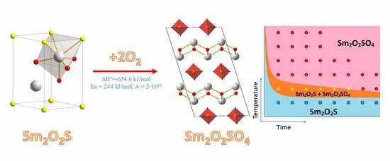
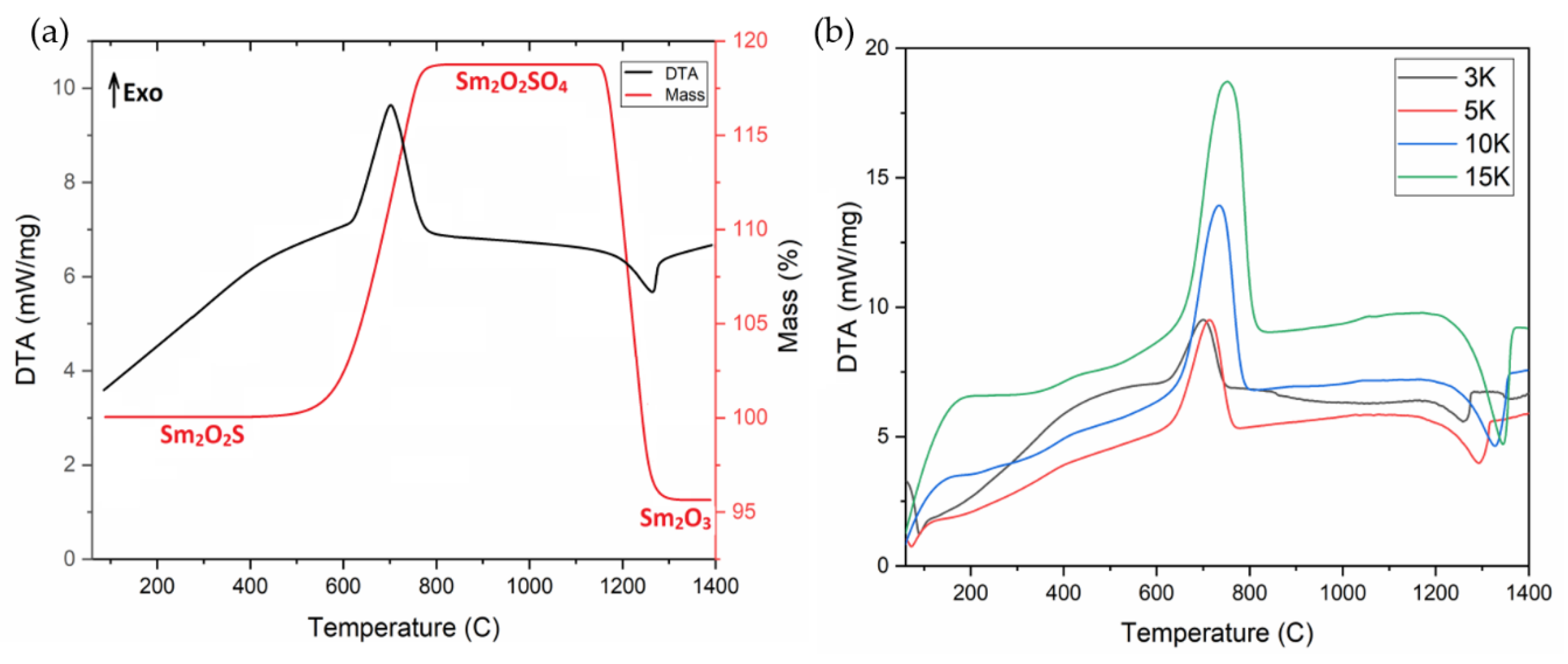
2.1. Dynamic Oxidation of Sm2O2S and Thermal Stability of Sm2O2SO4

According to the differential thermal analysis (Figure 1a), the samarium oxysulfide oxidation begins at the temperature of 550 °C, proceeds in one stage and ends at 775 °C. The mass gain corresponds to the samarium oxysulfate (Sm2O2SO4) formation. The process is described by the reaction equation:
Sm2O2S + 2O2 → Sm2O2SO4
The resulting samarium oxysulfate is stable up to 1100 °C, and, then, it decomposes in one stage with the Sm2O3formation.The process can be described by the equation:
Sm2O2SO4 → Sm2O3 + SO2 + 1/2O2
The certain enthalpies of the two reactions allow us to write thermochemical equations:
Sm2O2S (trig) + 2O2(gas) → Sm2O2SO4(monocl); ∆Hº = −654.6 kJ/mol
Sm2O2SO4(monocl) → Sm2O3(cubic) + SO2(gas) + 1/2O2(gas); ∆Hº = 170.8 kJ/mol
Using the data on the enthalpies of samarium oxide [47] and sulfur oxide (IV) [48] formation, the enthalpy of samarium oxysulfate formation was calculated by the Hess law and the value is equal to ∆Hºf(Sm2O2SO4 (monocl)) = −2294.0 kJ/mol.Substituting the enthalpy of Sm2O2SO4 formation in the equation for calculating the enthalpy of reaction 4 and using the samarium oxysulfide formation enthalpy ∆Hºf(Sm2O2S(trig)) = −1642.6 kJ/mol [49], we obtain the theoretical samarium oxysulfideoxidation enthalpy equal to −652.4 kJ/mol, which is perfectly compatible with the value determined according to the DTA measurements.
To study the kinetics of the Sm2O2SO4formation and decomposition processes, the thermal analysis of the samples was carried out at selected heating rates of 3, 5, 10, 15 °C/min (Figure 1b). Based on the DTA data at the pointed heating rates, the kinetic parameters of the processwere calculated.The temperature dependence of the oxidation rate of Sm2O2S to Sm2O2SO4 is characterized by relatively moderate parameters for such processes: Ea = 244 kJ/mol, A = 2 × 10¹⁰.The activation energy of the Sm2O2SO4decomposition to Sm2O3 is much higher and it is equalto 357 kJ/mol, but the preexponential factor is an order of magnitude lower and is equal to 1 × 10⁹. If we compare the parameters with those known for Eu2O2SO4 [14] (400 kJ/mol and 1 × 10¹², respectively), this corresponds to wider peaks in the DTA curves for the Eu2O2SO4decomposition, which indicates its higher kinetic stability, as compared to that of Sm2O2SO4. In addition, the significantly higher preexponential factor for the Eu2O2SO4decomposition, in comparison with that of Sm2O2SO4, suggests that the Sm2O2SO4 symmetry is, at least, not higher than that of Eu2O2SO4. The reduced kinetic stability of Sm2O2SO4, in comparison with that of Eu2O2SO4, is in a good agreement with the enthalpy values of compound decomposition.

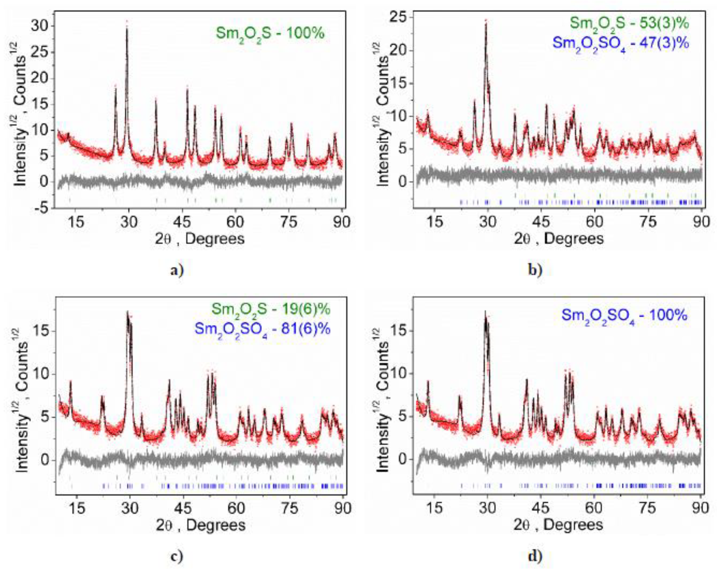
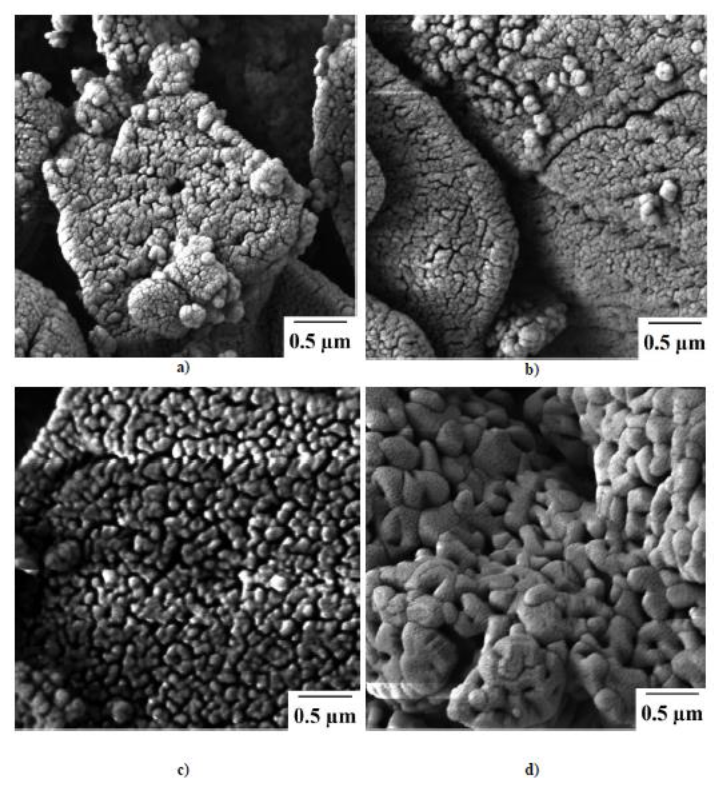
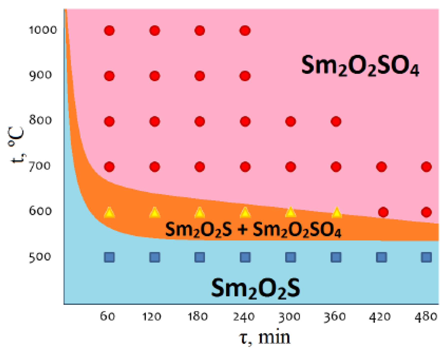
2.2. Isothermal Oxidation of Sm2O2S

At the temperature of 500 °C for 10 h, according to the results of X-ray phase analysis, there is no phase composition change of the Sm2O2S sample (Figure 2a). However, starting from 600 °C, the phase composition of the sample changes rapidly and, after only two hours, approximatelyhalf of Sm2O2S enters into the reaction (Figure 2b). After five hours, only about 20% of samarium oxysulfide remains in the sample (Figure 2c). In 7 h of the process, the sample contains only pure samarium oxysulfate (Figure 2d). The temperature increase to 700 °C leads to a sharp increase in the reaction rate, and the complete oxidation of the sample is reached for one hour. Such behavior differs significantly from the EuS oxidation process [35] where such pronounced rate temperature dependence is not observed. This effect is obviously related to the fact that only one reaction occurs during the Sm2O2S oxidation, in contrast to the EuS oxidation process, in which several parallel competing processes are realized. The samarium oxysulfide samples oxidation at 800, 900 and 1000 °C leads to the production of Sm2O2SO4 samples for one hour. An increase in the exposure time at these temperatures does not lead to a further change in the phase composition of the samples.
According to scanning electron microscopy, the samarium oxysulfide powder is formed by agglomerates sized 2–3 μm. The agglomerates have a clear granule structure. The initial granules have a size of about 50–100 nm (Figure 3a). Carrying out the oxidation process at 600 °C practically does not affect the change in the microstructure of the obtained Sm2O2SO4 samples (Figure 3b). A further increase in the process temperature leads to the agglomeration of the initial granules while maintaining the overall structure of the agglomerates (Figure 3c,d). In the Sm2O2SO4 sample obtained at 1000 °C, the initial granules have sizes from 250 nm to 0.5 μm. It should be pointed that the particlemicrostructure preservation is an important effect determining the possibility of applying the oxidation process to the synthesis of biocompatible materials based on rare earth oxysulfates [28,29,30].
Based on the analysis of available experimental data on the phase composition of the samples obtained in isothermal processes, a kinetic diagram was built for the chemical composition changes during the samarium oxysulfide oxidation with air oxygen (Figure 4). In the diagram, threephase statefields can be observed. Two single-phase fields related to the stability conditions for compounds Sm2O2S (blue) and Sm2O2SO4 (pink), and the intermediate two-phase field of Sm2O2S + Sm2O2SO4 (orange), whichboundaries are clearly governed by the thermodynamic and kinetic parameters of the process, are determined. As it is seen, the pure Sm2O2SO4 phase can be synthesized at temperatures ≥700 °C for the reaction time 60–480 min. The phase fieldposition in the diagram allows one to determine the conditions for the targeted preparation of the samples with specified phase compositions.

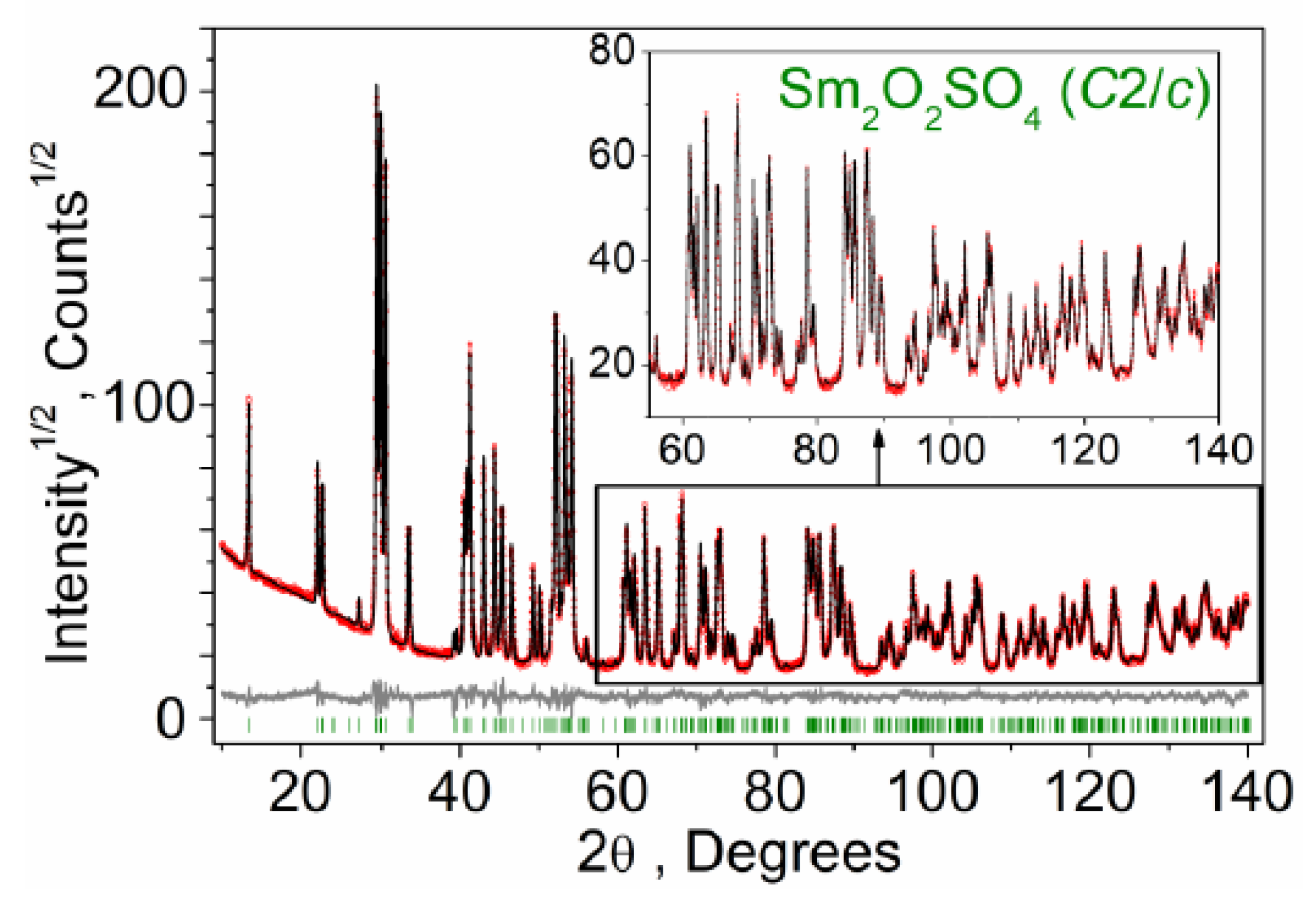
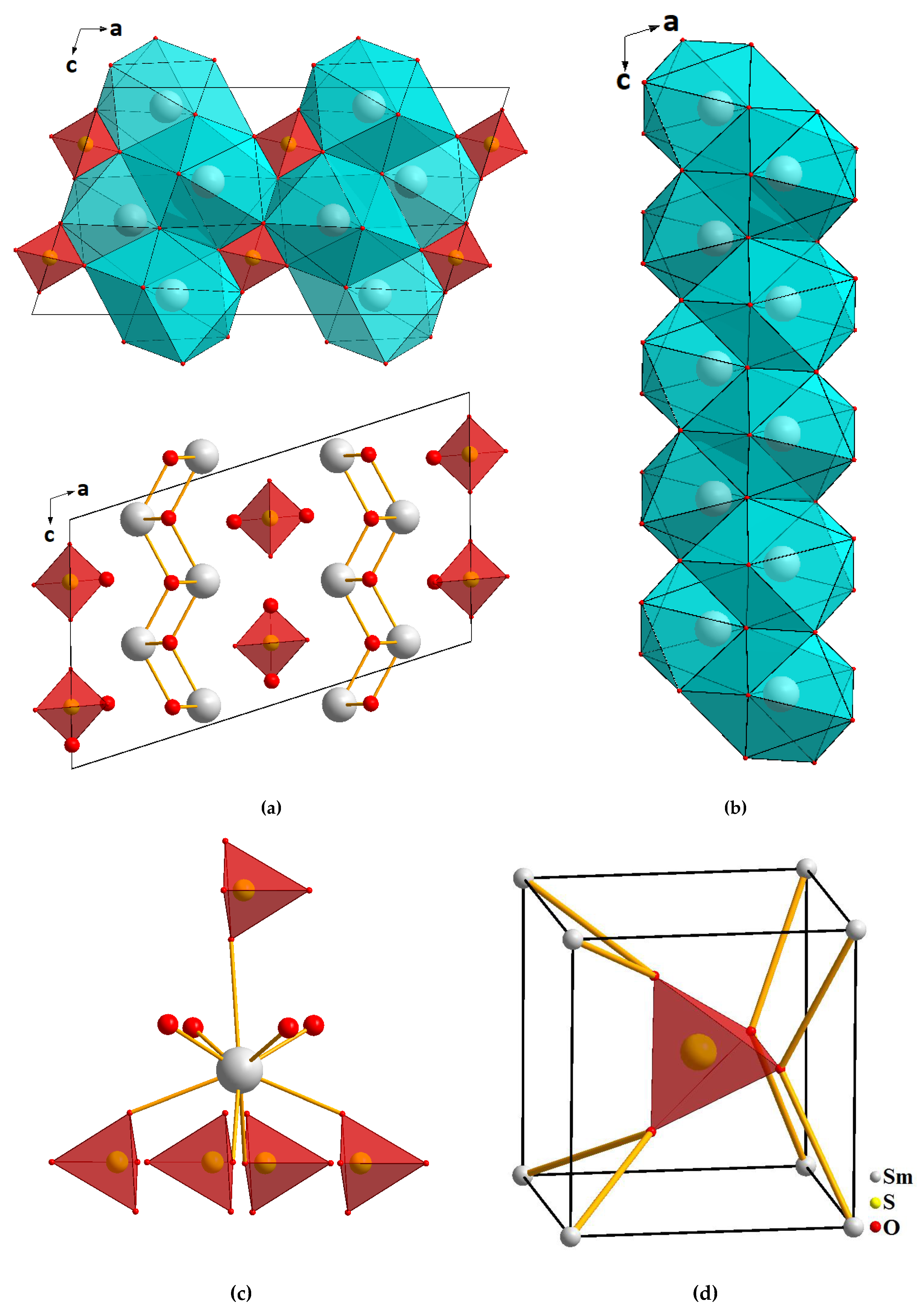
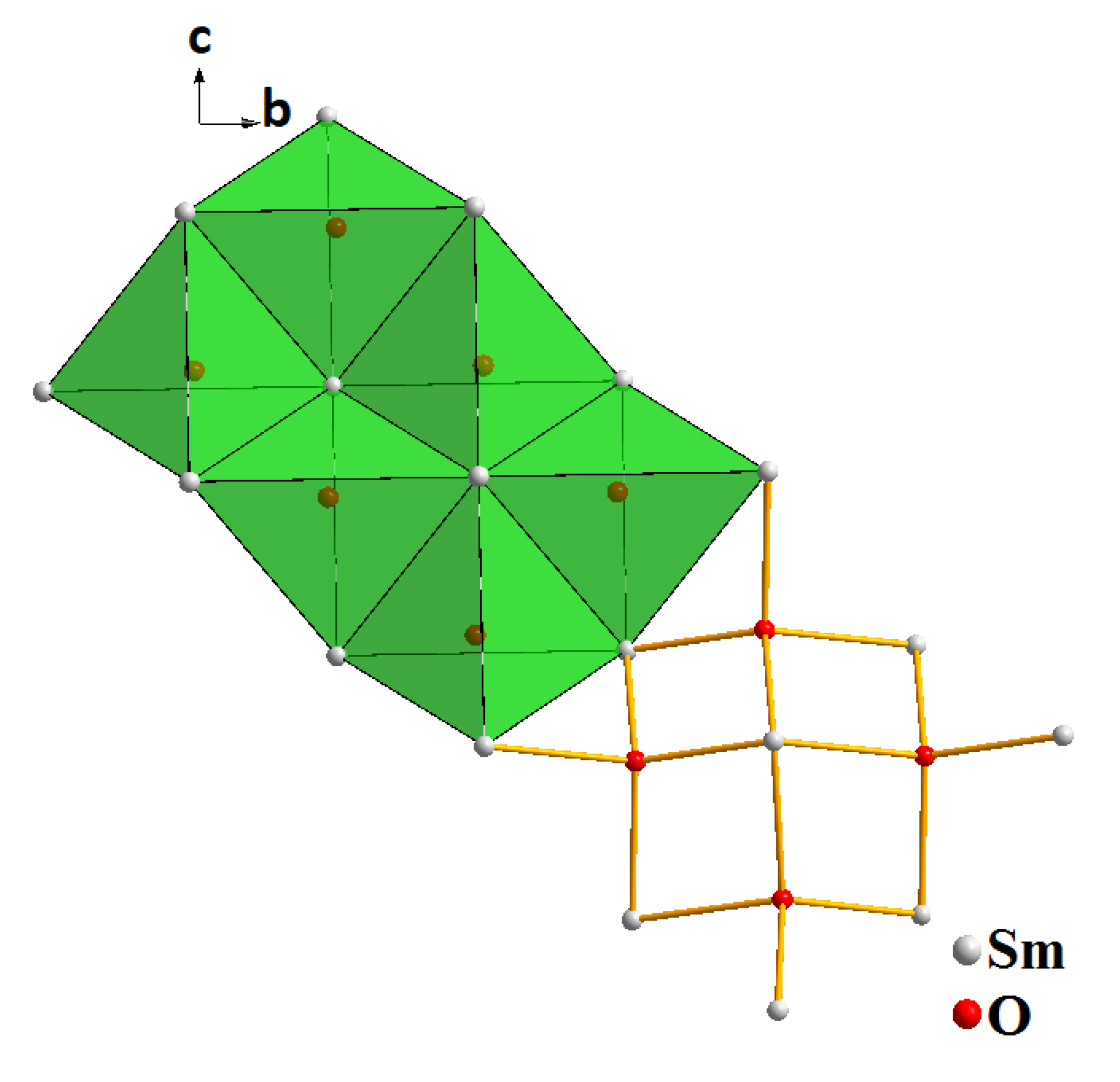
2.3. Structural Properties of Sm2O2SO4

A sample of Sm2O2SO4 for structural analysis was obtained by oxidizing samarium oxysulfide in the air at 900 °C for 10 h. The Rietveld refinement was carried out by using TOPAS 4.2 [50] which accounts the esd’s of each point by a special weight scheme. All peaks were indexed by a monoclinic cell (C2/c) with the parameters close to those of Eu2O2SO4 [35] and; therefore, the crystal structure of Eu2O2SO4was taken as a starting model for Rietveld refinement. The Eu3+ site in the Eu2O2SO4 structure was considered as occupied by the Sm3+ ion. In order to reduce the number of refined parameters, only one thermal parameter was refined for all O atoms. The refinement was stable and gave low R-factors (Table 1, Figure 5). The atom coordinates and main bond lengths obtained in Sm2O2SO4aresummarized in Table 2 and Table 3, respectively. The cif and checkcif files are given in Supplementary Materials. The crystallographic data are deposited in the Cambridge Crystallographic Data Centre (CSD # 1968636). The data can be downloaded from the site (www.ccdc.cam.ac.uk/data_request/cif).
The main difference of Eu2O2SO4 and Sm2O2SO4 structures is observed in their cell parameters and cell volumes. The former crystal has a = 13.65826(27), b = 4.188744(73), c = 8.14400(14) Å, β = 107.2819(21)°, V = 444.892(15) Å3, and the compound under investigation Sm2O2SO4 is characterized by a = 13.7442 (2), b = 4.20178 (4), c = 8.16711 (8) Å, β = 107.224 (1)°, V = 450.498 (9) Å3. It is clearly seen that the cell parameters and cell volume of Eu2O2SO4 are smaller than those of Sm2O2SO4, and it is consistent with the fact that ion radius IR(Eu, CN=9) = 1.12 Å is smaller than IR(Sm, CN=9) = 1.132 Å.
As shown in Figure 6, the structure is represented by the alternation of cationic layers [Sm2O22+]n with the anionic layers consisting of isolated [SO4]2− tetrahedra. Both layers are parallel to (100) (Figure 6a). All samarium atoms occupy identical crystallographic positions and are coordinated by nine oxygen atoms: five oxygen atoms belong to monodentate-bound sulfate groups, and the remaining oxygen atoms are bridging (Figure 6c). Thus, the samarium atom in the structure forms a coordination environment shaped as a three-cap trigonal prism. Two caps of the coordination polyhedron, connected along the edge at the angle of 180°, form a plane of four oxygen atoms. The trigonal prism and caps in the coordination polyhedron are deformed due to the difference in the Sm-O bond lengths. One Sm-O bond is much longer than the others. As a result, the coordination number of samarium is classified as 8 + 1. The SmO9 polyhedra join with each other forming an infinite chain along the c-axis (Figure 6b). The oxygen atoms of SO4 groups are coordinated by sulfur and samarium atoms. The sulfate tetrahedron is surrounded by eight samarium atoms, resulting in the formation of sphere-shaped coordination as almost a perfect cube (Figure 6d). Each bridging oxygen atom is coordinated by four samarium atoms, and it results in the formation of [OSm4] tetrahedra. These tetrahedra, sequentially pair wise connected with each other, form unlimited zigzag chains. The interconnected chains form continuous layers (Figure 7).

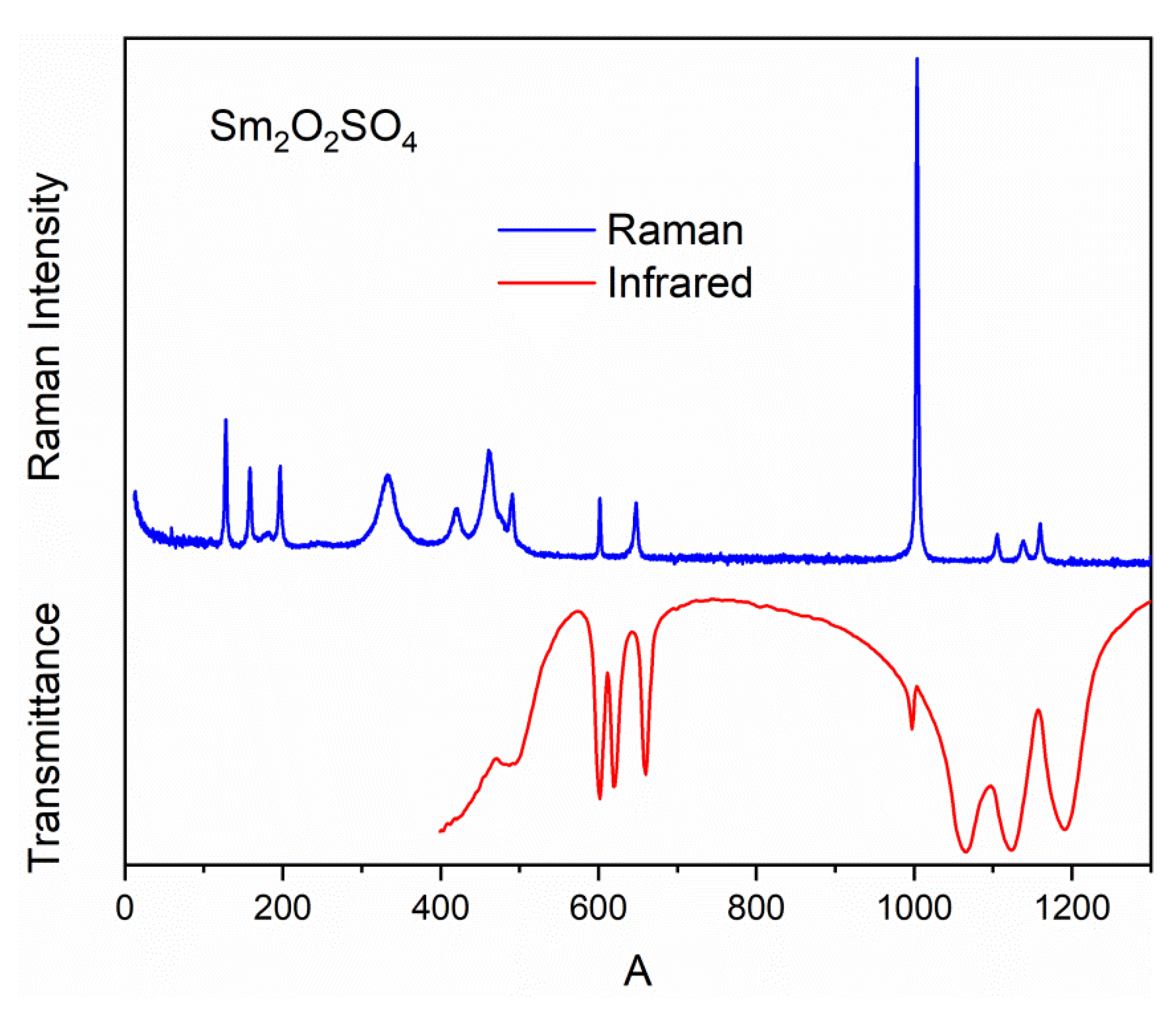
2.4. Vibrational Spectra of Sm2O2SO4

Raman and Infrared spectra of Sm2O2SO4 are shown in Figure 8. The irreducible vibrational representations for the monoclinic structure of Sm2O2SO4 at the center of the Brillouin zone is Γvibr = 13Ag + 13Au + 14Bg + 14Bu, where Au + 2Bu are acoustic modes and 13Ag + 14Bg are Raman-active modes, while the 12Au + 12Bu modes are active in IR spectra. The free tetrahedral [SO4]2− ion of the Td symmetry exhibits four internal vibrations. All four vibrations are Raman-active, whereas only ν3 and ν4 are Infrared-active. In the solid state, ν3 and ν4 may split into two or three bands because of the site effect [51]. The correlation diagram of internal vibrations between the free [SO4]2− ions of the Tdsymmetry, its site symmetry (C2) and the factor group symmetry (C2h) of a unit cell is given in Table 4.
From the correlation diagram, we can conclude that four spectral bands should be observed in the range of stretching vibrations of the SO4 tetrahedra (975–1225 cm−1) in the Raman spectrum. The IR spectrum of the Sm2O2SO4 structure should contain four bands in the range of stretching vibrations of [SO4]2− ions, too. Three of them are ν3 antisymmetric stretching and one is related to ν1 symmetric stretching vibration. The ν4 bending vibrations locate in the range of 575–675 cm−1. The relevant spectral bands can be seen in Figure S1 (Supplementary Materials) and Figure 8. The Raman bands associated with the ν4 bending vibrations of SO4tetrahedra are overlapped with bands related to Sm-O vibrations, and these vibrations locate in the range of 300–500 cm−1. The low-intensity bands in Raman spectra around 250 cm−1 should correspond to rotational vibrations of [SO4]2− ions [52]. The remaining spectral bands below 200 cm-1 are translational vibrations of SmO9polyhedra, SO4tetrahedra and Sm3+ ions.

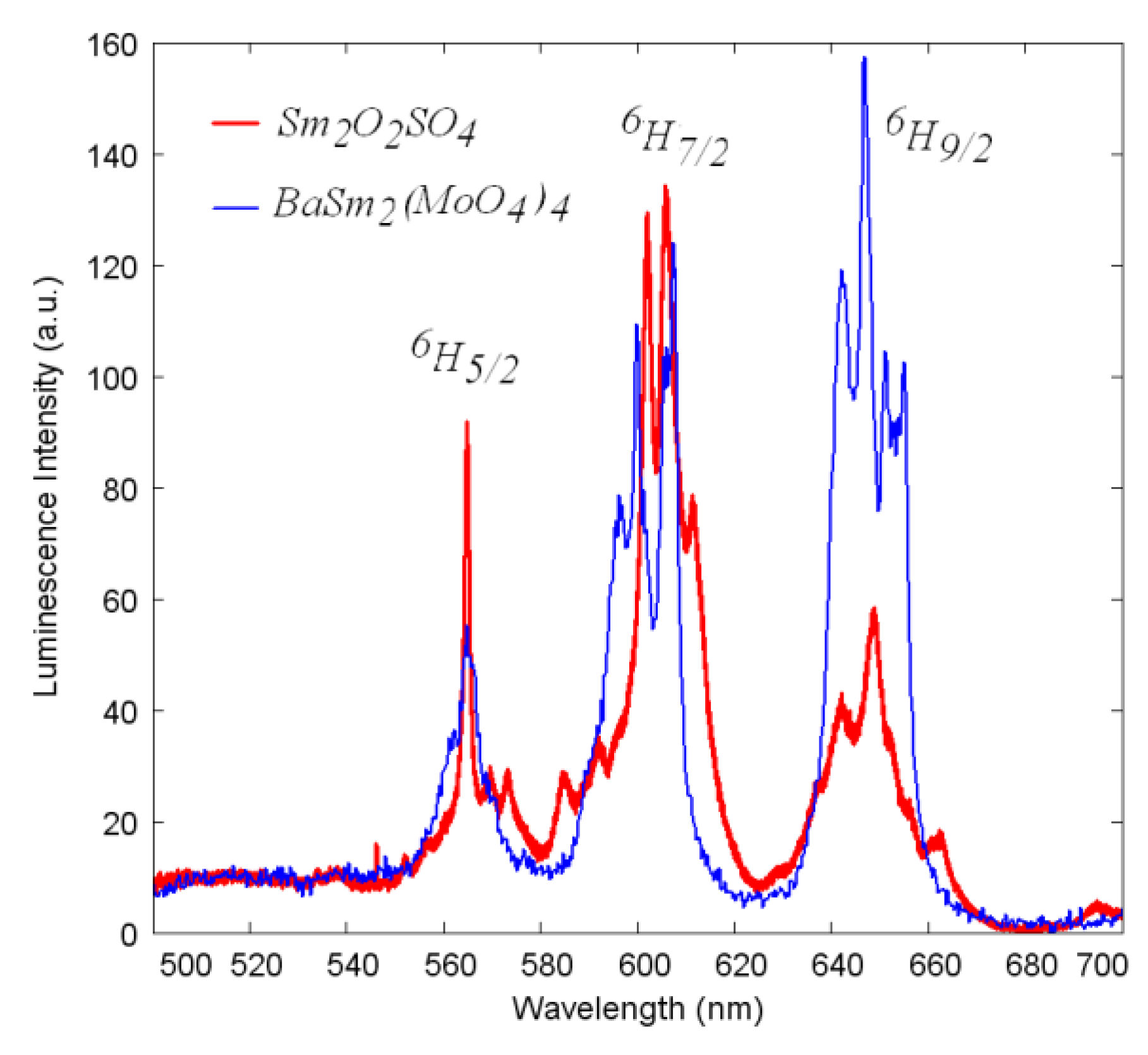
2.5. Luminescent Properties of Sm2O2SO4

The Sm2O2SO4 luminescence spectrum was recorded using the excitation by theGaN laser diode with the central wavelength 410 nm (24400 cm−1) falling into three closely-spaced Sm3+ transitions from the ground state 6H5/2 to 6P5/2, 4M19/2 and 4L13/2 excited states. The obtained spectrum is presented in Figure 9 in comparison with the luminescence spectrum of another highly-concentrated samarium-containing BaSm2(MoO4)4 crystal [39]. The structure of luminescence spectra of both Sm2O2SO4 and the reference crystal is rather similar and exhibits three main bands easily assignable to the transitions from the4G5/2 state to 6H5/2, 6H7/2 and 6H9/2multiplets. However, the distribution of the intensities between three mentioned channels in Sm2O2SO4 is slightly different from that of the reference crystal, while the red transition to the6H9/2 state dominates in the reference crystal, the orange transition to the 6H7/2 state prevails in Sm2O2SO4. This difference demonstrates the possibility of controlling the samarium ion emission chromaticity via the crystal field engineering that allows certain variation of Judd–Ofelt intensity parameters. We must note that the reference crystal spectrum was divided by 10 for a better comparison of the shapes. Therefore, we must deduce that concentration quenching of the luminescence in Sm2O2SO4 is rather high in comparison with (e.g., molybdate crystalline lattices).

3. Materials and Methods

3.1. Synthesis Methods

Samarium oxysulfide was obtained by the reduction of samarium sulfate Sm2(SO4)3 (99.9%, Merck Ltd., Germany) in the hydrogen atmosphere at the temperature of 700 °C. The installation scheme for carrying out the high-temperature recovery processes is shown in Figure S2 (Supplementary Materials). High-purity hydrogen was obtained by the electrolytic method in a SPECTR-6M hydrogen generator (Spectr, Moscow, Russia). The temperature control and regulation were carried out using a microprocessor controller (Thermoceramics, Moscow, Russia). The temperature measurement in the reaction zone was provided by a chromel–alumel thermocouple. A weighed amount of dry Sm2(SO4)3 was placed in a quartz reactor, and it was purged with hydrogen from the generator for 30 min at the rate of 6 L/h. After that, the reactor was placed in a heated vertical furnace and kept for 5 h. After the completion of the recovery process, the reactor was removed from the furnace and cooled to room temperature. The process proceeding during the recovery is described by the equation:
Sm2(SO4)3 + 12H2 → Sm2O2S + 2H2S↑ + 10H2O↑
To study the samarium oxysulfide oxidation with air oxygen, 0.5 g of Sm2O2S sample was uniformly distributed as a thin layer over a ceramic boat bottom with the area of 3 × 5 cm2. In order to prevent the tight layer formation during the oxidation process, all samarium oxysulfide samples were crushed in an agate mortar with acetone addition. After the filling, the ceramic boat was placed in a horizontal furnace (Thermoceramics, Moscow, Russia) heated to the required temperature and the processing was carried out in a continuous air flow. After the required time, the boat was removed from the oven and cooled to room temperature in a desiccator with the silica gel to avoid surface hydration. A study of the phase composition of obtained oxidized sample was carried out by the X-ray diffraction method. The isothermal oxidation experiments were carried out at the temperatures of 500, 600, 700, 800, 900 and 1000 °C. The total time of the oxidation process at each temperature did not exceed 10 h.

3.2. Methods of Physico-Chemical Analysis

The thermal analysis in the synthetic air (80% Ar-20% O2) flow was carried out on a Simultaneous Thermal Analysis (STA) equipment 499 F5 Jupiter NETZSCH (Netzsch, Selb, Germany). The powder samples were inserted into alumina crucibles. The heating rate was 3 °C/min. For the enthalpy determination, the equipment was calibrated with the use of standard metal substances, such as In, Sn, Bi, Zn, Al, Ag, Au and Ni. The heat effect peaks were determined with the package «Proteus 6 2012» (Netzsch, Selb, Germany). The peak temperature and area in parallel experiments were reproduced at an inaccuracy lower than 3%. The kinetic parameters determination was based on Kissinger formula [53] in the linearized form:
1 T = R E l n A R E 1 E R l n b T 2
where T is the temperature with a maximum reaction rate; b—heating rate; E—activation energy and A—preexponential factor. The representative examples of using the formula in topochemical processes can be found elsewhere [54,55,56].
To determine the phase composition of the samples at various oxidation stages, we used a BRUKER D2 PHASER X-ray diffractometer (Bruker, Billerica, MA, USA) with a linear detector LYNXEYE (CuKα radiation, Ni-filter, Bruker, Billerica, MA, USA). The crystal structure was refined using the Rietveld method in the TOPAS 4.2 program [50]. The powder diffraction data of Sm2O2SO4 for Rietveld analysis were collected at room temperature with a Bruker D8 ADVANCE powder diffractometer (Cu-Kα radiation, Bruker, USA) equipped with a linear detector VANTEC (Bruker, Billerica, MA, USA). The step size of 2θ was 0.016°, and the counting time was 5 s per step. The particle morphology analysis was carried out on an electron microscope JEOL JSM-6510LV (Japan). The X-ray energy-dispersive analyzer (Oxford Instruments, Abington, UK) was used to register the X-ray signal at recording the element spectrum in the selected regions of the sample surface. The possible inaccuracy of elemental content determination by this method was equal to ±0.2%. The Fourier-transform infrared spectroscopy (FTIR) analysis was carried out with the use of Fourier-Transform Infrared Spectrometer FSM 1201 (Infraspec, Moscow, Russia). The sample for the investigation was prepared in the tablet shape with the addition of annealed KBr. The Raman scattering spectra of Sm2O2SO4 were collected in backscattering geometry, using a triple monochromator Horiba JobinYvon T64000 Raman spectrometer (JobinYvon, France) operating in subtractive mode. The spectral resolution for the recorded Stokes side Raman spectra was about 1 cm−1 (this resolution was achieved by using gratings with 1800 grooves mm−1 and 100 micrometer slits). Single-mode krypton 647.1 nm of Lexel Kr+ laser of 3 mW on the sample was used as an excitation light source. The luminescence spectra at room temperature were recorded using a Horiba-Jobin-Yvon T64000 spectrometer (JobinYvon, France) and GaN laser diode with the central wavelength 410 nm. Spectral resolution of the measurement channel of the spectrometer was 2.7 cm−1.

4. Conclusions

A comprehensive study of the samariumoxysulfide oxidation process was carried out. The kinetic and thermodynamic characteristics of the process were established. The effect of oxidation temperature on the morphology of samarium oxysulfate samples was evaluated. The main structural and spectroscopic characteristics of samarium oxysulfatewere determined. According to the X-ray powder diffraction data, the monoclinic symmetrywasestablished.The main structural elements and their influence on the properties of the compound were analyzed. The theoretical calculations of vibration spectra confirm the adequacy of the structural model, which is important for such complex structures with the ambiguity in the choice of the structural model. The Sm2O2SO4luminescent-spectral characteristics were determined. The luminescence spectrum consists of three main luminescent bands originating from the 4G5/2 state, the transition to 6H7/2 in the orange part of the spectrum being dominant.

Supplementary Materials

The following are available online. The cif and checkcif files. Figure S1: Decomposition of the Sm2O2SO4 Raman spectrum in the range of ν4 vibrations of [SO4]2− ions, Figure S2: Installation scheme of processing substances in a stream of hydrogen: 1—a hydrogen generator; 2—power control unit of electricity supplied to the furnace; 3—Thermocouple; 4—electric heating furnace; 5—reactor with the processed substance.

Author Contributions

Conceptualization, Y.G.D. and V.V.A.; methodology, E.I.S.; software, S.A.B.; formal analysis, A.S.O., M.S.M., A.S.A.; investigation, A.S.O. and A.S.K.; resources, S.S.V.; data curation, A.S.A. and N.A.K.; writing—original draft preparation, Y.G.D.; writing—review and editing, V.V.A.; project administration, O.V.A. All authors have read and agreed to the published version of the manuscript.

Funding

This research was funded by the Russian Foundation for Basic Research (Grants 18-02-00754, 18-32-20011) and Russian Science Foundation (project 19-42-02003).

Acknowledgments

A.S.Aleksandrovsky, A.S.Krylov and A.S.Oreshonkov are grateful to the Basic Project of the Ministry of Science of the Russian Federation. Use of equipment of Krasnoyarsk Regional Center of Research Equipment of Federal Research Center «Krasnoyarsk Science Center SB RAS» is acknowledged.

Conflicts of Interest

The authors declare no conflicts of interest.

References

  1. Andreev, P.O.; Sal’nikova, E.I.; Kislitsyn, A.A. Kinetics of the transformation of Ln2O2SO4 into Ln2O2S (Ln = La, Pr, Nd, and Sm) in a hydrogen flow. Russ. J. Phys. Chem. A 2013, 87, 1482–1487. [Google Scholar] [CrossRef]
  2. Sal’nikova, E.I.; Andreev, P.O.; Antonov, S.M. Kinetic diagrams of Ln2O2SO4 phase transformations in a H2 flow (Ln = La, Pr, Nd, Sm). Russ. J. Phys. Chem. A 2013, 87, 1280–1283. [Google Scholar] [CrossRef]
  3. Sal’nikova, E.I.; Kaliev, D.I.; Andreev, P.O. Kinetics of phase formation upon the treatment of La2(SO4)3 and La2O2SO4 in a hydrogen flow. Russ. J. Phys. Chem. A 2011, 85, 2121–2125. [Google Scholar] [CrossRef]
  4. Machado, L.C.; de Azeredo, M.T.D.; Corrêa, H.P.S.; do Rosário Matos, J.; Mazali, Í.O. Formation of oxysulfide LnO2S2 and oxysulfate LnO2SO4 phases in the thermal decomposition process of lanthanide sulfonates (Ln = La, Sm). J. Therm. Anal. Calorim. 2012, 107, 305–311. [Google Scholar] [CrossRef]
  5. Kim, S.; Masui, T.; Imanaka, N. Synthesis of red-emitting phosphors based on gadolinium oxysulfate by a flux method. Electrochemistry 2009, 77, 611–613. [Google Scholar] [CrossRef] [Green Version]
  6. Kijima, T.; Shinbori, T.; Sekita, M.; Uota, M.; Sakai, G. Abnormally enhanced Eu3+ emission in Y2O2SO4: Eu3+ inherited from their precursory dodecylsulfate-templated concentric-layered nanostructure. J. Lumin. 2008, 128, 311–316. [Google Scholar] [CrossRef]
  7. Kijima, T.; Isayama, T.; Sekita, M.; Uota, M.; Sakai, G. Emission properties of Tb3+ in Y2O2SO4 derived from their precursory dodecylsulfate-templated concentric- and straight-layered nanostructures. J. Alloys Compd. 2009, 485, 730–733. [Google Scholar] [CrossRef]
  8. Machida, M.; Kawamura, K.; Ito, K.; Ikeue, K. Large-capacity oxygen storage by lanthanide oxysulfate/oxysulfide systems. Chem. Mater. 2005, 17, 1487–1492. [Google Scholar] [CrossRef]
  9. Machida, M.; Kawamura, K.; Kawano, T.; Zhang, D.; Ikeue, K. Layered Pr-dodecyl sulfate mesophases as precursors of Pr2O2SO4 having a large oxygen-storage capacity. J. Mater. Chem. 2006, 16, 3084–3090. [Google Scholar] [CrossRef]
  10. Machida, M.; Kawano, T.; Eto, M.; Zhang, D.; Ikeue, K. Ln dependence of the large-capacity oxygen storage/release property of Ln oxysulfate/oxysulfide systems. Chem. Mater. 2007, 19, 954–960. [Google Scholar] [CrossRef]
  11. Zhang, D.; Yoshioka, F.; Ikeue, K.; Machida, M. synthesis and oxygen release/storage properties of Ce-substituted La-oxysulfates, (La1−xCex)2O2SO4. Chem. Mater. 2008, 20, 6697–6703. [Google Scholar] [CrossRef]
  12. Denisenko, Y.G.; Aleksandrovsky, A.S.; Atuchin, V.V.; Krylov, A.S.; Molokeev, M.S.; Oreshonkov, A.S.; Shestakov, N.P.; Andreev, O.V. Exploration of structural, thermal and spectroscopic properties of self-activated sulfate Eu2(SO4)3 with isolated SO4 groups. J. Ind. Eng. Chem. 2018, 68, 109–116. [Google Scholar] [CrossRef] [Green Version]
  13. Hartenbach, I.; Schleid, T. Serendipitous Formation of Single-Crystalline Eu2O2[SO4]. Z. Anorg. Allg. Chem. 2002, 628, 2171. [Google Scholar] [CrossRef]
  14. Denisenko, Y.G.; Khritokhin, N.A.; Andreev, O.V.; Basova, S.A.; Sal’nikova, E.I.; Polkovnikov, A.A. Thermal decomposition of europium sulfates Eu2(SO4)3·8H2O and EuSO4. J. Solid State Chem. 2017, 255, 219–224. [Google Scholar] [CrossRef]
  15. Poston, J.A.; Siriwardane, R.V.; Fisher, E.P.; Miltz, A.L. Thermal decomposition of the rare earth sulfates of cerium (III), cerium (IV), lanthanum (III) and samarium (III). Appl. Surf. Sci. 2003, 214, 83–102. [Google Scholar] [CrossRef]
  16. Liang, J.; Ma, R.; Geng, F.; Ebina, Y.; Sasaki, T. Ln2(OH)4SO4·nH2O (Ln = Pr to Tb; n ∼ 2): A new family of layered rare-earth hydroxides rigidly pillared by sulfate ions. Chem. Mater. 2010, 22, 6001–6007. [Google Scholar] [CrossRef]
  17. Chen, F.; Chen, G.; Liu, T.; Zhang, N.; Liu, X.; Luo, H.; Li, J.; Chen, L.; Ma, R.; Qiu, G. Controllable fabrication and optical properties of uniform gadolinium oxysulfate hollow spheres. Sci. Rep. 2016, 5, 17934. [Google Scholar] [CrossRef] [Green Version]
  18. Zhang, D.; Kawada, T.; Yoshioka, F.; Machida, M. Oxygen gateway effect of CeO2/La2O2SO4 composite oxygen storage materials. ACS Omega 2016, 1, 789–798. [Google Scholar] [CrossRef] [Green Version]
  19. Xing, T.H.; Song, L.X.; Xiong, J.; Cao, H.B.; Du, P.F. Preparation and luminescent properties of Tb3+ doped Y2O2SO4microflakes. Adv. Appl. Ceram. 2013, 112, 455–459. [Google Scholar] [CrossRef]
  20. Sal’nikova, E.I.; Denisenko, Y.G.; Aleksandrovsky, A.S.; Kolesnikov, I.E.; Lähderanta, E.; Andreev, P.O.; Azarapin, N.O.; Andreev, O.V.; Basova, S.A.; Matigorov, A.V. Synthesis and optical properties RE2O2S: Ln (RE= La, Y.; Ln= Ce, Eu, Dy, Er). J. Solid State Chem. 2019, 279, 120964. [Google Scholar]
  21. Gaft, M.; Raichlin, Y.; Pelascini, F.; Panzer, G.; Motto Ros, V. Imaging rare-earth elements in minerals by laser-induced plasma spectroscopy: Molecular emission and plasma-induced luminescence. Spectrochim. Acta Part B At. Spectrosc. 2019, 151, 12–19. [Google Scholar] [CrossRef]
  22. Yu, M.; Xu, H.; Li, Y.; Dai, Q.; Wang, G.; Qin, W. Morphology luminescence and photovoltaic performance of lanthanide-doped CaWO4 nanocrystals. J. Colloid Interface Sci. 2020, 559, 162–168. [Google Scholar] [CrossRef] [PubMed]
  23. Zhang, J.; Chen, G.; Zhai, Z.; Chen, H.; Zhang, Y. Optical temperature sensing using upconversion luminescence in rare-earth ions doped Ca2Gd8(SiO4)6O2 phosphors. J. Alloys Compd. 2019, 771, 838–846. [Google Scholar] [CrossRef]
  24. Pejchal, J.; Barta, J.; Trojek, T.; Kucerkova, R.; Beitlerova, A.; Nikl, M. Luminescence and scintillation properties of rare-earth-doped LaAlO3 single crystals. Radiat. Meas. 2019, 121, 26–31. [Google Scholar] [CrossRef]
  25. Kichanov, S.E.; Gorshkova, Y.E.; Rachkovskaya, G.E.; Kozlenko, D.P.; Zakharevich, G.B.; Savenko, B.N. Structural evolution of luminescence nanoparticles with rare-earth ions in the oxyfluoride glass ceramics. Mater. Chem. Phys. 2019, 237, 121830. [Google Scholar] [CrossRef]
  26. Atuchin, V.V.; Aleksandrovsky, A.S.; Chimitova, O.D.; Gavrilova, T.A.; Krylov, A.S.; Molokeev, M.S.; Oreshonkov, A.S.; Bazarov, B.G.; Bazarova, J.G. Synthesis and spectroscopic properties of monoclinic α-Eu2(MoO4)3. J. Phys. Chem. C 2014, 118, 15404–15411. [Google Scholar] [CrossRef]
  27. Lim, C.S.; Atuchin, V.V.; Aleksandrovsky, A.S.; Denisenko, Y.G.; Molokeev, M.S.; Oreshonkov, A.S. Fabrication of microcrystalline NaPbLa(WO4)3:Yb3+/Ho3+ phosphors and their upconversionphotoluminescent characteristics. Korean J. Mater. Res. 2019, 29, 741–746. [Google Scholar] [CrossRef]
  28. Osseni, S.A.; Denisenko, Y.G.; Fatombi, J.K.; Sal’nikova, E.I.; Andreev, O.V. Synthesis and characterization of Ln2O2SO4 (Ln = Gd, Ho, Dy and Lu) nanoparticles obtained by coprecipitation method and study of their reduction reaction under H2 flow. J. Nanostructure Chem. 2017, 7, 337–343. [Google Scholar] [CrossRef] [Green Version]
  29. Osseni, S.A.; Lechevallier, S.; Verelst, M.; Dujardin, C.; Dexpert-Ghys, J.; Neumeyer, D.; Leclercq, M.; Baaziz, H.; Cussac, D.; Santran, V.; et al. New nanoplatform based on Gd2O2S:Eu3+ core: Synthesis, characterization and use for in vitro bio-labelling. J. Mater. Chem. 2011, 21, 18365–18372. [Google Scholar] [CrossRef]
  30. Osseni, S.A.; Lechevallier, S.; Verelst, M.; Perriat, P.; Dexpert-Ghys, J.; Neumeyer, D.; Garcia, R.; Mayer, F.; Djanashvili, K.; Peters, J.A.; et al. Gadolinium oxysulfide nanoparticles as multimodal imaging agents for T2-weighted MR, X-ray tomography and photoluminescence. Nanoscale 2014, 6, 555–564. [Google Scholar] [CrossRef]
  31. Sedykh, A.E.; Kurth, D.G.; Müller-Buschbaum, K. Two series of lanthanide coordination polymers and complexes with 4′-phenylterpyridine and their luminescence properties. Eur. J. Inorg. Chem. 2019, 2019, 4564–4571. [Google Scholar] [CrossRef]
  32. Ribbeck, T.; Kerpen, C.; Löw, D.; Sedykh, A.E.; Müller-Buschbaum, K.; Ignat’ev, N.V.; Finze, M. Lanthanide trifluoromethyltricyanoborates: Synthesis, crystal structures and thermal properties. J. Fluor. Chem. 2019, 219, 70–78. [Google Scholar] [CrossRef]
  33. Stangl, J.M.; Dietrich, D.; Sedykh, A.E.; Janiak, C.; Müller-Buschbaum, K. Luminescent MOF polymer mixed matrix membranes for humidity sensing in real status analysis. J. Mater. Chem. C 2018, 6, 9248–9257. [Google Scholar] [CrossRef]
  34. Atuchin, V.V.; Aleksandrovsky, A.S.; Bazarov, B.G.; Bazarova, J.G.; Chimitova, O.D.; Denisenko, Y.G.; Gavrilova, T.A.; Krylov, A.S.; Maximovskiy, E.A.; Molokeev, M.S.; et al. Exploration of structural, vibrational and spectroscopic properties of self-activated orthorhombic double molybdateRbEu(MoO4)2 with isolated MoO4 units. J. Alloys Compd. 2019, 785, 692–697. [Google Scholar] [CrossRef] [Green Version]
  35. Denisenko, Y.G.; Molokeev, M.S.; Krylov, A.S.; Aleksandrovsky, A.S.; Oreshonkov, A.S.; Atuchin, V.V.; Azarapin, N.O.; Plyusnin, P.E.; Sal’nikova, E.I.; Andreev, O.V. High-Temperature Oxidation of Europium (II) Sulfide. J. Ind. Eng. Chem. 2019, 79, 62–70. [Google Scholar] [CrossRef] [Green Version]
  36. Andrrev, O.V.; Razumkova, I.A.; Boiko, A.N. Synthesis and thermal stability of rare earth compounds REF3, REF3·nH2O and (H3O)RE3F10·nH2O (RE= Tb−Lu, Y), obtained from sulphide precursors. J. Fluor. Chem. 2018, 207, 77–83. [Google Scholar] [CrossRef]
  37. Razumkova, I.A.; Boiko, A.N.; Andreev, O.V.; Basova, S.A. Synthesis of [(H3O)Tm3F10]·nH2O, ErF3, and TmF3 powders and their physicochemical properties. Russ. J. Inorg. Chem. 2017, 62, 418–422. [Google Scholar]
  38. Razumkova, I.A. Synthesis of NaYF4 compounds from sulfide precursors. J. Fluor. Chem. 2018, 205, 1–4. [Google Scholar] [CrossRef]
  39. Atuchin, V.V.; Aleksandrovsky, A.S.; Molokeev, M.S.; Krylov, A.S.; Oreshonkov, A.S.; Zhou, D. Structural and spectroscopic properties of self-activated monoclinic molybdate BaSm2(MoO4)4. J. Alloys Compd. 2017, 729, 843–849. [Google Scholar] [CrossRef] [Green Version]
  40. Atuchin, V.V.; Aleksandrovsky, A.S.; Chimitova, O.D.; Diao, C.P.; Gavrilova, T.A.; Kesler, V.G.; Molokeev, M.S.; Krylov, A.S.; Bazarov, B.G.; Bazarova, J.G.; et al. Electronic structure of β-RbSm(MoO4)2 and chemical bonding in molybdates. Dalt. Trans. 2015, 44, 1805–1815. [Google Scholar] [CrossRef]
  41. Atuchin, V.V.; Subanakov, A.K.; Aleksandrovsky, A.S.; Bazarov, B.G.; Bazarova, J.G.; Gavrilova, T.A.; Krylov, A.S.; Molokeev, M.S.; Oreshonkov, A.S.; Stefanovich, S.Y. Structural and spectroscopic properties of new noncentrosymmetric self-activated borate Rb3EuB6O12 with B5O10 units. Mater. Des. 2018, 140, 488–494. [Google Scholar] [CrossRef] [Green Version]
  42. Ji, H.; Huang, Z.; Xia, Z.; Molokeev, M.S.; Jiang, X.; Lin, Z.; Atuchin, V.V. Comparative investigations of the crystal structure and photoluminescence property of eulytite-type Ba3Eu(PO4)3 and Sr3Eu(PO4)3. Dalt. Trans. 2015, 44, 7679–7686. [Google Scholar] [CrossRef] [PubMed]
  43. Biondo, V.; Sarvezuk, P.W.C.; Ivashita, F.F.; Silva, K.L.; Paesano, A.; Isnard, O. Geometric magnetic frustration in RE2O2S oxysulfides (RE= Sm, Eu and Gd). Mater. Res. Bull. 2014, 54, 41–47. [Google Scholar] [CrossRef]
  44. Yu, C.; Yu, M.; Li, C.; Liu, X.; Yang, J.; Yang, P.; Lin, J. Facile sonochemical synthesis and photoluminescent properties of lanthanide orthophosphate nanoparticles. J. Solid State Chem. 2009, 182, 339–347. [Google Scholar] [CrossRef]
  45. Cascales, C.; Lor, B.G.; Puebla, E.G.; Iglesias, M.; Monge, M.A.; Valero, C.R.; Snejko, N. Catalytic behavior of rare-earth sulfates: Applications in organic hydrogenation and oxidation reactions. Chem. Mater. 2004, 16, 4144–4149. [Google Scholar] [CrossRef]
  46. Xu, Z.; Li, C.; Hou, Z.; Peng, C.; Lin, J. Morphological control and luminescence properties of lanthanide orthovanadate LnVO4 (Ln= La to Lu) nano-/microcrystals via hydrothermal process. Cryst. Eng. Comm. 2011, 13, 474–482. [Google Scholar] [CrossRef]
  47. Baker, F.B.; Fitzgibbon, G.C.; Pavone, D.; Holley, C.E.; Hansen, L.D.; Lewis, E.A. Enthalpies of formation of Sm2O3 (monoclinic) and Sm2O3 (cubic). J. Chem. Thermodyn. 1972, 4, 621–636. [Google Scholar] [CrossRef]
  48. Eckman, J.R.; Rossini, F.D. The heat of formation of sulphur dioxide. Bur. Stand. J. Res. 1929, 3, 597. [Google Scholar] [CrossRef]
  49. Suponitskiy, Y.L. Thermal Chemistry of Oxygen-Containing Compounds of REE Elements and Elements of Group VI. Thesis of Doctor of Science in Chemistry, D. Mendeleev University of Chemical Technology of Russia, Moscow, Russia, 2002. [Google Scholar]
  50. Bruker AXS TOPAS V4; Bruker: Karlsruhe, Germany, 2008.
  51. Nakamoto, K. Infrared and Raman Spectra of Inorganic and Coordination Compounds, 6th ed.; Wiley: New York, NY, USA, 2009. [Google Scholar]
  52. Denisenko, Y.G.; Atuchin, V.V.; Molokeev, M.S.; Aleksandrovsky, A.S.; Krylov, A.S.; Oreshonkov, A.S.; Volkova, S.S.; Andreev, O.V. Structure, thermal stability, and spectroscopic properties of triclinic double sulfate AgEu(SO4)2 with isolated SO4 groups. Inorg. Chem. 2018, 57, 13279–13288. [Google Scholar] [CrossRef]
  53. Blaine, R.L.; Kissinger, H.E. Homer Kissinger and the Kissinger equation. Thermochim. Acta 2012, 540, 1–6. [Google Scholar] [CrossRef]
  54. Bukovec, N.; Bukovec, P.; Šiftar, J. Kinetics of the thermal decomposition of Pr2(SO4)3 to Pr2O2SO4. Thermochim. Acta 1980, 35, 85–91. [Google Scholar] [CrossRef]
  55. Lyadov, A.S.; Kurilkin, V.V. Reduction specifics of rare-earth orthovanadates (REE= La, Nd, Sm, Dy, Ho, Er, Tm, Yb, and Lu). Russ. J. Inorg. Chem. 2016, 61, 86–92. [Google Scholar] [CrossRef]
  56. Llópiz, J.; Romero, M.M.; Jerez, A.; Laureiro, Y. Kinetic analysis of thermogravimetric data. Thermochim. Acta 1995, 256, 205–211. [Google Scholar] [CrossRef]
Sample Availability: Samples of the Sm2O2SO4 are available from the authors.
Figure 1. DTA/TGof Sm2O2S in synthetic air (a) and the shift of the peaks of thermal effects depending on the heating rate (b).
Molecules 25 01330 g001
Figure 2. X-ray diffraction patterns of Sm2O2S (a) and the samples subjected to oxidation at 600 °C for 2 h (b), 5 h (c) and 7 h (d).
Molecules 25 01330 g002
Figure 3. SEM images of Sm2O2S (a) and of Sm2O2SO4 samples obtained at temperatures of 600 °C (b), 800 °C (c) and 1000 °C (d).
Molecules 25 01330 g003
Figure 4. Kinetic scheme of changes in the chemical composition during the samarium oxysulfide oxidation.
Molecules 25 01330 g004
Figure 5. Difference Rietveld plot of Sm2O2SO4.
Molecules 25 01330 g005
Figure 6. Projections of the Sm2O2SO4 crystal structure (a), the structure of zigzag chains [SmO9]n (b), coordination of samarium (c) and coordination of sulfate tetrahedra (d) in the structure.
Molecules 25 01330 g006
Figure 7. The cationic layers structure formed by the junction of tetrahedra [OSm4].
Molecules 25 01330 g007
Figure 8. Raman and Infrared spectra of Sm2O2SO4 powder.
Molecules 25 01330 g008
Figure 9. The luminescence spectra of Sm2O2SO4 (red) and of the reference crystal (BaSm2(MoO4)4, blue). For better comparison, the reference crystal spectrum is divided by 10.
Molecules 25 01330 g009
Table 1. Main parameters of processing and refinement of the Sm2O2SO4 sample.
CompoundSm2O2SO4
Space groupC2/c
a, Å13.7442 (2)
b, Å4.20178 (4)
c, Å8.16711 (8)
β, º107.224 (1)
V, Å3450.498 (9)
Z4
-interval, º10–140
Rwp, %6.16
Rp, %4.52
Rexp, %2.78
χ22.22
RB, %1.70
a, b, c and β—cell parameters; V—cell volume, Z—number of formula in unit cell; Rwp—weighted profile R-factor, Rp—profile R-factor; Rexp—expected R-factor; χ2—goodness of fit, RB—Bragg R-factor.
Table 2. Fractional atomic coordinates and isotropic displacement parameters (Å2) of Sm2O2SO4.
xyzBiso
Sm10.16930 (3)0.5015 (4)0.0850 (3)0.45 (2)
S100.0339 (15)0.251.63 (8)
O30.0904 (4)0.8717 (12)0.2840 (19)0.75 (7)
O20.9996 (8)0.2711 (12)0.0985 (8)0.75 (7)
O10.2474 (3)0.022 (2)0.120 (3)0.75 (7)
Biso—isotropic thermal parameter.
Table 3. Main bond lengths (Å) of Sm2O2SO4.
Sm1-O32.698 (9)Sm1-O1 v2.417 (10)
Sm1-O3 i2.846 (13)Sm1-O1 vi2.291 (14)
Sm1-O3 ii3.202 (3)Sm1-O1 vii2.346 (19)
Sm1-O2 iii2.558 (9)S1-O3 viii1.372 (5)
Sm1-O2 iv2.547 (8)S1-O2 iii1.588 (7)
Sm1-O12.259 (9)
Symmetry codes: (i) x, -y+1, z-1/2; (ii) 1/2-x, -1/2+y, 1/2-z; (iii) x-1, y, z; (iv) -x+1, -y+1, -z; (v) x, y+1, z; (vi) -x+1/2, -y+1/2, -z; (vii) -x+1/2, y+1/2, -z+1/2; (viii) x, y-1, z.
Table 4. Correlation diagram of internal vibrations of the [SO4]2− ions in the Sm2O2SO4.
Wavenumber (cm−1) [51]Td
Point Group
C2
Site Symmetry
C2h
Factor Group Symmetry
983A11)AAg+ Au
450E2)2A2Ag + 2Au
1105E3)A + 2BAg + Au+ 2Bg+ 2Bu
611E4)A+ 2BAg+ Au+ 2Bg+ 2Bu

