
Phytochemical Composition, Antioxidant Capacity, and Enzyme Inhibitory Activity in Callus, Somaclonal Variant, and Normal Green Shoot Tissues of Catharanthus roseus (L) G. Don

 ,
,  , ,
, , 

Abstract
1. Introduction
2. Results
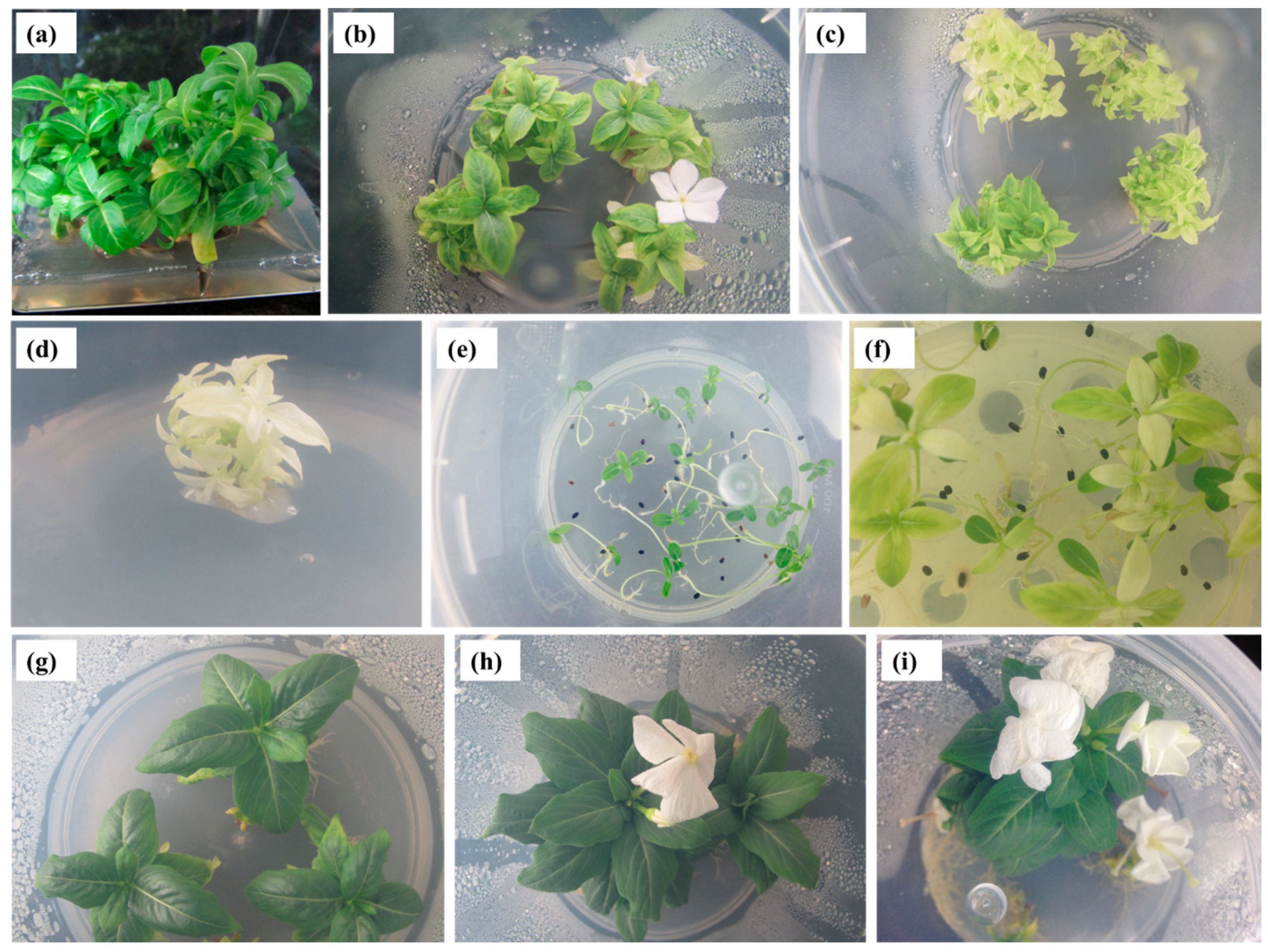
2.1. In Vitro Micropropagation
2.2. Phytochemical Composition
2.3. Antioxidant Effects
2.4. Enzyme Inhibitory Properties
3. Discussion
4. Materials and Methods
4.1. In Vitro Micropropagation
4.1.1. Plant Materials and Surface Decontamination
4.1.2. Axillary Shoot Multiplication
4.1.3. Rooting and Acclimatization
4.2. Phytochemical Analysis
4.2.1. Extract Preparation
4.2.2. Identification and Quantification of the Phytochemicals
4.2.3. Determination of Total Phenolics and Flavonoids
4.3. Biological Activities
4.3.1. Antioxidant Activity
4.3.2. Enzyme Inhibition Assay
5. Conclusions
Supplementary Materials
Author Contributions
Funding
Acknowledgments
Conflicts of Interest
References
- Alam, M.M.; Naeem, M.; Khan, M.M.A.; Uddin, M. Vincristine and Vinblastine Anticancer Catharanthus Alkaloids: Pharmacological Applications and Strategies for Yield Improvement. In Catharanthus Roseus; Springer: Cham, Switzerland, 2017; Volume 15, pp. 277–307. [Google Scholar]
- Qu, Y.; Safonova, O.; De Luca, V. Completion of the canonical pathway for assembly of anti-cancer drugs vincristine/vinblastine in Catharanthus roseus. Plant J. 2019, 97, 257–266. [Google Scholar] [CrossRef] [PubMed]
- Mustafa, N.R.; Verpoorte, R. Phenolic compounds in Catharanthus roseus. Phytochem. Rev. 2007, 6, 243–258. [Google Scholar] [CrossRef]
- Piovan, A.; Filippini, R. Anthocyanins in Catharanthus roseus in vivo and in vitro: A review. Phytochem. Rev. 2007, 6, 235–242. [Google Scholar] [CrossRef]
- Ferreres, F.; Pereira, D.M.; Valentão, P.; Andrade, P.B.; Seabra, R.M.; Sottomayor, M. New Phenolic Compounds and Antioxidant Potential Catharanthus roseus. J. Agric. Food Chem. 2008, 56, 9967–9974. [Google Scholar] [CrossRef] [PubMed]
- Verma, P.; Mathur, A.K.; Masood, N.; Luqman, S.; Shanker, K. Tryptophan over-producing cell suspensions of Catharanthus roseus (L) G. Don and their up-scaling in stirred tank bioreactor: Detection of phenolic compound with antioxidant potential. Protoplasma 2013, 250, 371–380. [Google Scholar] [CrossRef] [PubMed]
- Senbagalakshmi, P.; Rao, M.V.; Kumar, T.S. In Vitro Studies, Biosynthesis of Secondary Metabolites and Pharmacological Utility of Catharanthus roseus (L.) G. Don.: A Review. In Catharanthus Roseus; Springer Science: Cham, Switzerland, 2017; Volume 1, pp. 153–199. [Google Scholar]
- Pham, H.N.T.; Sakoff, J.A.; Vuong, Q.V.; Bowyer, M.C.; Scarlett, C.J. Screening phytochemical content, antioxidant, antimicrobial and cytotoxic activities of Catharanthus roseus (L.) G. Don stem extract and its fractions. Biocatal. Agric. Biotechnol. 2018, 16, 405–411. [Google Scholar] [CrossRef]
- Das, A.; Sarkar, S.; Bhattacharyya, S.; Gantait, S. Biotechnological advancements in Catharanthus roseus (L.) G. Don. Appl. Microbiol. Biotechnol. 2020, 104, 4811–4835. [Google Scholar] [CrossRef] [PubMed]
- Nejat, N.; Valdiani, A.; Cahill, D.; Tan, Y.-H.; Mahmood, M.; Abiri, R. Ornamental Exterior versus Therapeutic Interior of Madagascar Periwinkle (Catharanthus roseus): The Two Faces of a Versatile Herb. Sci. World J. 2015, 2015, 1–19. [Google Scholar] [CrossRef]
- Pietrosiuk, A.; Furmanowa, M.; Łata, B. Catharanthus roseus: Micropropagation and in vitro techniques. Phytochem. Rev. 2007, 6, 459–473. [Google Scholar] [CrossRef]
- Aslam, J.; Ajaz, S.; Nadim, M.M. Pharmacognosy, phytochemistry pharmacological and biotechnological approaches of Catharanthus roseus (L.) G. Don. In Recent Trends In Biotechnology and Therapeutic Application of Medicinal Plants; Shahid, M., Shahzad, A., Malik, A., Sahai, A., Eds.; Springer: New York, NY, USA, 2013; pp. 189–210. [Google Scholar]
- Barrales-Cureño, H.J.; Andrade-Hoyos, P.; Luna-Cruz, A.; Reyes, C.; Chávez-Salinas, S.; López-Valdez, L.G. In Vitro Biotechnological Production and Pharmacological Studies of Antileukemic Alkaloids of Catharanthus roseus. In Catharanthus roseus; Naeem, M., Aftab, T., Khan, M., Eds.; Springer: Cham, Switzerland, 2017; pp. 17–34. [Google Scholar]
- Miura, Y.; Hirata, K.; Kurano, N.; Miyamoto, K.; Uchida, K. Formation of Vinblastine in Multiple Shoot Culture of Catharanthus roseus. Planta Medica 1988, 54, 18–20. [Google Scholar] [CrossRef]
- Hirata, K.; Horiuchi, M.; Asada, M.; Ando, T.; Miyamoto, K.; Miura, Y. Stimulation of dimeric alkaloid production by near-ultraviolet light in multiple shoot cultures of Catharanthus roseus. J. Ferment. Bioeng. 1992, 74, 222–225. [Google Scholar] [CrossRef]
- Satdive, R.K.; Fulzele, D.P.; Eapen, S. Studies on Production of Ajmalicine in Shake Flasks by Multiple Shoot Cultures of Catharanthus roseus. Biotechnol. Prog. 2003, 19, 1071–1075. [Google Scholar] [CrossRef] [PubMed]
- Hernandez-Dominguez, E.; Campos-Tamayo, F.; Vázquez-Flota, F. Vindoline synthesis in in vitro shoot cultures of Catharanthus roseus. Biotechnol. Lett. 2004, 26, 671–674. [Google Scholar] [CrossRef]
- Campos-Tamayo, F.; Hernández-Domínguez, E.; Vázquez-Flota, F. Vindoline Formation in Shoot Cultures of Catharanthus roseus is Synchronously Activated with Morphogenesis Through the Last Biosynthetic Step. Ann. Bot. 2008, 102, 409–415. [Google Scholar] [CrossRef]
- Pati, P.K.; Kaur, J.; Singh, P. A liquid culture system for shoot proliferation and analysis of pharmaceutically active constituents of Catharanthus roseus (L.) G. Don. Plant Cell, Tissue Organ Cult. 2011, 105, 299–307. [Google Scholar] [CrossRef]
- Sharma, A.; Mathur, A.K.; Ganpathy, J.; Joshi, B.; Patel, P. Effect of abiotic elicitation and pathway precursors feeding over terpenoid indole alkaloids production in multiple shoot and callus cultures of Catharanthus roseus. Biology 2019, 74, 543–553. [Google Scholar] [CrossRef]
- Khan, M.; Liu, H.; Wang, J.; Sun, B. Inhibitory effect of phenolic compounds and plant extracts on the formation of advance glycation end products: A comprehensive review. Food Res. Int. 2020, 130, 108933. [Google Scholar] [CrossRef]
- Moreno, P.R.H.; Van Der Heijden, R.; Verpoorte, R. Elicitor-mediated induction of isochorismate synthase and accumulation of 2,3-dihydroxy benzoic acid in Catharanthus roseus cell suspension and shoot cultures. Plant Cell Rep. 1994, 14, 188–191. [Google Scholar] [CrossRef]
- Park, H.Y.; Kim, D.H.; Sivanesan, I. Micropropagation of Ajuga species: A mini review. Biotechnol. Lett. 2017, 39, 1291–1298. [Google Scholar] [CrossRef]
- Bakrudden, A.A.A.; Shanthi, G.S.; Gouthaman, T.; Kavitha, M.S.; Rao, M.V. In vitro micropropagation of Catharanthus roseus—An anti-cancer medicinal plant. Acta Bot. Hung. 2011, 53, 197–209. [Google Scholar] [CrossRef]
- Kumar, A.; Prakash, K.; Sinha, R.K.; Kumar, N. In Vitro Plant Propagation of Catharanthus roseus and Assessment of Genetic Fidelity of Micropropagated Plants by RAPD Marker Assay. Appl. Biochem. Biotechnol. 2013, 169, 894–900. [Google Scholar] [CrossRef]
- Mehta, J.; Upadhyay, D.; Paras, P.; Ansari, R.; Rathore, S.; Tiwari, S. Multiple shoots regeneration of (anti-cancer plant) Catharanthus roseus—An important medicinal plant. American J. PharmTech Res. 2013, 3, 785–793. [Google Scholar]
- Panigrahi, J.; Dholu, P.; Shah, T.J.; Gantait, S. Silver nitrate-induced in vitro shoot multiplication and precocious flowering in Catharanthus roseus (L.) G. Don, a rich source of terpenoid indole alkaloids. Plant Cell Tissue Organ Cult. 2018, 132, 579–584. [Google Scholar] [CrossRef]
- Amiri, S.; Fotovat, R.; Tarinejhad, A.; Panahi, B.; Mohammadi, S.A. In vitro regeneration of periwinkle (Catharanthus roseus L.) and fidelity analysis of regenerated plants with ISSR markers. J. Plant Physiol. Breed. 2019, 9, 129–138. [Google Scholar]
- Huetteman, C.A.; Preece, J.E. Thidiazuron: A potent cytokinin for woody plant tissue culture. Plant Cell Tissue Organ Cult. 1993, 33, 105–119. [Google Scholar] [CrossRef]
- Guo, B.; Bilal, H.A.; Zeb, A.; Xu, L.L.; Wei, Y.H. Thidiazuron: A multi-dimensional plant growth regulator. Afr. J. Biotechnol. 2011, 10, 8984–9000. [Google Scholar] [CrossRef]
- Unal, B.T. Thidiazuron as an Elicitor in the Production of Secondary Metabolite. In Thidiazuron: From Urea Derivative to Plant Growth Regulator; Ahmad, N., Faisal, M., Eds.; Springer: Singapore, 2018. [Google Scholar]
- Dewir, Y.H.; Nurmansyah; Naidoo, Y.; Da Silva, J.A.T. Thidiazuron-induced abnormalities in plant tissue cultures. Plant Cell Rep. 2018, 37, 1451–1470. [Google Scholar] [CrossRef] [PubMed]
- Jeong, B.R.; Sivanesan, I. Impact of light quality and sucrose on adventitious shoot regeneration and bioactive compound accumulation in Ajuga multiflora Bunge. Sci. Hortic. 2018, 236, 222–228. [Google Scholar] [CrossRef]
- Knobloch, K.-H.; Berlin, J. Influence of Medium Composition on the Formation of Secondary Compounds in Cell Suspension Cultures of Catharanthus roseus (L.) G. Don. Zeitschrift für Naturforschung C 1980, 35, 551–556. [Google Scholar] [CrossRef]
- Scragg, A.H.; Ashton, S.; York, A.; Stepan-Sarkissian, G.; Grey, D. Growth of Catharanthus roseus suspension from maximum biomass and alkaloids accumulation. Enzym. Microb. Technol. 1990, 12, 292–298. [Google Scholar] [CrossRef]
- Swanberg, A.; Dai, W. Plant Regeneration of Periwinkle (Catharanthus roseus) via Organogenesis. HortScience 2008, 43, 832–836. [Google Scholar] [CrossRef]
- Junaid, A.; Mujib, A.; Bhat, M.; Sharma, M. Somatic embryo proliferation, maturation and germination in Catharanthus roseus. Plant Cell Tissue Organ Cult. 2006, 84, 325–332. [Google Scholar] [CrossRef]
- Bairu, M.W.; Aremu, A.O.; Van Staden, J. Somaclonal variation in plants: Causes and detection methods. Plant Growth Regul. 2011, 63, 147–173. [Google Scholar] [CrossRef]
- Sivanesan, I.; Jeong, B.R. Identification of somaclonal variants in proliferating shoot cultures of Senecio cruentus cv. Tokyo Daruma. Plant Cell Tissue Organ Cult. 2012, 111, 247–253. [Google Scholar] [CrossRef]
- Jeong, B.R.; Sivanesan, I. Direct adventitious shoot regeneration, in vitro flowering, fruiting, secondary metabolite content and antioxidant activity of Scrophularia takesimensis Nakai. Plant Cell Tissue Organ Cult. 2015, 123, 607–618. [Google Scholar] [CrossRef]
- Kim, D.H.; Kang, K.W.; Sivanesan, I. Influence of auxins on somatic embryogenesis in Haworthia retusa Duval. Biol. 2018, 74, 25–33. [Google Scholar] [CrossRef]
- Ferreres, F.; Pereira, D.M.; Valentão, P.; Oliveira, J.M.; Faria, J.; Gaspar, L.; Sottomayor, M.; Andrade, P.B. Simple and reproducible HPLC–DAD–ESI-MS/MS analysis of alkaloids in Catharanthus roseus roots. J. Pharm. Biomed. Anal. 2010, 51, 65–69. [Google Scholar] [CrossRef]
- Zhou, H.; Tai, Y.; Sun, C.; Pan, Y. Rapid identification of vinca alkaloids by direct-injection electrospray ionisation tandem mass spectrometry and confirmation by high-performance liquid chromatography-mass spectrometry. Phytochem. Anal. 2005, 16, 328–333. [Google Scholar] [CrossRef]
- Heijden, R.; Jacobs, D.; Snoeijer, W.; Hallard, D.; Verpoorte, R. The Catharanthus Alkaloids:Pharmacognosy and Biotechnology. Curr. Med. Chem. 2004, 11, 607–628. [Google Scholar] [CrossRef]
- Chen, Q.; Lu, X.; Guo, X.-R.; Guo, Q.; Li, D. Metabolomics Characterization of Two Apocynaceae Plants, Catharanthus roseus and Vinca minor, Using GC-MS and LC-MS Methods in Combination. Molecules 2017, 22, 997. [Google Scholar] [CrossRef]
- Park, H.; Kim, D.H.; Saini, R.K.; Gopal, J.; Keum, Y.S.; Sivanesan, I. Micropropagation and Quantification of Bioactive Compounds in Mertensia maritima (L.) Gray. Int. J. Mol. Sci. 2019, 20, 2141. [Google Scholar] [CrossRef] [PubMed]
- Mason, M.G.; Ross, J.J.; Babst, B.A.; Wienclaw, B.N.; Beveridge, C.A. Sugar demand, not auxin, is the initial regulator of apical dominance. Proc. Natl. Acad. Sci. USA 2014, 111, 6092–6097. [Google Scholar] [CrossRef] [PubMed]
- Aragão, V.P.M.; Ribeiro, Y.R.D.S.; Reis, R.S.; Macedo, A.F.; Floh, E.I.S.; Silveira, V.; Santa-Catarina, C. In vitro organogenesis of Cedrela fissilis Vell. (Meliaceae): The involvement of endogenous polyamines and carbohydrates on shoot development. Plant Cell Tissue Organ Cult. 2015, 124, 611–620. [Google Scholar] [CrossRef]
- De Conti, D.; Corredor-Prado, J.P.; Junior, D.R.; Suzuki, R.M.; Guerra, M.P.; Pescador, R. Determination of endogenous IAA and carbohydrates during the induction and development of protocorm-like bodies of Cattleya tigrina A. Richard. Acta Sci. Biol. Sci. 2018, 40, 37874. [Google Scholar] [CrossRef]
- Barbier, F.; Péron, T.; Lecerf, M.; Perez-Garcia, M.-D.; Barrière, Q.; Rolčík, J.; Boutet-Mercey, S.; Citerne, S.; Lemoine, R.; Porcheron, B.; et al. Sucrose is an early modulator of the key hormonal mechanisms controlling bud outgrowth in Rosa hybrida. J. Exp. Bot. 2015, 66, 2569–2582. [Google Scholar] [CrossRef] [PubMed]
- Calamar, A.; De Klerk, G.-J. Effect of sucrose on adventitious root regeneration in apple. Plant Cell Tissue Organ Cult. 2002, 70, 207–212. [Google Scholar] [CrossRef]
- Mishra, B.S.; Singh, M.; Aggrawal, P.; Laxmi, A. Glucose and Auxin Signaling Interaction in Controlling Arabidopsis thaliana Seedlings Root Growth and Development. PLoS ONE 2009, 4, e4502. [Google Scholar] [CrossRef]
- Sami, F.; Siddiqui, H.; Hayat, S. Interaction of glucose and phytohormone signaling in plants. Plant Physiol. Biochem. 2019, 135, 119–126. [Google Scholar] [CrossRef]
- Barbier, F.F.; Dun, E.A.; Kerr, S.C.; Chabikwa, T.G.; Beveridge, C.A. An Update on the Signals Controlling Shoot Branching. Trends Plant Sci. 2019, 24, 220–236. [Google Scholar] [CrossRef]
- Vaz, A.P.; Figueiredo-Ribeiro, R.D.C.L.; Kerbauy, G.B. Photoperiod and temperature effects on in vitro growth and flowering of P. pusilla, an epiphytic orchid. Plant Physiol. Biochem. 2004, 42, 411–415. [Google Scholar] [CrossRef]
- Da Silva, J.A.T.; Nhut, D.T. Thin Cell Layers and Floral Morphogenesis, Floral Genetics and in Vitro Flowering. In Thin Cell Layer Culture System: Regeneration and Transformation Applications; Springer: Dordrecht, The Netherlands, 2003; pp. 285–342. [Google Scholar]
- Sivanesan, I.; Park, S.W. Optimizing factors affecting adventitious shoot regeneration, in vitro flowering and fruiting of Withania somnifera (L.) Dunal. Ind. Crop. Prod. 2015, 76, 323–328. [Google Scholar] [CrossRef]
- Upadhye, A.S.; Datar, M.N.; Waghamode, P.B.; Rajopadhye, A.A. In vitro flowering and fruiting of critically endangered plant Ceropegia rollae Hemadri. Ind. J. Biotechnol. 2016, 15, 112–115. [Google Scholar]
- Yorgancilar, M.; Erisen, S. The effect of thidiazuron (TDZ) on shoot regeneration of Astragalus schizopterus. J. Animal Plant Sci. 2011, 21, 519–524. [Google Scholar]
- Han, B.-H.; Park, B.-M. In vitro micropropagation of Philodendron cannifolium. J. Plant Biotechnol. 2008, 35, 203–208. [Google Scholar] [CrossRef][Green Version]
- Sivanesan, I.; Park, S.W. Effect of thidiazuron on axillary shoot multiplication and subsequent rooting of Sphagneticola trilobata (L.) Pruski. Propag. Ornam. Plants 2014, 14, 147–151. [Google Scholar]
- Khan, P.S.V.; Hausman, J.; Rao, K. Effect of Agar, MS Medium Strength, Sucrose and Polyamines on in vitro Rooting of Syzygium alternifolium. Biol. Plant. 1999, 42, 333–340. [Google Scholar] [CrossRef]
- Moon, S.H.; Pandurangan, M.; Kim, D.H.; Venkatesh, J.; Patel, R.V.; Mistry, B.M. A rich source of potential bioactive compounds with anticancer activities by Catharanthus roseus cambium meristematic stem cell cultures. J. Ethnopharmacol. 2018, 217, 107–117. [Google Scholar] [CrossRef]
- Sánchez-Rangel, J.C.; Benavides, J.; Heredia, J.B.; Cisneros-Zevallos, L.; Jacobo-Velázquez, D.A. The Folin–Ciocalteu assay revisited: Improvement of its specificity for total phenolic content determination. Anal. Methods 2013, 5, 5990–5999. [Google Scholar] [CrossRef]
- Fernandes, A.J.D.; Ferreira, M.R.A.; Randau, K.P.; De Souza, T.P.; Soares, L.A.L. Total Flavonoids Content in the Raw Material and Aqueous Extractives fromBauhinia monandraKurz (Caesalpiniaceae). Sci. World J. 2012, 2012, 1–7. [Google Scholar] [CrossRef]
- Neha, K.; Haider, R.; Pathak, A.; Yar, M.S. Medicinal prospects of antioxidants: A review. Eur. J. Med. Chem. 2019, 178, 687–704. [Google Scholar] [CrossRef]
- Sarikurkcu, C.; Andrade, J.C.; Ozer, M.S.; Silva, J.M.F.D.L.; Ceylan, O.; De Sousa, E.O.; Coutinho, H.D.M. LC-MS/MS profiles and interrelationships between the enzyme inhibition activity, total phenolic content and antioxidant potential of Micromeria nervosa extracts. Food Chem. 2020, 328, 126930. [Google Scholar] [CrossRef] [PubMed]
- Vidal-Gutiérrez, M.; Robles-Zepeda, R.E.; Vilegas, W.; Gonzalez-Aguilar, G.A.; Torres-Moreno, H.; López-Romero, J.C. Phenolic composition and antioxidant activity of Bursera microphylla A. Gray. Ind. Crop. Prod. 2020, 152, 112412. [Google Scholar] [CrossRef]
- Pereira, D.M.; Faria, J.; Gaspar, L.; Ferreres, F.; Valentão, P.; Sottomayor, M.; Andrade, P.B. Exploiting Catharanthus roseus roots: Source of antioxidants. Food Chem. 2010, 121, 56–61. [Google Scholar] [CrossRef]
- Rauf, A.; Jehan, N. Natural Products as a Potential Enzyme Inhibitors from Medicinal Plants. In Enzyme Inhibitors and Activators; IntechOpen: Rijeka, Croatia, 2017; pp. 165–177. [Google Scholar]
- Ramsay, R.R.; Tipton, K.F. Assessment of Enzyme Inhibition: A Review with Examples from the Development of Monoamine Oxidase and Cholinesterase Inhibitory Drugs. Molecules 2017, 22, 1192. [Google Scholar] [CrossRef] [PubMed]
- Chinsembu, K.C. Diabetes mellitus and nature’s pharmacy of putative antidiabetic plants. J. Herb. Med. 2019, 15, 100230. [Google Scholar] [CrossRef]
- Mishra, P.; Kumar, A.; Panda, G. Anti-cholinesterase hybrids as multi-target-directed ligands against Alzheimer’s disease (1998–2018). Bioorg. Med. Chem. 2019, 27, 895–930. [Google Scholar] [CrossRef] [PubMed]
- Prasansuklab, A.; Brimson, J.M.; Tencomnao, T. Potential Thai medicinal plants for neurodegenerative diseases: A review focusing on the anti-glutamate toxicity effect. J. Tradit. Complement. Med. 2020, 10, 301–308. [Google Scholar] [CrossRef]
- Pereira, D.M.; Ferreres, F.; Oliveira, J.M.; Gaspar, L.; Faria, J.; Valentão, P.; Sottomayor, M.; Andrade, P.B. Pharmacological effects of Catharanthus roseus root alkaloids in acetylcholinesterase inhibition and cholinergic neurotransmission. Phytomedicine 2010, 17, 646–652. [Google Scholar] [CrossRef]
- Mukherjee, P.K.; Biswas, R.; Sharma, A.; Banerjee, S.; Biswas, S.; Katiyar, C. Validation of medicinal herbs for anti-tyrosinase potential. J. Herb. Med. 2018, 14, 1–16. [Google Scholar] [CrossRef]
- Murashige, T.; Skoog, F. A revised medium for rapid growth and bio assays with tobacco tissue cultures. Physiol. Plant 1962, 15, 473–497. [Google Scholar] [CrossRef]
- Dall’Acqua, S.; Uysal, A.; Diuzheva, A.; Gunes, E.; Jekő, J.; Cziáky, Z.; Picot-Allain, C.M.N.; Mahomoodally, M.F. Characterization of phytochemical components of Ferula halophila extracts using HPLC-MS/MS and their pharmacological potentials: A multi-functional insight. J. Pharm. Biomed. Anal. 2018, 160, 374–382. [Google Scholar] [CrossRef]
- Slinkard, K.; Singleton, V.L. Total phenol analysis: Automation and comparison with manual methods. Am. J. Enol. Viticult. 1977, 28, 49–55. [Google Scholar]
- Zengin, G.; Sarikurkcu, C.; Aktumsek, A.; Ceylan, R. Sideritis galatica Bornm.: A source of multifunctional agents for the management of oxidative damage, Alzheimer’s’s and diabetes mellitus. J. Funct. Foods 2014, 11, 538–547. [Google Scholar] [CrossRef]
- Uysal, S.; Zengin, G.; Locatelli, M.; Bahadori, M.B.; Mocan, A.; Bellagamba, G.; De Luca, E.; Mollica, A.; Aktumsek, A. Cytotoxic and Enzyme Inhibitory Potential of Two Potentilla species (P. speciosa L. and P. reptans Willd.) and Their Chemical Composition. Front. Pharmacol. 2017, 8, 290. [Google Scholar] [CrossRef] [PubMed]
Sample Availability: Samples are not available from the authors. |


| Cytokinin | Conc. (µM) | Shoot Induction (%) | Shoot Number | Shoot Length (cm) |
|---|---|---|---|---|
| Control (MS) | 0 | 18.3 ± 2.7 j | 1.3 ± 0.3 i | 1.0 ± 0.5 h,i |
| BA | 1 | 23.1 ± 4.8 i | 2.6 ± 0.7 h | 2.5 ± 0.4 d-f |
| 2 | 55.8 ± 3.9 d | 4.0 ± 1.0 e,f | 3.1 ± 0.4 d | |
| 4 | 66.4 ± 3.7 b | 6.3 ± 1.3 c | 2.9 ± 0.6 d,e | |
| 8 | 60.3 ± 2.9 c | 3.7 ± 1.0 e-g | 2.4 ± 0.4 e-g | |
| 16 | 44.2 ± 4.0 f | 2.7 ± 1.2 g,h | 1.5 ± 0.3 h | |
| Kinetin | 1 | 25.7 ± 2.8 i | 2.3 ± 0.9 h | 1.8 ± 0.5 g,h |
| 2 | 32.7 ± 2.8 h | 3.1 ± 0.8 f-h | 2.1 ± 0.4 f,g | |
| 4 | 59.5 ± 2.4 c | 5.7 ± 1.0 c | 3.6 ± 0.8 c | |
| 8 | 54.2 ± 3.0 d | 4.4 ± 1.1 d,e | 2.2 ± 0.4 f,g | |
| 16 | 48.7 ± 3.3 e | 2.9 ± 1.1 g,h | 1.5 ± 0.3 h | |
| TDZ | 1 | 37.8 ± 2.8 g | 4.3 ± 0.9 d,e | 4.4 ± 1.1 b |
| 2 | 75.2 ± 3.7 a | 10.1 ± 1.5 a | 5.0 ± 0.7 a | |
| 4 | 67.1 ± 3.4 b | 7.7 ± 1.2 b | 3.0 ± 0.4 d | |
| 8 | 61.7 ± 5.5 c | 5.3 ± 0.7 c,d | 1.4 ± 0.3 h | |
| 16 | 53.3 ± 3.1 d | 3.7 ± 1.0 e-g | 0.8 ± 0.2 i | |
| f-value | ||||
| F-test | Cytokinin | 196.6 | 83.0 | 18.7 |
| Conc. | 393.4 | 60.5 | 73.8 | |
| Cytokinin * Conc. | 50.8 | 15.8 | 30.8 | |
| p-value | ||||
| Cytokinin | <0.001 | <0.001 | <0.001 | |
| Conc. | <0.001 | <0.001 | <0.001 | |
| Cytokinin * Conc. | <0.001 | <0.001 | <0.001 | |
| Factors | Shoot Induction (%) | Shoot Number | Shoot Length (cm) |
|---|---|---|---|
| BA | 49.9 ± 17.1 b | 3.8 ± 1.5 b | 2.5 ± 0.6 b |
| Kinetin | 44.2 ± 14.4 c | 3.7 ± 1.4 b | 2.2 ± 0.8 b |
| TDZ | 59.0 ± 14.3 a | 6.2 ± 2.7 a | 2.9 ± 1.8 a |
| 1 µM | 28.9 ± 7.8 e | 3.1 ± 1.1 d | 2.9 ± 1.3 b |
| 2 µM | 54.6 ± 21.3 c | 5.7 ± 3.8 b | 3.4 ± 1.5 a |
| 4 µM | 64.4 ± 4.2 a | 6.6 ± 1.0 a | 3.1 ± 0.4 ab |
| 8 µM | 58.7 ± 4.0 b | 4.5 ± 0.8 c | 1.9 ± 0.5 c |
| 16 µM | 48.7 ± 4.6 d | 3.1 ± 0.5 d | 1.3 ± 0.4 d |
| Concentration (µM) | Shoot Induction (%) | Shoot Number | Shoot Length (cm) | |||
|---|---|---|---|---|---|---|
| TDZ | IAA | IBA | NAA | |||
| 0 | 0 | 0 | 0 | 18.3 ± 2.7 r | 1.3 ± 0.3 r | 1.0 ± 0.5 k |
| 2 | 0.5 | 0 | 0 | 79.1 ± 2.4 c,d | 11.0 ± 1.2 e,f | 3.8 ± 0.5 c,d |
| 4 | 0.5 | 0 | 0 | 69.2 ± 3.4 h-k | 8.0 ± 1.7 j-l | 3.3 ± 0.6 e,f |
| 8 | 0.5 | 0 | 0 | 66.3 ± 3.3 k,l | 5.2 ± 1.3 n-p | 1.7 ± 0.5 j |
| 2 | 1 | 0 | 0 | 82.3 ± 2.7 b,c | 12.9 ± 1.5 c,d | 4.1 ± 0.6 b,c |
| 4 | 1 | 0 | 0 | 80.4 ± 4.7 c | 10.0 ± 1.7 f,g | 2.8 ± 0.4 g |
| 8 | 1 | 0 | 0 | 70.1 ± 3.5 g-j | 7.3 ± 1.2 k-m | 1.9 ± 0.4 h-j |
| 2 | 2 | 0 | 0 | 63.6 ± 3.2 l,m | 8.9 ± 1.6 g-j | 3.0 ± 0.2 f,g |
| 4 | 2 | 0 | 0 | 43.7 ± 3.2 p | 6.3 ± 0.9 m,n | 2.8 ± 0.3 g |
| 8 | 2 | 0 | 0 | 38.2 ± 3.5 q | 4.1 ± 1.2 p,q | 1.1 ± 0.3 k |
| 2 | 0 | 0.5 | 0 | 80.8 ± 4.1 c | 13.3 ± 1.7 c,d | 4.5 ± 0.4 b |
| 4 | 0 | 0.5 | 0 | 70.9 ± 2.9 g-i | 9.4 ± 1.3 f-j | 3.6 ± 0.3 d,e |
| 8 | 0 | 0.5 | 0 | 67.3 ± 4.4 j,k | 7.1 ± 1.5 k-m | 1.8 ± 0.2 i,j |
| 2 | 0 | 1 | 0 | 84.1 ± 2.8 b | 14.9 ± 1.8 b | 4.1 ± 0.3 b,c |
| 4 | 0 | 1 | 0 | 76.4 ± 3.4 d,e | 10.4 ± 1.2 f,g | 4.4 ± 0.5 b |
| 8 | 0 | 1 | 0 | 73.1 ± 3.6 f,g | 8.1 ± 1.1 i-l | 2.3 ± 0.3 h |
| 2 | 0 | 2 | 0 | 68.2 ± 3.5 i-k | 6.8 ± 1.2 l,m | 3.3 ± 0.4 e,f |
| 4 | 0 | 2 | 0 | 60.4 ± 2.2 m,n | 5.9 ± 0.8 n-o | 2.2 ± 0.4 h,i |
| 8 | 0 | 2 | 0 | 45.8 ± 3.8 p | 4.7 ± 1.2 o-q | 1.0 ± 0.3 k |
| 2 | 0 | 0 | 0.5 | 79.0 ± 3.3 c,d | 14.3 ± 1.7 b,c | 5.1 ± 0.3 a |
| 4 | 0 | 0 | 0.5 | 75.2 ± 3.7 e,f | 10.2 ± 1.6 f,g | 4.4 ± 0.5 b |
| 8 | 0 | 0 | 0.5 | 67.1 ± 4.0 j,k | 8.4 ± 1.7 h-k | 2.8 ± 0.4 g |
| 2 | 0 | 0 | 1 | 91.1 ± 2.7 a | 19.2 ± 2.0 a | 4.9 ± 0.4 a |
| 4 | 0 | 0 | 1 | 79.3 ± 2.9 c,d | 12.3 ± 2.1 d,e | 3.5 ± 0.5 d,e |
| 8 | 0 | 0 | 1 | 69.9 ± 3.6 g-j | 9.7 ± 1.9 f-i | 2.7 ± 0.3 g |
| 2 | 0 | 0 | 2 | 72.6 ± 2.6 f-h | 9.6 ± 1.9 f-j | 3.3 ± 0.4 e,f |
| 4 | 0 | 0 | 2 | 60.1 ± 3.3 g-j | 9.5 ± 1.8 f-j | 2.1 ± 0.2 h,i |
| 8 | 0 | 0 | 2 | 52.7 ± 2.7 o | 3.2 ± 1.6 q | 1.1 ± 0.3 k |
| Sucrose (%) | Shoot Induction (%) | Shoot Number | Shoot Length (cm) | Flowering (%) | Flower Number |
|---|---|---|---|---|---|
| 0 | 0.0 ± 0.0 e | 0.0 ± 0.0 e | 0.0 ± 0.0 d | 0.0 ± 0.0 c | 0.0 ± 0.0 c |
| 2 | 85.6 ± 2.9 c | 13.7 ± 1.3 d | 3.6 ± 0.5 c | 0.0 ± 0.0 c | 0.0 ± 0.0 c |
| 3 | 91.1 ± 2.7 b | 19.2 ± 2.0 b | 4.9 ± 0.4 a | 0.0 ± 0.0 c | 0.0 ± 0.0 c |
| 4 | 95.8 ± 2.0 a | 23.6 ± 2.7 a | 4.5 ± 0.4 b | 23.8 ± 3.7 b | 2.0 ± 0.7 b |
| 5 | 67.8 ± 3.4 d | 15.8 ± 2.3 c | 3.9 ± 0.7 c | 35.3 ± 4.0 a | 2.9 ± 1.2 a |
| No. of Subculture | Shoot Induction (%) | Normal Shoot Number | Variant Shoot Number |
|---|---|---|---|
| 0 | 95.8 ± 2.0 c | 23.6 ± 2.7 d | 0.0 ± 0.0 d |
| 1 | 98.3 ± 1.5 b | 28.3 ± 2.7 c | 0.0 ± 0.0 d |
| 2 | 100 ± 0.0 a | 31.7 ± 3.3 b | 0.0 ± 0.0 d |
| 3 | 100 ± 0.0 a | 39.0 ± 2.9 a | 0.0 ± 0.0 d |
| 4 | 100 ± 0.0 a | 20.4 ± 2.6 e | 7.4 ± 1.7 c |
| 5 | 100 ± 0.0 a | 14.8 ± 1.9 f | 11.8 ± 1.6 b |
| 6 | 100 ± 0.0 a | 7.9 ± 1.1 g | 13.4 ± 2.1 a |
| IBA (µM) | Rooted Shoot (%) | Number of Roots | Root Length (cm) |
|---|---|---|---|
| 0 | 0.0 ± 0.0 e | 0.0 ± 0.0 e | 0.0 ± 0.0 d |
| 2 | 57.7 ± 6.3 c | 5.1 ± 1.4 c | 3.5 ± 0.8 b |
| 4 | 90.9 ± 5.2 a | 9.3 ± 1.3 a | 6.2 ± 1.8 a |
| 8 | 78.4 ± 6.2 b | 6.4 ± 1.4 b | 5.2 ± 1.1 a |
| 12 | 26.7 ± 4.9 d | 2.9 ± 1.1 d | 2.1 ± 0.8 c |
| Sucrose (%) | Rooted Shoot (%) | Number of Roots | Root Length (cm) | Flowering (%) | Flower Number |
|---|---|---|---|---|---|
| 0 | 39.8 ± 7.2 e | 2.7 ± 1.2 e | 2.9 ± 0.9 d | 0.0 ± 0.0 d | 0.0 ± 0.0 d |
| 2 | 96.7 ± 3.8 a | 15.2 ± 2.5 a | 8.3 ± 1.4 a | 0.0 ± 0.0 d | 0.0 ± 0.0 d |
| 3 | 90.9 ± 5.2 b | 9.3 ± 1.3 b | 6.2 ± 1.9 b | 32.6 ± 9.6 c | 1.3 ± 0.5 c |
| 4 | 79.6 ± 5.7 c | 6.1 ± 1.9 c | 4.9 ± 1.5 bc | 56.4 ± 7.9 b | 2.3 ± 0.7 b |
| 5 | 67.3 ± 6.2 d | 4.4 ± 1.3 d | 4.4 ± 1.7 c | 67.6 ± 6.2 a | 3.9 ± 1.2 a |
| Total Phenolic Content (mg GAE/g) | Total Flavonoid Content (mg RE/g) | |
|---|---|---|
| Somaclonal variant shoot | 26.45 ± 0.17 | 1.21 ± 0.07 |
| Callus | 14.66 ± 0.06 | 0.28 ± 0.09 |
| Normal green shoot | 30.58 ± 0.66 | 2.47 ± 0.07 |
| No. | Name | Formula | Rt | [M + H]+ | [M – H]− | Fragment 1 | Fragment 2 | Fragment 3 | Fragment 4 | Fragment 5 | References |
|---|---|---|---|---|---|---|---|---|---|---|---|
| 1 | Neochlorogenic acid (5-O-Caffeoylquinic acid) | C16H18O9 | 10.12 | 355.10291 | 163.0387 | 145.0283 | 135.0440 | 117.0337 | 89.0389 | ||
| 2 1 | Chlorogenic acid (3-O-Caffeoylquinic acid) | C16H18O9 | 14.85 | 355.10291 | 163.0388 | 145.0283 | 135.0440 | 117.0335 | 89.0389 | ||
| 3 | 3-O-Feruloylquinic acid cis isomer | C17H20O9 | 14.87 | 367.10291 | 193.0498 | 191.0550 | 173.0443 | 134.0362 | |||
| 4 | 3-O-Feruloylquinic acid | C17H20O9 | 15.11 | 367.10291 | 193.0498 | 191.0556 | 173.0443 | 134.0360 | 93.0330 | ||
| 5 | Methylcoumarin isomer 1 | C10H8O2 | 15.71 | 161.06026 | 133.0647 | 105.0701 | 103.0545 | 91.0545 | 79.0547 | ||
| 6 | Loganic acid | C16H24O10 | 15.72 | 375.12913 | 213.0761 | 169.0858 | 151.0753 | 113.0229 | 69.0329 | ||
| 7 | Chryptochlorogenic acid (4-O-Caffeoylquinic acid) | C16H18O9 | 16.09 | 355.10291 | 163.0387 | 145.0283 | 135.0440 | 117.0336 | 89.0388 | ||
| 8 | Vindolinine | C21H24N2O2 | 17.11 | 337.19161 | 320.1641 | 276.1383 | 177.0909 | 144.0807 | 117.0700 | [42,43] | |
| 9 | Secologanoside | C16H22O11 | 17.27 | 389.10839 | 345.1190 | 209.0448 | 165.0545 | 121.0644 | 69.0329 | ||
| 10 | Unidentified alkaloid | C20H24N2O2 | 18.08 | 325.19161 | 307.1801 | 277.1320 | 186.0914 | 174.0912 | 138.0913 | ||
| 11 | 19-S-Vindolinine | C21H24N2O2 | 18.16 | 337.19161 | 320.1640 | 276.1380 | 177.0908 | 144.0807 | 117.0700 | [42,43] | |
| 12 | Unidentified alkaloid | C20H22N2O2 | 18.17 | 323.7596 | 248.1437 | 219.1039 | 173.1072 | 144.0807 | 79.0548 | ||
| 13 | Dihydrositsirikine | C21H28N2O3 | 18.37 | 357.21782 | 339.2061 | 311.1382 | 251.1178 | 234.0910 | 136.1120 | [44] | |
| 14 | 5-O-Feruloylquinic acid | C17H20O9 | 18.47 | 367.10291 | 193.0499 | 191.0552 | 173.0442 | 134.0360 | 93.0329 | ||
| 15 | Unidentified alkaloid | C21H28N2O3 | 18.94 | 357.21782 | 253.1694 | 226.1434 | 214.1435 | 144.0807 | 110.0966 | ||
| 16 | 4-O-Feruloylquinic acid | C17H20O9 | 18.99 | 367.10291 | 193.0498 | 191.0551 | 173.0443 | 134.0360 | 93.0329 | ||
| 17 | Loganin | C17H26O10 | 19.05 | 391.16043 | 229.1067 | 197.0820 | 179.0703 | 151.0752 | 109.0649 | [45] | |
| 18 | Methylcoumarin isomer 2 | C10H8O2 | 19.06 | 161.06026 | 133.0648 | 105.0702 | 103.0545 | 91.0547 | 79.0546 | ||
| 19 | Unidentified alkaloid | C20H22N2O2 | 19.67 | 323.17596 | 216.1017 | 184.0758 | 156.0807 | 129.0699 | |||
| 20 | Antirhine isomer | C19H24N2O | 19.83 | 297.19669 | 280.1698 | 236.1428 | 166.1221 | 154.1225 | 144.0807 | ||
| 21 | 11-Hydroxycyclolochnerine or Lochneridine | C20H24N2O3 | 19.91 | 341.18652 | 323.1751 | 281.1640 | 264.1386 | 218.0808 | 200.0703 | [44] | |
| 22 | Vinervine | C20H22N2O3 | 20.07 | 339.17087 | 307.1435 | 279.1484 | 250.1215 | 185.0704 | |||
| 23 | Panarine | C20H22N2O2 | 20.32 | 323.17596 | 305.1643 | 166.0860 | 156.0804 | 148.1119 | 144.0806 | ||
| 24 | Secologanol | C17H26O10 | 20.36 | 391.16043 | 229.1068 | 211.0963 | 193.0859 | 179.0701 | 167.0702 | ||
| 25 | 5-O-Feruloylquinic acid cis isomer | C17H20O9 | 20.51 | 367.10291 | 193.0490 | 191.0552 | 173.0445 | 134.0360 | 93.0330 | ||
| 26 | Ammocalline | C19H22N2 | 20.64 | 279.18612 | 248.1431 | 219.1039 | 149.0232 | 144.0807 | 107.0858 | [44] | |
| 27 | Antirhine | C19H24N2O | 20.82 | 297.19669 | 280.1689 | 196.1122 | 166.1224 | 154.1225 | 144.0807 | [44] | |
| 28 | Unidentified alkaloid | C21H24N2O2 | 20.95 | 337.19161 | 305.1639 | 222.1276 | 180.1018 | 156.0806 | 144.0807 | ||
| 29 | Quercetin-O-dirhamnosylhexoside | C33H40O20 | 21.05 | 755.20347 | 301.0354 | 300.0275 | 299.0198 | 271.0247 | 255.0296 | ||
| 30 | Cathenamine or Vallesiachotamine | C21H22N2O3 | 21.26 | 351.17087 | 321.1592 | 289.1330 | 247.1226 | 233.1069 | 182.0836 | [44] | |
| 31 | 11-Hydroxycyclolochnerine or Lochneridine | C20H24N2O3 | 21.48 | 341.18652 | 323.1749 | 279.1491 | 264.1381 | 198.0913 | 138.1277 | [44] | |
| 32 | Cathenamine or Vallesiachotamine | C21H22N2O3 | 21.59 | 351.17087 | 321.1590 | 289.1333 | 247.1225 | 196.0752 | 168.0805 | [44] | |
| 33 | Akuammicine | C20H22N2O2 | 21.60 | 323.17596 | 294.1484 | 291.1487 | 280.1330 | 263.1538 | 234.1279 | [44] | |
| 34 | Catharanthine | C21H24N2O2 | 21.76 | 337.19161 | 173.1071 | 165.0907 | 144.0806 | 133.0648 | 93.0702 | [43,45] | |
| 35 | Ajmalicine | C21H24N2O3 | 22.06 | 353.18652 | 321.1593 | 222.1113 | 210.1121 | 178.0862 | 144.0807 | [44] | |
| 36 | 3-epi-Ajmalicine or 19-epi-3-iso-Ajmalicine | C21H24N2O3 | 22.34 | 353.18652 | 321.1593 | 222.1112 | 210.1121 | 178.0859 | 144.0806 | [44] | |
| 37 | 7-Deoxyloganic acid | C16H24O9 | 22.35 | 359.13421 | 197.0810 | 153.0907 | 135.0803 | 109.0643 | 89.0228 | ||
| 38 | Kaempferol-O-dirhamnosylhexoside | C33H40O19 | 22.40 | 739.20856 | 285.0402 | 284.0325 | 283.0244 | 255.0294 | 227.0343 | ||
| 39 | Coronaridine | C21H26N2O2 | 22.42 | 339.20725 | 307.1795 | 262.1585 | 209.1072 | 144.0807 | 130.0653 | [44] | |
| 40 | Akuammicine isomer | C20H22N2O2 | 22.75 | 323.17596 | 294.1487 | 291.1490 | 280.1330 | 263.1538 | 234.1289 | ||
| 41 | Strictosidine | C27H34N2O9 | 22.80 | 531.23426 | 514.2064 | 352.1535 | 334.1432 | 165.0545 | 144.0806 | [44] | |
| 42 | Tubotaiwine | C20H24N2O2 | 22.95 | 325.19161 | 293.1643 | 265.1333 | 236.1421 | 222.1271 | 194.0958 | [44] | |
| 43 | Unidentified alkaloid | C21H24N2O3 | 23.06 | 353.18652 | 321.1593 | 228.1015 | 214.0859 | 196.0754 | 168.0805 | ||
| 44 | Unidentified alkaloid | C20H24N2O2 | 23.50 | 325.19161 | 296.1642 | 293.1644 | 236.1427 | 216.1016 | 156.0806 | ||
| 45 | 3-epi-Ajmalicine or 19-epi-3-iso-Ajmalicine | C21H24N2O3 | 23.71 | 353.18652 | 321.1605 | 222.1113 | 210.1121 | 178.0862 | 144.0806 | [44] | |
| 46 | Tabersonine or isomer | C21H24N2O2 | 23.76 | 337.19161 | 305.1646 | 277.1695 | 228.1016 | 196.0756 | 168.0806 | [42] | |
| 47 | Serpentine or Alstonine | C21H20N2O3 | 23.80 | 349.15522 | 317.1280 | 263.0811 | 261.0653 | 235.0862 | 206.0829 | [45] | |
| 48 | Serpentine or Alstonine | C21H20N2O3 | 24.44 | 349.15522 | 317.1280 | 263.0810 | 261.0654 | 235.0861 | 206.0832 | [45] | |
| 49 | Tabersonine or isomer | C21H24N2O2 | 24.61 | 337.19161 | 305.1642 | 277.1693 | 228.1016 | 196.0758 | 168.0807 | [42] | |
| 50 | Vindoline | C25H32N2O6 | 24.80 | 457.23387 | 439.2197 | 397.2116 | 337.1886 | 222.1125 | 188.1068 | [43,45] | |
| 51 | Vindolidine | C24H30N2O5 | 25.23 | 427.22330 | 409.2113 | 367.2011 | 158.0963 | 143.0730 | [43,44] | ||
| 52 | Isorhamnetin-O-hexoside | C22H22O12 | 25.29 | 477.10330 | 315.0512 | 314.0434 | 285.0407 | 271.0250 | 243.0293 | ||
| 53 | Isorhamnetin-3-O-rutinoside (Narcissin) | C28H32O16 | 25.56 | 623.16122 | 315.0508 | 314.0432 | 300.0276 | 299.0196 | 271.0246 | ||
| 54 | Rosicine | C19H20N2O3 | 29.32 | 325.15522 | 293.1281 | 265.1328 | 249.1381 | 230.1171 | 170.0962 | [44] | |
| 55 1 | Isorhamnetin (3′-Methoxy-3,4′,5,7-tetrahydroxyflavone) | C16H12O7 | 30.41 | 315.05048 | 300.0270 | 151.0026 | 107.0123 |
| No. | Name | Formula | Rt | [M + H]+ | [M – H]− | Fragment 1 | Fragment 2 | Fragment 3 | Fragment 4 | Fragment 5 | References |
|---|---|---|---|---|---|---|---|---|---|---|---|
| 1 | Pantothenic acid | C9H17NO5 | 6.11 | 220.11850 | 202.1073 | 184.0968 | 174.1122 | 116.0344 | 90.0553 | ||
| 2 1 | Tryptamine | C10H12N2 | 9.65 | 161.10788 | 144.0807 | 143.0730 | 117.0701 | 103.0546 | 91.0547 | ||
| 3 | Unidentified alkaloid | C20H24N2O2 | 13.25 | 325.19161 | 307.1801 | 277.1329 | 160.1120 | 152.1068 | 135.1041 | ||
| 4 | Norharman (β-Carboline) | C11H8N2 | 14.52 | 169.07658 | 115.0542 | [44] | |||||
| 5 | Loganic acid | C16H24O10 | 15.70 | 375.12913 | 213.0760 | 169.0857 | 151.0750 | 113.0228 | 69.0329 | ||
| 6 | Vindolinine | C21H24N2O2 | 17.02 | 337.19161 | 320.1639 | 276.1384 | 177.0908 | 144.0807 | 117.0700 | [42,43] | |
| 7 | Secologanoside | C16H22O11 | 17.25 | 389.10839 | 345.1187 | 209.0444 | 165.0544 | 121.0643 | 69.0329 | ||
| 8 | Sweroside or isomer | C16H22O9 | 18.00 | 359.13421 | 197.0807 | 179.0702 | 151.0751 | 127.0390 | 111.0806 | ||
| 9 | 19-S-Vindolinine | C21H24N2O2 | 18.05 | 337.19161 | 320.1640 | 276.1383 | 177.0909 | 144.0807 | 117.0700 | [42,43] | |
| 10 | Unidentified alkaloid | C20H24N2O2 | 18.08 | 325.19161 | 307.1802 | 277.1330 | 186.0913 | 174.0912 | 138.0914 | ||
| 11 | Unidentified alkaloid | C25H32N2O6 | 18.74 | 457.23387 | 439.1856 | 295.1801 | 277.1703 | 185.1084 | 144.0814 | ||
| 12 | Loganin | C17H26O10 | 19.02 | 391.16043 | 229.1068 | 197.0818 | 179.0703 | 151.0752 | 109.0651 | [45] | |
| 13 | Unidentified alkaloid | C25H32N2O6 | 19.36 | 457.23387 | 325.1898 | 307.1802 | 270.1330 | 174.0914 | 122.0963 | ||
| 14 | Unidentified alkaloid | C20H22N2O2 | 19.60 | 323.17596 | 216.1016 | 184.0755 | 156.0806 | 129.0700 | |||
| 15 | Harmine isomer | C13H12N2O | 19.63 | 213.10279 | 198.0786 | 170.0833 | 88.0760 | ||||
| 16 | Vinervine | C20H22N2O3 | 20.02 | 339.17087 | 307.1436 | 279.1490 | 250.1216 | 185.0705 | |||
| 17 | Panarine | C20H22N2O2 | 20.25 | 323.17596 | 305.1641 | 166.0861 | 156.0805 | 148.1120 | 144.0807 | ||
| 18 | Secologanol | C17H26O10 | 20.34 | 391.16043 | 229.1065 | 211.0961 | 193.0858 | 179.0700 | 167.0700 | ||
| 19 | Unidentified alkaloid | C21H24N2O2 | 20.35 | 337.19161 | 305.1643 | 277.1690 | 234.1276 | 196.0995 | 144.0805 | ||
| 20 | Antirhine | C19H24N2O | 20.83 | 297.19669 | 280.1689 | 196.1122 | 166.1227 | 154.1225 | 144.0807 | [44] | |
| 21 | Unidentified alkaloid | C21H24N2O2 | 20.93 | 337.19161 | 305.1647 | 277.1700 | 222.1274 | 180.1019 | 156.0807 | ||
| 22 | Cathenamine or Vallesiachotamine | C21H22N2O3 | 21.22 | 351.17087 | 321.1590 | 289.1335 | 247.1226 | 233.1069 | 182.0838 | [44] | |
| 23 | 11-Hydroxycyclolochnerine or Lochneridine | C20H24N2O3 | 21.46 | 341.18652 | 323.1748 | 279.1488 | 264.1354 | 198.0911 | [44] | ||
| 24 | Cathenamine or Vallesiachotamine | C21H22N2O3 | 21.56 | 351.17087 | 321.1593 | 289.1331 | 247.1226 | 168.0804 | [44] | ||
| 25 | Akuammicine | C20H22N2O2 | 21.65 | 323.17596 | 294.1487 | 291.1487 | 280.1311 | 263.1538 | 234.1280 | [44] | |
| 26 | Catharanthine | C21H24N2O2 | 21.82 | 337.19161 | 173.1071 | 165.0906 | 144.0806 | 133.0648 | 93.0702 | [43,45] | |
| 27 | Ajmalicine | C21H24N2O3 | 22.01 | 353.18652 | 321.1586 | 222.1112 | 210.1121 | 178.0860 | 144.0807 | [44] | |
| 28 | 7-Deoxyloganic acid | C16H24O9 | 22.33 | 359.13421 | 197.0811 | 153.0907 | 135.0801 | 109.0644 | 89.0227 | ||
| 29 | 3-epi-Ajmalicine or 19-epi-3-iso-Ajmalicine | C21H24N2O3 | 22.36 | 353.18652 | 321.1593 | 222.1112 | 210.1122 | 178.0860 | 144.0806 | [44] | |
| 30 | Strictosidine | C27H34N2O9 | 22.63 | 531.23426 | 514.2069 | 352.1545 | 334.1433 | 165.0544 | 144.0807 | [44] | |
| 31 | Tubotaiwine | C20H24N2O2 | 22.83 | 325.19161 | 293.1643 | 265.1325 | 236.1427 | 222.1264 | 194.0966 | [44] | |
| 32 | Tabersonine or isomer | C21H24N2O2 | 23.68 | 337.19161 | 305.1645 | 277.1696 | 228.1014 | 196.0759 | 168.0805 | [42] | |
| 33 | Serpentine or Alstonine | C21H20N2O3 | 23.70 | 349.15522 | 317.1278 | 263.0810 | 261.0652 | 235.0862 | 206.0832 | [45] | |
| 34 | Serpentine or Alstonine | C21H20N2O3 | 24.40 | 349.15522 | 317.1279 | 263.0810 | 261.0653 | 235.0860 | 206.0827 | [45] | |
| 35 | Vindoline | C25H32N2O6 | 24.84 | 457.23387 | 439.2195 | 397.2118 | 337.1883 | 222.1122 | 188.1069 | [43,45] | |
| 36 | Unidentified alkaloid | C21H24N2O3 | 24.93 | 353.18652 | 321.1592 | 293.1629 | 250.1233 | 212.0932 | 199.0865 | ||
| 37 | Vindolidine | C24H30N2O5 | 25.37 | 427.22330 | 409.2098 | 367.2010 | 158.0962 | 143.0727 | [43,44] | ||
| 38 | Unidentified alkaloid | C21H24N2O3 | 26.34 | 353.18652 | 321.1595 | 278.1180 | 210.1122 | 170.0959 | 144.0807 | ||
| 39 | Rosicine | C19H20N2O3 | 29.33 | 325.15522 | 293.1280 | 265.1329 | 249.1381 | 230.1171 | 170.0962 | [44] |
| No. | Name | Formula | Rt | [M + H]+ | [M – H]− | Fragment 1 | Fragment 2 | Fragment 3 | Fragment 4 | Fragment 5 | References |
|---|---|---|---|---|---|---|---|---|---|---|---|
| 1 | Neochlorogenic acid (5-O-Caffeoylquinic acid) | C16H18O9 | 10.12 | 355.10291 | 163.0387 | 145.0283 | 135.0440 | 117.0336 | 89.0388 | ||
| 2 | Unidentified alkaloid | C20H24N2O2 | 13.27 | 325.19161 | 307.1799 | 277.1329 | 160.1117 | 152.1068 | 135.1042 | ||
| 3 | 3-O-Feruloylquinic acid cis isomer | C17H20O9 | 14.84 | 367.10291 | 193.0498 | 191.0550 | 173.0444 | 134.0360 | |||
| 4 1 | Chlorogenic acid (3-O-Caffeoylquinic acid) | C16H18O9 | 14.87 | 355.10291 | 163.0387 | 145.0283 | 135.0440 | 117.0337 | 89.0389 | ||
| 5 | 3-O-Feruloylquinic acid | C17H20O9 | 15.09 | 367.10291 | 193.0497 | 191.0552 | 173,0443 | 134.0360 | 93.0329 | ||
| 6 | Loganic acid | C16H24O10 | 15.70 | 375.12913 | 213.0760 | 169.0858 | 151.0751 | 113.0229 | 69.0329 | ||
| 7 | Chryptochlorogenic acid (4-O-Caffeoylquinic acid) | C16H18O9 | 16.11 | 355.10291 | 163.0388 | 145.0284 | 135.0441 | 117.0336 | 89.0389 | ||
| 8 | Vindolinine | C21H24N2O2 | 17.02 | 337.19161 | 320.1640 | 276.1380 | 177.0910 | 144.0807 | 117.0700 | [42,43] | |
| 9 | Secologanoside | C16H22O11 | 17.25 | 389.10839 | 345.1189 | 209.0446 | 165.0543 | 121.0643 | 69.0329 | ||
| 10 | 5-O-(4-Coumaroyl)quinic acid | C16H18O8 | 17.40 | 337.09235 | 191.0552 | 173.0443 | 163.0388 | 119.0487 | 93.0329 | ||
| 11 | 4-O-Feruloylquinic acid cis isomer | C17H20O9 | 17.59 | 367.10291 | 193.0496 | 191.0556 | 173.0443 | 134.0360 | 93.0329 | ||
| 12 | Sweroside or isomer | C16H22O9 | 17.98 | 359.13421 | 197.0807 | 179.0701 | 151.0752 | 127.0390 | 111.0806 | ||
| 13 | Unidentified alkaloid | C20H24N2O2 | 17.99 | 325.19161 | 307.1800 | 277.1325 | 186.0914 | 174.0912 | 138.0913 | ||
| 14 | 4-O-(4-Coumaroyl)quinic acid | C16H18O8 | 18.04 | 337.09235 | 191.0550 | 173.0443 | 163.0387 | 119.0486 | 93.0329 | ||
| 15 | 19-S-Vindolinine | C21H24N2O2 | 18.07 | 337.19161 | 320.1640 | 276.1385 | 177.0908 | 144.0807 | 117.0700 | [42,43] | |
| 16 | Unidentified alkaloid | C20H22N2O2 | 18.10 | 323.17596 | 248.1431 | 219.1040 | 173.1070 | 144.0806 | 79.0547 | ||
| 17 | 5-O-Feruloylquinic acid | C17H20O9 | 18.45 | 367.10291 | 193.0499 | 191.0552 | 173.0443 | 134.0359 | 93.0329 | ||
| 18 | Unidentified alkaloid | C21H28N2O3 | 18.88 | 357.21782 | 253.1695 | 226.1434 | 214.1434 | 144.0806 | 110.0966 | ||
| 19 | 4-O-Feruloylquinic acid | C17H20O9 | 18.95 | 367.10291 | 193.0497 | 191.0548 | 173.0443 | 134.0360 | 93.0329 | ||
| 20 | Unidentified alkaloid | C20H22N2O2 | 19.59 | 323.17596 | 216.1016 | 184.0757 | 156.0806 | 129.0702 | |||
| 21 | 5-O-(4-Coumaroyl)quinic acid cis isomer | C16H18O8 | 19,63 | 337.09235 | 191.0552 | 173.0440 | 163.0391 | 119.0487 | 93.0328 | ||
| 22 | Antirhine isomer | C19H24N2O | 19.77 | 297.19669 | 280.1697 | 236.1425 | 166.1225 | 154.1224 | 144.0807 | ||
| 23 | 11-Hydroxycyclolochnerine or Lochneridine | C20H24N2O3 | 19.84 | 341.18652 | 323.1751 | 281.1640 | 264.1386 | 218.0808 | 200.0703 | [44] | |
| 24 | Vinervine | C20H22N2O3 | 19.98 | 339.17087 | 307.1436 | 279.1487 | 250.1258 | 185.0707 | |||
| 25 | Methyl caffeoylquinate | C17H20O9 | 19.99 | 367.10291 | 193.0499 | 179.0340 | 173.0443 | 161.0232 | 135.0438 | ||
| 26 | Panarine | C20H22N2O2 | 20.26 | 323.17596 | 305.1644 | 166.0860 | 156.0805 | 148.1119 | 144.0807 | ||
| 27 | Secologanol | C17H26O10 | 20.33 | 391.16043 | 229.1069 | 211.0963 | 193.0859 | 179.0702 | 167.0703 | ||
| 28 | 5-O-Feruloylquinic acid cis isomer | C17H20O9 | 20.48 | 367.10291 | 193.0499 | 191.0552 | 173.0448 | 134.0361 | 93.0329 | ||
| 29 | Ammocalline | C19H22N2 | 20.54 | 279.18612 | 248.1429 | 219.1041 | 149.0231 | 144.0806 | 107.0858 | [44] | |
| 30 | Antirhine | C19H24N2O | 20.75 | 297.19669 | 280.1687 | 196.1117 | 166.1225 | 154.1225 | 144.0807 | [44] | |
| 31 | Unidentified alkaloid | C21H24N2O2 | 20.81 | 337.19161 | 305.1640 | 222.1275 | 180.1017 | 156.0807 | 144.0806 | ||
| 32 | Quercetin-O-dirhamnosylhexoside | C33H40O20 | 21.02 | 755.20347 | 301.0352 | 300.0275 | 299.0216 | 271.0247 | 255.0294 | ||
| 33 | Cathenamine or Vallesiachotamine | C21H22N2O3 | 21.17 | 351.17087 | 321.1596 | 289.1333 | 247.1226 | 233.1069 | 182.0837 | [44] | |
| 34 | 11-Hydroxycyclolochnerine or Lochneridine | C20H24N2O3 | 21.38 | 341.18652 | 323.1749 | 279.1491 | 264.1279 | 198.0913 | 138.1277 | [44] | |
| 35 | Cathenamine or Vallesiachotamine | C21H22N2O3 | 21.42 | 351.17087 | 321.1592 | 289.1331 | 247.1223 | 196.0756 | 168.0806 | [44] | |
| 36 | Akuammicine | C20H22N2O2 | 21.49 | 323.17596 | 294.1485 | 291.1487 | 280.1332 | 263.1538 | 234.1279 | [44] | |
| 37 | Catharanthine | C21H24N2O2 | 21.57 | 337.19161 | 173.1071 | 165.0908 | 144.0806 | 133.0648 | 93.0702 | [43,45] | |
| 38 | Desacetylvindoline | C23H30N2O5 | 21.91 | 415.22330 | 397.2130 | 365.1854 | 355.2009 | 188.1069 | 173.0830 | ||
| 39 | Ajmalicine | C21H24N2O3 | 21.99 | 353.18652 | 321.1596 | 222.1119 | 210.1122 | 178.0863 | 144.0807 | [44] | |
| 40 | Coronaridine | C21H26N2O2 | 22.29 | 339.20725 | 307.1802 | 262.1590 | 209.1062 | 144.0808 | 130.0646 | [44] | |
| 41 | 7-Deoxyloganic acid | C16H24O9 | 22.33 | 359.13421 | 197.0811 | 153.0907 | 135.0802 | 109.0643 | 89.0228 | ||
| 42 | Kaempferol-O-dirhamnosylhexoside | C33H40O19 | 22.38 | 739.20856 | 285.0403 | 284.0326 | 283.0246 | 255.0295 | 227.0341 | ||
| 43 | Akuammicine isomer | C20H22N2O2 | 22.70 | 323.17596 | 294.1486 | 291.1487 | 280.1325 | 263.1538 | 234.1281 | ||
| 44 | Strictosidine | C27H34N2O9 | 22.77 | 531.23426 | 514.2066 | 352.1537 | 334.1431 | 165.0543 | 144.0807 | [44] | |
| 45 | Tubotaiwine | C20H24N2O2 | 22.89 | 325.19161 | 293.1642 | 265.1330 | 236.1440 | 222.1274 | 194.0963 | [44] | |
| 46 | Unidentified alkaloid | C21H24N2O3 | 23.02 | 353.18652 | 321.1590 | 228.1014 | 214.0865 | 196.0755 | 168.0805 | ||
| 47 | Unidentified alkaloid | C20H24N2O2 | 23.45 | 325.19161 | 296.1636 | 293.1644 | 236.1434 | 216.1016 | 156.0806 | ||
| 48 | Serpentine or Alstonine | C21H20N2O3 | 23.71 | 349.15522 | 317.1280 | 263.0810 | 261.0653 | 235.0863 | 206.0832 | [45] | |
| 49 | Tabersonine or isomer | C21H24N2O2 | 23.72 | 337.19161 | 305.1646 | 277.1698 | 228.1016 | 196.0755 | 168.0806 | [42] | |
| 50 | Unidentified alkaloid | C21H24N2O3 | 24.13 | 353.18652 | 336.1828 | 308.1645 | 229.1096 | 165.0908 | 144.0807 | ||
| 51 | Serpentine or Alstonine | C21H20N2O3 | 24.32 | 349.15522 | 317.1278 | 263.0810 | 261.0654 | 235.0862 | 206.0825 | [45] | |
| 52 | Tabersonine or isomer | C21H24N2O2 | 24.54 | 337.19161 | 305.1643 | 277.1692 | 228.1017 | 196.0753 | 168.0807 | [42] | |
| 53 | Vindoline | C25H32N2O6 | 24.65 | 457.23387 | 439.2211 | 397.2116 | 337.1901 | 222.1122 | 188.1068 | [43,45] | |
| 54 | Vindolidine | C24H30N2O5 | 24.96 | 427.22330 | 409.2123 | 367.2012 | 158.0963 | 143.0727 | [43,44] | ||
| 55 | Isorhamnetin-O-hexoside | C22H22O12 | 25.28 | 477.10330 | 315.0509 | 314.0433 | 285.0404 | 271.0247 | 243.0294 | ||
| 56 | Isorhamnetin-3-O-rutinoside (Narcissin) | C28H32O16 | 25.56 | 623.16122 | 315.0511 | 314.0433 | 300.0275 | 299.0197 | 271.0249 | ||
| 57 | Methoxy-trihydroxyflavanone | C16H14O6 | 27.83 | 303.08686 | 179.0337 | 177.0546 | 163.0394 | 153.0181 | 145.0284 | ||
| 58 | Rosicine | C19H20N2O3 | 29.33 | 325.15522 | 293.1284 | 265.1332 | 249.1384 | 230.1173 | 170.0962 | [44] | |
| 59 1 | Isorhamnetin (3′-Methoxy-3,4′,5,7-tetrahydroxyflavone) | C16H12O7 | 30.40 | 315.05048 | 300.0270 | 283.0252 | 271.0257 | 151.0022 | 107.0122 |
| DPPH | ABTS | CUPRAC | FRAP | PBD | Chelating | |
|---|---|---|---|---|---|---|
| Somaclonal variant shoot | 1.65 ± 0.05 | 1.45 ± 0.01 | 1.33 ± 0.01 | 1.00 ± 0.01 | 1.44 ± 0.04 | 0.69 ± 0.02 |
| Callus | >3 | 1.85 ± 0.05 | 2.41 ± 0.01 | 1.35 ± 0.02 | >3 | 0.96 ± 0.02 |
| Normal green shoot | 1.57 ± 0.08 | 1.44 ± 0.03 | 1.16 ± 0.01 | 0.97 ± 0.01 | 1.13 ± 0.06 | 0.82 ± 0.02 |
| Trolox | 0.06 ± 0.01 | 0.09 ± 0.01 | 0.11 ± 0.01 | 0.04 ± 0.01 | 0.52 ± 0.02 | nt |
| EDTA | nt | nt | nt | nt | nt | 0.02 ± 0.001 |
| AChE | BChE | Tyrosinase | Amylase | |
|---|---|---|---|---|
| Somaclonal variant shoot | 0.74 ± 0.01 | 1.02 ± 0.03 | 0.86 ± 0.02 | 1.39 ± 0.02 |
| Callus | 0.65 ± 0.01 | 1.04 ± 0.03 | 1.05 ± 0.03 | 1.29 ± 0.07 |
| Normal green shoot | 0.72 ± 0.01 | 0.96 ± 0.06 | 0.83 ± 0.01 | 1.30 ± 0.01 |
| Galantamine | 0.003 ± 0.001 | 0.007 ± 0.002 | nt | nt |
| Kojic acid | nt | Nt | 0.08 ± 0.001 | nt |
| Acarbose | nt | Nt | nt | 0.68 ± 0.01 |
Publisher’s Note: MDPI stays neutral with regard to jurisdictional claims in published maps and institutional affiliations. |
© 2020 by the authors. Licensee MDPI, Basel, Switzerland. This article is an open access article distributed under the terms and conditions of the Creative Commons Attribution (CC BY) license (http://creativecommons.org/licenses/by/4.0/).
Share and Cite
Lee, O.N.; Ak, G.; Zengin, G.; Cziáky, Z.; Jekő, J.; Rengasamy, K.R.R.; Park, H.Y.; Kim, D.H.; Sivanesan, I. Phytochemical Composition, Antioxidant Capacity, and Enzyme Inhibitory Activity in Callus, Somaclonal Variant, and Normal Green Shoot Tissues of Catharanthus roseus (L) G. Don. Molecules 2020, 25, 4945. https://doi.org/10.3390/molecules25214945
Lee ON, Ak G, Zengin G, Cziáky Z, Jekő J, Rengasamy KRR, Park HY, Kim DH, Sivanesan I. Phytochemical Composition, Antioxidant Capacity, and Enzyme Inhibitory Activity in Callus, Somaclonal Variant, and Normal Green Shoot Tissues of Catharanthus roseus (L) G. Don. Molecules. 2020; 25(21):4945. https://doi.org/10.3390/molecules25214945
Chicago/Turabian StyleLee, O. New, Gunes Ak, Gokhan Zengin, Zoltán Cziáky, József Jekő, Kannan R.R. Rengasamy, Han Yong Park, Doo Hwan Kim, and Iyyakkannu Sivanesan. 2020. "Phytochemical Composition, Antioxidant Capacity, and Enzyme Inhibitory Activity in Callus, Somaclonal Variant, and Normal Green Shoot Tissues of Catharanthus roseus (L) G. Don" Molecules 25, no. 21: 4945. https://doi.org/10.3390/molecules25214945
APA StyleLee, O. N., Ak, G., Zengin, G., Cziáky, Z., Jekő, J., Rengasamy, K. R. R., Park, H. Y., Kim, D. H., & Sivanesan, I. (2020). Phytochemical Composition, Antioxidant Capacity, and Enzyme Inhibitory Activity in Callus, Somaclonal Variant, and Normal Green Shoot Tissues of Catharanthus roseus (L) G. Don. Molecules, 25(21), 4945. https://doi.org/10.3390/molecules25214945