Article

Design and Synthesis of Novel Hybrid 8-Hydroxy Quinoline-Indole Derivatives as Inhibitors of Aβ Self-Aggregation and Metal Chelation-Induced Aβ Aggregation

by
Suresh K. Bowroju
1,
Nirjal Mainali
2,
Srinivas Ayyadevara
3,
Narsimha R. Penthala
1,
Sesha Krishnamachari
3,
Samuel Kakraba
2,
Robert J. Shmookler Reis
2,3,4 and
Peter A. Crooks
1,*
1
Department of Pharmaceutical Sciences, College of Pharmacy, University of Arkansas for Medical Sciences, Little Rock, AR 72205, USA
2
Bioinformatics Program, University of Arkansas at Little Rock and University of Arkansas for Medical Sciences, Little Rock, AR 72205, USA
3
Central Arkansas Veterans Healthcare Service, University of Arkansas for Medical Sciences, Little Rock, AR 72205, USA
4
Department of Geriatrics, College of Medicine, University of Arkansas for Medical Sciences, Little Rock, AR 72205, USA
*
Author to whom correspondence should be addressed.
Molecules 2020, 25(16), 3610; https://doi.org/10.3390/molecules25163610
Submission received: 16 July 2020 / Revised: 5 August 2020 / Accepted: 7 August 2020 / Published: 8 August 2020
(This article belongs to the Special Issue A Special Issue in Honor of Prof. Duane D. Miller)

Abstract

:
A series of novel hybrid 8-hydroxyquinoline-indole derivatives (7a–7e, 12a–12b and 18a–18h) were synthesized and screened for inhibitory activity against self-induced and metal-ion induced Aβ1–42 aggregation as potential treatments for Alzheimer’s disease (AD). In vitro studies identified the most inhibitory compounds against self-induced Aβ1–42 aggregation as 18c, 18d and 18f (EC50 = 1.72, 1.48 and 1.08 µM, respectively) compared to the known anti-amyloid drug, clioquinol (1, EC50 = 9.95 µM). The fluorescence of thioflavin T-stained amyloid formed by Aβ1–42 aggregation in the presence of Cu2+ or Zn2+ ions was also dramatically decreased by treatment with 18c, 18d and 18f. The most potent hybrid compound 18f afforded 82.3% and 88.3% inhibition, respectively, against Cu2+- induced and Zn2+- induced Aβ1–42 aggregation. Compounds 18c, 18d and 18f were shown to be effective in reducing protein aggregation in HEK-tau and SY5Y-APPSw cells. Molecular docking studies with the most active compounds performed against Aβ1–42 peptide indicated that the potent inhibitory activity of 18d and 18f were predicted to be due to hydrogen bonding interactions, π–π stacking interactions and π–cation interactions with Aβ1–42, which may inhibit both self-aggregation as well as metal ion binding to Aβ1–42 to favor the inhibition of Aβ1–42 aggregation.

Graphical Abstract

1. Introduction

Alzheimer’s disease (AD) is the most debilitating age-associated neurodegenerative disorder leading to dementia, affecting millions of elderly people, and the number of patients is expected to reach 130 million worldwide by 2050 [1,2]. AD remains incurable due to the low efficacy and the very limited number of available drugs to treat this neurodegenerative disease. Salient features of AD are the accumulation of amyloid-β (Aβ) plaques and tangles containing hyperphosphorylated tau protein. These proteins misfold and aggregate in the brains of affected individuals as amyloid senile plaques outside the neurons and neurofibrillary tangles (NFTs) within neurons, which are diagnostic for AD but accompanied by dyshomeostasis of biometals [3,4]. Current treatments for AD include acetylcholinesterase (AChE) inhibitors (i.e., tacrine, donepezil, rivastigmine, and galantamine), and N-methyl-D-aspartate (NMDA) antagonists, the only drugs approved for AD which provide a symptomatic relief strategy for mild forms of AD [5]. Unfortunately, there is currently no means to cure or even slow the progression of AD [6], spurring increased efforts to develop more effective drugs to prevent or treat AD. Due to the complexity of AD and the multitude of factors potentially involved in its progression, a strategy that uses multi-target directed ligands (MTDLs) has drawn much attention as a mainstream therapeutic approach for treatment of this disease [7,8,9,10,11].
The deposition of Aβ plays a crucial role in the pathogenesis of AD [12]. Many studies have shown that Aβ forms consist of several different types of aggregates, such as oligomers and fibrils [13,14]. Among these aggregates, soluble Aβ oligomers produce the most potent neurotoxicity and are generally regarded as the main neurotoxins in AD. Soluble Aβ oligomers not only lead to cognitive impairment in rodents but also induce neuronal death in primary neurons and neuronal cell cultures [15]. Soluble Aβ oligomers rapidly interact with neuronal cell membranes, produce free radicals, and increase the levels of intracellular reactive oxygen species (ROS) [16]. Moreover, Aβ oligomers induce neuronal apoptosis by regulating signaling pathways, such as glycogen synthase kinase 3β (GSK3β) and mitogen-activated protein kinase (MEK)/extracellular signal-regulated kinase (ERK) signaling [16]. In addition, metal ions such as Cu2+, Zn2+, Fe2+, and Fe3+ are required for neuronal activity within synapses. Due to the necessity of these metal ions, cells have a sophisticated system to maintain metal-ion homeostasis. Breakdown of these mechanisms alters the ionic balance and can result in aggregation of Aβ peptide into plaques, and production of reactive oxygen species induced by Aβ. Elevated levels of these metal ions can readily bind to Aβ via histidine residues H6, H13, and H14, facilitating Aβ aggregation [17,18,19,20,21] and generating reactive oxygen species (ROS) via Fenton-like reactions, which lead to oxidative stress and eventual neuronal death in AD patients [22]. Thus, clearance of Aβ amyloid in the brain by targeting metal ions effectively detoxifies the Aβ plaques, and has become a promising approach for inhibition of Aβ aggregation [17,20,22]. These metal ions are also implicated in the formation of hyperphosphorylated tau and tau tangles [23].
Clioquinol (CQ, 1, Figure 1), and PBT2 (2,Figure 1) exhibit moderate affinity for Cu2+ and Zn2+ and can inhibit metal-induced Aβ aggregation and ROS generation in vitro [24,25,26,27]. Recently, 8-hydroxyquinoline-based multi-target-directed ligands (MTDLs), such as M30, HLA20, WBQ5187, and tacrine-8-hydroxyquinoline hybrids, have been developed for treatment of AD. These compounds significantly inhibit Aβ aggregation in vitro and improve cognition in vivo in mouse models [28,29,30,31]. Interestingly, the melatonin-N-benzylamine hybrids (3, Figure 1), which have an indole framework, can promote the effective development of neural stem cells into the neuronal phenotype [32,33]. The current study focuses on a series of molecules that incorporate the indole moiety and the 8-hydroxyquinoline moiety into a novel 8-hydroxyquinoline-indole hybrid structure (Figure 2; left panel). The fifth and seventh position of the 8-hydroxyquinoline moiety has also been varied by replacing the hydrogen atoms with chloro or bromo groups, followed by conjugation of these 8-hydroxyquinoline moieties to indole or 5 fluoro-, 5-chloro- or 5-methoxyindole scaffolds to improve binding affinity and metal ion selectivity. In addition, recent studies have reported that incorporating a piperazine moiety into these MTDL molecules affords significant inhibition of Aβ aggregation [34], which prompted us to design a second set of hybrid molecules by inserting a piperazine bridging moiety between the 8-hydroxyquinoline and indole scaffolds (Figure 2; right panel) to afford a series of novel 8-hydroxyquinoline indole ester and amide analogs as multitarget-directed drugs in AD.
Taking into account neuronal loss, deficits in adult neurogenesis, and the formation of plaques and tangles associated with AD, the present work is focused on the identification of pharmacophores containing the 8-hydroxyquinoline moiety conjugated to an indole scaffold, in order to generate a series of hybrid 8-hydroxyquinoline indole esters and amides without (7a–7e and 12a–12b) and with a piperazine bridging moiety (18a–18h). Fifteen novel hybrid 8-hydroxyquinoline-indole analogs of this type were prepared and investigated for their inhibition of Aβ1–42 self-aggregation and metal ion-induced aggregation. Compounds which showed inhibitory activity against self-induced Aβ1–42 aggregation were also evaluated against HEK-tau and SY5Y-APPSw cells to determine their effects on cellular protein aggregation.
For the synthesis of hybrid 8-hydroxyquinoline indoles (7a–7e), we initially prepared tert-butyl (2-(hydroxymethyl)quinolin-8-yl) carbonates 4a and 4b utilizing literature procedures [35], and carried out EDC coupling of each compound with a variety of indole-2-carboxylic acids (5a–5c) to afford Boc-protected ester intermediates 6a–6e. Boc-deprotection of intermediates 6a–6e was carried out with trifluoroacetic acid in dichloromethane at room temperature to afford 7a–7e (Scheme 1).
For the synthesis of hybrid N-((5,7-dichloro-8-hydroxyquinolin-2-yl)methyl)-1H-indole-2-carboxamides (12a–12b), the key intermediate 2-(aminomethyl)-5,7-dichloroquinolin-8-yl tert-butyl carbonate (10) was prepared by reacting intermediate 4b [35] with iodine in the presence of PPh3 and imidazole in dichloromethane, for 1 h at room temperature, to afford iodo-intermediate 8. The product was then reacted with sodium azide, at reflux temperature in acetone for 6 h, to afford the azido intermediate 9. Compound 9 was then reduced to the amino compound 10 by treatment with PPh3 in water at 60 °C for 12 h (Scheme 2).
Finally, EDC coupling of 10 with indole-2-carboxylic acids 5a or 5b afforded the Boc-protected amide intermediates 11a and 11b. Boc-deprotection of 11a and 11b was carried out with trifluoroacetic acid in dichloromethane at room temperature to afford 12a and 12b, respectively (Scheme 3).
For the synthesis of hybrid (8-hydroxyquinolin-2-yl)methyl-4-(1H-indole-2-carbonyl)piperazine-1-carboxylates 18a–18h, initially we synthesized 8-(tert-butoxycarbonyloxyquinolin-2-yl)methyl 1H-1,2,4-triazole-1- carboxylate intermediates 14a–14c by reacting tert-butyl (2-(hydroxymethyl)quinolin-8-yl) carbonates 4a–4c with di-(1H-1,2,4-triazol-1-yl)methanone (13, CDT) in dichloromethane at room temperature. Compounds 14a–14c were then each reacted with piperazine (15) in dichloromethane at room temperature to afford the corresponding piperazine-1-carboxylates 16a–16c, which were subjected to EDC coupling with indole-2-carboxylic acids 5a–5c to afford the Boc-protected intermediates 17a–17h. Subsequent deprotection of 17a–17h with trifluoroacetic acid in dichloromethane at room temperature over 4 h afforded the desired series of (8-hydroxyquinolin-2-yl)methyl-4-(1H-indole-2-carbonyl)piperazine-1-carboxylates 18a–18h in 7590% yields (Scheme 4). Structural confirmation of all the above intermediates and final synthetic products was obtained from 1H-NMR, 13C-NMR, and HR-MS analysis (See Supplementary Materials).

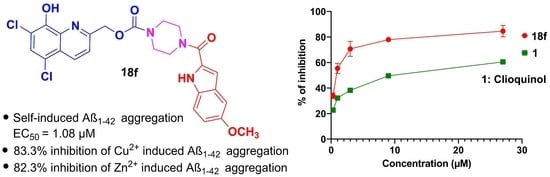
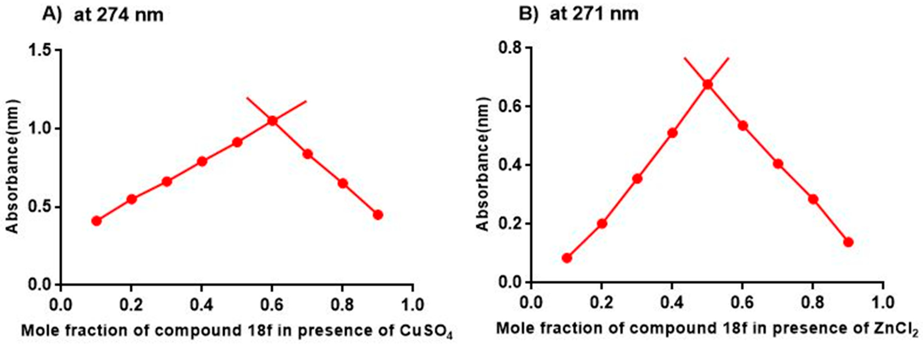
2. Results and Discussion

The novel hybrid 8-hydroxyquinoline-indole derivatives, 7a–7e, 12a, 12b, and the (8-hydroxyquinolin-2-yl)methyl-4-(1H-indole-2-carbonyl)piperazine-1-carboxylate analogs 18a–18h were evaluated for inhibitory activity against self-induced Aβ1–42 aggregation at five different concentrations (0.3, 1.0, 3, 9 and 27 µM) with 25 µM Aβ1–42, utilizing a thioflavin T (ThT) fluorescence assay with clioquinol, 1 as a positive control (Figure 3). In the absence of inhibitor, the self-induced Aβ1–42 aggregation ThT fluorescence was recorded as 115 arbitrary units (a.u.). The self-induced Aβ1–42 aggregation results for all the above synthetic analogs from the ThT fluorescence assay are shown in Table 1. The results indicate that analogs lacking a piperazino bridging moiety in their structure (i.e., 7a–7e and 12a–12b) were found to be either weak inhibitors or inactive against self-induced Aβ1–42 aggregation. Of the piperazino-containing compounds 18a–18h, five analogs exhibited EC50 values >3 μM. 8-Hydroxyquinolin-2-yl compounds containing the piperazine bridge linked to indole (18c), or 5-methoxy indole (18d and 18f) (EC50 = 1.72, 1.48 and 1.08 µM, respectively) were the most potent inhibitors of self-induced Aβ1–42 and were superior to both clioquinol (1, EC50 = 9.95 µM) and 1H-indole-2-carboxylic acid (5a, EC50 > 20 µM). The structure activity study (SAR) revealed that incorporation of the piperazino bridging moiety between a 5-methoxy indole group and a 5,7-dichloro-8-hydroxyquinoline group affords the most potent compound, 18f, which exhibited a 10-fold improvement in inhibitory potency over 1.
Analogs 18c, 18d, and 18f, showed potent self-induced Aβ1–42 aggregation (<2 µM) and were selected for further investigation for inhibition of Cu2+- and Zn2+-induced Aβ1–42 aggregation. Metal ions such as Cu2+ and Zn2+ can react with Aβ to form Aβ oligomers, and then eventually to form Aβ amyloid plaques [8,14]. Due to their inhibitory activity against Aβ1–42, 18c, 18d, and 18f were expected to decrease metal ion-induced Aβ1–42 aggregation.
Initially, CuSO4 or ZnCl2 was added to Aβ1–42 peptides at room temperature and the mixture incubated with or without drugs for 24 h at 37 °C. The fluorescence intensity of Aβ1–42 peptide alone was designated as 100%. The fluorescence intensity of Aβ1–42 peptide after treatment with Cu2+ was 135.1% and after treatment with Zn2+ was 129.3% compared to Aβ1–42 alone (Figure 4A,B), indicating that both Cu2+ and Zn2+ ions significantly enhanced Aβ1–42 aggregation.
In contrast, the fluorescence produced by Aβ1–42 treated with Cu2+ or Zn2+ ions in the presence of 18c, 18d, and 18f dramatically decreased (Figure 4), indicating inhibition of metal ion-induced Aβ1–42 aggregation (i.e., 18c, 76.6% decrease (Cu2+) and 70.3% decrease (Zn2+); 18d, 66.1% decrease (Cu2+) and 59.3% decrease (Zn2+); 18f, 88.3% decrease (Cu2+) and 82.3% decrease (Zn2+); and for comparison compound 1, 62.5% decrease (Cu2+) and 57.2% decrease (Zn2+)). These results show that 18f exhibited significantly greater inhibitory activity in Cu2+- and Zn2+-induced Aβ1–42 aggregation than compound 1.
Based on the above results from self-induced Aβ1–42 aggregation and metal-induced Aβ1–42 aggregation studies, the most potent compounds 18f was selected for further investigation into its ability to chelate metal ions such as Cu2+, Zn2+, and Fe2+ using UV-vis spectroscopy at wavelengths ranging from 200 to 500 nm. Compound 18f (50 μM) was treated with 50 μM concentrations of CuSO4, ZnCl2, or FeSO4 for 30 min in HEPES buffer at room temperature, and displayed maximum absorption wavelength shifts from 252 to 274, 269 and 255 nm in the presence of Cu2+, Zn2+, and Fe2+, respectively (Figure 5). These wavelength shifts are indicative of the formation of 18f-Cu2+, 18f-Zn2+, and 18f-Fe+2 and show that Zn2+ and Cu2+ undergo significant chelation with compound 18f. The spectrum of 18f-Fe2+ showed only a moderate increase in wavelength. Furthermore, the stoichiometry of formation of metal complexes 18f-Cu2+ and 18f-Zn2+ was also determined by using Job’s method (Figure 6) [36]. The isosbestic points shown in Figure 6A,B at 0.6 and 0.5 imply a stoichiometry of 1.5:1 for 18f-Cu2+ and 1:1 for 18f-Zn2+.
Copper and zinc dysregulation in the brain is considered to arise as a consequence of age-associated neurodegenerative disease such as Alzheimer’s disease. Zinc has also been shown to play an important role in pre- and post-synaptic responses [37]. Our drug design approach is to target dysregulation of metal homeostasis and protein aggregation. Dysregulation of metal ions is implicated in the elevated formation of reactive oxygen species and in susceptibility to oxidative stress. These metal ions were shown to interact with protein aggregation seed proteins such as tau and Aβ1–42, increasing misfolding and promoting protein aggregation. The 8-hydroxyquinoline compound, PBT2, has been shown to influence trans-synaptic effects of copper and zinc ions in a cell culture model and has been considered as a treatment for AD [38]. PBT2 (2) was also shown to induce amyloid degradation by inhibiting glycogen synthase kinase-3 activity via phosphorylation [38].
In our study, we have shown that restoring metal homeostasis by treatment with hybrid 8-hydroxyquinoline-indole analogs reduces tau- and amyloid-mediated protein aggregation, and their associated neurotoxicity. Analogs 7a–7d, 18c–18f, and 18h were evaluated for their ability to reduce protein aggregation in HEK-tau cells. HEK-tau cells were exposed to the above Aβ142 aggregation inhibitors and parent drug clioquinol (1) at 1 µM for 48 h at 37 °C. Cells were assessed for amyloid deposits by staining with 0.1% w/v thioflavin T, counterstaining nuclei with DAPI, and mean ThT fluorescence signal per nucleus was calculated over multiple fields [39]. All the analogs reduced fluorescence intensity (indicative of protein aggregation) by 2662% (P < 0.001 by 2-tailed t-test). Among these analogs, compounds 7b–7d, 18c, and 18f were the most effective as protein aggregation inhibitors in HEK-tau cells, and were more potent than clioquinol (1) (Figure 7A,B).
In another study compounds 7a–7d, 18c–18f, and 18h were also evaluated for their ability to reduce protein aggregation in SY5Y-APPSw cells. SY5Y-APPSw cells were exposed to the above protein aggregation inhibitors and parent compound 1 at 1 µM for 48 h at 37 °C. Cells were assessed for amyloid deposits by staining with 0.1% w/v ThT, counterstaining nuclei with DAPI, and mean ThT fluorescence signal per nucleus calculated over multiple fields. All the analogs reduced fluorescence intensity (indicative of protein aggregation) by 22–67% (P < 0.001 by 2-tailed t-test). Analogs 7a and 18d were the most effective in SY5Y-APPSw cells and were more potent than the parent compound, clioquinol (1) (Figure 7C,D). Thioflavin-T fluorescence assays for in vitro amyloid aggregation and cell culture models of amyloid aggregation overlap but are not identical. This is due to the fact that thioflavin in cell-culture stains not just amyloid aggregates but also other amyloid-like fibrillar structures. Another important difference is that in vitro aggregation is assessed after 24 h, whereas aggregation in cell culture is assessed after 48 h.
The binding interactions of the active compounds 18d and 18f were also investigated utilizing a crystal structure of the target Aβ1–42 peptide (PDB code: 1IYT) [40] employing Schrodinger Maestro 11.4 software (Figure 8) [41]. For analogue 18d, the quinoline ester carbonyl group of this hybrid molecule is involved in a hydrogen bonding interaction with Lys16 at a distance of 3.12 Ao units and the 8-hydroxyquinoline moiety is involved in a π–π stacking interaction with residue Phe20; this is similar to that observed with the deoxyvasicinone-donepezil hybrid molecule binding region on Aβ1–42 reported in the literature [41]. In addition, the quinolone and indole ring systems of compound 18d are involved in π–cation interactions with Lys16. With the potent analog 18f, the hydroxyl group of the 8-hydroxyquinoline moiety is involved in a hydrogen bonding interaction with the His13 residue at 2.17 Ao units, and the 8-hydroxyquinoline moiety of 18f is also involved in a π–π stacking interaction with Phe20, similar to the binding mode of compound 18d. In addition, the quinoline ring system of 18f is involved in a π–cation interaction with Lys16. These docking results indicate that the effective inhibition of both self-induced and metal ion-induced Aβ1–42 aggregation by 18d and 18f may be due to hydrogen bonding interactions, π–π stacking interactions, and π–cation interactions of these molecules. Of particular interest is the interaction the most potent compound 18f with the His13 residue on the Aβ1–42 peptide. His13 has been implicated in the binding of metal ions with Aβ1–42 (17–21); thus, 18f may compete with metal ions for this binding site on Aβ1–42, thereby inhibiting metal ion induction of Aβ1–42 aggregation.
In summary, a series of hybrid 8-hydroxyquinoline-indole derivatives 7a–7e, 12a–12b, and 18a–18h have been synthesized and evaluated as inhibitors of Aβ1–42 self-aggregation and metal chelation-induced aggregation, and have also been evaluated against HEK-tau and SY5Y-APPSw cells to determine their effect on cellular protein aggregation. In vitro studies showed that hybrid 8-hydroxyquinoline-indole analogs lacking the piperazine bridging unit (i.e., compounds 7a–7e and 12a–12b) exhibited poor-to-moderate inhibitory activities against Aβ1–42 peptide self-aggregation. However, the majority of the hybrid 8-hydroxyquinoline-indole compounds incorporating the piperazine bridge unit (i.e., 18c–18f, and 18h) were potent inhibitors against Aβ1–42 self-aggregation. Analogue 18f was the most potent inhibitor of Aβ1–42 aggregation in both self-induced and metal chelation-induced assays, and in protein aggregation assays in HEK-tau cells. Compound 18f afforded 82.3% and 88.3% inhibition against Cu2+-induced and Zn2+-induced Aβ1–42 aggregation assays, and was also shown to be a chelator of Cu2+, Zn2+, and Fe2+ ions in physiological buffer solutions. Molecular docking studies with 18f showed that this molecule has the potential to interact with the His13 residue on Aβ1–42 peptide, which suggests that its potent anti-aggregation properties may also be due in part to its ability to competitively inhibit the binding of metal ions that interact with this site to induce Aβ1–42 aggregation. Thus, the present study has identified 18f, an analog that incorporates a piperazine bridging group between a 5-methoxyindole moiety and a 5,7-dichloro-8-hydroxyquinoline moiety, as the most potent Aβ1-42 aggregation inhibitor; this analog exhibited a 10-fold improvement in inhibitory potency over clioquinol (1) as an inhibitor of Aβ1–42 self-aggregation. We consider 18f to be a lead compound worthy of further structural optimization and preclinical development as a treatment for AD.

3. Experimental Section
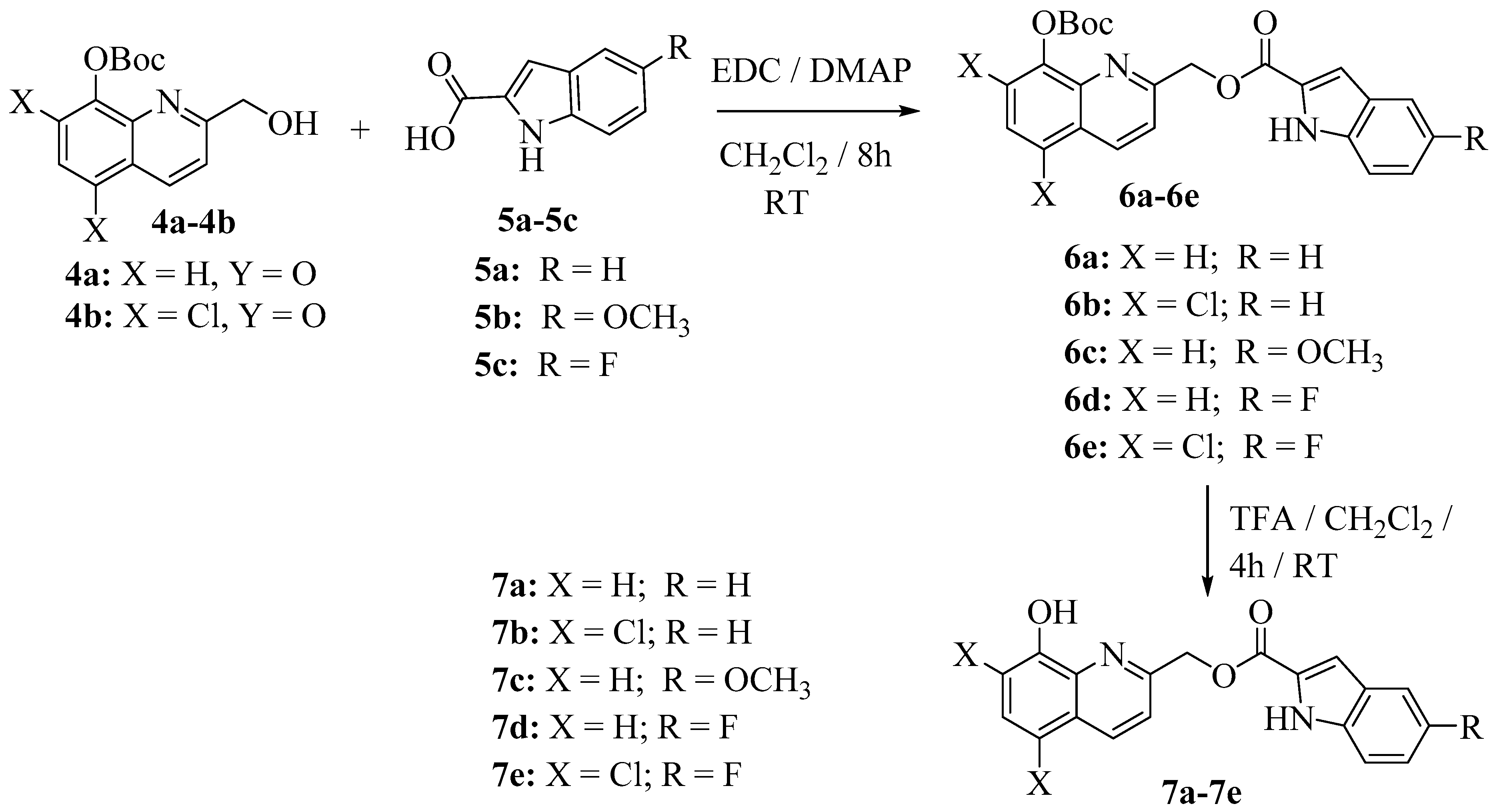
3.1. Chemistry

All reagents, solvents, and chemicals utilized in the synthesis of the hybrid 8-hydroxyquinoline-indole analogs were purchased from Oakwood Chemicals Fisher Scientific and Adooq Bioscience. 1H and 13C-NMR spectra were recorded on a Varian 400 MHz spectrometer equipped with a Linux workstation running on vNMRj software. Spectral analyses were carried out in CDCl3 or DMSO-d6 for both 1H and 13C spectra. Chemical shifts were measured in δ parts per million (ppm) and coupling constants (J) were measured in hertz (Hz). High-resolution mass spectra (HR-MS) were recorded on an Agilent 6545 ESI/APCI TOF MS. Thin-layer chromatography (TLC) was carried out on pre-coated silica gel glass plates (F254 Merck).

3.2. Experimental Procedure for the Synthesis of Hybrid 8-Hydroxyquinoline-Indole Analogs

Synthesis of (8-hydroxyquinolin-2-yl)methyl-1H-indole-2-carboxylate hybrid analogs 7a–7e
To a stirred solution of an appropriate indole-2-carboxylic acid (5a–5c) (0.15 mmol) in dry dichloromethane (5 mL) under an argon atmosphere at 0 °C was added 1-ethyl-3-(3-dimethylamino propyl)carbodiimide (EDCI, 0.181 mmol), and the mixture allowed to stir for 15 min. A catalytic amount of 4-dimethylaminopyridine (DMAP, 0.045 mmol) was then added, followed by addition of an appropriate tert-butyl (2-(hydroxymethyl)quinolin-8-yl) carbonate (4a–4b) (0.151 mmol) in dry dichloromethane (3 mL) at 0 °C. The resulting mixture was stirred for 8 h at room temperature. After completion of the reaction (monitored by TLC), the reaction mixture was washed with water (10 mL), and the dichloromethane layer separated, dried over anhydrous sodium sulfate, filtered, concentrated, and purified by silica gel column chromatography using 25% methanolic dichloromethane as eluent to afford the corresponding Boc-protected 8-hydroxyquinoline-indole derivatives (6a–6e).
The above Boc-protected 8-hydroxyquinoline-indole derivatives (6a–6e) were each dissolved in a mixture of DCM (10 mL) and TFA (1.0 mL) and stirred for 6 h until the Boc-deprotection was completed. A saturated NaHCO3 solution (20 mL) was then added and the resulting mixture was extracted with dichloromethane. The organic layer was separated and washed with water, followed by brine solution, dried over anhydrous Na2SO4, and concentrated to afford the appropriate hybrid 8-hydroxyquinoline indole derivatives (7a–7e).
(8-Hydroxyquinolin-2-yl)methyl-1H-indole-2-carboxylate (7a): 1H-NMR (400 MHz, DMSO-d6): δ 12.00 (s, 1H), 9.67 (s, 1H), 8.38 (d, J = 8.4 Hz, 1H), 7.7663 (m, 2H), 7.527.37 (m, 3H), 7.357.22 (m, 2H), 7.13–7.07 (m, 2H), 5.64 (s, 2H) ppm. 13C-NMR (100 MHz, DMSO-d6): δ 161.44, 154.70, 153.40, 138.08, 138.01, 137.56, 128.57, 127.99, 127.16, 127.14, 125.34, 122.59, 120.73, 120.35, 118.15, 113.06, 112.19, 108.99, 67.50 ppm. HR-MS (ESI) m/z calcd for C19H14N2O3 (M + H)+ 319.1075, found 319.1076.
(5,7-Dichloro-8-hydroxyquinolin-2-yl)methyl-1H-indole-2-carboxylate (7b): 1H-NMR (400 MHz, DMSO-d6): δ 12.02 (s, 1H), 10.67 (s, 1H), 8.58 (d, J = 8.8 Hz, 1H), 7.91 (d, J = 8.8 Hz, 1H), 7.84 (s, 1H), 7.7 (d, J = 8.0 Hz, 1H), 7.49 (d, J = 8.8 Hz, 1H), 7.33 (s, 1H), 7.29 (t, J = 7.6 Hz, 1H), 7.10 (t, J = 7.6 Hz, 1H), 5.70 (s, 2H) ppm. 13C-NMR (100 MHz, DMSO-d6): δ 161.35, 156.98, 149.23, 138.68, 138.05, 134.50, 128.27, 127.15, 126.97, 125.42, 124.61, 122.62, 121.61, 120.77, 119.58, 116.56, 113.06, 109.14, 67.02 ppm. HR-MS (ESI) m/z calcd for C19H12Cl2N2O3 (M + H)+ 387.0291, found 387.0296.
(8-Hydroxyquinolin-2-yl)methyl-5-methoxy-1H-indole-2-carboxylate (7c): 1H-NMR (400 MHz, DMSO-d6): δ 11.87 (s, 1H), 9.67 (s, 1H), 8.38 (d, J = 8.4 Hz, 1H), 7.70 (d, J = 8.4 Hz, 1H), 7.46–7.35 (m, 3H), 7.2 (s, 1H), 7.12 (d, J = 2.8 Hz, 2H), 6.95 (d, J = 8.8 Hz, 1H), 5.63 (s, 2H), 3.76 (s, 3H) ppm.13C-NMR (100 MHz, DMSO-d6): δ 161.36, 154.76, 154.44, 153.40, 138.08, 137.55, 133.40, 128.57, 127.98, 127.49, 127.30, 120.33, 118.15, 117.03, 113.98, 112.18, 108.55, 102.39, 67.40, 55.65 ppm. HR-MS (ESI) m/z calcd for C20H16N2O4 (M + H)+ 349.1174, found 349.1178.
(8-Hydroxyquinolin-2-yl)methyl-5-fluoro-1H-indole-2-carboxylate (7d): 1H-NMR (400 MHz, DMSO-d6): δ 12.13 (s, 1H), 9.67 (s, 1H), 8.38 (d, J = 8.4 Hz, 1H), 7.71 (d, J = 8.4 Hz, 1H), 7.50–7.40 (m, 4H), 7.28 (s, 1H), 7.19–7.11 (m, 2H), 5.64 (s, 2H) ppm. 13C-NMR (100 MHz, DMSO-d6): δ 161.17, 158.86, 156.54, 154.58, 153.41, 138.09, 137.57, 134.75, 128.80, 128.58, 128.01, 127.24, 127.13, 120.37, 118.15, 114.49, 114.39, 114.20, 112.20, 108.88, 108.83, 106.66, 106.43, 67.62 ppm. HR-MS (ESI) m/z calcd for C19H13FN2O3 (M + H)+ 337.0969, found 337.0978.
(5,7-Dichloro-8-hydroxyquinolin-2-yl)methyl-5-fluoro-1H-indole-2-carboxylate (7e): 1H-NMR (400 MHz, DMSO-d6): δ 12.14 (s, 1H), 10.64 (s, 1H), 8.57 (d, J = 8.8 Hz, 1H), 7.91 (d, J = 8.8 Hz, 1H), 7.83 (s, 1H), 7.50 (m, 2H), 7.30 (d, J = 2.0 Hz, 1H), 7.16 (td, J = 9.2, 2.4 Hz, 1H), 5.67 (s, 2H) ppm; 13C-NMR (100 MHz, DMSO-d6): δ 161.09, 158.87, 156.83, 156.54, 149.20, 138.66, 134.78, 134.47, 128.62, 128.26, 127.21, 127.11, 124.59, 121.59, 119.57, 116.55, 114.54, 114.49, 114.39, 114.27, 109.01, 108.95, 106.67, 106.44, 67.13 ppm. HR-MS (ESI) m/z calcd for C19H11Cl2FN2O3 (M + H)+ 405.0187, found 405.0199.
Synthesis of tert-butyl (5,7-dichloro-2-(iodomethyl)quinolin-8-yl) carbonate (8):
To a stirred solution of tert-butyl-(5,7-dichloro-2-(hydroxymethyl)quinolin-8-yl) carbonate 4b (1 mmol) was added triphenyl phosphine (1.2 mmol), imidazole (1.3 mmol) in dichloromethane, and iodine (1.2 mmol) at room temperature. The resulting reaction mixture was stirred for 1 h. After completion of the reaction, aqueous sodium dithionite solution was added and the resulting mixture extracted with dichloromethane, the separated organic layer was dried over Na2SO4 and concentrated under reduced pressure. The crude product was purified by column chromatography (silica gel, 10% ethyl acetate in hexanes) to afford compound 8.
1H-NMR (400 MHz, CDCl3): δ 8.42 (d, J = 8.4 Hz, 1H), 7.62 (s, 1H), 7.61 (d, J = 8.8 Hz, 1H), 4.61 (s, 2H), 1.60 (s, 9H) ppm. 13C-NMR (101 MHz, CDCl3): δ 160.22, 150.41, 142.71, 141.54, 134.27, 128.76, 127.40, 127.34, 124.70, 122.56, 84.43, 27.67, 5.41 ppm.
Synthesis of 2-(azidomethyl)-5,7-dichloroquinolin-8-yl tert-butyl carbonate (9):
To a stirred solution of tert-butyl-(5,7-dichloro-2-(iodomethyl)quinolin-8-yl) carbonate 8 (1 mmol) in acetone was added sodium azide (3 mmol). The resulting mixture was stirred at reflux temperature for 6 h. After completion of the reaction, the reaction mixture was cooled to room temperature then solvent was evaporated. Water was added to the crude mass and mixture extracted with dichloromethane, the separated organic layer dried over Na2SO4 and concentrated under reduced pressure. The crude product was purified by column chromatography (silica gel, 1% methanol in dichloromethane) to afford compound 9.
1H-NMR (CDCl3, 400 MHz): δ 8.49 (d, J = 8.8 Hz, 1H), 8.00 (s, 1H), 7.60 (d, J = 8.4 Hz, 1H), 4.64 (s, 2H), 1.59 (s, 9H) ppm. 13C-NMR (CDCl3, 101 MHz): δ 13C-NMR (101 MHz, CDCl3): δ 158.07, 150.27, 144.91, 141.76, 137.04, 133.13, 126.94, 121.31, 118.93, 117.11, 84.41, 77.30, 76.98, 76.67, 55.60, 27.61 ppm.
Synthesis of 2-(aminomethyl)-5,7-dichloroquinolin-8-yl tert-butyl carbonate (10):
To a stirred solution of 2-(azidomethyl)-5,7-dichloroquinolin-8-yl tert-butyl carbonate 9 (1 mmol) in THF/H2O (3 mL, 9:1) was added PPh3 (1.2 mmol) at room temperature. The resulting reaction mixture was stirred at 60 °C for 12 h. After completion of reaction, the reaction mixture was concentrated under vacuum and 1 M aqueous HCl added to the residue. The mixture was stirred for 30 min, then the mixture was filtered to get aqueous phase. The aqueous phase was basified to pH 10 with NaHCO3 and extracted with dichloromethane. The combined organic layer was washed with brine, dried over anhydrous Na2SO4, and the solution was evaporated under vacuum to afford target compound 10.
1H-NMR (400 MHz, CDCl3): δ 8.41 (d, J = 8.7 Hz, 1H), 7.63 (s, 1H), 7.39 (d, J = 8.7 Hz, 1H), 4.89 (s, 2H), 1.58 (s, 9H) ppm. 13C-NMR (101 MHz, CDCl3): δ 161.16, 150.50, 142.37, 140.93, 134.02, 129.02, 127.64, 127.17, 125.30, 119.75, 84.65, 77.29, 77.17, 76.97, 76.65, 64.14, 27.58 ppm.
Synthesis of N-((8-hydroxyquinolin-2-yl)methyl)-1H-indole-2-carboxamide hybrid analogs 12a and 12b:
To a stirred solution of indole-2-carboxylic acid 5a or 5b (0.15 mmol) in dry dichloromethane (5 mL) under an argon atmosphere at 0 °C was added EDCI (0.181 mmol) and the mixture allowed to stir for 15 min. A catalytic amount of DMAP (0.045 mmol) was then added followed by addition of 2-(aminomethyl)-5,7-dichloroquinolin-8-yl tert-butyl carbonate (10) (0.151 mmol) in dry dichloromethane (3 mL) at 0 °C, and the resulting mixture was stirred for 8 h at room temperature. After completion of the reaction (monitored by TLC), the reaction mixture was washed with water (10 mL), and the dichloromethane layer separated, dried over anhydrous sodium sulfate, filtered, concentrated, and purified by silica gel column chromatography, using 2–5% methanolic dichloromethane as eluent, to afford the corresponding Boc-protected 8-hydroxyquinoline-indole derivatives 11a or 11b as a solid. Products were obtained in 56–74% yield.
The above Boc-protected 8-hydroxyquinoline-indole derivatives 11a and 11b were each dissolved in a mixture of DCM (10 mL) and trifluoroacetic acid (TFA, 1.0 mL) and the resulting mixture stirred for 6 h until the Boc-deprotection was completed. A saturated NaHCO3 solution (20 mL) was then added and the resulting mixture extracted with dichloromethane. The organic layer was separated and washed with water, followed by brine solution, dried over anhydrous Na2SO4, and concentrated to afford the appropriate hybrid 8-hydroxyquinoline indole derivatives 12a and 12b.
N-((5,7-Dichloro-8-hydroxyquinolin-2-yl)methyl)-1H-indole-2-carboxamide (12a): 1H-NMR (400 MHz, DMSO-d6): δ 12.01 (s, 1H), 10.71 (s, 1H), 10.07 (s, 1H), 8.52(d, J = 8.4 Hz, 1H), 8.07 (s, 1H), 7.93 (d, J = 8.8 Hz, 1H), 7.70 (d, J = 8.0 Hz, 1H), 7.49 (d, J = 8.0 Hz, 1H), 7.33 (s, 1H), 7.28 (t, J = 7.6 Hz, 1H), 7.12 (t, J = 7.6 Hz, 1H), 5.71 (s, 2H) ppm; 13C-NMR (100 MHz, DMSO-d6): δ 161.34, 156.89, 151.03, 138.59, 138.05, 137.00, 133.70, 127.14, 126.97, 126.25, 125.41, 122.61, 122.08, 120.77, 113.06, 109.39, 109.14, 106.03, 66.91 ppm. HR-MS (ESI) m/z calcd for C19H13Cl2N3O2 (M + H)+ 386.0379, found 386.0382.
N-((5,7-Dichloro-8-hydroxyquinolin-2-yl)methyl)-5-methoxy-1H-indole-2-carboxamide (12b): 1H-NMR (400 MHz, DMSO-d6): δ 11.87 (s, 1H), 10.64 (s, 1H), 10.02 (s, 1H), 8.56 (d, J = 8.8 Hz, 1H), 7.87 (d, J = 8.8 Hz, 1H), 7.8 (s, 1H), 7.35 (d, J = 9.2 Hz, 1H), 7.2–7.19 (m, 1H), 7.11 (d, J = 2.4 Hz, 1H), 6.94 (dd, J = 8.8, 2.4 Hz, 1H), 5.67 (s, 2H), 3.74 (s, 3H) ppm; 13C-NMR (100 MHz, DMSO-d6): δ 161.26, 157.04, 154.46, 149.21, 138.68, 134.49, 133.43, 128.26, 127.47, 127.10, 124.60, 121.59, 119.58, 117.13, 116.55, 113.98, 108.68, 102.37, 66.93, 55.65 ppm. HR-MS (ESI) m/z calcd for C20H15Cl2N3O3 (M + H)+ 416.0485, found 416.0492.
Synthesis of (8-((tert-butoxycarbonyl)oxy)quinolin-2-yl)methyl-1H-1,2,4-triazole-1-carboxylates 14a–14c:
To a stirred solution of an appropriate tert-butyl (2-(hydroxymethyl)quinolin-8-yl) carbonate (4a–4c) (1 mmol) in dichloromethane was added carbonylditriazole (2 mmol) at room temperature. The reaction mixture was stirred for 30 min. After completion of the reaction, water was added and the resulting mixture was extracted with dichloromethane, the separated organic layer dried over Na2SO4 and concentrated under reduced pressure to afford triazole intermediates 14a–14c.
(8-((tert-Butoxycarbonyl)oxy)quinolin-2-yl)methyl-1H-1,2,4-triazole-1-carboxylate (14a):
1H-NMR (400 MHz, CDCl3): δ 8.88 (s, 1H), 8.27 (d, J = 8.5 Hz, 1H), 8.11 (s, 1H), 7.74 (dd, J = 6.6, 3.0 Hz, 1H), 7.63 (d, J = 8.5 Hz, 1H), 7.58–7.50 (m, 2H), 5.77 (s, 2H), 1.59 (s, 9H) ppm. 13C-NMR (101 MHz, CDCl3): δ 153.83, 153.68, 151.83, 147.28, 145.79, 140.60, 137.46, 128.89, 126.90, 125.61, 121.67, 119.87, 83.70, 71.17, 27.62 ppm.
(8-((tert-Butoxycarbonyl)oxy)-5,7-dichloroquinolin-2-yl)methyl-1H-1,2,4-triazole-1-carboxylate (14b):
1H-NMR (400 MHz, CDCl3): δ 8.89 (s, 1H), 8.59 (d, J = 8.8 Hz, 1H), 8.11 (s, 1H), 7.71 (d, J = 8.4 Hz, 2H), 5.77 (s, 2H), 1.57 (s, 9H) ppm. 13C-NMR (101 MHz, CDCl3): δ 155.61, 153.90, 150.35, 145.84, 142.78, 141.72, 134.77, 128.95, 127.86, 125.42, 120.29, 84.57, 70.38, 27.54 ppm.
(8-((tert-Butoxycarbonyl)oxy)-5,7-dibromoquinolin-2-yl)methyl-1H-1,2,4-triazole-1-carboxylate (14c):
1H-NMR (400 MHz, CDCl3): δ 8.89 (s, 1H), 8.54 (d, J = 8.7 Hz, 1H), 8.08 (d, J = 30.9 Hz, 2H), 7.71 (d, J = 8.8 Hz, 1H), 5.78 (s, 2H), 1.58 (s, 9H) ppm. 13C-NMR (1010 MHz, CDCl3): δ 155.53, 153.88, 150.16, 147.26, 145.86, 144.92, 141.67, 137.30, 133.51, 127.12, 120.74, 118.92, 117.35, 84.52, 70.23, 27.55 ppm.
Synthesis of hybrid (8-((tert-butoxycarbonyl)oxy)quinolin-2-yl)methylpiperazine-1- carboxylates 16a–16c:
An appropriate triazole intermediate (14a–14c) (1 mmol) was dissolved in dichloromethane (2 mL) and treated with piperazine (15) (1.0 mmol) at room temperature for 1 h. Completion of the reaction was monitored by TLC. Water (2 mL) was then added and the resulting mixture was extracted with dichloromethane (2 × 3 mL), the organic layer separated, dried over anhydrous Na2SO4 and concentrated to afford the appropriate Boc-protected product (16a–16c). The crude product was purified by column chromatography (silica gel, 2% methanol in dichloromethane) to afford the desired product (16a–16c).
(8-((tert-Butoxycarbonyl)oxy)quinolin-2-yl)methyl-piperazine-1-carboxylate (16a): 1H-NMR (400 MHz, DMSO-d6): δ 8.33 (d, J = 8.4 Hz, 1H), 7.52 (d, J = 8.4Hz, 1H), 7.45–7.35 (m, 2H), 7.10 (dd, J = 7.2, 1.2 Hz, 1H), 5.33 (s, 2H), 3.37 (m, 4H), 2.72–2.61 (m, 4H), 1.58 (s, 9H) ppm; 13C-NMR (101 MHz, DMSO-d6): δ 155.59, 154.75, 153.35, 153.22, 137.99, 137.42, 128.43, 127.80, 119.91, 118.08, 112.07, 67.84, 45.81, 27.52 ppm.
(8-((tert-Butoxycarbonyl)oxy)-5,7-dichloro-8-hydroxyquinolin-2-yl)methyl-piperazine-1-carboxylate (16b): 1H-NMR (400 MHz, CDCl3): δ 8.39 (d, J = 8.4 Hz, 1H), 7.52 (d, J = 8.8 Hz, 1H), 7.48 (s, 1H), 5.37 (s, 2H), 3.59 (m, 4H), 2.88 (br-s, 4H), 1.57 (s, 9H) ppm; 13C-NMR (101 MHz, CDCl3): δ 156.66, 153.64, 154.82, 147.78, 137.93, 134.32, 128.07, 124.18, 120.40, 120.31, 115.78, 67.22, 53.42, 45.49, 27.52 ppm.
(8-((tert-Butoxycarbonyl)oxy)-5,7-dibromo-8-hydroxyquinolin-2-yl)methyl-piperazine-1-carboxylate (16c): 1H-NMR (400 MHz, DMSO-d6): δ 8.40 (d, J = 8.8 Hz, 1H), 7.97 (s, 1H), 7.68 (d, J = 8.8 Hz, 1H), 5.38 (s, 2H), 3.46 (m, 4H), 2.80 (br-s, 4H), 1.58 (s, 9H) ppm; 13C-NMR (101 MHz, DMSO-d6): δ 156.92, 154.60, 153.55 153.13, 139.44, 136.71, 133.63, 126.37, 121.45, 106.98, 106.32, 67.49, 45.01, 44.33, 27.51 ppm.
Synthesis of (8-hydroxyquinolin-2-yl)methyl-4-(1H-indole-2-carbonyl)piperazine-1-carboxylate hybrid derivatives (18a–18h):
To the stirred solution of an appropriate indole-2-carboxylic acid (5a–5c) (0.15 mmol) in dry dichloromethane (5 mL) under an argon atmosphere at 0 °C was added EDCI (0.181 mmol), and the mixture allowed to stir for 15 min. A catalytic amount of DMAP (0.045 mmol) was then added, followed by addition of an appropriate (8-((tert-butoxycarbonyloxy)-quinolin-2-yl)methyl piperazine-1-carboxylate (16a–16c) (0.151 mmol) in dry dichloromethane (3 mL) at 0 °C. The resulting mixture was stirred for 8 h at room temperature. After completion of the reaction (monitoring by TLC), the reaction mixture was washed with water (10 mL) and the dichloromethane layer separated, dried over anhydrous sodium sulfate, filtered, and concentrated. The crude material was purified by silica gel column chromatography, using 2–5% methanolic dichloromethane as eluent, to afford the corresponding (8-((tert-butoxycarbonyl)oxy) quinolin-2-yl)methyl-4-(1H-indole-2-carbonyl)piperazine-1-carboxylate hybrid derivatives (17a–17h), which was dissolved in a mixture of DCM (10 mL) and TFA (1.0 mL) and stirred for 6 h until Boc-deprotection was completed. Saturated NaHCO3 solution (20 mL) was then added to the resulting mixture which was extracted with dichloromethane. The organic layer was separated and washed with water, followed by brine solution, dried over anhydrous Na2SO4 and concentrated to afford the corresponding (8-hydroxyquinolin-2-yl)methyl-4-(1H-indole-2-carbonyl)piperazine-1- carboxylate hybrid derivative (18a–18h).
(8-Hydroxyquinolin-2-yl)methyl-4-(1H-indole-2-carbonyl)piperazine-1-carboxylate (18a): 1H-NMR (400 MHz, CDCl3): δ 9.67 (s, 1H), 8.19 (d, J = 8.4 Hz, 1H), 7.65 (d, J = 8.0 Hz, 1H), 7.52–7.42 (m, 3H), 7.35–7.2 (m, 2H), 7.20–7.12 (m, 2H), 6.78 (s, 1H), 5.45 (s, 2H), 3.96 (s, 4H), 3.70 (s, 4H) ppm; 13C-NMR (100 MHz, CDCl3): δ 162.82, 154.94, 154.26, 151.88, 137.07, 127.85, 124.64, 121.87, 120.66, 120.06, 117.73, 111.71, 110.44, 105.49, 68.12, 43.74 ppm. HR-MS (ESI) m/z calcd for C24H22N4O4 (M + H)+ 431.1708, found 431.1712.
(5,7-Dibromo-8-hydroxyquinolin-2-yl)methyl-4-(1H-indole-2-carbonyl)piperazine-1-carboxylate (18b): 1H-NMR (400 MHz, CDCl3): δ 9.50 (s, 1H), 8.44 (d, J = 8.8 Hz, 1H), 7.86 (s, 1H), 7.61 (dd, J = 15.2, 8.4 Hz, 2H), 7.41 (d, J = 8.0 Hz, 1H), 7.28 (t, J = 7.6 Hz, 1H), 7.13 (t, J = 7.6 Hz, 1H), 6.76 (s, 1H), 5.46 (s, 2H), 3.96 (s, 4H), 3.68 (s, 4H) ppm; 13C-NMR (100 MHz, CDCl3): δ 162.75, 156.36, 154.69, 149.35, 137.77, 137.09, 135.75, 133.78, 128.62, 127.32, 125.92, 124.69, 121.91, 121.05, 120.73, 111.78, 110.07, 105.50, 104.33, 67.38, 44.03 ppm. HR-MS (ESI) m/z calcd for C24H20Br2N4O4 (M + H)+ 588.9879, found 588.9885.
(5,7-Dichloro-8-hydroxyquinolin-2-yl)methyl-4-(1H-indole-2-carbonyl)piperazine-1-carboxylate (18c): 1H-NMR (400 MHz, CDCl3): δ 10.08 (s, 1H), 8.41 (d, J = 8.8 Hz, 1H), 7.63 (d, J = 7.6 Hz, 1H), 7.56 (d, J = 8.4 Hz, 1H), 7.5 (s, 1H), 7.42 (d, J = 7.6 Hz, 1H), 7.27 (d, J = 7.6 Hz, 1H), 7.13 (t, J = 7.2 Hz, 1H), 6.76 (s, 1H), 5.45 (s, 2H), 3.99 (s, 4H), 3.7 (s, 4H) ppm. 13C-NMR (100 MHz, CDCl3): δ 162.98, 156.40, 154.75, 147.29, 137.69, 135.95, 134.42, 128.61, 128.14, 127.22, 124.54, 124.13, 121.84, 120.68, 120.61, 120.45, 115.68, 111.89, 105.52, 67.48, 43.82 ppm. HR-MS (ESI) m/z calcd for C24H20Cl2N4O4 (M + H)+ 499.0925, found 499.0929.
(8-Hydroxyquinolin-2-yl)methyl-4-(5-methoxy-1H-indole-2-carbonyl)piperazine-1-carboxylate (18d): 1H-NMR (400 MHz, DMSO-d6): δ 11.46 (s, 1H), 9.64 (s, 1H), 8.34 (d, J = 8.4 Hz, 1H), 7.58 (d, J = 8.4 Hz, 1H), 7.45–7.38 (m, 2H), 7.33 (d, J = 8.8 Hz, 1H), 7.12 (d, J = 6.8 Hz, 1H), 7.07 (s, 1H), 6.86 (dd, J = 8.8, 2.4 Hz, 1H), 6.75 (s, 1H), 5.38 (s, 2H), 3.81 (br-s, 4H), 3.75 (s, 3H), 3.63 (br-s, 2H), 3.56 (br-s, 2H) ppm; 13C-NMR (100 MHz, DMSO-d6): δ 162.65, 155.37, 154.78, 154.18, 153.37, 138.03, 137.43, 131.66, 130.37, 128.50, 127.83, 127.52, 120.01, 118.13, 114.89, 113.36, 112.13, 104.51, 102.31, 68.09, 55.66, 44.04 ppm. HR-MS (ESI) m/z calcd for C25H24N4O5 (M + H)+ 461.1811, found 461.1813.
(5,7-Dibromo-8-hydroxyquinolin-2-yl)methyl-4-(5-methoxy-1H-indole-2-carbonyl)piperazine-1-carboxylate (18e): 1H-NMR (400 MHz, DMSO-d6): δ 11.46 (s, 1H), 8.48 (d, J = 8.8 Hz, 1H), 8.05 (s, 1H), 7.8 (d, J = 8.8 Hz, 1H), 7.33 (d, J = 9.2 Hz, 1H), 7.07 (d, J = 2.0 Hz, 1H), 6.85 (dd, J = 8.8, 2.4 Hz, 1H), 6.75 (s, 1H), 5.45 (s, 2H), 3.821 (s, 4H), 3.75 (s, 3H), 3.65 (br-s, 4H) ppm. 13C-NMR (100 MHz, DMSO-d6): δ 162.66, 157.61, 154.62, 154.18, 151.04, 138.57, 136.89, 133.57, 131.66, 130.35, 127.51, 126.16, 121.75, 114.90, 113.36, 109.33, 105.98, 104.52, 102.30, 67.54, 55.67 ppm. HR-MS (ESI) m/z calcd for C25H22Br2N4O5 (M + H)+ 618.9998, found 619.0000.
(5,7-Dichloro-8-hydroxyquinolin-2-yl)methyl-4-(5-methoxy-1H-indole-2-carbonyl)piperazine-1-carboxylate (18f): 1H-NMR (400 MHz, DMSO-d6): δ 11.46 (s, 1H), 8.54 (d, J = 8.8 Hz, 1H), 7.80 (s, 1H), 7.78 (d, J = 8.8 Hz, 1H), 7.33 (d, J = 8.8 Hz, 1H), 7.06 (d, J = 2.4 Hz, 1H), 6.85 (dd, J = 8.8, 2.4 Hz, 1H), 6.74 (s, 1H), 5.44 (s, 2H), 4.04 (s, 2H), 3.82 (s, 2H), 3.75 (s, 3H), 3.66 (s, 2H), 3.54 (s, 2H) ppm; 13C-NMR (100 MHz, DMSO-d6): δ 162.66, 162.61, 157.66, 154.61, 154.17, 149.09, 138.58, 134.32, 131.65, 131.50, 130.32, 130.18, 128.09, 127.49, 127.45, 124.49, 121.23, 119.58, 116.43, 114.88, 113.34, 113.29, 104.49, 104.45, 102.25, 67.63, 44.09, 40.54, 40.33, 40.12, 39.91, 39.70, 39.49, 39.28 ppm. HR-MS (ESI) m/z calcd for C25H22Cl2N4O5 (M + H)+ 529.1026, found 529.1031.
(5,7-Dichloro-8-hydroxyquinolin-2-yl)methyl-4-(5-fluoro-1H-indole-2-carbonyl)piperazine-1-carboxylate (18g): 1H-NMR (400 MHz, DMSO-d6): δ 11.73 (s, 1H), 8.52 (d, J = 8.8 Hz, 1H), 7.80 (s, 1H), 7.77 (d, J = 8.8 Hz, 1H), 7.43 (dd, J = 8.8, 4.8 Hz, 1H), 7.37 (d, J = 10.0 Hz, 1H), 7.05 (t, J = 9.2 Hz, 1H), 6.83 (s, 1H), 5.44 (s, 2H), 3.82 (s, 4H), 3.66 (br-s, 2H), 3.55 (br-s, 2H) ppm. 13C-NMR (100 MHz, DMSO-d6): δ 162.35, 158.72, 157.60, 156.40, 154.62, 149.28, 138.66, 134.30, 133.13, 131.83, 128.11, 127.33, 127.22, 124.50, 121.22, 119.42, 116.47, 113.77, 113.67, 112.57, 112.31, 106.04, 105.81, 104.72, 104.67, 67.66, 43.92 ppm. HR-MS (ESI) m/z calcd for C24H19Cl2FN4O4 (M + H)+ 517.0829, found 517.0833.
(5,7-Dibromo-8-hydroxyquinolin-2-yl)methyl-4-(5-fluoro-1H-indole-2-carbonyl)piperazine-1-carboxylate (18h): 1H-NMR (400 MHz, DMSO-d6): δ 11.72 (s, 1H), 9.65 (s, 1H), 8.35 (d, J = 8.4 Hz, 1H), 7.58 (d, J = 8.4 Hz, 1H), 7.45–7.35 (m, 4H), 7.12 -7.03 (m, 2H), 6.83 (d, J = 1.6 Hz, 1H), 5.38 (s, 2H), 3.81 (br-s, 4H), 3.63 (br-s, 2H), 3.54 (br-s, 2H) ppm. 13C-NMR (101 MHz, DMSO-d6): δ 162.33, 158.71, 156.39, 155.35, 154.78, 153.35, 138.01, 137.44, 133.12, 131.83, 128.50, 127.84, 127.33, 127.22, 120.01, 118.14, 113.77, 113.67, 112.57, 112.31, 112.14, 106.05, 105.82, 104.74, 104.69, 68.09, 44.0. HR-MS (ESI) m/z calcd for C24H21FN4O4 (M + H)+ 449.1621, found 449.1616.

3.3. Methodology for the In Vitro Self-Induced Aβ1–42 Aggregation Assay

The thioflavin T (ThT)-fluorescence assay was used to measure the inhibition of Aβ aggregation [42]. Aβ1–42 (Adooq Bioscience, CA, USA) was pretreated with 1 mL of hexafluoroisopropanol (HFIP) to afford a stock solution, which was aliquoted into small samples. The solvent was evaporated at room temperature, and samples were stored at −80 °C. For Aβ1–42 aggregation inhibition experiments, phosphate buffer (pH 7.4) was added to the Aβ stock solution to afford a 50 µM concentration before use. A mixture of the Aβ1–42 peptide (10 µL, 25 µM final concentration) with or without the test compound (10 µL; 0.3, 1.0, 3, 9, and 27 µM) was incubated at 37 °C for 48 h. Blanks using phosphate buffer (pH 7.4) instead of Aβ, with or without test compound, were also assessed. Then 50 mM glycine-NaOH buffer (pH 8.0) containing ThT (5 µM) was added to 20 µL of the sample to afford a final volume of 200 µL. The fluorescence intensities were recorded (excitation, 450 nm; emission, 485 nm). The fluorescence intensities were background corrected to the no enzyme control and 100% of intensity reflected no inhibition of aggregation. To obtain the normalized amyloid aggregation inhibition relative to the control group, the amyloid aggregation/cell values were further divided by the total amyloid fluorescence of the control group. Using GraphPad Prism, the amyloid aggregation values (relative to control group) were entered along the corresponding concentrations for the triplicates and then a log transformation was obtained. Using the dose–response simulation module in GraphPad Prism, nonlinear regression fit (curve) was then generated for each small molecule with various concentrations used. Curves were then compared to determine if they were statistically different, by performing the best-fit values of selected unshared parameters between data sets and extra sum-of-squares F test, allowing for selection of the simpler model unless the P value was less than 0.05. The logEC50 output parameter was then selected to obtain the EC50 for each of the dose–response curves for the small molecules.

3.4. Methodology for the In Vitro Metal-Induced Aβ1–42 Aggregation Assay

For the inhibition of copper- and zinc-mediated Aβ1–42 aggregation, 20 µM HEPES (pH 6.6) in 150 µM NaCl was added to the Aβ1–42 stock solution to afford a 25 µM solution. Mixtures of the peptide (10 µL, 25 µM final concentration) with or without Cu2+ or Zn2+ (10 µL, 25 μM final concentration) and the test compound (10 µL, 50 μM final concentration) were incubated at 37 °C for 24 h. Then 50 mM glycine-NaOH buffer (pH 8.0) containing ThT (5 µM) was added to 20 µL of the sample and diluted to afford a final volume of 200 µL. Fluorescence intensities of the solutions were recorded (excitation, 450 nm; emission, 485 nm). The percentage of aggregation was calculated by the expression (1 − IFi/IFc) × 100, in which IFi and IFc are the fluorescence intensities obtained for Aβ in the presence and absence of inhibitors, respectively.

3.5. Methodology for the Metal-Chelation Study

The chelating studies were performed with a UV–vis spectrophotometer. The absorption spectra of test compound (50 μM, final concentration) alone or in the presence of CuSO4, FeSO4, or ZnCl2 (50 μM, final concentration) in buffer (20 mM HEPES, 150 mM NaCl, pH 7.4) were incubated for 30 min and then recorded at room temperature, respectively. For the stoichiometry of the test compound–Cu2+ complex or the test compound–Zn2+ complex, a fixed amount of test compound (50 μM) was mixed with increasing amounts of copper ion or Zn ion (0100 μM), and the UV–vis difference spectra were analyzed to determine the ratio of ligand/metal ion in the complex.

3.6. Methodology for In Vitro HEK-Tau and SY5Y-APPSw Cell Aggregation Assays

Human Embryonic Kidney cells that overexpress tau protein (HEK-tau) cells or SY5Y neuroblastoma cells that express a familial-AD mutant of amyloid precursor protein (SY5Y-APPSw cells) were each seeded in 96-well plates at 8000 cells per well. After 24 h, cells were supplemented with test compound at 1 µM after diluting with medium. Vehicle only control was also included for each experiment. The cells were cultured for an additional 48 h at 37 °C. Both the cell lines were able to reach approximately 80% confluency in 48 h. Cells were assessed for amyloid deposits by staining with 0.1% w/v ThT, counterstaining nuclei with DAPI, and calculating mean ThT fluorescence signal per nucleus over multiple fields.

3.7. Methodology for Molecular Docking

Molecular docking was performed using Schrodinger software Maestro 11.4 suite. The ligands were prepared using the LigPrep module. The protein crystal structure PDB ID: 1IYT was downloaded from the RCSB Protein Data Bank (https://www.rcsb.org/). The downloaded protein crystal structure was prepared in Protein Preparation Wizard wherein the missing hydrogens were added and bond orders assigned. A grid for docking the ligand was generated with the centroid of the co-crystallized ligand from PDB ID: 1IYT as the center of the grid with no constraints. The prepared ligands 18d and 18f were docked into this grid using Glide module. Molecular docking was performed at XP precision and results were analyzed in the Maestro visualizer.

Supplementary Materials

The following are available online.

Author Contributions

S.K.B.: Design, Synthesis and In vitro self-induced and metal-Induced Aβ1–42 aggregation assay; N.M. and S.K. (Sesha Krishnamachari): In vitro HEK-Tau and SY5Y-APPSw cell culture experiments; S.A.: Design of screening the drugs against Aβ1–42 aggregation and cell culture experiments; N.R.P.: Molecular docking studies; S.K. (Samuel Kakraba): Calculations of in vitro self-induced and metal-Induced Aβ1–42 aggregation assay results; R.J.S.R.: Supervision and design of overall biological experiments; P.A.C.: Supervision and design of overall project; S.K.B. and N.R.P.: Manuscript preparation with additional inputs from P.A.C. All authors have read and agreed to the published version of the manuscript.

Funding

We are grateful for research support from the National Institutes of Health, NIA grant P01 AG012411 (S. Griffin, P.I.) and from the U.S. Dept. of Veteran Affairs, Merit Award BX001655 (R. J. Shmookler Reis, P.I.); also a Senior Research Career Scientist Award BX004851 to R.J.S.R.; and an Arkansas Research Alliance Scholar award to P.A.C.

Conflicts of Interest

The authors declare no conflicts of interest.

References

  1. Förstl, H.; Kurz, A. Clinical features of Alzheimer’s disease. Eur. Arch. Psy. Clin. Neurosci. 1999, 249, 288–290. [Google Scholar] [CrossRef] [PubMed]
  2. Torre, J.D.; Aliev, G.; Perry, G. Drug Therapy in Alzheimer’s Disease. N. Engl. J. Med. 2004, 351, 1911–1913. [Google Scholar]
  3. Querfurth, H.W.; LaFerla, F.M. Alzheimer’s disease. N. Engl. J. Med. 2010, 362, 329–344. [Google Scholar] [CrossRef] [Green Version]
  4. Walsh, D.M.; Selkoe, D.J. Deciphering the molecular basis of memory failure in Alzheimer’s disease. Neuron 2004, 44, 181–193. [Google Scholar] [CrossRef] [Green Version]
  5. Munoz-Torrero, D. Acetylcholinesterase inhibitors as disease-modifying therapies for Alzheimer’s disease. Curr. Med. Chem. 2008, 15, 2433–2455. [Google Scholar] [CrossRef]
  6. Young, A.B. Four decades of neurodegenerative disease research: How far we have come! J. Neurosci. 2009, 29, 12722–12728. [Google Scholar] [CrossRef] [Green Version]
  7. Spangenberg, E.E.; Green, K.N. Inflammation in Alzheimer’s disease: Lessons learned from microglia-depletion models. Brain Behav. Immun. 2017, 61, 1–11. [Google Scholar] [CrossRef] [Green Version]
  8. Deora, G.S.; Kantham, S.; Chan, S.; Dighe, S.N.; Veliyath, S.K.; McColl, G.; Parat, M.O.; McGeary, R.P.; Ross, B.P. Multifunctional Analogs of Kynurenic Acid for the Treatment of Alzheimer’s Disease: Synthesis, Pharmacology, and Molecular Modeling Studies. ACS Chem. Neurosci. 2017, 8, 2667–2675. [Google Scholar] [CrossRef] [PubMed]
  9. Hiremathad, A.; Keri, R.S.; Esteves, A.R.; Cardoso, S.M.; Chaves, S.; Santos, M.A. Novel Tacrine-Hydroxyphenylbenzimidazole hybrids as potential multitarget drug candidates for Alzheimer’s disease. Eur. J. Med. Chem. 2018, 148, 255–267. [Google Scholar] [CrossRef] [PubMed]
  10. Kumar, B.; Dwivedi, A.R.; Sarkar, B.; Gupta, S.K.; Krishnamurthy, S.; Mantha, A.K.; Parkash, J.; Kumar, V. 4,6-Diphenylpyrimidine Derivatives as Dual Inhibitors of Monoamine Oxidase and Acetylcholinesterase for the Treatment of Alzheimer’s Disease. ACS Chem. Neurosci. 2019, 10, 252–265. [Google Scholar] [CrossRef]
  11. Li, X.; Wang, H.; Xu, Y.; Liu, W.; Gong, Q.; Wang, W.; Qiu, X.; Zhu, J.; Mao, F.; Zhang, H.; et al. Novel Vilazodone-Tacrine Hybrids as Potential Multitarget-Directed Ligands for the Treatment of Alzheimer’s Disease Accompanied with Depression: Design, Synthesis, and Biological Evaluation. ACS Chem. Neurosci. 2017, 8, 2708–2721. [Google Scholar] [CrossRef] [PubMed]
  12. Sevigny, J.; Chiao, P.; Bussiere, T.; Weinreb, P.H.; Williams, L.; Maier, M.; Dunstan, R.; Salloway, S.; Chen, T.; Ling, Y.; et al. The antibody aducanumab reduces Abeta plaques in Alzheimer’s disease. Nature 2016, 537, 50–56. [Google Scholar] [CrossRef] [PubMed]
  13. Mohamed, T.; Shakeri, A.; Rao, P.P. Amyloid cascade in Alzheimer’s disease: Recent advances in medicinal chemistry. Eur. J. Med. Chem. 2016, 113, 258–272. [Google Scholar] [CrossRef] [PubMed]
  14. Viola, K.L.; Klein, W.L. Amyloid-β oligomers in Alzheimer’s disease pathogenesis, treatment, and diagnosis. Acta Neuropathol. 2015, 129, 183–206. [Google Scholar] [CrossRef]
  15. Chang, L.; Cui, W.; Yang, Y.; Xu, S.; Zhou, W.; Fu, H.; Hu, S.; Mak, S.; Hu, J.; Wang, Q.; et al. Protection against β-amyloid-induced synaptic and memory impairments via altering β-amyloid assembly by bis(heptyl)-cognitin. Sci. Rep. 2015, 5, 10256. [Google Scholar] [CrossRef]
  16. Zhao, L.N.; Long, H.; Mu, Y.; Chew, L.Y. The toxicity of amyloid β oligomers. Int. J. Mol. Sci. 2012, 13, 7303–7327. [Google Scholar] [CrossRef] [Green Version]
  17. Barnham, K.J.; Bush, A.I. Metals in Alzheimer’s and Parkinson’s diseases. Curr. Opin. Chem. Biol. 2008, 12, 222–228. [Google Scholar] [CrossRef]
  18. Himes, R.A.; Park, G.Y.; Siluvai, G.S.; Blackburn, N.J.; Karlin, K.D. Structural studies of copper(I) complexes of amyloid-β peptide fragments: Formation of two-coordinate bis(histidine) complexes. Angew. Chem. Int. Ed. Engl. 2008, 47, 9084–9087. [Google Scholar] [CrossRef]
  19. Hou, L.; Zagorski, M.G. NMR reveals anomalous copper(II) binding to the amyloid Abeta peptide of Alzheimer’s disease. J. Am. Chem. Soc. 2006, 128, 9260–9261. [Google Scholar] [CrossRef]
  20. Duce, J.A.; Bush, A.I. Biological metals and Alzheimer’s disease: Implications for therapeutics and diagnostics. Prog. Neurobiol. 2010, 92, 1–18. [Google Scholar] [CrossRef]
  21. Kepp, K.P. Bioinorganic Chemistry of Alzheimer’s Disease. Chem. Rev. 2012, 112, 5193–5239. [Google Scholar] [CrossRef] [PubMed] [Green Version]
  22. Savelieff, M.G.; DeToma, A.S.; Derrick, J.S.; Lim, M.H. The ongoing search for small molecules to study metal-associated amyloid-β species in Alzheimer’s disease. Acc. Chem. Res. 2014, 47, 2475–2482. [Google Scholar] [CrossRef]
  23. Kim, A.C.; Lim, S.; Kim, Y.K. Metal Ion Effects on Aβ and Tau Aggregation. Int. J. Mol. Sci. 2018, 19, 128. [Google Scholar] [CrossRef] [PubMed] [Green Version]
  24. Ritchie, C.W.; Bush, A.I.; Mackinnon, A.; Macfarlane, S.; Mastwyk, M.; MacGregor, L.; Kiers, L.; Cherny, R.; Li, Q.X.; Tammer, A.; et al. Metal-protein attenuation with iodochlorhydroxyquin (clioquinol) targeting Abeta amyloid deposition and toxicity in Alzheimer disease: A pilot phase 2 clinical trial. Arch. Neurol. 2003, 60, 1685–1691. [Google Scholar] [CrossRef] [PubMed]
  25. Cherny, R.A.; Atwood, C.S.; Xilinas, M.E.; Gray, D.N.; Jones, W.D.; McLean, C.A.; Barnham, K.J.; Volitakis, I.; Fraser, F.W.; Kim, Y.; et al. Treatment with a copper-zinc chelator markedly and rapidly inhibits β-amyloid accumulation in Alzheimer’s disease transgenic mice. Neuron 2001, 30, 665–676. [Google Scholar] [CrossRef] [Green Version]
  26. Adlard, P.A.; Cherny, R.A.; Finkelstein, D.I.; Gautier, E.; Robb, E.; Cortes, M.; Volitakis, I.; Liu, X.; Smith, J.P.; Perez, K.; et al. Rapid restoration of cognition in Alzheimer’s transgenic mice with 8-hydroxy quinoline analogs is associated with decreased interstitial Abeta. Neuron 2008, 59, 43–55. [Google Scholar] [CrossRef] [Green Version]
  27. Faux, N.G.; Ritchie, C.W.; Gunn, A.; Rembach, A.; Tsatsanis, A.; Bedo, J.; Harrison, J.; Lannfelt, L.; Blennow, K.; Zetterberg, H.; et al. PBT2 rapidly improves cognition in Alzheimer’s Disease: Additional phase II analyses. J. Alzheimers Dis. 2010, 20, 509–516. [Google Scholar] [CrossRef] [Green Version]
  28. Fernandez-Bachiller, M.I.; Perez, C.; Gonzalez-Munoz, G.C.; Conde, S.; Lopez, M.G.; Villarroya, M.; Garcia, A.G.; Rodriguez-Franco, M.I. Novel tacrine-8-hydroxyquinoline hybrids as multifunctional agents for the treatment of Alzheimer’s disease, with neuroprotective, cholinergic, antioxidant, and copper-complexing properties. J. Med. Chem. 2010, 53, 4927–4937. [Google Scholar] [CrossRef]
  29. Zheng, H.; Gal, S.; Weiner, L.M.; Bar-Am, O.; Warshawsky, A.; Fridkin, M.; Youdim, M.B. Novel multifunctional neuroprotective iron chelator-monoamine oxidase inhibitor drugs for neurodegenerative diseases: In vitro studies on antioxidant activity, prevention of lipid peroxide formation and monoamine oxidase inhibition. J. Neurochem. 2005, 95, 68–78. [Google Scholar] [CrossRef]
  30. Zheng, H.; Youdim, M.B.; Fridkin, M. Site-activated multifunctional chelator with acetylcholinesterase and neuroprotective-neurorestorative moieties for Alzheimer’s therapy. J. Med. Chem. 2009, 52, 4095–4098. [Google Scholar] [CrossRef]
  31. Wang, Z.; Wang, Y.; Wang, B.; Li, W.; Huang, L.; Li, X. Design, Synthesis, and Evaluation of Orally Available Clioquinol-Moracin M Hybrids as Multitarget-Directed Ligands for Cognitive Improvement in a Rat Model of Neurodegeneration in Alzheimer’s Disease. J. Med. Chem. 2015, 58, 8616–8637. [Google Scholar] [CrossRef] [PubMed]
  32. López-Iglesias, B.; Pérez, C.; Morales-García, J.A.; Alonso-Gil, S.; Pérez-Castillo, A.; Romero, A.; López, M.G.; Villarroya, M.; Conde, S.; Rodríguez-Franco, M.I. New Melatonin–N,N-Dibenzyl(N-methyl)amine Hybrids: Potent Neurogenic Agents with Antioxidant, Cholinergic, and Neuroprotective Properties as Innovative Drugs for Alzheimer’s Disease. J. Med. Chem. 2014, 57, 3773–3785. [Google Scholar] [CrossRef] [PubMed]
  33. Ramirez-Rodriguez, G.; Klempin, F.; Babu, H.; Benitez-King, G.; Kempermann, G. Melatonin modulates cell survival of new neurons in the hippocampus of adult mice. Neuropsychopharmacology 2009, 34, 2180–2191. [Google Scholar] [CrossRef]
  34. Yang, X.; Cai, P.; Liu, Q.; Wu, J.; Yin, Y.; Wang, X.; Kong, L. Novel 8-hydroxyquinoline derivatives targeting β-amyloid aggregation, metal chelation and oxidative stress against Alzheimer’s disease. Bioorg. Med. Chem. 2018, 26, 3191–3201. [Google Scholar] [CrossRef] [PubMed]
  35. Liang, S.H.; Southon, A.G.; Fraser, B.H.; Krause-Heuer, A.M.; Zhang, B.; Shoup, T.M.; Lewis, R.; Volitakis, I.; Han, Y.; Greguric, I.; et al. Novel Fluorinated 8-Hydroxyquinoline Based Metal Ionophores for Exploring the Metal Hypothesis of Alzheimer’s Disease. ACS Med. Chem. Lett. 2015, 6, 1025–1029. [Google Scholar] [CrossRef] [PubMed] [Green Version]
  36. Gil, V.M.S.; Oliveira, N.C. On the use of the method of continuous variations. J. Chem. Educ. 1990, 67, 473. [Google Scholar] [CrossRef]
  37. Sensi, S.L.; Paoletti, P.; Koh, J.Y.; Aizenman, E.; Bush, A.I.; Hershfinkel, M. The neurophysiology and pathology of brain zinc. J. Neurosci. 2011, 31, 16076–16085. [Google Scholar] [CrossRef]
  38. Crouch, P.J.; Savva, M.S.; Hung, L.W.; Donnelly, P.S.; Mot, A.I.; Parker, S.J.; Greenough, M.A.; Volitakis, I.; Adlard, P.A.; Cherny, R.A.; et al. The Alzheimer’s therapeutic PBT2 promotes amyloid-β degradation and GSK3 phosphorylation via a metal chaperone activity. J. Neurochem. 2011, 119, 220–230. [Google Scholar] [CrossRef]
  39. Biancalana, M.; Koide, S. Molecular mechanism of Thioflavin-T binding to amyloid fibrils. Biochim. Biophys. Acta. 2010, 1804, 1405–1412. [Google Scholar] [CrossRef] [Green Version]
  40. Crescenzi, O.; Tomaselli, S.; Guerrini, R.; Salvadori, S.; D’Ursi, A.M.; Temussi, P.A.; Picone, D. Solution structure of the Alzheimer amyloid β-peptide (1–42) in an apolar microenvironment. Similarity with a virus fusion domain. Eur. J. Biochem. 2002, 269, 5642–5648. [Google Scholar] [CrossRef]
  41. Du, H.; Liu, X.; Xie, J.; Ma, F. Novel Deoxyvasicinone–Donepezil Hybrids as Potential Multitarget Drug Candidates for Alzheimer’s Disease. ACS Chem. Neurosci. 2019, 10, 2397–2407. [Google Scholar] [CrossRef] [PubMed]
  42. Lu, C.; Guo, Y.; Yan, J.; Luo, Z.; Luo, H.-B.; Yan, M.; Huang, L.; Li, X. Design, Synthesis, and Evaluation of Multitarget-Directed Resveratrol Derivatives for the Treatment of Alzheimer’s Disease. J. Med. Chem. 2013, 56, 5843–5859. [Google Scholar] [CrossRef] [PubMed]
Sample Availability: Samples of the compounds are available from the authors.
Figure 1. Chemical structures of multitarget-directed drugs clioquinol (1), PBT2 (2), and melatonin-N-benzylamine (3).
Molecules 25 03610 g001
Figure 2. Design strategy for hybrid 8-hydroxyquinoline-indole ester and amide analogs with and without a piperazine-bridging moiety as multitarget-directed drugs for Alzheimer’s disease (AD) therapy.
Molecules 25 03610 g002
Scheme 1. Synthesis of (8-hydroxyquinolin-2-yl)methyl 1H-indole-2-carboxylate analogs 7a–7e.
Molecules 25 03610 sch001
Scheme 2. Synthesis of 2-(aminomethyl)-5,7-dichloroquinolin-8-yl tert-butyl carbonate intermediate 10.
Molecules 25 03610 sch002
Scheme 3. Synthesis of N-((5,7-dichloro-8-hydroxyquinolin-2-yl)methyl)-1H-indole-2-carboxamide analogs (12a and 12b).
Molecules 25 03610 sch003
Scheme 4. Synthesis of (8-hydroxyquinolin-2-yl)methyl-4-(1H-indole-2-carbonyl)piperazine-1- carboxylate analogs 18a–18h.
Molecules 25 03610 sch004
Figure 3. Dose–response curves of (A) compounds 7a7e, 12a12b and 1; (B) compounds 18a18h and 1 against self-induced Aβ1–42 aggregation.
Molecules 25 03610 g003
Figure 4. Effect of (A) Zn2+- induced and (B) Cu2+-induced Aβ1–42 aggregation in the presence clioquinol (1), 18c, 18d, and 18f at 50 µM. Data shown are presented as the mean ±SD of three independent experiments.
Molecules 25 03610 g004
Figure 5. UV-absorbance spectra of compound 18f (50 µM) in the presence of Cu2+, Zn2+, or Fe2+ (each at 50 µM) in buffer (20 mM HEPES, 150 mM NaCl; pH = 7.4). 18f displayed maximum absorption wavelength shifts from 252 to 274, 269 and 255 nm in the presence of Cu2+, Zn2+, and Fe2+, respectively.
Molecules 25 03610 g005
Figure 6. Determination of the stoichiometry of (A) 18f-Cu2+ complex and (B) the 18f-Zn2+ complex by Job’s method [34].
Molecules 25 03610 g006
Figure 7. Amyloid-like aggregate intensity reduction by several analogs of hybrid 8-hydroxy quinoline-indoles in HEK-tau cells (A,B) and in SY5Y APPSw (neuroblastoma) cells (C,D).
Molecules 25 03610 g007
Figure 8. Proposed binding modes of (A) compound 18d and (B) 18f with Aβ1–42 peptide.
Molecules 25 03610 g008
Table 1. EC50 (µM) values of compound 1, and hybrid 8-hydroxyquinoline-indole derivatives 7a–7e, 12a–12b and 18a–18h against self-induced Aβ1–42 aggregation in thioflavin T (ThT)-fluorescence assays.
Compd.17a7b7c7d7e12a12b
1–429.954.269.283.226.349.52>10>10
Compd.18a18b18c18d18e18f18g18h
1–42>10>101.721.482.491.08>102.58

