Article

New Methyl Threonolactones and Pyroglutamates of Spilanthes acmella (L.) L. and Their Bone Formation Activities

by
Retno Widyowati
1,2,*,
Melanny Ika Sulistyowaty
2,3,
Nguyen Hoang Uyen
2,
Sachiko Sugimoto
2,
Yoshi Yamano
2,
Hideaki Otsuka
4 and
Katsuyoshi Matsunami
2,*
1
Department of Pharmacognosy and Phytochemistry, Faculty of Pharmacy, Airlangga University, Gedung Nanizar Zaman Joenoes, Kampus C Unair, Surabaya 60115, Indonesia
2
Graduate School of Biomedical & Health Sciences, Hiroshima University; 1-2-3 Kasumi, Minami-ku, Hiroshima 734-8551, Japan
3
Department of Pharmaceutical Chemistry, Faculty of Pharmacy, Airlangga University, Gedung Nanizar Zaman Joenoes, Kampus C Unair, Surabaya 60115, Indonesia
4
Graduate School of Pharmacy, Yasuda Women’s University, Hiroshima 734-8551, Japan
*
Authors to whom correspondence should be addressed.
Molecules 2020, 25(11), 2500; https://doi.org/10.3390/molecules25112500
Submission received: 4 May 2020 / Revised: 24 May 2020 / Accepted: 26 May 2020 / Published: 28 May 2020

Abstract

:
In our continuing research for bioactive constituents from natural resources, a new methyl threonolactone glucopyranoside (1), a new methyl threonolactone fructofuranoside (2), 2 new pyroglutamates (3 and 4), and 10 known compounds (514) were isolated from the whole plant of Spilanthes acmella (L.) L. The structures of these compounds were determined based on various spectroscopic and chemical analyses. All of the isolated compounds were evaluated on bone formation parameters, such as ALP (alkaline phosphatase) and mineralization stimulatory activities of MC3T3-E1 cell lines. The results showed that the new compound, 1,3-butanediol 3-pyroglutamate (4), 2-deoxy-d-ribono-1,4-lactone (6), methyl pyroglutamate (7), ampelopsisionoside (10), icariside B1 (11), and benzyl α-l-arabinopyranosyl-(1→6)-β-d-glucopyranoside (12) stimulated both ALP and mineralization activities.

Graphical Abstract

1. Introduction

Osteoporosis is an age-related chronic disease characterized by a decrease of bone mineral density and an increased risk of bone fracture. As the population in the world ages, osteoporosis is becoming an important social problem. In the world, more than 200 million people are suffering from osteoporosis. Approximately 50% of women over 50 years old will have an osteoporosis-related fracture in their lifetime. An imbalance of bone remodeling causes osteoporosis through bone resorption by osteoclasts and bone formation by osteoblasts. Insufficient bone formation is an essential cause of osteoporosis. Mesenchymal stem cells differentiate to osteoblasts with activation of alkaline phosphatase (ALP) and bone mineralization, which are regulated by a variety of molecules such as Runt-related transcription factor 2 (Runx2), bone morphogenetic proteins (BMPs), and estrogen [1].
Spilanthes is a genus comprising over 60 species that are widely distributed in tropical and subtropical regions of the world, such as Africa, America, Borneo, India, Sri Lanka, and Asia [2,3]. Spilanthes acmella (L.) L. (Asteraceae) has commonly been used as a folk remedy, e.g., for toothaches, skin diseases, sexual deficiencies [4], rheumatism, fever, antioxidant [5], dysentery, snakebite, stammering in children [3], antiseptics, antibacterials, antifungals, antimalarials, influenza, cough, rabies, and tuberculosis [6,7]. It is also known to have been used as a panacea (Sumatra, Indonesia), the remedy of toothaches (Sudan), stomatitis (Java, Indonesia), and wound healing (India) [8].
The main constituents from the whole aerial parts, flower heads, and roots of this plant are spilanthol and acmellonate used to reduce toothaches, to induce saliva secretion [4,6,9], as powerful insecticides [10,11], and as local anesthetics [3]. It is also an important source of highly valuable bioactive compounds, such as phenolics, coumarins, triterpenoids [12], and flavonoids [13].
In our previous study, a combination of 70% ethanol extract of this plant and physical exercise increased testosterone level and osteoblast cell differentiation against glucocorticoid-induced osteoporotic male mice [14]. In addition, the 1-butanol and water layers of a 70% ethanol extract of this plant stimulated an osteoblast cell marker, ALP, of MC3T3-E1 osteoblast-like cells (126% and 127%, respectively) [15]. Based on these results, this plant extract seems to have the potential to be used as osteoporotic therapy by increasing bone formation. Therefore, it is important to know which compounds support these activities to understand the molecular basis and future development of the anti-osteoporotic remedy.
In this study, our further investigation on the chemical constituents of the 1-butanol layer demonstrated the presence of 2 new methyl threonolactone glycosides, 2-C-methyl-d-threono-1,4-lactone-3-O-β-d-glucopyranoside (1) and 2-C-methyl-d-threono-1,4-lactone-2-O-α-d-fructofuranoside (2); 2 new pyroglutamates, 1,3-butanediol 1-pyroglutamate (3) and 1,3-butanediol 3-pyroglutamate (4); and 10 known compounds, 2-C-methyl-d-threono-1,4-lactone (5) [16], 2-deoxy-d-ribono-1,4-lactone (6) [17], methyl pyroglutamate (7) [18], dendranthemoside A (8) [19], dendranthemoside B (9) [19], ampelopsisionoside (10) [20], icariside B1 (11) [21], benzyl α-l-arabinopyranosyl-(1→6)-β-d-glucopyranoside (12) [22], chicoriin (13) [23], and uridine (14) [24] (Figure 1). These compounds were isolated using various chromatographic techniques, such as silica gel, octadecylsilylated silica gel (ODS), and HPLC. The chemical structures were then determined by spectroscopic analyses using infrared (IR), high-resolution electrospray ionization mass spectrometry (HR-ESI-MS), 1D, and 2D NMR (Figures S1–S32). Besides, the isolated compounds were evaluated on bone formation parameters. Among them, 4, 6, 7, 10, 11, and 12 showed osteoblast stimulation activities.

2. Results and Discussion

2.1. Identification of Compounds

The 1-butanol layer of a methanol extract of S. acmella was fractionated with the guide of osteoblastic activity. As a result, 14 compounds (114) were isolated using several types of chromatography. Among the four new compounds, two methyl threonolactone were found to be glycosylated with a different monosaccharide, D-glucose, and D-fructose (1 and 2), respectively, and the other two were positional isomers of glutamate with 1,3-butanediol (3 and 4).

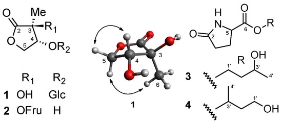
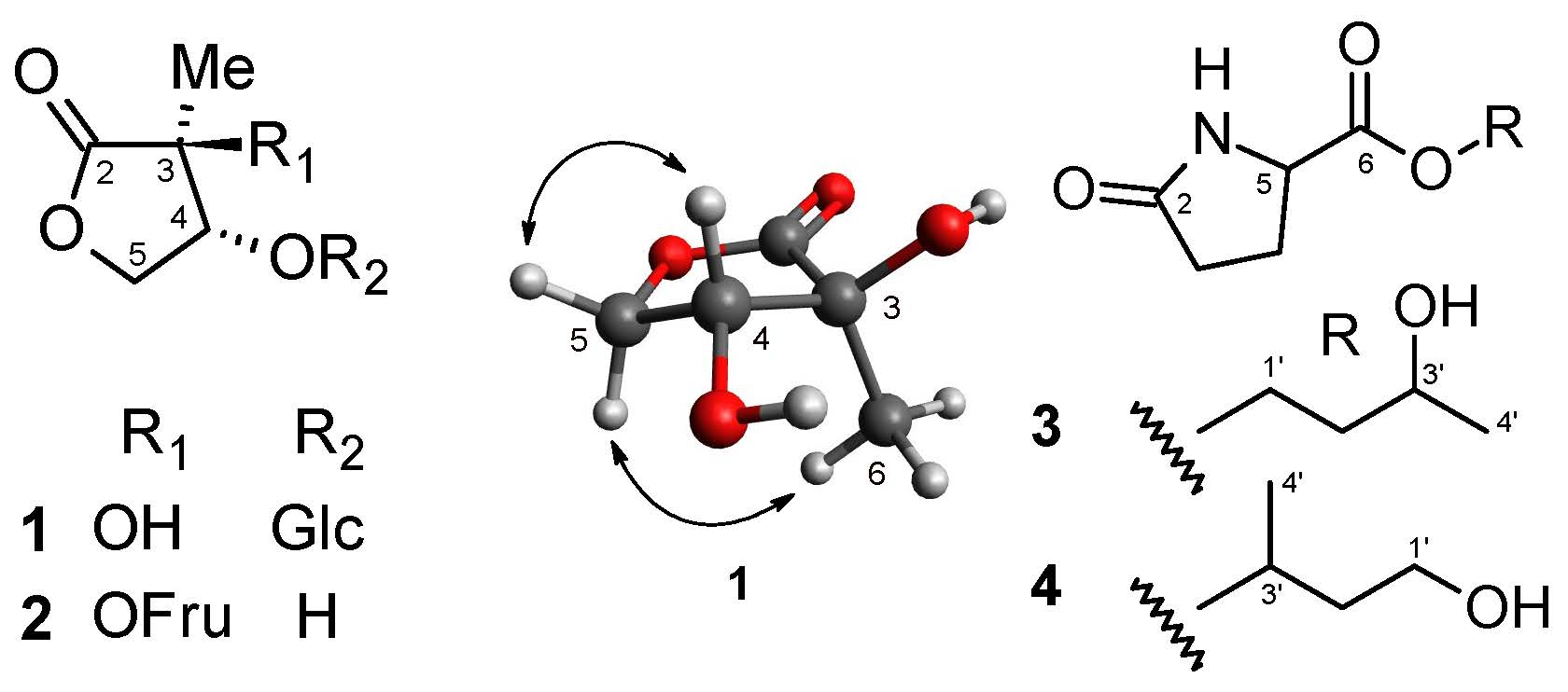
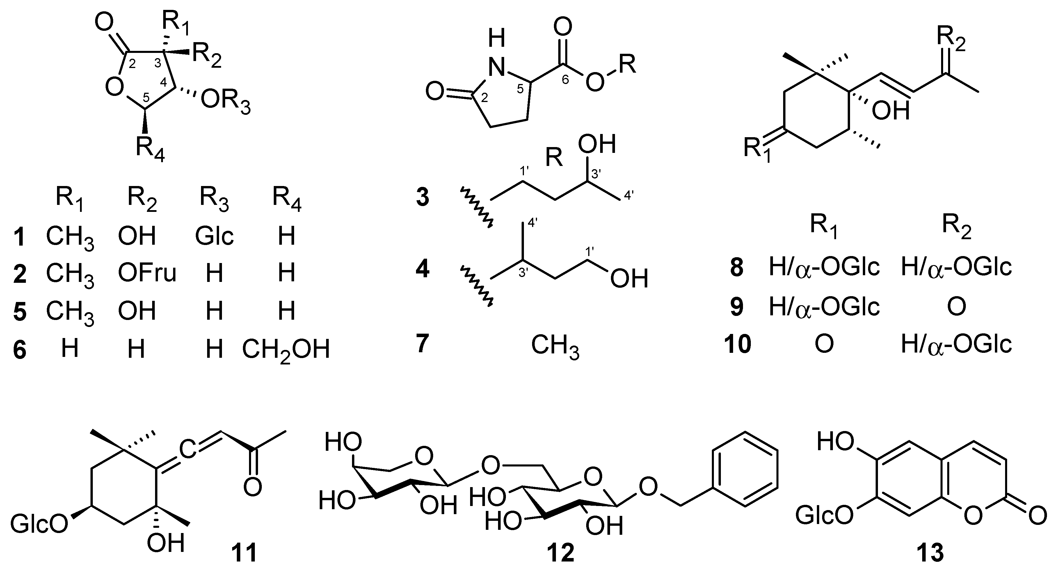
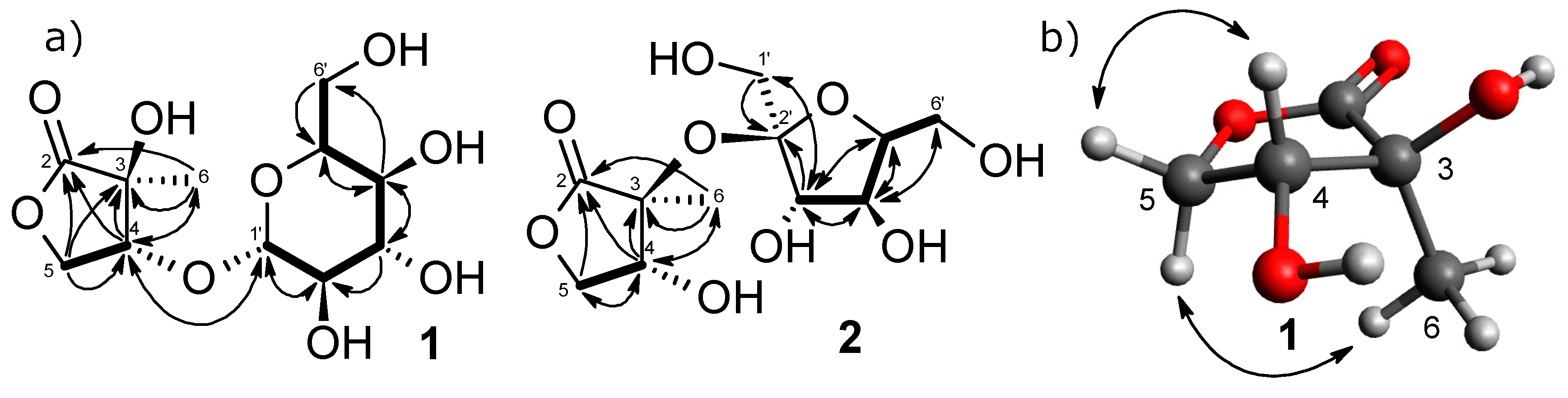
2.1.1. Structure of 2-C-methyl-d-threono-1,4-lactone-3-O-β-d-glucopyranoside (1)

Compound 1 was obtained as a colorless amorphous powder with a molecular formula of C11H18O9 as determined by HR-ESI-MS at an m/z of 317.0845 [M + Na]+ (calcd for C11H18O9Na: 317.0843). The IR spectrum implied the presence of carbonyl (1777 cm−1), hydroxy groups (3392 cm−1), and C-O linkage (1210 and 1078 cm−1). The 1H-NMR spectrum (Table 1) displayed signals due to a methyl proton at δH 1.41 (3H, s); four oxygenated methylene protons at δH 3.60 (1H, dd, J = 11.6, 7.4 Hz), 3.91 (1H, dd, J = 11.6, 2.5 Hz), 4.10 (1H, dd, J = 9.5, 6.4 Hz), and 4.52 (1H, dd, J = 9.5, 6.5 Hz); five oxygenated methine protons at δH 3.238 (1H, dd, J = 9.2, 7.7 Hz), 3.237 (1H, dd, J = 9.8, 9.1 Hz), 3.34 (1H, ddd, J = 9.8, 7.4, 2.5 Hz), 3.36 (1H, t-like, J = 9.1 Hz), and 4.43 (1H, t-like, J = 6.5 Hz); and an anomeric proton at δH 4.37 (1H, d, J = 7.7 Hz) that indicated the presence of a glycosyl moiety. A lactone moiety was also indicated from three degrees of unsaturation, the relatively higher wavenumber of carbonyl (1777 cm−1), the esterified low field-shifted chemical shifts, and non-equivalency of H-5α (δH 4.10, 1H, dd, J = 9.5, 6.4 Hz) and H-5β (δH 4.52, 1H, dd, J = 9.5, 6.5 Hz).
The 13C-NMR spectrum (Table 2) of 1 showed 11 carbon resonances that were classified by chemical shift values and the HSQC spectrum into a methyl carbon (δC 19.2), two oxygenated methylene carbons (δC 63.2 and 69.6), five oxygenated methine carbons (δC 72.2, 74.9, 78.1, 78.2, and 83.4), a quaternary carbon (δC 75.7), an anomeric carbon (δC 104.0) and a carbonyl carbon at δC 179.4. The NMR spectroscopic data of 1 closely resembled that of 2-C-methyl-d-threono-1,4-lactone (5) [16,17], except for a large difference in the chemical shift values at C-4. The deshielded proton at δH 4.43 and the carbon at δC 83.4 of 1 suggested that the glycosyl group was attached to C-4. This was confirmed by an HMBC experiment from the correlation between H-4 (δH 4.43) and the carbon signal at δC 104.0 (Figure 2a). While five signals corresponding to C2′-C6′ and an anomeric carbon signal resonating at δC 104.0 were indicative of glucopyranoside. The relative configuration of the aglycone moiety was established using NOESY analysis. The correlations between H-5α/Me-6 and H-4/H-5β suggested α-orientation for H-5α and Me-6, and β-orientation for H-4 and H-5β (Figure 2b). Acid hydrolysis of 1 with 1N HCl liberated glucose and aglycone (1a). The glucose was determined to be D-series from the result of HPLC analysis with an optical rotation detector [25]. The coupling constant (J = 7.7 Hz) of the anomeric proton signal (δH 4.37) suggested a β-glycosidic linkage. The aglycone (1a) was identified to be 2-C-methyl-d-threono-1,4-lactone (5) [16] by spectroscopic (IR, HR-ESI-MS, 1D NMR, and [α]D) analyses. The 4R configuration was also supported by the application of the β-d-glucosylation-induced shift-trend rule, i.e., Δδ glucoside-aglycone values of C-3 (±0), C-4 (+7.4), and C5 (−3.4) suggested C-5 is Pro-S [26]. Based on these results, the structure of 1 was determined to be 2-C-methyl-d-threono-1,4-lactone-3-O-β-d-glucopyranoside.

2.1.2. Structure of 2-C-methyl-d-threono-1,4-lactone-3-O-α-d-fructofuranoside (2)

Compound 2 was obtained as a colorless amorphous powder whose molecular formula was determined to be C11H18O9 from positive-ion HR-ESI-MS at an m/z of 317.0844 [M + Na]+ (calcd 317.0843). The 1H and 13C-NMR spectra (Table 1 and Table 2) of 2 were similar to those of 1 and 2-C-methyl-d-threono-1,4-lactone (5). The 13C-NMR also showed two secondary carbons at δC 60.6 and 62.9; three tertiary carbons at δC 79.0, 82.6, and 84.7; and a quaternary carbon at δC 109.2, indicating a fructofuranose moiety. Zhang et al. (2009) described that α and β orientations of D-fructofuranose were distinguishable by the J value of H-3 and the chemical shift of C-2. The characteristic values of α orientation are 3JH3,H4 = 3–4 Hz and C-2 (δC 107–109), while the β orientation is 3JH3,H4 = 7–9 Hz and C-2 (δC 103–106) [27], which suggested the α-fructofuranosylation to the aglycone. The 2D NMR analyses (COSY, HSQC, and HMBC) supported the planar structure of 2 (Figure 2a). The upfield-shift of C-4, compared to 1, suggested the quarternary C-3/C-2′ connectivity. Acid hydrolysis of 2 yielded aglycone (2-C-methyl-d-threono-1,4-lactone) (2a) and D-fructose. Thus, 2 was estimated to be 2-C-methyl-d-threono-1,4-lactone-3-O- α -d-fructofuranoside, although a chemical synthesis may support our conclusion.

2.1.3. Structure of 1,3-butanediol-1-pyroglutamate (3)

Compound 3 was obtained as a colorless amorphous powder and displayed an [M + Na]+ ion at an m/z of 224.0890 (calcd 224.0893) corresponding to a molecular formula of C9H15O4N. The IR spectrum showed a strong absorption band for the hydroxy (3331 cm−1) and carbonyl (1735 and 1684 cm−1). The 1H and 13C-NMR spectra of 3 showed signals assignable to two methylenes (δH 2.31 (1H, ddd, J = 17.1, 9.9, 5.5 Hz)) and δH 2.37 (1H, ddd, J = 17.1, 9.4, 7.3 Hz) / δC 30.4 (C-3), and δH 2.16 (1H, m) and 2.48 (1H, m)/δC 26.0 (C-4)), a methine (δH 4.29 (1H, dd, J = 9.1, 4.4 Hz)/δC 57.3 (C-5)), and two carbonyls (δC 181.2 (C-2) and 174.2 (C-6)) (Table 1 and Table 2). The chemical shift values and coupling patterns of these signals suggested a pyroglutamate moiety [18]. In addition, the 1H and 13C-NMR spectra also revealed a 1,3-butanediol framework, with a methyl (δH 1.20 (3H, d, J = 6.3 Hz)/δC 23.9 (Me-10)), a methylene (δH 1.75 (1H, m) and 1.79 (1H, m)/δC 38.8 (C-2′)), an oxygenated methine [δH 3.86 (1H, dqd, J = 8.2, 6.3, 4.4 Hz)/δC 65.5 (C-3′)), and an oxygenated methylene (δH 4.27 (2H, m)/δC 64.0 (C-1′)). The relatively lower chemical shift value of H-1′ (δH 4.27) indicated the ester linkage with pyroglutamate at C-1′. The 1H–1H COSY spectrum displayed correlations through H-3, H-4, and H-5, and through H-1′, H-2′, H-3′, and H-4′. The HMBC spectrum demonstrated correlations of C-2 with H-3, and H-4, and correlations of C-6 with H-4 and H-5. Furthermore, the strong correlation of H-1′ with C-6 established that the 1,3-butanediol moiety was located at the C-6 (Figure 3) to form an ester linkage with primary alcohol. Therefore, the structure of 3 was elucidated to be 1,3-butanediol-1-pyroglutamate.

2.1.4. Structure of 1,3-butanediol 3-pyroglutamate (4)

Compound 4 was isolated as a colorless amorphous powder. The NMR spectra together with the molecular ion at an m/z 224.0893 [M + Na]+ (calcd for C9H15O4NNa: 224.0893) in HR-ESI-MS indicated that 4 was an isomer of 3. The shielding of H-1′ (δH 3.65 (1H, dt-like, J = 10.6, 6.3 Hz) and δH 3.68 (1H, dt-like, J = 10.6, 6.7 Hz)/C-1′ (δC 60.5)) and deshielding of H-3′ (δH 3.89 (1H, dqd, J = 7.7, 6.3, 4.8 Hz)/C-3′ (δC 65.5)) suggested that the ester linkage of 1,3-butanediol must be changed from primary alcohol (C-1′) to the secondary alcohol (C-3′) of 1,3-butanediol in 4 (Table 1 and Table 2). The planar structure was supported by COSY and HMBC correlations (Figure 3). The difficulty of observing the correlation between C-3′ and C-6 coincided with the abundant multiplicity of H-3′. As a result, the structure of 4 was estimated to be 1,3-butanediol 3-pyroglutamate.
The absolute stereochemistry of compounds 3 and 4 remains to be elucidated because of the insufficient amount for further analysis.

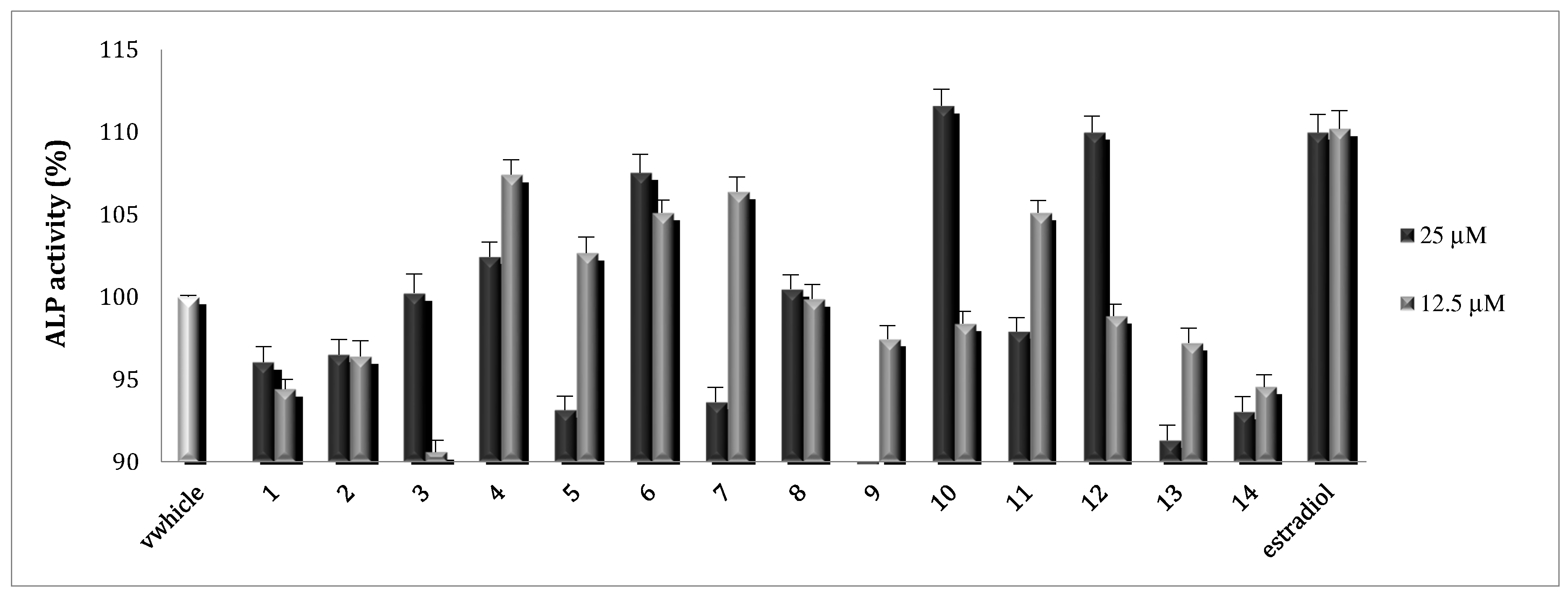
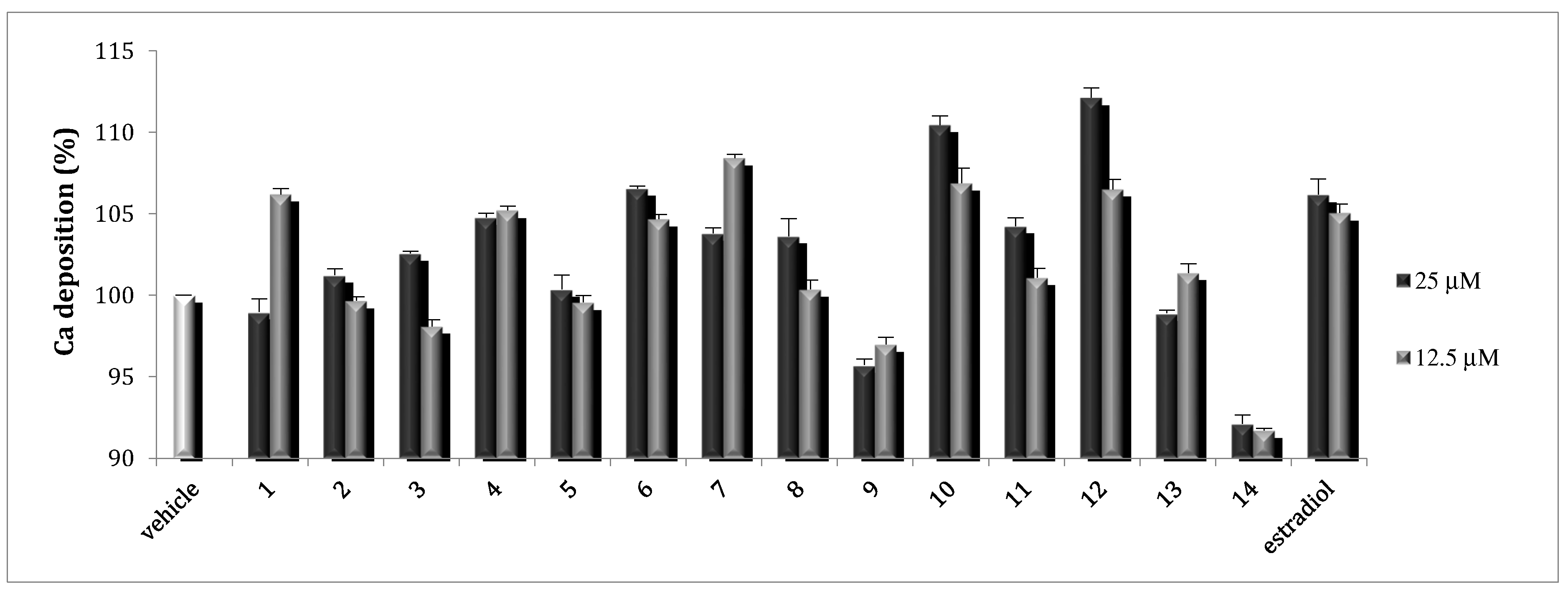
2.2. Osteoblast Activity

Osteoblasts are the most important cells in bone tissue and are critical for bone formation through proliferation and differentiation. During osteoblast differentiation, BMPs induce the expression of osteoblastic markers, such as ALP. Proliferating osteoblasts show ALP activity, which is greatly enhanced during in vitro bone formation. ALP is a membrane-bound enzyme that is often used as a marker for osteogenic differentiation. 17β-estradiol has a significant impact on bone mineral metabolisms. It affects osteoblast proliferation through modulating the release of several local regulators of bone turnover from monocytes and enhanced BMP-4 induced osteoblastic marker expression and mineralization [28].
To evaluate the effects of 114 on osteoblast function, ALP activity, which is related to the osteoid formation and initiation of the deposition of minerals, was evaluated. In this study, it was found that 4, 6, 7, 10, 11, and 12 stimulated ALP activity, which markedly increased osteoblast growth and differentiation of osteoblastic MC3T3-E1 cells. Compounds 7 and 11 did not show concentration dependency, probably due to the toxicity at higher concentrations. At concentrations of 25 µM, 6, 10, and 12 stimulated ALP activity up to 112% comparable to the positive control, 17β-estradiol at 0.02 and 0.01 µM (Figure 4).
Osteoblasts can be induced to produce vast extracellular calcium deposition in vitro. This process is called mineralization. Calcium deposition is an indication of successful in vitro bone formation and can specifically be stained bright orange-red using Alizarin Red S. The effects of 114 were then examined by measuring the calcium deposition by Alizarin Red staining. As was found for the ALP activity study above, 4, 6, 7, 10, 11, and 12 showed stimulatory effects on mineralization. Compounds 6, 10, and 12 stimulated the mineralization up to 112% at 25 μM, comparable to that of the positive control, 17β-estradiol, at 0.02 and 0.01 μM (Figure 5).
In bone formation, osteoblasts are key cells in bone matrix formation and calcification. Osteogenesis starts with osteoblast production and secretion of type I collagen, which makes up about 90% of the organic bone matrix or the osteoid. Osteoblast also becomes high in alkaline phosphatase. Alkaline phosphatase is released into the osteoid to initiate the deposition of minerals. After mineralization, the bone becomes hard and rigid with necessary mechanical properties to withstand the external forces to support the body and protect the internal organs. Our study demonstrated that 4, 6, 7, 10, 11, and 12 stimulated both ALP activity and calcium deposition in osteoblastic MC3T3-E1 cell in vitro, which suggests that the extract of S. acmella and these compounds have potential to be a remedy for osteoporosis as osteoblastic bone formation stimulant.

3. Materials and Methods

3.1. General Experimental Procedures

1H and 13C-NMR spectra were taken on a Bruker Ultrashield Avance 600 spectrometer at 600 MHz and 150 MHz, respectively, with TMS as an internal standard. IR and UV spectra were measured on a HORIBA FT-720 FT-IR spectrophotometer and JASCO V-520 UV–vis spectrophotometer, respectively. Optical rotation was measured on a JASCO P-1030 digital polarimeter. Positive ion HR-ESI-MS was recorded using an LTQ Orbitrap XL mass spectrometer (Thermo Fisher Scientific, Waltham, MA, USA). Silica gel open column chromatography (CC) and reversed-phase (ODS) CC were performed on silica gel 60 (E. Merck, Darmstadt, Germany), and Cosmosil 75C18-OPN (Nacalai Tesque, Kyoto, Japan; Φ = 35 mm, L = 350 mm), respectively. HPLC was performed on an ODS column (Inertsil ODS-3, GL Science, Tokyo, Japan; Φ = 6 mm, L = 250 mm, 1.5 mL/min), and the eluate was monitored with a JASCO RI-930 intelligent detector and a JASCO PU-1580 intelligent pump unless otherwise specified.

3.2. Plant Material

Whole plants of Spilanthes acmella (L.) L. were collected in late June 2007 in Kebun Raya Purwodadi, Malang, Indonesia (07°46′09″–07°47′23″ South latitude and 112°16′23″–112°17′17″ East Longitude), and voucher specimens were deposited at the Department of Pharmacognosy and Phytochemistry, Faculty of Pharmacy, Airlangga University as SA30062007 [29,30].

3.3. Extraction and Isolation

The air-dried plants (2.0 kg) were extracted with methanol (MeOH, 10.0 l × 3). The methanol solution was concentrated and adjusted to 95% aq. methanol by the addition of water and then partitioned with n-hexane (1.0 l × 3, 23.5 g). The remaining aqueous methanol layer was evaporated and resuspended in 0.5 l of water and then partitioned with ethyl acetate (1.0 l × 3, 40.4 g) and 1-butanol (1.0 l × 3, 47.5 g), successively.
The 1-butanol layer (40.0 g) was subjected on silica gel (300 g) CC with increasing amounts of MeOH in CHCl3 [Hexane-CHCl3 (1:1), 4 l, CHCl3-MeOH (50:1, 40:1, 30:1, 20:1, 15:1, 10:1, 7:1, 5:1, 3:1, 2:1, 2 l), 500 mL fractions being collected], yielding 12 fractions (Fr. Sab1–Sab12). The fraction Sab11 (2.75 g) was subjected to ODS CC in 10% aq. MeOH (400 mL)–100% MeOH (400 mL), linear-gradient, led 10 fractions (Fr. Sab11-1–Sab11-10). The fraction Sab11-3 (442 mg) and Sab11-4 (123 mg) were purified by HPLC with 35% aq. MeOH to give 1 (10.1 mg) and 8 (dendranthemoside A, 3.71 mg), respectively. The fraction Sab10 (1.81 g) was subjected to ODS CC in 10% aq. MeOH (400 mL)–100% MeOH (400 mL) and led seven fractions (Fr. Sab10-1–Sab10-7). The fraction Sab10-1 (770 mg) was purified by HPLC with 100% H2O, YMC Triart C18 column. Three peaks appeared at 5, 18, and 35 minutes and were collected to give 2 (7.62 mg), 5 (2-C-methyl-d-threono-1,4-lactone, 9.31 mg), and 14 (uridine, 27.5 mg). The fraction Sab10-2 (193 mg) was purified by HPLC (20% aq. MeOH) to give 11 (icariside B1, 6.12 mg) and 13 (chicoriin, 2.99 mg). The fraction Sab10-3 (142 mg) was purified by HPLC (35% aq. MeOH) to give 9 (dendranthemoside B, 4.31 mg). The fraction Sab5 (710 mg) was subjected to ODS CC in 10% aq. MeOH (400 mL)–100% MeOH (400 mL) and led 10 fractions (Fr. Sab5-1–Sab5-10). The fraction Sab5-1 (483 mg) and Sab5-2 (68.3 mg) were purified by HPLC (100% H2O, YMC Triart C18 column) to give 3 (7.80 mg), 4 (4.21 mg), and 7 (methyl pyroglutamate, 6.63 mg), respectively. The mixture of fraction Sab6, Sab7, Sab8, and Sab9 (2.06 g) was subjected to ODS CC in 10% aq. MeOH (400 mL)–100% MeOH (400 mL) and led 10 fractions (Fr. Sab6-9-1–Sab6-9-10). The fraction Sab6-9-1 (340 mg) was purified by HPLC (100% H2O, YMC Triart C18 column) to give 6 (2-deoxy-d-ribono-1,4-lactone, 6.01 mg). The fraction, Sab6-9-4 (114 mg), was purified by HPLC (40% aq. MeOH) to give 10 (ampelopsisionoside, 5.43 mg). The fraction Sab12 (5.36 g) was subjected to ODS CC in 10% aq. MeOH (400 mL)–100% MeOH (400 mL) and led 10 fractions (Fr. Sab12-1–Sab12-10). The fraction Sab12-3 (129 mg) was purified by HPLC (35% aq. MeOH) to give 12 (benzyl α-l-arabinopyranosyl (1→6)-β-d-glucopyranoside, 4.51 mg).

3.3.1. 2-C-methyl-d-threono-1,4-lactone-4-O-β-d-glucopyranoside (1)

Colorless amorphous powder; [α]D 26.7 -28.6 (c 0.78, MeOH); IR (film) max cm−1: 3392, 2924, 1777, 1713, 1650, 1557, 1456, 1391, 1210, 1078, 899, 647; 1H-NMR and 13C-NMR, see Table 1 and Table 2; positive HR-ESI-MS (m/z): 317.0845 [M + Na]+(calcd for C11H18O9Na: 317.0843).

3.3.2. 2-C-methyl-d-threono-1,4-lactone-3-O-α-d-fructofuranoside (2)

Colorless amorphous powder; [α]D 26.9 -10.4 (c 0.74, MeOH); IR (film) max cm−1: 3386, 2938, 1774, 1732, 1651, 1540, 1456, 1339, 1206, 1073, 870, 669; 1H-NMR and 13C-NMR, see Table 1 and Table 2; positive HR-ESI-MS (m/z): 317.0844 [M + Na]+(calcd for C11H18O9Na: 317.0843).

3.3.3. 1,3-butanediol-1-pyroglutamate (3)

Colorless amorphous powder; [α]D 27.7 −0.86 (c 0.42, MeOH); IR (film) max cm−1: 3331, 2966, 2926, 1735, 1684, 1557, 1457, 1338, 1207, 1052, 670; 1H-NMR and 13C-NMR, see Table 1 and Table 2; positive HR-ESI-MS (m/z): 224.0890 [M + Na]+(calcd for C9H15O4NNa: 224.0893).

3.3.4. 1,3-butanediol-3-pyroglutamate (4)

Colorless amorphous powder; [α]D 27.1 +1.40 (c 0.41, MeOH); IR (film) max cm−1: 3314, 2969, 2931, 1735, 1683, 1557, 1457, 1338, 1229, 1054, 669; 1H-NMR and 13C-NMR, see Table 1 and Table 2; positive HR-ESI-MS (m/z): 224.0893 [M + Na]+ (calcd for C9H15O4NNa: 224.0893).

3.3.5. Acid Hydrolysis for Identification of Sugar Moiety of 1 and 2

A solution of 1 or 2 (1 mg) in 1 N aq. HCl (0.1 mL) was heated at 80 °C for 2 h. The mixture was neutralized by the addition of amberlite IRA400 (OH- form), and the resin was removed by filtration. Then, the filtrates were extracted with EtOAc. The aqueous layers were subjected to HPLC analysis (column: Shodex Asahipak NH 2P-50 4E, 250 × 4.6 mm i.d.; mobile phase: 75% CH3CN in water; detection: optical rotation (JASCO OR-2090Plus); flow rate: 1.0 mL/min) to identify D-glucose and D-fructose, which were identified by the comparison of their retention times with those of authentic samples; tR: 5.11 (D-fructose, negative optical rotation) and tR: 6.10 (D-glucose, positive optical rotation). They also yielded the aglycon of 2-C-methyl-d-threono-1,4-lactone (1a and 2a) [26]. (1a): Colorless amorphous powder; [α]D 27.5 −15.0 (c 0.06, MeOH); IRmax (film) cm−1: 3286, 2923, 1729, 1649, 1558, 1456, 1339, 1205, 1092, 667; 1H-NMR (methanol-d4, δH): 1.35 (3H, s, H-6), 3.96 (1H, dd, J = 9.0, 4.0 Hz, H-5a), 4.17 (1H, dd, J = 5.0, 4.0 Hz, H-4), 4.49 (1H, dd, J = 9.0, 5.0 Hz, H-5b); positive HR-ESI-MS m/z 155.0312 [M + Na]+(calcd. for C5H8O4Na: 155.0315) and tR = 10.20 (HPLC, 100% H2O, YMC Triart C18 column). (2a): Colorless amorphous powder; [α]D 27.9 −15.7 (c 0.08, MeOH); IRmax (film) cm−1: 3373, 2927, 1717, 1649, 1558, 1455, 1338, 1207, 1098, 668; 1H-NMR (methanol-d4, δH): 1.35 (3H, s, H-6), 3.96 (1H, dd, J = 9.0, 4.0 Hz, H-5a), 4.17 (1H, dd, J = 5.0, 4.0 Hz, H-4), 4.49 (1H, dd, J = 9.0, 5.0 Hz, H-5b); positive HR-ESI-MS m/z 155.0319 [M + Na]+ (calcd. for C5H8O4Na: 155.0315) and tR = 10.20 (HPLC, 100% H2O, YMC Triart C18 column) together with authentic sample 5.

3.4. Osteoblast Activities

3.4.1. Cell Culture

An osteoblast-like cell of MC3T3-E1 was purchased from Riken Cell Bank, Tsukuba, Japan. The cells were cultured in α-MEM medium containing 10% FBS in a CO2 incubator at 37 °C and sub-cultured every 3 days by trypsin (0.25%) treatment. The 5 × 10⁴ cells were seeded in 24-well plates and incubated for the following ALP and mineralization assays.

3.4.2. Alkaline Phosphatase (ALP) Activity

The cells were treated at 90% confluence with the culture medium containing 10 mM β-glycerophosphate and 50 µg/mL ascorbic acid to initiate in vitro proliferation. The medium was changed every 2–3 d. After 6 days, the cells were cultured individually for 3 days with medium containing 0.3% bovine serum and isolated compounds (114). On harvesting, the medium was removed, and the cell monolayer was gently washed twice with phosphate-buffered saline. The cells were lysed with 0.2% Triton X-100, and centrifuged at 14,000 × g for 5 min. The clear supernatant was used to measure ALP activity using p-nitrophenylphosphate [31,32].

3.4.3. Mineralization of MC3T3-E1

The cells were treated, at 90% confluence, with culture medium containing 10 mM β-glycerophosphate and 50 µg/mL ascorbic acid, to initiate in vitro mineralization. After 12 days, the cells were cultured individually for 2 days with medium containing 0.3% bovine serum and isolated compounds (114). On harvesting, the cells were fixed with 70% ethanol for 1 hour and then stained with 40 mM Alizarin Red S for 10 min with gentle shaking. To quantify the bound dye, the stain was solubilized with 10% cetylpyridinium chloride by shaking for 15 min. The absorbance of the solubilized stain was measured at 561 nm [33].

4. Conclusions

The chemical investigation of the 1-butanol layer of the methanol extract of Spilanthes acmella (L.) L. gave 14 compounds (114), including 2 new methyl threonolactone glycosides, 2-C-methyl-d-threono-1,4-lactone-3-O-β-d-glucopyranoside (1) and 2-C-methyl-d-threono-1,4-lactone-2-O-α-d-fructofuranoside (2); 2 new pyroglutamates, 1,3-butanediol 1-pyroglutamate (3) and 1,3-butanediol 3-pyroglutamate (4); and 10 known compounds, 2-C-methyl-d-threono-1,4-lactone (5), 2-deoxy-d-ribono-1,4-lactone (6), methyl pyroglutamate (7), dendranthemoside A (8), dendranthemoside B (9), ampelopsisionoside (10), icariside B1 (11), benzyl α-l-arabinopyranosyl-(1→6)-β-d-glucopyranoside (12), chicoriin (13), and uridine (14). All the isolated compounds were evaluated for ALP and calcium deposition as markers for osteogenic differentiation and mineralization stimulatory activities. Our study demonstrated that compounds 4, 6, 7, 10, 11, and 12 stimulated ALP activity and calcium deposition in osteoblastic MC3T3-E1 cell in vitro. Compounds 6, 10, and 12 stimulated ALP and mineralization up to 112% at 25 µM, comparable to that of the positive control, 17β-estradiol, at 0.02 and 0.01 μM. Of these active compounds, compounds 4, 6, 7, 10, and 11 are the first time to be reported their osteoblastic activity. While compound 12 has already isolated as an osteoblastic active compound from the fruit of Prunus mume [34]. The isolation of the same active compound (12) from the different plants through independent study strengthened and supported the reliability of our results. Overall, the compounds 4, 6, 7, 10, 11, and 12 are the active principle of S. acmella on the stimulation of osteoblastic bone formation and may play an important role in bone remodeling as drug seeds in osteoporosis therapy.

Supplementary Materials

The following are available online: Figure S1–S32: 1D and 2D NMR data for 14.

Author Contributions

Conceptualization, K.M. and R.W.; Methodology, S.S.; Software, Y.Y.; Validation, H.O., K.M. and S.S.; Formal analysis, K.M.; Investigation, R.W., M.I.S., and N.H.U.; Resources, R.W.; Data curation, N.H.U.; Writing—original draft preparation, R.W.; Editing, K.M.; Visualization, R.W.; Supervision, H.O. and K.M.; Project administration, M.I.S.; Funding acquisition, R.W. and K.M. All authors have read and agreed to the published version of the manuscript.

Funding

This research was funded by PTUPT 2017 research grant of the Indonesia Government, JSPS KAKENHI grant nos. 17K08336, 16K18896, 17K15465, and 15H04651, and the cosmetology research foundation.

Acknowledgments

The authors are grateful for access to the superconducting NMR instrument, UV, and ESI-MS at the Analytical Center of Molecular Medicine, the Analysis Center of Life Science, and the Natural Science Center for Basic Research and Development of the Graduate School of Biomedical and Health Sciences, Hiroshima University.

Conflicts of Interest

The authors declare no conflict of interest.

References

  1. Vandenbroucke, A.; Luyten, F.P.; Flamaing, J.; Gielen, E. Pharmacological treatment of osteoporosis in the oldest old. Clin. Interv. Aging 2017, 12, 1065–1077. [Google Scholar] [CrossRef] [Green Version]
  2. Sahu, J.; Jain, K.; Jain, B.; Sahu, R.K. A review on phytopharmacology and micropropagation of Spilanthes acmella. Pharmacologyonline 2011, 2, 1105–1110. [Google Scholar]
  3. Tiwari, K.L.; Jadhav, S.K.; Joshi, V. An updated review on medicinal herb genus Spilanthes. Chin. J. Integr. Med. 2011, 9, 1170–1178. [Google Scholar] [CrossRef]
  4. Sharma, V.; Boonen, J.; Chauhan, N.S.; Thakur, M.; Spiegeleer, B.D.; Dixit, V.K. Spilanthes acmella ethanolic flower extract: LC-MS alkylamide profiling and its effects on sexual behavior in male rats. Phytomedicine 2011, 18, 1161–1169. [Google Scholar] [CrossRef] [PubMed] [Green Version]
  5. Wongsawatkul, O.; Prachayasittikul, S.; Chartchalerm, I.N.A.; Satayavivad, J.; Ruchirawat, S.; Prachayasittikul, V. Vasorelaxant and antioxidant activities of Spilanthes acmella Murr. Int. J. Mol. Sci. 2008, 9, 2724–2744. [Google Scholar] [CrossRef] [PubMed]
  6. Ramsewak, R.S.; Erickson, A.J.; Nair, M.G. Bioactive N-isobutylamides from the flower buds of Spilanthes acmella. Phytochemistry 1999, 51, 729–732. [Google Scholar] [CrossRef]
  7. Storey, C.; Salem, J.I. Lay use of Amazonian plants for the treatment of tuberculosis. Acta Amaz. 1997, 27, 175–182. [Google Scholar] [CrossRef] [Green Version]
  8. Hossan, S.; Agarwala, B.; Sarwar, S.; Karim, M.; Jahan, R.; Rahmatullah, M. Traditional use of medicinal plants in Bangladesh to treat urinary tract infections and sexually transmitted diseases. Ethnobot. Res. Appl. 2010, 8, 61–74. [Google Scholar] [CrossRef] [Green Version]
  9. Arora, S.; Vijay, S.; Kumar, D. Phytochemical and antimicrobial studies on the leaves of Spilanthes acmella. J. Chem. Pharm. Res. 2011, 3, 145–150. [Google Scholar]
  10. Jondiko, I.J.O. A mosquito larvicide in Spilanthes mauritiana. Phytochemistry 1986, 25, 2289–2290. [Google Scholar] [CrossRef]
  11. Kadir, H.A.; Zakaria, M.B.; Kechil, A.A.; Azirun, M.S. Toxicity and electrophysiological effects of Spilanthes acmella Murr. extracts on Periplaneta americana L. Pest. Manag. Sci 1989, 25, 329–335. [Google Scholar] [CrossRef]
  12. Prachayasittikul, S.; Suphapong, S.; Worachartcheewan, A.; Lawung, R.; Ruchirawat, S.; Prachayasittikul, V. Bioactive metabolites from Spilanthes acmella Murr. Molecules 2009, 14, 850–867. [Google Scholar] [CrossRef] [PubMed] [Green Version]
  13. Narayana, K.R.; Reddy, M.S.; Chaluvadi, M.R.; Krishna, D.R. Bioflavonoids classification, pharmacological, biochemical effects and therapeutic potential. Indian J. Pharmacol. 2001, 33, 2–16. [Google Scholar]
  14. Laswati, H.; Subadi, I.; Widyowati, R.; Agil, M.; Pangkahila, J.A. Spilanthes acmella and physical exercise increased testosterone levels and osteoblast cells in glucocorticoid-induced osteoporosis male mice. Bali Med. J. 2015, 4, 76–81. [Google Scholar] [CrossRef]
  15. Widyowati, R. Alkaline phosphatase activity of Graptophyllum pictum and Spilanthes acmella fractions against MC3T3-E1 cells as marker of osteoblast differentiation cells. Int. J. Pharm. Pharm. Sci. 2011, 3, 34–37. [Google Scholar]
  16. Uriburu, M.L.; Gil, R.R.; Sosa, V.E.; Fuente, J.R. Caffeoyl esters of threonic acid and its lactone from Viguiera pazensis. J. Arg. Chem. Soc. 2008, 96, 55–61. [Google Scholar]
  17. Kitajima, J.; Ishikawa, T.; Tanaka, T.; Ida, Y. Water-soluble constituents of fennel IX. Glucides and nucleosides. Chem. Pharm. Bull. 1999, 47, 988–992. [Google Scholar] [CrossRef] [Green Version]
  18. Barraclough, P.; Dieterich, P.; Spray, C.A.; Young, D.W. Two separate and distinct syntheses of stereospecifically deuterated samples of (2S)-proline. Org. Biomol. Chem. 2006, 4, 1483–1491. [Google Scholar] [CrossRef]
  19. Otsuka, H.; Takeda, Y.; Yamasaki, K.; Takeda, Y. Structural elucidation of Dendranthemosides A and B: Two new β-ionone glucosides from Dendranthema shiwogiku. Planta Med. 1992, 58, 373–375. [Google Scholar] [CrossRef]
  20. Marino, S.D.; Borbone, N.; Zollo, F.; Lanaro, A.; Meglio, P.D.; Lorizzi, M. Megastigmane and phenolic components from Laurus nobilis L. leaves and their inhibitory effects on nitric oxide production. J. Agric. Food Chem. 2004, 52, 7525–7531. [Google Scholar] [CrossRef]
  21. Miyase, T.; Ueno, A.; Takizawa, N.; Kobayashi, H.; Karasawa, H. Studies on the glycosides of Epimedium grandiflorum MORR. Var. thunbergianum (MIQ.) NAKAI. I. Chem. Pharm. Bull. 1987, 35, 1109–1117. [Google Scholar] [CrossRef] [Green Version]
  22. Rosa, S.; Giulio, A.; Tommonaro, G. Aliphatic and aromatic glycosides from the cell cultures of Lycopersicon esculentum. Phytochemistry 1996, 42, 1031–1034. [Google Scholar] [CrossRef]
  23. Zhou, H.; Qin, M.; Hong, J.; Ni, Y.; Wu, G. Chemical constituents of Viola yedoensis. Chin. J. Nat. Med. 2009, 7, 290–292. [Google Scholar] [CrossRef]
  24. Chen, H.; Geng, C.; Cao, T.; Zhang, X.; Ma, Y.; Huang, X.; Chen, J. Chemical structure of capsicuoside A from fruits of Capsium annum. China J. Chin. Mater. Med. 2013, 38, 1934–1937. [Google Scholar]
  25. Tanaka, T.; Nakashima, T.; Ueda, T.; Tomii, K.; Kouno, I. Facile discrimination of aldose enantiomers by reversed-phase HPLC. Chem. Pharm. Bull. 2007, 55, 899–901. [Google Scholar] [CrossRef] [Green Version]
  26. Kasai, R.; Suzuno, M.; Asakawa, J.; Tanaka, O. Carbon-13 chemical shifts of isoprenoid-β-d-glucopyranosides and -β-d-mannopyranosides. Stereochemical influences of aglycone alcohols. Tetrahedron. Lett. 1977, 18, 175–178. [Google Scholar] [CrossRef]
  27. Zhang, Z.; Wang, D.; Zhao, Y.; Gao, H.; Hu, Y.; Hu, J. Fructose-derived carbohydrates from Alisma orientalis. Nat. Prod. Res. 2009, 23, 1013–1020. [Google Scholar] [CrossRef]
  28. Stein, G.S.; Lian, J.B. Molecular mechanisms mediating developmental and hormone-regulated expression of genes in osteoblasts: An integrated relationship of cell growth and differentiation. In Cellular and Molecular Biology of Bone; Noda, M., Ed.; Academic Press: Tokyo, Japan, 1993; pp. 47–95. [Google Scholar]
  29. Backer, C.A.; Bakhuizen van den Brink Jr, R.C. Flora of Java (Spermatophytes only); Noordhoff: Groningen, The Netherlands, 1965; p. 408. [Google Scholar]
  30. Heyne, K. Tumbuhan Berguna Indonesia III; Badan Litbang Departemen Kehutanan: Jakarta, Indonesia, 1987; p. 1456. [Google Scholar]
  31. Cuong, N.X.; Tuan, T.A.; Kiem, P.V.; Minh, C.V.; Choi, E.M.; Kim, Y.H. New cembranoid diterpenes from the Vietnamese soft coral Sarcophyton mililatensis stimulate osteoblastic differentiation in MC3T3-E1 cells. Chem. Pharm. Bull. 2008, 56, 988–992. [Google Scholar] [CrossRef] [Green Version]
  32. Hamazaki, T.; Suzuki, N.; Widyowati, R.; Miyahara, T.; Kadota, S.; Ochiai, H.; Hamazaki, K. The depressive effects of 5,8,11-eicosatrienoic acid (20:3n-9) on osteoblasts. Lipids 2009, 44, 97–102. [Google Scholar] [CrossRef]
  33. Widyowati, R.; Tezuka, Y.; Miyahara, T.; Awale, S.; Kadota, S. Alkaline phosphatase (ALP) enhancing iridoid glucosides from the Indonesian medicinal plant Barleria lupulina. Nat. Prod. Commun. 2010, 5, 1711–1716. [Google Scholar] [CrossRef] [Green Version]
  34. Yan, X.T.; Lee, S.H.; Li, W.; Sun, Y.N.; Yang, S.Y.; Jang, H.D.; Kim, Y.H. Evaluation of the antioxidant and anti-osteoporosis activities of chemical constituents of the fruits of Prunus mume. Food Chem. 2014, 156, 408–415. [Google Scholar] [CrossRef] [PubMed]
Sample Availability: Samples of the compounds 114 are available from the authors.
Figure 1. Structures of compounds 113.
Molecules 25 02500 g001
Figure 2. (a) Molecules 25 02500 i001 1H–1H COSY and Molecules 25 02500 i002 HMBC correlations; (b) NOESY correlations.
Molecules 25 02500 g002
Figure 3. HMBC and COSY correlations of 3 and 4.
Molecules 25 02500 g003
Figure 4. ALP activity of 114 toward MC3T3-E1 cell lines.
Molecules 25 02500 g004
Figure 5. Calcium deposition of 1–14 toward MC3T3-E1 cell lines.
Molecules 25 02500 g005
Table 1. 1H-NMR spectroscopic data for compounds 14.
Position1234
3--2.31 ddd (17.1, 9.9, 5.5)
2.37 ddd (17.1, 9.4, 7.3)
2.32 ddd (16.9, 9.7, 5.6)
2.37 ddd (16.9, 9.4, 7.1)
44.43 t-like (6.5)4.17 dd (5.5, 4.4)2.16 m
2.48 m
2.17 dddd (13.0, 9.4, 5.6, 4.7)
2.49 dddd (13.0, 9.7, 9.2, 7.1)
54.10 dd (9.5, 6.4, α)
4.52 dd (9.5, 6.5, β)
3.97 dd (9.4, 4.4, α)
4.48 dd (9.4, 5.5, β)
4.29 dd (9.1, 4.4)4.24 dd (9.2, 4.7)
61.41 s (3H)1.35 s (3H)--
1′4.37 d (7.7)3.63 d (12.1)
3.71 d (12.1)
4.27 m (2H)3.65 dt-like (10.6, 6.3)
3.68 dt-like (10.6, 6.7)
2′3.238 dd (9.2, 7.7)-1.75 m
1.79 m
1.62 dtd (13.8, 6.7, 4.8)
1.67 ddt (13.8, 7.7, 6.3)
3′3.36 t-like (9.1)4.04 d (4.2)3.86 dqd (8.2, 6.3, 4.4)3.89 dqd (7.7, 6.3, 4.8)
4′3.237 dd (9.8, 9.1)3.89 dd (6.4, 4.2)1.20 d (3H, 6.3)1.18 d (3H,6.3)
5′3.34 ddd (9.8, 7.4, 2.5)3.84 ddd (6.4, 4.9, 3.1)
6′3.60 dd (11.6, 7.4)
3.91 dd (11.6, 2.5)
3.64 dd (11.9, 4.9)
3.78 dd (11.9, 3.1)
600 MHz (methanol-d4). Chemical shifts (δ) in ppm. m: multiplet or overlapped signals.
Table 2. 13C-NMR spectroscopic data for compounds 14.
Position12341a
2179.4179.3181.2181.3180.2
375.7 (±0) *75.930.430.675.7
483.4 (+7.4) *75.626.026.376.0
569.6 (−3.4) *72.957.357.473.0
619.217.6174.2176.217.7
1’104.060.664.060.5
2’74.9109.238.842.6
3’78.282.665.566.3
4’72.279.023.923.9
5’78.184.7
6’63.262.9
150 MHz (methanol-d4). Chemical shifts (δ) in ppm. *: Δδ glucoside - aglycone.

