Article

Seven New Drimane-Type Sesquiterpenoids from a Marine-Derived Fungus Paraconiothyrium sporulosum YK-03

1
Department of Medicinal Chemistry and Natural Medicine Chemistry, College of Pharmacy, Harbin Medical University, Harbin 150081, China
2
School of Traditional Chinese Materia Medica, Shenyang Pharmaceutical University, Shenyang 110016, China
3
State Key Laboratory of Bioactive Substance and Function of Natural Medicines, Institute of Materia Medica, Chinese Academy of Medical Sciences & Peking Union Medical College, Beijing 100050, China
4
Institute of Chinese Materia Medica, China Academy of Chinese Medical Sciences, Beijing 100700, China
*
Author to whom correspondence should be addressed.
Molecules 2019, 24(9), 1817; https://doi.org/10.3390/molecules24091817
Submission received: 9 April 2019 / Revised: 29 April 2019 / Accepted: 30 April 2019 / Published: 10 May 2019
(This article belongs to the Section Natural Products Chemistry)

Abstract

:
Seven new drimane-type sesquiterpenoids, namely the sporulositols AD (14), 6-hydroxydiaporol (5), seco-sporulositol (6) and sporuloside (7) were isolated from the ethyl acetate extract of fermentation broth for a marine-derived fungus Paraconiothyrium sporulosum YK-03. Their structures were elucidated by analysis of extensive spectroscopic data, and the absolute configurations were established by crystal X-ray diffraction analysis and comparisons of circular dichroism data. Among them, sporulositols AE (14) and seco-sporulositol (6) represent the first five examples of a unique class of drimanic mannitol derivatives, while compounds 6 and 7 may represent two new series of natural drimanes, possessing an aromatic ring with a rare 4,5-secodrimanic skeleton and an unusual CH3-15 rearranged drimanic α-D-glucopyranside, respectively. Furthermore, the origin of mannitol moiety was investigated by reliable HPLC and NMR analyses.

1. Introduction

Marine fungi afforded chemically diverse and pharmacologically active metabolites, and have become a remarkable source of marine drugs [1,2,3,4]. Paraconiothyrium genus is a new genus specified by Verkley et al. through analysis of 18S rRNA and ITS sequences [5], and has been known as a plant pathogen similar to Phoma and rarely as a human pathogen [6]. Verkley proposed that two Coniothyrium species (C. fuckelii and C. sporulosum) should be combined into the genus Paraconiothyrium, and united as P. sporulosum [5,6]. So far, structural diverse terpenoids including brasilamides A–N [7,8,9], hawaiinolides A–G [10,11], epoxyphomalins A–E [12], sporulaminals A–B [13] and six isopimarane diterpenoid glycosides [14], and several polyketides [15,16,17,18,19,20,21,22,23,24,25,26] have been identified from the genus Paraconiothyrium.
Drimanes, a type of sesquiterpenoid with a bicyclic scaffold, are widely distributed in plants, liverworts, fungi and certain marine organisms (sponges), possessing diverse structural features [27,28] and extensive biological activities, such as antibacterial [29,30], antifungal [31,32], antiviral [29,33], cytotoxic [34,35], antifeedant [36,37], plant-growth [38,39], and so on. Natural rearranged drimanes only occurred with a 1,2 methyl shift from C-4 to C-3 [40]. In 2013, the first drimanic compound with an aromatic ring was synthesized [41], while a synthesized seco-drimanic compound was reported for the first time in 2016 [42]. Because of their interesting structural features and bioactivities, they have attracted increasing attention of biologists and chemists for further research [36,41,43].
In course of our continuous exploration for novel marine natural products, seven new drimane-type sesquiterpenoids (Figure 1), namely sporulositols AD (14), 6-hydroxydiaporol (5), seco-sporulositol (6) and sporuloside (7), were isolated from the ethyl acetate extract of a fermentation broth of a marine-derived fungus P. sporulosum YK-03 (Genbank Accession Number KC416199), which was collected from the sea mud in the intertidal zone of Bohai Bay (Liaoning Province, China). Among them, sporulositols AD (14) and seco-sporulositol (6) represent the first five examples of unique drimanic mannitol derivatives, seco-sporulositol (6) possesses a 4,5-secodrimanic skeleton, and sporuloside (7) is a CH3-15 rearrangement derivative of drimanic glucoside. Compounds 6 and 7 may represent two new series of natural drimanes with an aromatic ring (compound 6), or a drimane-type sesquiterpene glycoside with an α-D-glucose moiety (compound 7). Herein, we report the isolation and structure elucidation of the isolated compounds, together with the origin of mannitol moiety.

2. Results and Discussion

The fermentation broth of P. sporulosum YK-03 was concentrated and extracted with ethyl acetate and n-butanol, successively. Then the ethyl acetate extract of the fermentation broth of P. sporulosum YK-03 was subjected to various modern chromatographic isolation methods (including silica gel/Sephadex LH-20 column chromatography and reversed-phase C18 preparative high performance liquid chromatography) to give seven new compounds 17 (4.3 mg, 93.3 mg, 89.8 mg, 2.6 mg, 2.3 mg, 2.5 mg and 2.0 mg). Their structures and the absolute configurations were elucidated by analysis of HRESIMS, 1D/2D NMR, circular dichroism (CD) and X-ray diffraction analyses.

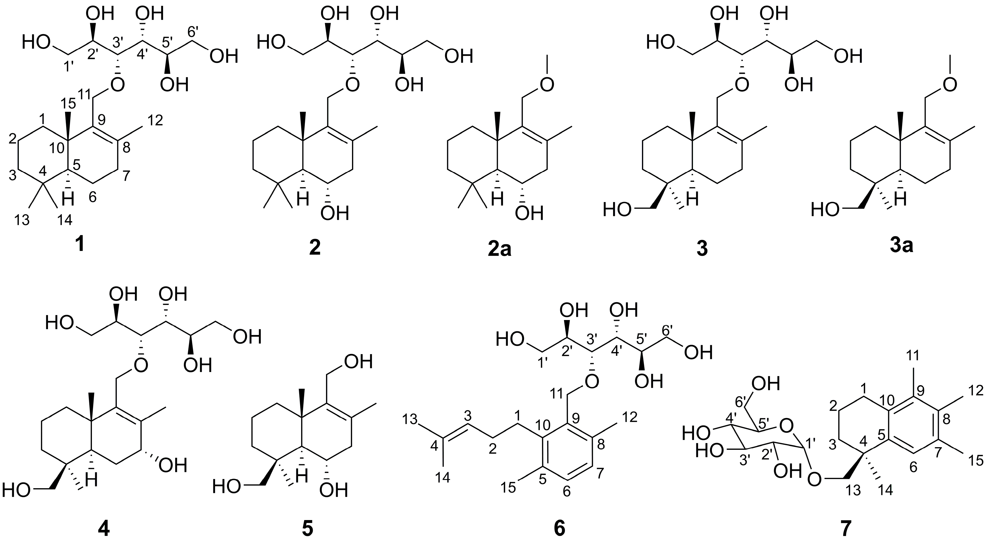
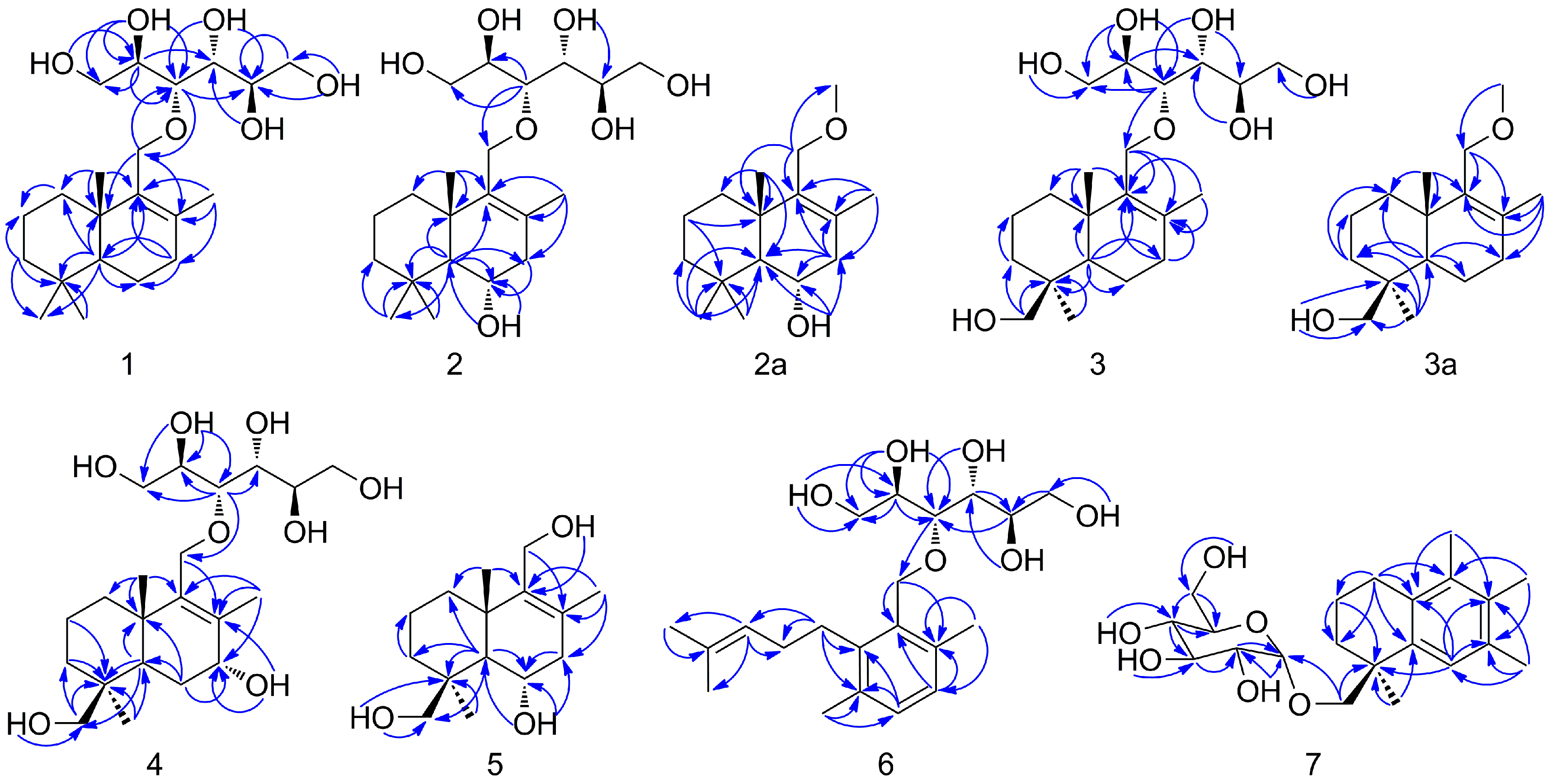
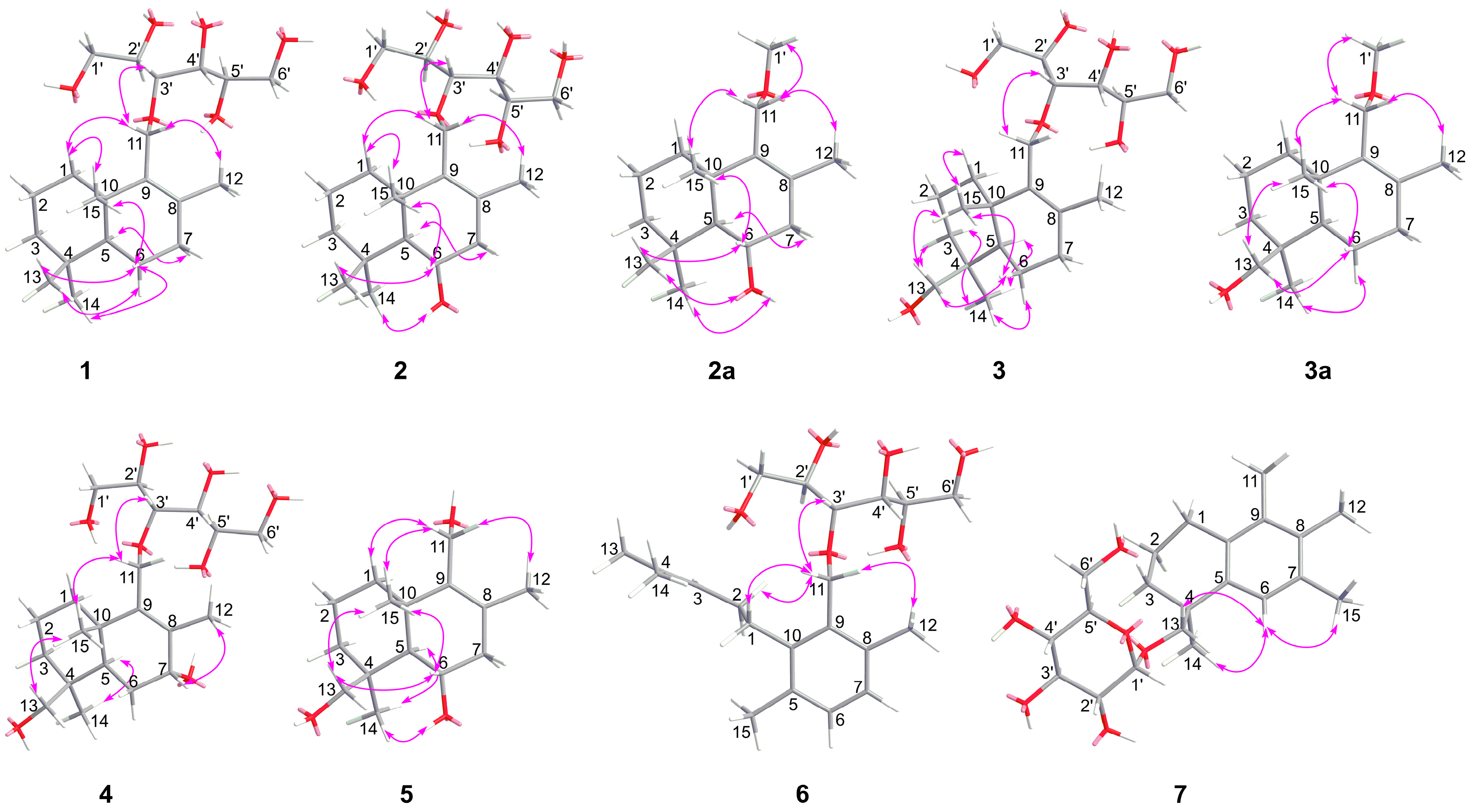
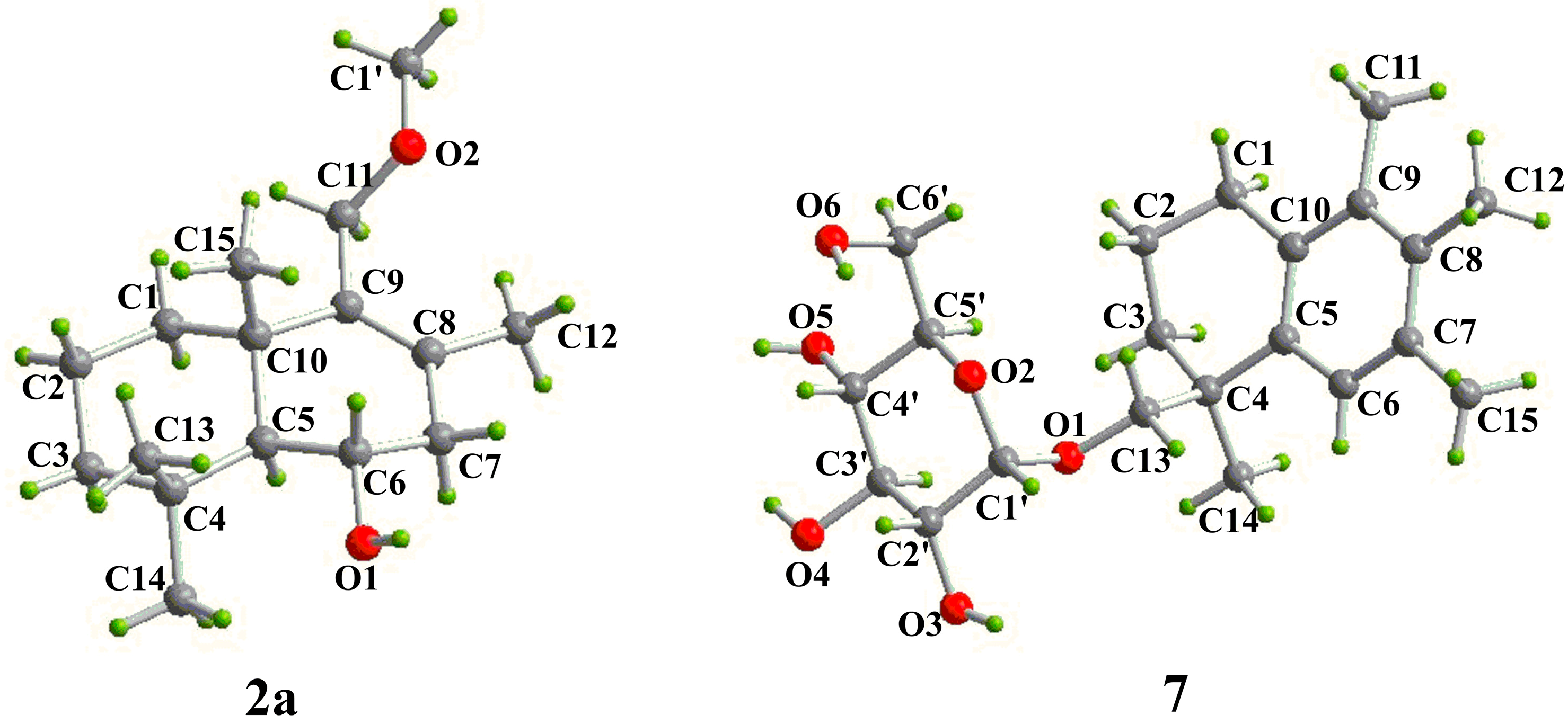
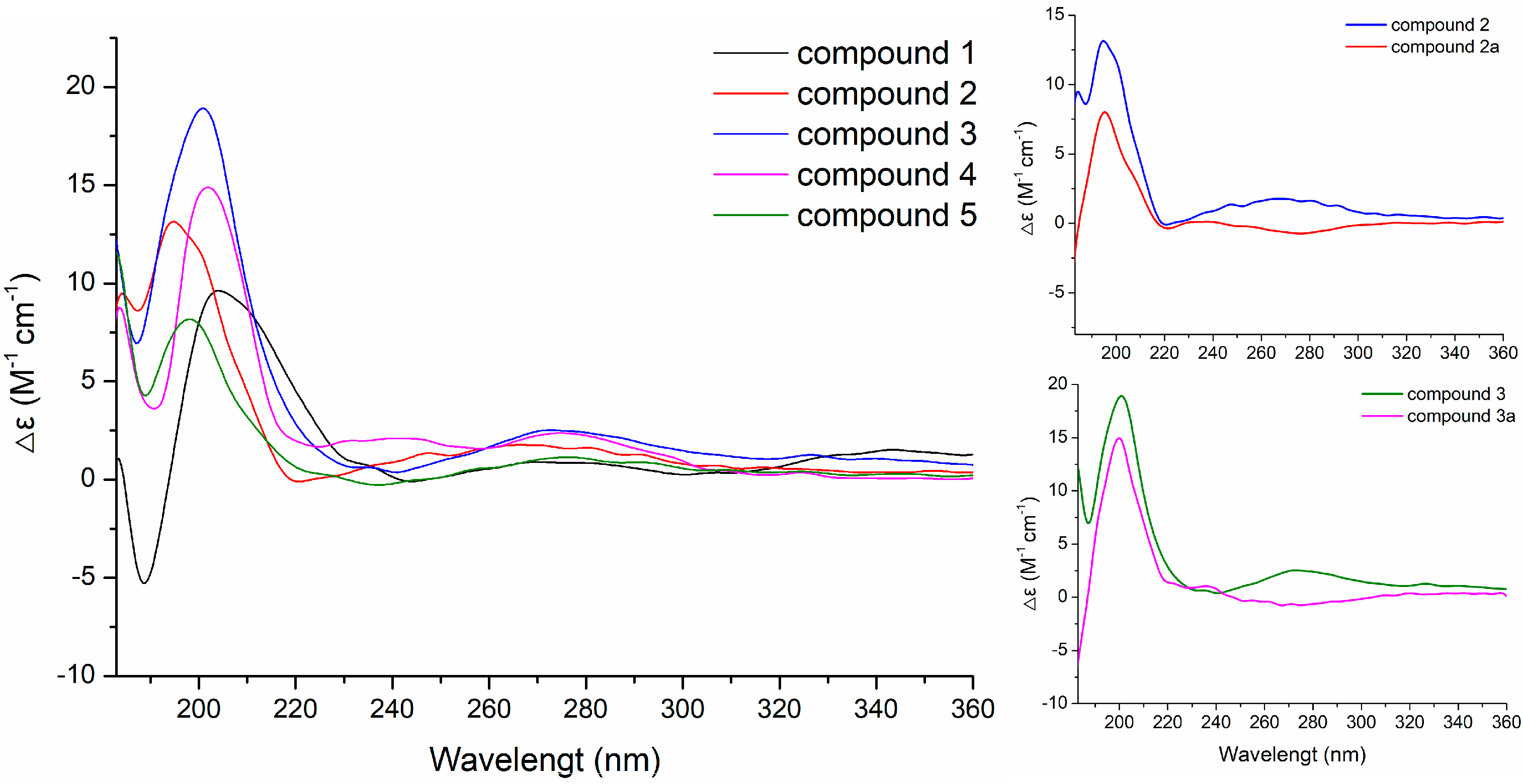
2.1. Structural Elucidation

Sporulositols AD (14) and seco-sporulositol (6) were isolated as colorless oils. Their respective molecular formulas of C21H38O6, C21H38O7, C21H38O7, C21H38O8, and C21H34O6 were established by analyses of their positive HRESIMS data, in which the sodium adduct ions ([M + Na]+) peaks appeared at m/z 409.2568, m/z 425.2512, m/z 425.2512, m/z 441.2452, and m/z 405.2239, respectively. Further, the above determination of molecular formulas was supported by analysis of the NMR data (Table 1 and Table 2), indicating three, three, three, three, and five indices of hydrogen deficiency, respectively, for 14 and 6. The IR spectra of sporulositols AD (14) and seco-sporulositol (6) showed the presence of hydroxyl (νmax 3384.7–3416.6 cm−1) and olefinic (νmax 1632.0–1659.0 cm−1) groups in their structures, and beyond these, there was an aromatic (νmax 1597.8, 1554.4, and 1432.7 cm−1) group in 6.
The NMR spectra (Table 1 and Table 2) of 14 and 6 all displayed very similar signals for a hexitol moiety (δH 3.51–3.54, 3.40–3.42/δC 62.9–63.0, δH 3.65–3.68/δC 72.6–72.8, δH 3.54–3.57/δC 77.6–77.7, δH 3.50–3.51/δC 71.0–71.1, δH 3.49–3.51/δC 71.3–71.4, and δH 3.60–3.61, 3.35–3.36/δC 63.9–64.0 for 14; δH 3.56, 3.43/δC 63.0, δH 3.72/δC 72.3, δH 3.76/δC 78.1, δH 3.58/δC 70.9, δH 3.54/δC 71.3, and δH 3.63, 3.41/δC 63.8 for 6) and the corresponding free hydroxyl groups (δH 4.20–4.77), indicating that the hexitol moieties existed in 14 and 6 share the same structures with similar configurations, and the same etherification positions with the remaining skeletons. The deduction was also supported by the HMBC correlations (Figure 2) of 1 from δH 3.68 (H-2′) to δC 63.0 (C-1′), 77.6 (C-3′) and 71.1 (C-4′); from δH 4.57 (HO-1′) to δC 72.8 (C-2′); from δH 4.72 (HO-2′) to δC 63.0, 72.8 and 77.6; from 3.57 (H-3′) to δC 63.0, 72.8 and 71.4 (C-5′); from δH 3.68 (H-2′) to δC 63.0, 77.6 and 71.4 (C-5′); from δH 4.20 (HO-4′) to δC 77.6, 71.1 and 71.4; from δH 3.61, 3.35 (H2-6′) to δC 71.4 and from δH 4.32 (HO-6′) to δC 71.4 and 64.0 (C-6′).
The remaining 13C-NMR data (Table 1) of 1 included fifteen carbon signals, attributable to four methyls (δC 19.8, 20.7, 21.5, 33.1), six methylenes (δC 18.6, 18.7, 33.3, 36.1, 41.4, 66.8), one methine (δC 51.1) and four quaternary carbons (δC 33.2, 37.5, 131.7, 138.4), supported by analyses of the 1H-NMR and HSQC data. Among these carbons, there were one oxygenated methylene {δH 4.00 (d, J = 15.0 Hz), 4.02 (d, J = 15.0 Hz)/δC 66.8} and one methyl {δH 1.65 (s)/δC 19.8} located at a pair of olefinic carbons (δC 131.7 and 138.4). The 11-hydroxyldrimane-8-en moiety in 1 was then established by HMBC correlations (Figure 2) from δH 4.00, 4.02 (H2-11) to δC 131.7 (C-8), δC 138.4 (C-9) and δC 37.5 (C-10), from δH 1.65 (H3-12) to δC 33.3 (C-7), C-8 and C-9, from δH 0.92 (H3-15) to δC 36.1 (C-1), C-9 and C-10, from δH 1.05 (H-5) to C-1, C-9, C-10, δC 33.2 (C-4), δC 21.5 (C-13) and δC 18.6 (C-6), from δH 2.00, 2.01 (H2-7) to C-6, C-8, C-9 and δC 51.1 (C-5), from δH 1.89, 1.18 (H2-1) to C-10 and δC 18.7 (C-2), from δH 1.36, 1.11 (H2-3) to C-2, C-4 and δC 21.5 (C-13), and from δH 0.81 (H3-13), 0.86 (H3-14) to C-4. Then, the HMBC correlations from δH 3.57 (H-3′) to δC 66.8 (C-11) and from δH 4.00, 4.02 (H2-11) to δC 77.6 (C-3′) established the above deduced hexitol and drimane-type sesquiterpenoid moieties to afford the planar structure of 1 through C11-O-C3.
The NMR data (Table 1) of 2 at δH/δC 1.28, 1.10/43.5 (CH2-3), 1.09/55.7 (CH-5), 3.88/66.1 (-O-CH-6), 2.29, 1.98/45.0 (CH2-7), 0.99/22.0 (CH3-13), 1.12/36.5 (CH3-14), 0.94/22.0 (CH3-15) and δC 130.4 (C-8), 40.0 (C-10), were quite different from those of 1, indicating that C-6 was hydroxylated in 2 due to the significant downfield shift of δC 66.1. The assignment was further confirmed by the key HMBC correlations (Figure 2) from δH 4.22 (HO-6) to δC 55.7 (C-5) and 66.1 (C-6), from both δH 1.09 (H-5) and δH 2.29, 1.98 (H-7) to C-6. Finally, the HMBC correlation from δH 3.54 (H-3′) to δC 66.7 (C-11) indicated that C-11 of the 6,11-dihydroxyldrimane-8-en moiety and C-3’ of the hexitol formed the planar structure of 2 by an O-ether bridge.
Comparison of NMR data (Table 1) of 3 with those of 1 and 2 revealed that CH3-13 of 3 was hydroxylated instead of CH2-6 in 2, in clue of the differences at δH/δC 1.77, 0.78/35.1 (CH2-3), 1.14/51.9 (CH-5), 3.52, 3.14/62.6 (-O-CH2-13), 0.87/27.3 (CH3-14), 0.89/21.3 (CH3-15) and δC 38.4 (C-4). The deduction was also supported by the key HMBC correlations (Figure 2) from δH 4.14 (HO-13) to δC 38.4 (C-4) and from δH 3.52, 3.14 (H2-13) to δC 35.1 (C-3) and C-4. Further, the HMBC correlation of δH 3.55 (H-3′)/δC 66.8 (C-11) suggested that the 11,13-dihydroxyldrimane-8-en (viz. diaporol [35]) and hexitol moieties in 3 formed the planar structure in the same way as compounds 1 and 2.
Different from NMR data (Table 1) of 3, the signals in the NMR data of 4 at δH/δC 1.49/45.4 (CH-5), 1.68, 1.57/28.8 (CH2-6), 3.69/68.2 (-O-CH-7), 1.74/17.6 (CH3-12), 0.85/19.5 (CH3-15) and δC 133.5 (C-8), 140.6 (C-9), 38.2 (C-10) suggested that CH2-7 was hydroxylated in 4, which was also supported by the key HMBC correlations from δH 4.56 (HO-7) to δC 68.2 (C-7) and C-8, from δH 3.69 (H-7) to C-8, and from both δH 1.74 (H3-12) and δH 1.68, 1.57 (H2-6) to C-7. The planar structure of 4 was finally established by HMBC correlation (Figure 2) from δH 3.57 (H-3′) to δC 66.8 (C-11), combining the 7,11,13-trihydroxyldrimane-8-en and hexitol moieties in 4 at the same positions as compounds 13.
6-Hydroxydiaporol (5) was obtained as a colorless oil. Its molecular formula of C15H26O3 was determined by analyses of its NMR data (Table 1) and positive HRESIMS (m/z 277.1814 [M + Na]+, calcd for C15H26O3, 254.1882) data. Based on HSQC correlations, NMR spectra of 5 showed the presence of three methyls (δH 1.61/δC19.2, δH 1.13/δC 31.4, and δH 0.98/δC 22.9), six methylenes (δH 1.78, 1.29/δC 37.0, δH 1.45, 1.33/δC 18.6, δH 1.58, 0.90/δC 38.1, δH 2.20, 1.94/δC 44.5, δH 3.91, 3.81/δC 56.5, and δH 3.78, 3.37/δC 65.1), two methines (δH 1.21/δC 56.5 and δH 3.96/δC 66.9) and four quaternary carbons (δC 38.7, 40.4, 128.5, 140.8). Among the deduced groups, there were two oxygenated methylenes {δH 3.91 (d, J = 11.6, 4.4 Hz), 3.81 (d, J = 11.6, 4.8 Hz)/δC 56.5 and δH 3.78 (d, J = 10.4, 4.8 Hz), 3.37 (d, J = 10.4, 4.4 Hz)/δC 65.1} and one oxygenated methine {δH 3.96 (m)/δC 66.9}. Then, the planar structure of 5 was constructed by the key HMBC correlations (Figure 2) from δH 1.61 (H3-12) to δC 44.5 (C-7), 128.5 (C-8) and 140.8 (C-9), from δH 3.91, 3.81 (H2-11) to C-8, C-9 and δC 40.4 (C-10), from δH 4.11 (HO-11) to δC 140.8, from δH 0.98 (H3-13) to δC 140.8, 40.4 and 37.0 (C-1), from δH 1.21 (H-5) to δC 37.0, 40.4, 38.1 (C-3), 38.7 (C-4), and 66.9 (C-6), from δH 4.43 (HO-6) to δC 56.5 (C-5), 66.9 and 44.5, from δH 2.20, 1.94 (H2-7) to δC 66.9 and 128.5, from δH 4.78 (HO-13) to δC 65.1 (C-13) and 38.7, from δH 1.13 (H3-14) to δC 38.7, and from δH 1.58, 0.91 (H2-3) to δC 38.7, 18.6 (C-2). Thus, 5 tuned out to be 6,11,13-trihydroxyldrimane-8-en, similar to the sesquiterpenoid cores in 14.
The relative configurations of compounds 15 were assigned by 1H-NMR J-values and NOE correlations (Figure 3). In these drimane-type sesquiterpenoids, the doublet J values (11.1–13.2 Hz) of H-5 suggested a trans-junction of the two cyclohexatomic ring system [35], H3-15 and H3-13 (or H2-13) adopt the same β-orientation, whereas H3-14, H-5 and HO-6 (or HO-7) oriented in the opposite α-direction. To determine the absolute configurations of 15, compounds 2 and 3 were selected and subjected to acid hydrolysis (5% trifluoroacetic acid in methanol; Figure 4), due to their abundant amounts.
Acid hydrolysis of 2 and 3 afforded a hexitol, together with 2a and 3a, respectively, whose structures were elucidated by extensive NMR spectroscopic data (Figure 2, Figure 3 and Figures S19–S29, S38–S45 in supplementary materials). Furthermore, NMR (Figure S28 and S29) and optical rotation data ( [ α ] D 25 +135.5 (c 0.380, CH3OH)) of the hexitol were quite similar as those reported of D-mannitol [44], which was also isolated from the same strain P. sporulosum YK-03. NOESY spectra of 2a and 3a (Figure 3) indicated that they share the same relative configurations as compounds 15. After many attempts, crystal of 2a suitable for single-crystal X-ray diffraction (Cu Kα) analysis (Figure 5) was successfully obtained upon slow evaporation of the solvent mixture (methanol-water, 20:1) by keeping the sample at room temperature for nearly one month. Thus, the absolute configuration of 2a was unambiguously determined as 5S,6S,10S. Based on the fact that the CD patterns of 15 and 3a (Figure 6) were identical to that of 2a, the absolute configurations of drimane-type sesquiterpenoid were assigned as 5S,10S in 1, 5S,6S,10S in 2, 4S,5R,10S in 3a, 4S,5R,10S in 3, 4S,5R,7R,10S in 4, and 4S,5R,6S,10S in 5.
Besides the NMR data of the hexitol, the remaining NMR signals of 6 (Table 2) showed the existence of a prenyl methyl {δH/δC 1.55 (s)/17.5, 1.66 (s)/25.6, 5.25 (t, J = 6.0 Hz)/124.3, 2.05 (m)/29.5, 2.71 (t, J = 8.4 Hz) /29.2 and δC 131.0} and a tetrasubstituted benzene {δH/δC 6.90 (d, J = 7.8 Hz)/127.6, 6.98 (d, J = 7.8 Hz)/129.7 and δC 133.2, 134.9, 135.6, 140.2} groups, assisted by HMBC correlations (Figure 2).
Then, the HMBC correlations (Figure 2) from δH 2.25 (s) to δC 129.7 (C-7) and 140.2 (C-8), and from δH 2.34 (s) to δC 127.6 (C-6) and 134.9 (C-5), from δH 4.67 (d, J = 10.2 Hz), 4.60 (d, J = 10.2 Hz) to δC 133.2 (C-10), 135.6 (C-9) and 140.2, and from δH 2.71 to δC 133.2 and 134.9 led to the assignment of the tetrasubstituted benzene with two methyls located at C-5 and C-8, an oxygenated methyl located at C-9, and a prenyl methyl located at C-10. Further, the HMBC correlation of δH 3.76 (m, H-3′) to δC 67.4 (C-11) indicated C-3’ was connected to C-11 by an ether O to afford the structure of 6. Based on the NMR data and biosynthetic homology, the hexitol moiety of 6 was presumed to be D-mannitol, the same as that of 15.
Sporuloside (7) was obtained as colorless oil. The molecular formular of C21H32O6 was established by its positive HRESIMS data at m/z 403.2103 [M + Na]+. Analysis of the NMR data of 7 (Table 2) indicated the existence of an α-glucosyl {δH/δC 4.64 (d, J = 3.6 Hz < 7.0 Hz)/99.3, 3.20 (m)/72.6, 3.40 (m)/73.7, 3.05 (td, J = 9.1, 5.2 Hz)/70.7, 3.28 (m)/73.3, and 3.56 (m), 3.40 (m)/61.4} moiety, a pentasubstituted benzene ring {δH/δC 7.01 (s)/126.1 and δC 132.1, 132.7, 133.0, 134.1, 139.2}, and three adjacent aromatic methyls {δH/δC 2.07 (s)/15.8, 2.09 (s)/15.9, 2.18(s)/21.0}, supported by the key HMBC correlations (Figure 2) from δH 2.18 (H3-15) to δC 126.1 (C-6), 133.0 (C-7), 132.1 (C-8), from δH 2.09 (H3-12) to δC 133.0, 132.1, 134.1 (C-9), from δH 2.07 (H3-11) to δC 132.1, 134.1, 132.7 (C-10), and from δH 7.01(H-6) to δC 133.0, 132.1, 132.7. The rest of NMR signals attributed to four methylenes {δH/δC 2.53 (t, J = 8.4 Hz)/28.2, 1.72 (m)/19.2, 1.84, 1.44 (each m)/33.0, 3.59, 3.24 (each d, J = 9.2 Hz)/75.6}, a methyl {δH/δC 1.25 (s)/27.1} and one quaternary carbon {δC 37.8}. The HMBC correlations of δH 2.53 (H2-1)/δC 19.2 (C-2), 33.0 (C-3), 139.2 (C-5), 134.1, 132.7, δH 1.72 (H2-2)/δC 33.0, 37.8 (C-4), δH 1.25 (H3-14)/δC 37.8, 139.2, δH 7.01/δC 37.8, and δH 3.59, 3.24 (H2-13)/δC 37.8, 99.3 (C-1′) combined the abovementioned groups to afford the planar structure of 7, in which C-13 was glycosidated by a α-glucose. Luckily, crystals of 7 suitable for single-crystal X-ray diffraction (Cu Kα) analysis (Figure 5) were successfully obtained upon slow evaporation of the solvent mixture (methanol-water, 20:1) by keeping the sample at room temperature for nearly one month, so the absolute configuration of C-4 was assigned as S, and α-glucosyl group as D-form.

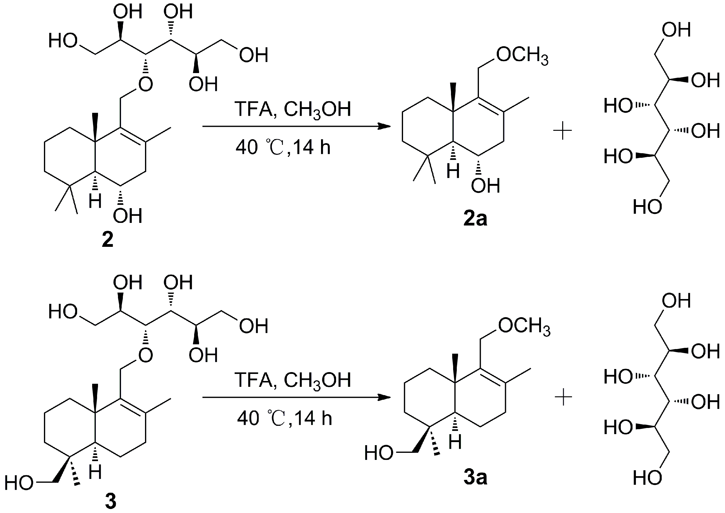
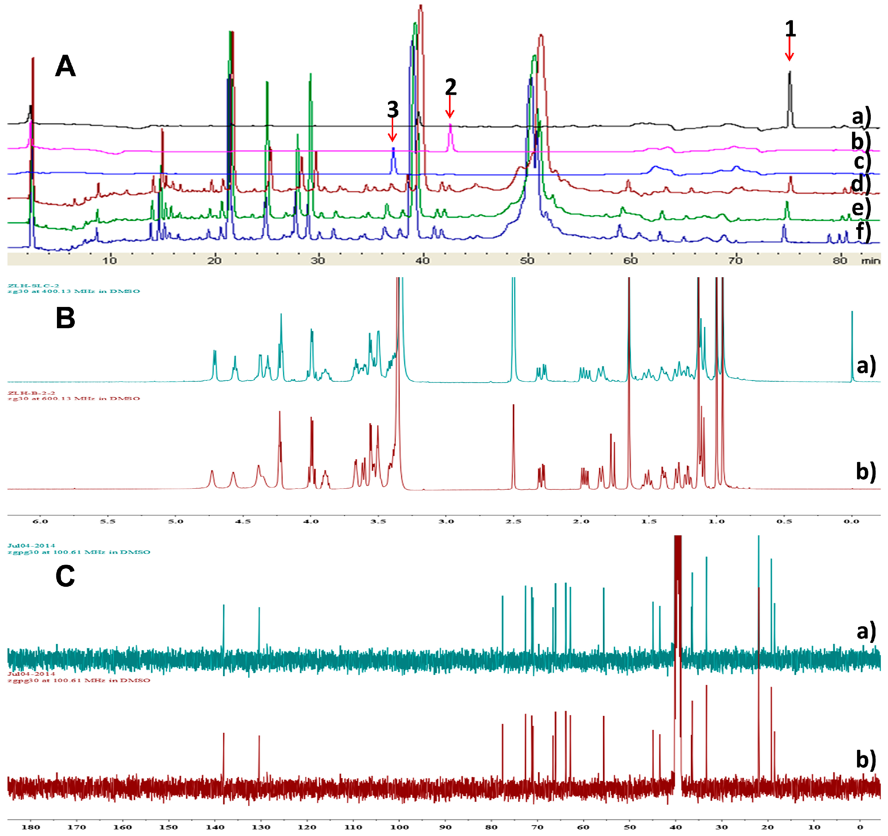
2.2. Investigation on the Origin of Mannitol Moiety

Sporulositols AD (14) and seco-sporulositol (6) represent the first examples of unique drimanic mannitol derivatives. To find out whether their mannitol moiety was formed intrinsically or derived from the medium, the normal medium (mannitol-contained, control group), modified medium No.1 (no mannitol, blank group) and modified medium No.2 (mannitol replaced by sorbitol, experimental group) were included for simultaneous cultivation of P. sporulosum YK-03 and HPLC analysis of the metabolites. Compounds 13 could be detected in all the three groups (Figure 7A), and compound 2 was isolated from the extract of experimental group (Figure 7B,C), revealing that the fungus can produce the mannitol moiety intrinsically, no matter whether the medium contains mannitol or not.
Compounds 17 were tested for cytotoxicity against two cell lines A549 (human lung adenocarcinoma cells) and MCF-7 (human breast cancer cells). Unfortunately, compounds 17 did not show any detectable cytotoxicity.

3. Materials and Methods

3.1. General Experimental Procedures

Optical rotations were measured using a Perkin-Elmer Model 241 polarimeter (Perkin Elmer, Inc. Waltham, MA, USA). UV spectra were obtained on a Shimadzu UV-1601 (Shimadzu Corp., Kyoto, Japan). IR spectra were taken on a Bruker IFS-55 infrared spectrophotometer (Bruker Optik BmbH, Ettlingen, Germany) with KBr disks. The HRESIMS data were obtained on a microTOF-Q Bruker mass instrument (Bruker Daltonics, Billerica, MA, USA). CD spectra were recorded with a Biologic MOS-450 spectrometer (BioLogic Science Instruments, Grenoble, French) using CH3CN as solvent. 1D and 2D NMR spectra were recorded on Bruker ARX-400 and AV-600 spectrometers (1H/13C, 400/100 MHz 600/150 MHz, Bruker, Zurich, Switzerland) using TMS as an internal standard. Chemical shifts (δ) were expressed in ppm. HPLC was performed using a Shimadzu LC-20AB HPLC pump equipped with a SPD-20A detector (Shimadzu Corp.) for new compound analysis, employing a YMC-Pack ODS-A column (250 mm × 4.6 mm, 5 µm), and for metabolite analysis in Figure 7, employing a CHIRALPAK AD-H column (250 mm × 4.6 mm, 5 μm). Reversed-phase HPLC was performed using a Shimadzu LC-8A HPLC pump equipped with SPD-10A detector for the purification of new compounds, employing a YMC-Pack ODS-A column (250 mm × 10 mm, 5 µm). Column chromatography (CC) was carried out on silica gel (200–300/400–500 mesh, Qingdao Marine Chemical, Inc., Qingdao, China) and sephadex LH-20 (Pharmacia, Uppsala, Sweden). Column fractions were monitored by TLC (Silica gel GF254, 200–300 mesh, Qingdao Haiyang Chemical Factory, Qingdao, China), and the spots were visualized by heating the plates after spraying with 10% H2SO4 in ethanol. All reagents of HPLC or analytical grade were purchased from Shangdong Yuwang Reagent Co., Ltd. (Shangdong, China).

3.2. Fungal Material

Paraconiothyrium sporulosum YK-03 was isolated from the sea mud collected from the intertidal zone of Bohai Bay in Liaoning Province of China. It was identified based on the analysis of ITS sequence (GenBank accession No. KC416199) and has been deposited in the School of Traditional Chinese Materia Medica, Shenyang Pharmaceutical University.

3.3. Fermentation

The strain was cultured on PDA (potato 20%, glucose 2% and agar 2%) medium in Petri dishes at 28 °C for 3 days, and were inoculated in a 500 mL Erlenmeyer flask containing 150 mL of media (maltose 2%, monosodium glutamate 1%, glucose 1%, yeast cream 0.3%, corn steep liquor 0.1%, maltose 2%, KH2PO4 0.05%, MgSO4.7H2O 0.03%). After incubation at 28 °C and 180 rpm for 4 days, a 5 mL cultural solution was transferred as a seed into each of 500 mL flask containing 150 mL liquid medium (maltose 2%, monosodium glutamate 1%, glucose 1%, yeast cream 0.5%, east cream 0.3%, corn steep liquor 0.1%, mannitol 2%, KH2PO4 0.05%, MgSO4.7H2O 0.03%, CaCO3 2%, sea water element 3.3%, pH6.5). The flasks were subsequently incubated at the same conditions for 8 days.

3.4. Extraction and Isolation

Following incubation, the fermentation broth of P. sporulosum YK-03 (70 L) was concentrated and extracted with ethyl acetate and n-butanol, successively. The ethyl acetate extract (20 g) was subjected to a silica gel column (10 cm × 120 cm), eluted with CHCl3-CH3OH (100:1–0:1), yielding 14 fractions AN. Fraction L (350 mg) was firstly subjected to a Sephadex LH-20 column (2.5 cm × 100 cm), eluted with CHCl3-CH3OH (1:1) to remove pigment, then purified LPLC using a gradient of increasing methanol (20%–100%) in water to afford three subfractions (L1L3). Fraction L1 (48 mg) afforded compound 1 (4.3 mg, tR 36.4 min) and compound 6 (2.5 mg, tR = 58.9 min) by using preparative HPLC (CH3OH-H2O 53:47, flow rate 3 mL/min, wavelength 210 nm), employing a YMC-Pack ODS-A column (250 mm × 10 mm, 5 µm). Fraction M (628 mg) was subjected to Sephadex LH-20 column (3 cm × 120 cm), eluted with CHCl3-CH3OH (1:1) and preparative HPLC (CH3OH-H2O 44:56, flow rate 3 mL/min, wavelength 210 nm) to obtain compound 2 (93.3 mg, tR 40.2 min) compound 3 (89.8 mg, tR = 45.5 min), and compound 4 (2.6 mg, tR = 58.2 min). Fraction N (1.2 g) was subjected to a silica gel column (4 cm × 80 cm), eluted with CHCl3-CH3OH (100:1–0:1), yielding 7 fractions (N1N7). Then, fraction N4 (89 mg) afforded compound 8 (3.2 mg) through recrystallization, and the rest solution was purified by preparative HPLC (CH3OH-H2O 30:70, flow rate 3 mL/min, wavelength 210 nm) to obtain compound 5 (2.3 mg, tR = 42.0 min) and compound 7 (2.0 mg, tR = 49.2 min).
Sporulositol A (1): colorless oil; [ α ] D 25 +101.6 (c 0.43, CH3OH); UV (CH3OH λmax 204.4 nm; IR (KBr) νmax 3416.6, 2928.1, 1659.0, 1461.7, 1384.4, 1080.7 cm−1; CD (c 0.10, CH3CN) λ(Δε) 203 (+7.75) nm; 1H-NMR (600 MHz, DMSO-d6) and 13C NMR (150 MHz, DMSO-d6), see Table 1; HRESIMS m/z 409.2568 [M + Na]+, (calcd for C21H38O6 386.2668).
Sporulositol B (2): colorless oil; [ α ] D 25 +113.9 (c 1.00, CH3OH); UV (CH3OH) λmax 206.4 nm; IR (KBr) νmax 3396.3, 2927.4, 1632.3 1434.2, 1384.2, 1075.0, 1043.1 cm−1; CD (c 0.10, CH3CN) λ(Δε) 203 (+6.36) nm; 1H-NMR (600 MHz, DMSO-d6) and 13C NMR (150 MHz, DMSO-d6), see Table 1; HRESIMS m/z 425.2512 [M + Na]+, (calcd for C21H38O7 402.2617).
Sporulositol C (3): colorless oil; [ α ] D 25 +101.8 (c 1.00, CH3OH); UV (CH3OH) λmax 206.2,243.6 nm; IR (KBr) νmax 3384.7, 2928.2, 1650.1, 1451.1, 1384.0, 1079.0, 1032.4 cm−1; CD (c 0.10, CH3CN) λ(Δε) 202 (+10.79) nm; 1H-NMR (600 MHz, DMSO-d6) and 13C NMR (150 MHz, DMSO-d6), see Table 1, HRESIMS m/z 425.2512 [M + Na]+, (calcd for C21H38O7 402.2617).
Sporulositol D (4): colorless oil; [ α ] D 25 +90.3 (c 0.26, CH3OH); UV (CH3OH) λmax 207.2 nm; IR (KBr) νmax 3405.7, 2922.7, 1632.3, 1597.6, 1552.5, 1430.0, 1384.4, 1121.3, 1053.1, 1033.0 cm−1; CD (c 0.10, CH3CN) λ(Δε) 201 (+11.07) nm;1 H-NMR (600 MHz, DMSO-d6) and 13C NMR (150 MHz, DMSO-d6), see Table 1; HRESIMS m/z 441.2452 [M + Na]+, (calcd for C21H38O8 418.2566).
6-hydroxyl diaporol (5): colorless oil; [ α ] D 25 +98.3 (c 0.23, CH3OH); UV (CH3OH) λmax 207.2 nm; IR (KBr) νmax 3418.1, 2924.9, 1658.1, 1554.1, 1433.8, 1384.3 cm−1; CD (c 0.10, CH3CN) λ(Δε) 196.5 (+6.12) nm; 1H-NMR (600 MHz, DMSO-d6) and 13C NMR (150 MHz, DMSO-d6), see Table 1; HRESIMS m/z 277.1814 [M + Na]+, (calcd for C15H26O3 254.1882).
seco-sporulositol (6): colorless oil; [ α ] D 25 +55.0 (c 0.25, CH3OH); UV (CH3OH) λmax 205.0 nm; IR (KBr) νmax 3405.4, 2923.0, 1632.0, 1597.8, 1554.4, 1432.7, 1384.4, 1127.7, 1033.1 cm−1; 1H-NMR (600 MHz, DMSO-d6) and 13C NMR (150 MHz, DMSO-d6), see Table 2; HRESIMS m/z 405.2239 [M + Na]+, (calcd for C21H34O6 382.2355).
Sporuloside (7): colorless oil; [ α ] D 25 +117.6 (c 0.26, CH3OH); UV (CH3OH) λmax 205.0 nm; IR (KBr) νmax 3405.4, 2921.6, 1634.3, 1457.7, 1384.4, 1148.2, 1123.5, 1025.5 cm−1; 1H-NMR (600 MHz, DMSO-d6) and 13C NMR (150 MHz, DMSO-d6), see Table 2; HRESIMS m/z 403.2103 [M + Na]+, (calcd for C21H32O6 380.2199).
D-mannitol (8): white powder; [ α ] D 25 +145.5 (c 0.50, CH3OH); 1H-NMR (DMSO-d6, 400 MHz), δH 3.60 (2H, m), 3.54 (2H, t, J = 8.0 Hz), 3.46 (2H, m), 3.38 (2H, m), 4.41 (2H, d, J = 5.6 Hz, HO ×2 ), 4.33(2H, t, J = 6.0 Hz, HO×2), 4.14 (2H, d, J = 7.2 Hz, HO × 2); 13C-NMR (DMSO-d6, 100 MHz) δC 64.2 × 2, 70.1 × 2, 71.7 × 2.

3.5. X-ray Crystallographic Analysis of Compounds 2a and 7

Crystal Data of 2a: C16H28O2, M =252.38, orthorhombic, a = 10.0481 (3) Å, b = 10.4089(4) Å, c = 28.5282(9) Å, U = 2983.76(17) Å3, T = 100.6, space group P212121 (no. 19), Z = 8, μ(Cu Kα) = 0.554, 10773 reflections measured, 5657 unique (Rint = 0.0286) which were used in all calculations. The final wR (F2) was 0.1314 (all data). The crystallographic data for the structure of 2a have been deposited with the Cambridge Crystallographic Data Centre as supplementary publication CCDC No. 1905155.
Crystal Data of 7: C21H32O6, M = 380.47, orthorhombic, a = 5.39286(9) Å, b = 7.54344 (11) Å, c = 48.7570(7) Å, U = 1983.47(5) Å3, T = 102.3, space group P212121 (no. 19), Z = 4, μ (Cu Kα) = 0.753, 6585 reflections measured, 3754 unique (Rint = 0.0252) which were used in all calculations. The finalwR (F2) was 0.0996 (all data). The crystallographic data for the structure of 7 have been deposited with the Cambridge Crystallographic Data Centre as supplementary publication CCDC No. 1905156.
CCDC-1905155 and CCDC-1905156 contain the supplementary crystallographic data, which can be obtained free of charge from the Cambridge Crystallographic Data Centre via http://www.ccdc.cam.ac.uk/conts/retrieving.html.

3.6. Acid Hydrolysis of Compounds 2 and 3

Compounds 2 and 3 (each 10 mg) were dissolved in CH3OH (1 mL) with 1 mL trifluoroacetic acid (TFA), and heated in a H2O bath at 40 °C for 14 h to give an acid hydrolysate. The acid hydrolysate was then vacuum evaporated to remove the residual TFA. Then, the hydrolysate was suspended in H2O, and extracted with CHCl3. Finally, the H2O solution afforded sugar alcohol through recrystallization, and the CHCl3 solution was purified by preparative HPLC to obtain compound 2a (1.8 mg) and compound 3a (2.0 mg), respectively.

3.7. Analysis of the Refrence Compounds 13 and Metabolites of P. Sporulosum YK-03 in Different Mediums Using HPLC and NMR Methods

P. sporulosum YK-03 was simultaneously cultured in three following liquid media at 28 °C and 180 rpm for 4 days. Liquid media: (1) Normal medium (mannitol-containing, control group): maltose 2%, monosodium glutamate 1%, glucose 1%, yeast cream 0.5%, east cream 0.3%, corn steep liquor 0.1%, mannitol 2%, KH2PO4 0.05%, MgSO4.7H2O 0.03%, CaCO3 2%, sea water element 3.3%, pH 6.5. (2) Modified medium No.1 (no mannitol, blank group): maltose 2%, monosodium glutamate 1%, glucose 1%, yeast cream 0.5%, east cream 0.3%, corn steep liquor 0.1%, KH2PO4 0.05%, MgSO4.7H2O 0.03%, CaCO3 2%, sea water element 3.3%, pH 6.5. (3) Modified medium No. 2 (mannitol replaced by sorbitol, experimental group): maltose 2%, monosodium glutamate 1%, glucose 1%, yeast cream 0.5%, east cream 0.3%, corn steep liquor 0.1%, sorbitol 2%, KH2PO4 0.05%, MgSO4.7H2O 0.03%, CaCO3 2%, sea water element 3.3%, pH 6.5.
After incubation, their fermentation broths were concentrated and extracted with ethyl acetate. Then their metabolites were analyzed by gradient HPLC analysis. HPLC chromatographic condition: (1) Instrument: Shimadzu LC-20AB HPLC pump equipped with an SPD-20A detector (Shimadzu Corp.); (2) Column: CHIRALPAK AD-H column (250 mm × 4.6 mm, 5 μm); (3) Mobile phase: CH3OH (B) and H2O (A); (4) Wavelength: 210 nm; (3) Flow rate: 1 mL min−1; (5) gradient elution program: 10% B; 0–6 min, linearly changed to 24% B; 6–26 min, linearly changed to 35% B; 26–40 min, linearly changed to 60% B; 40–60 min, linearly changed to 70% B; 60–72 min, linearly changed to 80% B; 72–82 min, linearly changed to 100%. The sample injection volume was 5.0 μL.
The strain was incubated in twenty Erlenmeyer flasks (500 mL) containing 150 mL Modified medium No. 2 medium at 28 °C and 180 rpm for 8 days. Then the fermentation broth was concentrated and extracted with ethyl acetate. The ethyl acetate extract was subjected to a silica gel column eluted with CHCl3-CH3OH (100:1–0:1) and preparative HPLC (CH3OH-H2O 44:56, flow rate 3 mL/min, wavelength 210 nm) to yield a drimane-type sesquiterpenoid, guiding by TLC and HPLC. Then its NMR spectra was compared with those of compounds 13.

3.8. Cytotoxic Activity Assay

The cytotoxicity was evaluated by using the MTT assay as described previously [45]. Doxorubicin hydrochloride was used as a positive control. The A549 and MCF-7 Cells (China Infrastructure of Cell Lines Resources, Beijing, China were cultured in McCoy’s 5A medium and DMEM basic medium (1×) at 37 °C under an atmosphere of 5% CO2, and were seeded on each well of 96-well plates containing 200 μL of tumor cell suspension (1 × 10⁴ cells). After 24 h, each well was added 2 μL of test solution and incubated for another 72 h. 50 μL of MTT solution (1 mg/mL, Beijing Cellchip Biotechnology Co., Ltd., Beijing, China was added to each well, and the plate was incubated for 3h under the same condition. Then, the plate was centrifuged and the supernatants were removed and cells were dissolved in 150 μL of DMSO to determine the IC50 values.

4. Conclusions

Seven new drimane-type sesquiterpenoids, including sporulositols AD (14), 6-hydroxy- diaporol (5), seco-sporulositol (6) and sporuloside (7), were isolated from a marine-derived fungus P. sporulosum YK-03. Their structures were established by extensive NMR experiments, X-ray diffraction analysis and comparisons of circular dichroism data. Compounds 14 and 6 are rare new drimanic hexitol derivatives containing a D-mannitol moiety. seco-sporulositol (6) and sporuloside (7) may represent two new series of natural drimanes, possessing an aromatic ring with a rare 4,5-secodrimanic skeleton and an unusual CH3-15 rearranged drimanic α-D-glucopyranside, respectively. Then, the cultivation medium was evaluated for the origin of D-mannitol moiety by HPLC and NMR analyses. These isolates enriched the structural diversity of natural drimanic sesquiterpenoids.

Supplementary Materials

The following are available online, Figures S1–S79: HRESIMS, 1D and 2D NMR, IR, UV and CD spectra of compounds 18, 2a and 3a; Figures S80–S81: 1D NMR of compound 2 isolated from the modified medium No.2.

Author Contributions

Y.-H.P. designed and guided the experiments. L.-H.Z. conducted the isolation, structural determination and wrote the manuscript. G.C. completed the preliminary original investigation on the mannitol moiety of 14 and 6, and revised the manuscript. Y.S. contributed to the biological activity evaluation. H.-F.W. performed the acid hydrolysis of compounds 2 and 3. J.B. and H.-M.H. performed the isolation, identification and fermentation of the fungal species. All authors reviewed the manuscript.

Funding

Financially Supported by Laboratory for Marine Drugs and Bioproducts of Qingdao National Laboratory for Marine Science and Technology (LMDBKF201701) and National Natural Science Foundation of China (No. 81202425).

Acknowledgments

We gratefully acknowledge Yi Sha and Wen Li, Department of Analytical Testing Center, Shenyang Pharmaceutical University, for measurements of the NMR data. We thank Jian Hao, Department of Analytical Testing Center, Beijing University of Chemical Technology, for the test of the X-ray diffraction.

Conflicts of Interest

The authors declare no conflict of interest.

References

  1. Blunt, J.W.; Carroll, A.R.; Copp, B.R.; Davis, R.A.; Keyzers, R.A.; Prinsep, M.R. Marine natural products. Nat. Prod. Rep. 2018, 35, 8–53. [Google Scholar] [CrossRef] [PubMed] [Green Version]
  2. Bugni, T.S.; Ireland, C.M. Marine-derived fungi: A chemically and biologically diverse group of microorganisms. Nat. Prod. Rep. 2004, 21, 143–163. [Google Scholar] [CrossRef]
  3. Barbero, H.; Diez-Poza, C.; Barbero, A. The Oxepane Motif in Marine Drugs. Mar. Drugs 2017, 15, 361. [Google Scholar] [CrossRef]
  4. Overy, D.P.; Bayman, P.; Kerr, R.G.; Bills, G.F. An assessment of natural product discovery from marine (sensu strictu) and marine-derived fungi. Mycology 2014, 5, 145–167. [Google Scholar] [CrossRef] [PubMed]
  5. Verkley, G.J.M.; Silva, M. d.; Wicklow, D.T.; Crou, P.W. Paraconiothyrium, a new genus to accommodate the mycoparasite Coniothyrium minitans, anamorphs of Paraphaeosphaeria, and four new species. Stud. Mycol. 2004, 50, 323–335. [Google Scholar]
  6. Takano, K.; Itoh, Y.; Ogino, T.; Kurosawa, K.; Sasaki, K. Phylogenetic analysis of manganese-oxidizing fungi isolated from manganese-rich aquatic environments in Hokkaido, Japan. Limnology 2006, 7, 219–223. [Google Scholar] [CrossRef]
  7. Liu, L.; Gao, H.; Chen, X.; Cai, X.; Yang, L.; Guo, L.; Yao, X.; Che, Y. Brasilamides A-D: Sesquiterpenoids from the Plant Endophytic Fungus Paraconiothyrium brasiliense. Eur. J. Org. Chem. 2010, 3302–3306. [Google Scholar] [CrossRef]
  8. Liu, L.; Chen, X.; Li, D.; Zhang, Y.; Li, L.; Guo, L.; Cao, Y.; Che, Y. Bisabolane Sesquiterpenoids from the Plant Endophytic Fungus Paraconiothyrium brasiliense. J. Nat. Prod. 2015, 78, 746–753. [Google Scholar] [CrossRef] [PubMed]
  9. Guo, Z.; Ren, F.; Che, Y.; Liu, G.; Liu, L. New Bergamotane Sesquiterpenoids from the Plant Endophytic Fungus Paraconiothyrium brasiliense. Molecules 2015, 20, 14611–14620. [Google Scholar] [CrossRef] [PubMed]
  10. Chen, S.; Zhang, Y.; Zhao, C.; Ren, F.; Liu, X.; Che, Y. Hawaiinolides E-G, cytotoxic cassane and cleistanthane diterpenoids from the entomogenous fungus Paraconiothyrium hawaiiense. Fitoterapia 2014, 99, 236–242. [Google Scholar] [CrossRef] [PubMed]
  11. Chen, S.; Zhang, Y.; Niu, S.; Liu, X.; Che, Y. Cytotoxic cleistanthane and cassane diterpenoids from the entomogenous fungus Paraconiothyrium hawaiiense. J. Nat. Prod. 2014, 77, 1513–1518. [Google Scholar] [CrossRef]
  12. Mohamed, I.E.; Kehraus, S.; Krick, A.; Konig, G.M.; Kelter, G.; Maier, A.; Fiebig, H.-H.; Kalesse, M.; Malek, N.P.; Gross, H. Mode of Action of Epoxyphomalins A and B and Characterization of Related Metabolites from the Marine-Derived Fungus Paraconiothyrium sp. J. Nat. Prod. 2010, 73, 2053–2056. [Google Scholar] [CrossRef] [PubMed]
  13. Zhang, L.-H.; Feng, B.-M.; Chen, G.; Li, S.-G.; Sun, Y.; Wu, H.-H.; Bai, J.; Hua, H.-M.; Wang, H.-F.; Pei, Y.-H. Sporulaminals A and B: A pair of unusual epimeric spiroaminal derivatives from a marine-derived fungus Paraconiothyrium sporulosum YK-03. RSC Adv. 2016, 6, 42361–42366. [Google Scholar] [CrossRef]
  14. Shiono, Y.; Kikuchi, M.; Koseki, T.; Murayama, T.; Kwon, E.; Aburai, N.; Kimura, K.-i. Isopimarane diterpene glycosides, isolated from endophytic fungus Paraconiothyrium sp. MY-42. Phytochemistry 2011, 72, 1400–1405. [Google Scholar] [CrossRef] [PubMed]
  15. Du, Y.; Chen, Z.; Li, H.; Wang, Y.; Fu, P.; Zhu, W. Pafuranones A and B, two dimeric polyketides from a rare marine algae-derived fungus Paraconiothyrium sp. Chin. Chem. Lett. 2019. [Google Scholar] [CrossRef]
  16. Suzuki, T.; Ariefta, N.R.; Koseki, T.; Furuno, H.; Kwon, E.; Momma, H.; Harneti, D.; Maharani, R.; Supratman, U.; Kimura, K.I.; et al. New polyketides, paralactonic acids A-E produced by Paraconiothyrium sp. SW-B-1, an endophytic fungus associated with a seaweed, Chondrus ocellatus Holmes. Fitoterapia 2019, 132, 75–81. [Google Scholar] [PubMed]
  17. Zhao, C.; Fu, P.; Zhang, Y.; Liu, X.; Ren, F.; Che, Y. Sporulosol, a New Ketal from the Fungus Paraconiothyrium sporulosum. Molecules 2018, 23, 1263. [Google Scholar] [CrossRef] [PubMed]
  18. Quang, T.H.; Nhiem, N.X.; Tai, B.H.; Yen, P.H.; Dung, D.T.; Ngan, N.T.T.; Le Tuan Anh, H.; Van Minh, C.; Van Kiem, P. Secondary metabolites from the marine-derived fungus Paraconiothyrium sp. VK-13. Vietnam J. Chem. 2018, 56, 434–439. [Google Scholar]
  19. Ren, F.; Chen, S.; Zhang, Y.; Zhu, S.; Xiao, J.; Liu, X.; Su, R.; Che, Y. Hawaiienols A-D, Highly Oxygenated p-Terphenyls from an Insect-Associated Fungus, Paraconiothyrium hawaiiense. J. Nat. Prod. 2018, 81, 1752–1759. [Google Scholar] [CrossRef]
  20. Almeida, C.; El Aouad, N.; Martin, J.; Perez-Victoria, I.; Gonzalez-Menendez, V.; Platas, G.; de la Cruz, M.; Monteiro, M.C.; de Pedro, N.; Bills, G.F.; et al. Graminin B, a furanone from the fungus Paraconiothyrium sp. J. Antibiot. 2014, 67, 421–423. [Google Scholar] [CrossRef]
  21. Xu, G.-B.; Mi, J.; Yang, T.; Wu, L.-W.; Yuan, X.-H.; Li, G.-Y. Two new polyketide metabolites isolated from Paraconiothyrium brasiliense. Chem. Nat. Compd. 2017, 53, 870–873. [Google Scholar] [CrossRef]
  22. Cho, N.; Ransom, T.T.; Sigmund, J.; Tran, T.; Cichewicz, R.H.; Goetz, M.; Beutler, J.A. Growth Inhibition of Colon Cancer and Melanoma Cells by Versiol Derivatives from a Paraconiothyrium Species. J. Nat. Prod. 2017, 80, 2037–2044. [Google Scholar] [CrossRef]
  23. Zhang, L.-H.; Li, S.-G.; Wu, H.-H.; Chen, G.; Li, L.; Bai, J.; Hua, H.-M.; Wang, H.-F.; Pei, Y.-H. 3,4-Dihydroisocoumarin derivatives from the marine-derived fungus Paraconiothyrium sporulosum YK-03. Phytochem. Lett. 2017, 20, 200–203. [Google Scholar] [CrossRef]
  24. De Gusmño, N.B.; Kaouadji, M.; Steiman, R.; Seigle-murandi, F.; Ulrichc, J. Coniothyriol, an uncommon polyketide from Coniothyrium sporulosum. Nat. Prod. Lett. 1993, 2, 287–292. [Google Scholar] [CrossRef]
  25. Guiraud, P.; Steiman, R.; Seigle-Murandi, F.; de Gusmao, N.B. Antimicrobial and Antitumor Activities of Mycosporulone. J. Nat. Prod. 1999, 62, 1222–1224. [Google Scholar] [CrossRef]
  26. Quang, T.H.; Kim, D.C.; Van Kiem, P.; Van Minh, C.; Nhiem, N.X.; Tai, B.H.; Yen, P.H.; Thi ThanH-Ngan, N.; Kim, H.J.; Oh, H. Macrolide and phenolic metabolites from the marine-derived fungus Paraconiothyrium sp. VK-13 with anti-inflammatory activity. J. Antibiot. 2018, 71, 826–830. [Google Scholar] [CrossRef]
  27. Jansen, B.J.M.; De Groot, A. The occurrence and biological activity of drimane sesquiterpenoids. Nat. Prod. Rep. 1991, 8, 309–318. [Google Scholar] [CrossRef]
  28. Jansen, B.J.M.; de Groot, A. Occurrence, biological activity and synthesis of drimane sesquiterpenoids. Nat. Prod. Rep. 2004, 21, 449–477. [Google Scholar] [CrossRef] [PubMed]
  29. Erkel, G.; Lorenzen, K.; Anke, T.; Velten, R.; Gimenez, A.; Steglich, W. Kuehneromycins A and B, two new biological active compounds from a Tasmanian Kuehneromyces sp. (Strophariaceae, Basidiomycetes). Naturforsch. C Biosci. 1995, 50, 1–10. [Google Scholar] [CrossRef]
  30. Fleck, W.F.; Schlegel, B.; Hoffmann, P.; Ritzau, M.; Heinze, S.; Graefe, U. Isolation of isodrimenediol, a possible intermediate of drimane biosynthesis from Polyporus arcularius. J. Nat. Prod. 1996, 59, 780–781. [Google Scholar] [CrossRef]
  31. Derita, M.; Montenegro, I.; Garibotto, F.; Enriz, R.D.; Fritis, M.C.; Zacchino, S.A. Structural requirements for the antifungal activities of natural drimane sesquiterpenes and analogues, supported by conformational and electronic studies. Molecules 2013, 18, 2029–2051. [Google Scholar] [CrossRef] [PubMed]
  32. De Almeida Alves, T.M.; Ribeiro, F.L.; Kloos, H.; Zani, C.L. Polygodial, the fungitoxic component from the Brazilian medicinal plant Polygonum punctatum. Mem. Inst. Oswaldo Cruz 2001, 96, 831–833. [Google Scholar] [CrossRef]
  33. Grabley, S.; Thiericke, R.; Zerlin, M.; Goert, A.; Phillips, S.; Zeeck, A. Secondary metabolites by chemical screening. 33. New albrassitriols from Aspergillus sp. (FH-A 6357). J. Antibiot. 1996, 49, 593–595. [Google Scholar] [CrossRef] [PubMed]
  34. Anke, H.; Sterner, O. Comparison of the antimicrobial and cytotoxic activities of twenty unsaturated sesquiterpene dialdehydes from plants and mushrooms. Planta Med. 1991, 57, 344–346. [Google Scholar] [CrossRef] [PubMed]
  35. Chen, C.-J.; Liu, X.-X.; Zhang, W.-J.; Zang, L.-Y.; Wang, G.; Ng, S.W.; Tan, R.-X.; Ge, H.-M. Sesquiterpenoids isolated from an endophyte fungus Diaporthe sp. RSC Adv. 2015, 5, 17559–17565. [Google Scholar] [CrossRef]
  36. Abad, A.; Agulló, C.; Cuñat, A.C.; González-Coloma, A.; Pardo, D. Preparation of 9α-Fluorinated Sesquiterpenic Drimanes and Evaluation of Their Antifeedant Activities. Eur. J. Org. Chem. 2010, 2182–2198. [Google Scholar] [CrossRef]
  37. Montenegro, I.; Pino, L.; Werner, E.; Madrid, A.; Espinoza, L.; Moreno, L.; Villena, J.; Cuellar, M. Comparative study on the larvicidal activity of drimane sesquiterpenes and nordrimane compounds against Drosophila melanogaster til-til. Molecules 2013, 18, 4192–4208. [Google Scholar] [CrossRef] [PubMed]
  38. Powell, G.; Hardie, J.; Pickett, J.A. Laboratory evaluation of antifeedant compounds for inhibiting settling by cereal aphids. Entomol. Exp. Appl. 1997, 84, 189–193. [Google Scholar] [CrossRef]
  39. Ying, B.-P.; Peiser, G.; Ji, Y.-Y.; Mathias, K.; Tutko, D.; Hwang, Y.-S. Phytotoxic sesquiterpenoids from Canella winterana. Phytochem. Lett. 1995, 38, 909–915. [Google Scholar] [CrossRef]
  40. Minale, L.; Riccio, R.; Sodano, G. Avarol, a novel sesquiterpenoid hydroquinone with a rearranged drimane skeleton from the sponge Disidea avara. Tetrahedron Lett. 1974, 3401–3404. [Google Scholar] [CrossRef]
  41. Ciocarlan, A.; Edu, C.; Biriiac, A.; Lungu, L.; Aricu, A.; D’Ambrosio, M.; Shova, S.; Nicolescu, A.; Deleanu, C.; Vornicu, N. Synthesis of Polyfunctional Drimanes from Drim-7,9(11)-diene and Drim-8-en-7-one. Synth. Commun. 2013, 43, 3020–3033. [Google Scholar] [CrossRef]
  42. Domingo, V.; Prieto, C.; Siva, L.; Rodilla, J.M.; Quilez del Moral, J.F.; Barrero, A.F. Iodine, a Mild Reagent for the Aromatization of Terpenoids. J. Nat. Prod. 2016, 79, 831–837. [Google Scholar] [CrossRef]
  43. Henquet, M.G.L.; Prota, N.; van der Hooft, J.J.J.; Varbanova-Herde, M.; Hulzink, R.J.M.; de Vos, M.; Prins, M.; de Both, M.T.J.; Franssen, M.C.R.; Bouwmeester, H.; et al. Identification of a drimenol synthase and drimenol oxidase from Persicaria hydropiper, involved in the biosynthesis of insect deterrent drimanes. Plant J. 1052, 90, 1052–1063. [Google Scholar] [CrossRef] [PubMed]
  44. Schnarr, G.W.; Vyas, D.M.; Szarek, W.A. Carbon-13 nuclear magnetic resonance spectra of acyclic carbohydrate derivatives: Alditols, 1,-bis(phenylhydrazones), and dithioacetals. J. Chem. Soc. Perkin Trans. 1979, 2, 496–503. [Google Scholar] [CrossRef]
  45. Ueoka, R.; Nakao, Y.; Kawatsu, S.; Yaegashi, J.; Matsumoto, Y.; Matsunaga, S.; Furihata, K.; van Soest, R.W.M.; Fusetani, N. Gracilioethers A-C, Antimalarial Metabolites from the Marine Sponge Agelas gracilis. J. Org. Chem. 2009, 74, 4203–4207. [Google Scholar] [PubMed]
Sample Availability: Not Available.
Figure 1. The structures of 17.
Molecules 24 01817 g001
Figure 2. The key HMBC correlations for 17.
Molecules 24 01817 g002
Figure 3. Selected NOE correlations for 17.
Molecules 24 01817 g003
Figure 4. Acid hydrolysis of 2 and 3.
Molecules 24 01817 g004
Figure 5. Diamond plot for X-ray crystal structures of 2a and 7.
Molecules 24 01817 g005
Figure 6. Experimental CD spectra of 15 in CH3CN.
Molecules 24 01817 g006
Figure 7. Analysis of the reference compounds 13 and metabolites of P. sporulosum YK-03 in different mediums using HPLC and NMR methods. (A) HPLC Analysis of the reference compounds 13 and metabolites of P. sporulosum YK-03 in different mediums: (a) compound 1; (b) compound 2; (c) compound 3; (d) metabolites from blank group (medium with-No mannitol); (e) metabolites from control group (Normal medium, mannitol-contained medium); (f) metabolites from experimental group (medium with mannitol replaced by sorbitol); 1H-NMR (B) and 13C-NMR (C) spectra of compound 2 isolated from the experimental and control groups: (a) experimental group; (b) control group.
Molecules 24 01817 g007
Table 1. NMR spectroscopic data of 15 a.
No.122a b 33a b 45
δCδH, mult (J, Hz)δCδH, mult (J, Hz)δCδH, mult (J, Hz)δCδH, mult (J, Hz)δCδH, mult (J, Hz)δCδH, mult (J, Hz)δCδH, mult (J, Hz)
136.11.89, d (13.2);
1.18, td (13.2, 2.4)
36.71.84, d (13.2);
1.20, td (13.2, 3.0)
36.91.71, d (12.6);
1.18, td (13.2, 3.6)
36.21.89, d (13.2);
1.19, dd (10.8, 3.6)
36.41.74, d (13.2);
1.16, d (13.2)
35.81.88, d (13.2);
1.17, td (13.2, 3.6)
37.01.78, m;
1.29, m
218.71.55, m;
1.43, m
18.61.51, qt (13.6, 3.8);
1.37, dt (13.6, 3.5)
18.91.50, qt (13.2, 3.6);
1.38, dt (13.2, 3.6)
18.51.49, m;
1.38, m
18.61.50, m;
1.37, m
18.51.55, m;
1.39, m
18.61.45, m;
1.33, m
341.41.36, m;
1.11, td (12.6, 3.6)
43.51.28, d (13.2);
1.10, d (10.8)
43.91.29, d (13.2);
1.11, m
35.11.77, d (13.2);
0.78, td (13.2, 3.6)
35.41.77, d (13.2);
0.78, td (13.2, 3.6)
35.21.78, d (13.8);
0.82, td (13.2, 3.6)
38.11.58, d (13.2);
0.90, td (13.2, 4.0)
433.2 33.4 33.8 38.4 38.7 38.0 38.7
551.11.05, d (12.6)55.71.09, d (11.1)56.11.08, d (10.8)51.91.14, d (12.6)52.21.13, d (12.6)45.41.49, d (13.2)56.51.21, d (11.2)
618.61.61, m;
1.37, m
66.13.88, m66.53.89, m18.71.67, m;
1.36, m
19.01.68, m;
1.37, m
28.81.68, d (12.6);
1.57, dd (12.6, 4.8)
66.93.96, m
733.32.00, m;
2.01, m
45.02.29, dd (17.8, 6.3);
1.98, dd (17.8, 8.9)
45.22.30, dd (18.0, 6.0);
1.96, dd (18.0, 9.0)
33.81.94, m;
1.95, m
33.91.95, m68.23.69, m44.52.20, dd (17.2, 6.0);
1.94, dd (17.2, 9.6)
8131.7 130.4 131.0 131.6 131.9 133.5 128.5
9138.4 138.2 137.6 138.4 137.8 140.6 140.8
1037.5 40.0 40.5 37.5 37.7 38.2 40.4
1166.84.02, d (15.0);
4.00, d (15.0)
66.73.98, d (16.7);
3.97, d (16.7)
67.83.80, d (10.5);
3.69, d (10.5)
66.84.00, d (12.6);
3.99, d (12.6)
67.93.80, d (10.2);
3.68, d (10.2)
66.84.02, d (10.2);
3.99, d (10.2)
56.53.91, dd (11.6, 4.4);
3.81, dd (11.6, 4.8)
1219.81.65, s19.31.64, s19.41.59, s19.71.63, s19.61.57, s17.61.74, s19.21.61, s
1321.50.81, s22.00.99, s22.40.94, s62.63.52, m;
3.14, m
62.93.52, m;
3.13, m
62.83.54, m;
3.13, dd (10.8, 5.4)
65.13.78, dd (10.4, 4.8);
3.37, dd (10.4, 4.4)
1433.10.86, s36.51.12, s36.81.13, s27.30.87, s27.60.87, s27.00.86, s31.41.13, s
1520.70.92, s22.00.94, s22.30.94, s21.30.89, s21.40.88, s19.50.85, s22.90.98, s
6-OH 4.22, d (6.6) 4.43, d (4.8)
7-OH 4.56, d (5.4) 4.11, t (4.8)
13-OH 4.14, t (4.8) 4.16, t (5.4)
1′63.03.54, dd (10.2, 4.8);
3.41, m
63.03.52, m;
3.40, m
63.03.53, m;
3.40, m
62.93.51, m;
3.42, m
2′72.83.68, m72.63.65, m 72.73.66, m 72.83.67, m
3′77.63.57, d (4.8)77.73.54, d (4.8) 77.63.55, d (4.8) 77.63.57, d (4.8)
4′71.13.51, m71.03.51, m 71.03.50, m 71.13.51, m
5′71.43.51, m71.33.49, m 71.33.50, m 71.33.51, m
6′64.03.61, dd (10.2, 4.2); 3.35, m63.93.60, d (10.8);
3.36, m
63.93.60, m;
3.36, m
63.93.61, dd (9.6, 6.0);
3.36, m
1’-OH 4.57, t (5.4) 4.57, br s 4.54, t (4.8) 4.61, t (5.4)
2’-OH 4.72, d (4.8) 4.72, br s 4.70, d (4.8) 4.77, d (5.4)
4’-OH 4.20, br s 4.23, d (5.4) 4.20, d (4.8) 4.25, br d (4.8)
5’-OH 4.37, br s 4.38, br s 4.36, br s 4.42, br d (4.8)
6’-OH 4.32, t (5.4) 4.35, br s 4.30, t (4.8) 4.34, t (5.4)
a The spectra were recorded at 600 (1H) and 100 MHz (13C) in DMSO-d6; Assignments were made by a combination of 1D and 2D NMR experiments. b 2a and 3a was obtained by acid hydrolysis of 2 and 3, respectively.
Table 2. NMR spectroscopic data of 6 and 7.
No.6 a7 b
δCδH, mult (J, Hz)δCδH, mult (J, Hz)
129.22.71, t (8.4)28.22.53, t (8.4)
229.52.05, m19.21.72, m
3124.35.25, t (6.0)33.01.84, m
1.44, m
4131.0 37.8
5134.9 139.2
6127.66.90, d (7.8)126.17.01, s
7129.76.98, d (7.8)133.0
8140.2 132.1
9135.6 134.1
10133.2 132.7
1167.44.67, d (10.2)
4.60, d (10.2)
15.82.07, s
1219.52.25, s15.92.09, s
1325.61.66, s75.65.59, d (9.2)
3.24, d (9.2)
1417.51.55, s27.11.25, s
1519.72.34, s21.02.18, s
1′63.03.56, m
3.43, m
99.34.64, d (3.6)
2′72.33.72, m72.63.20, m
3′78.13.76, d (5.1)73.73.40, m
4′70.93.58, m70.73.05, td (9.1, 5.2)
5′71.33.54, m73.33.28, m
6′63.83.63, m
3.41, m
61.43.56, m
3.40, m
1’-OH 4.59, t (5.4)
2’-OH 4.75, d (5.4) 4.60, d (6.4)
3’-OH 4.74, d (4.8)
4’-OH 4.30, d (5.4) 4.85, d (5.2)
5’-OH 4.40, d (5.4)
6’-OH 4.35, t (5.4) 4.38, t (6.0)
a The NMR spectra were recorded at 600 (1H) and 100 MHz (13C) in DMSO-d6. b The NMR spectra were recorded at 400 (1H) and 100 MHz (13C) in DMSO-d6. Assignments were made by a combination of 1D and 2D NMR experiments.

