
DNA-Templated Fluorescent Nanoclusters for Metal Ions Detection


Abstract
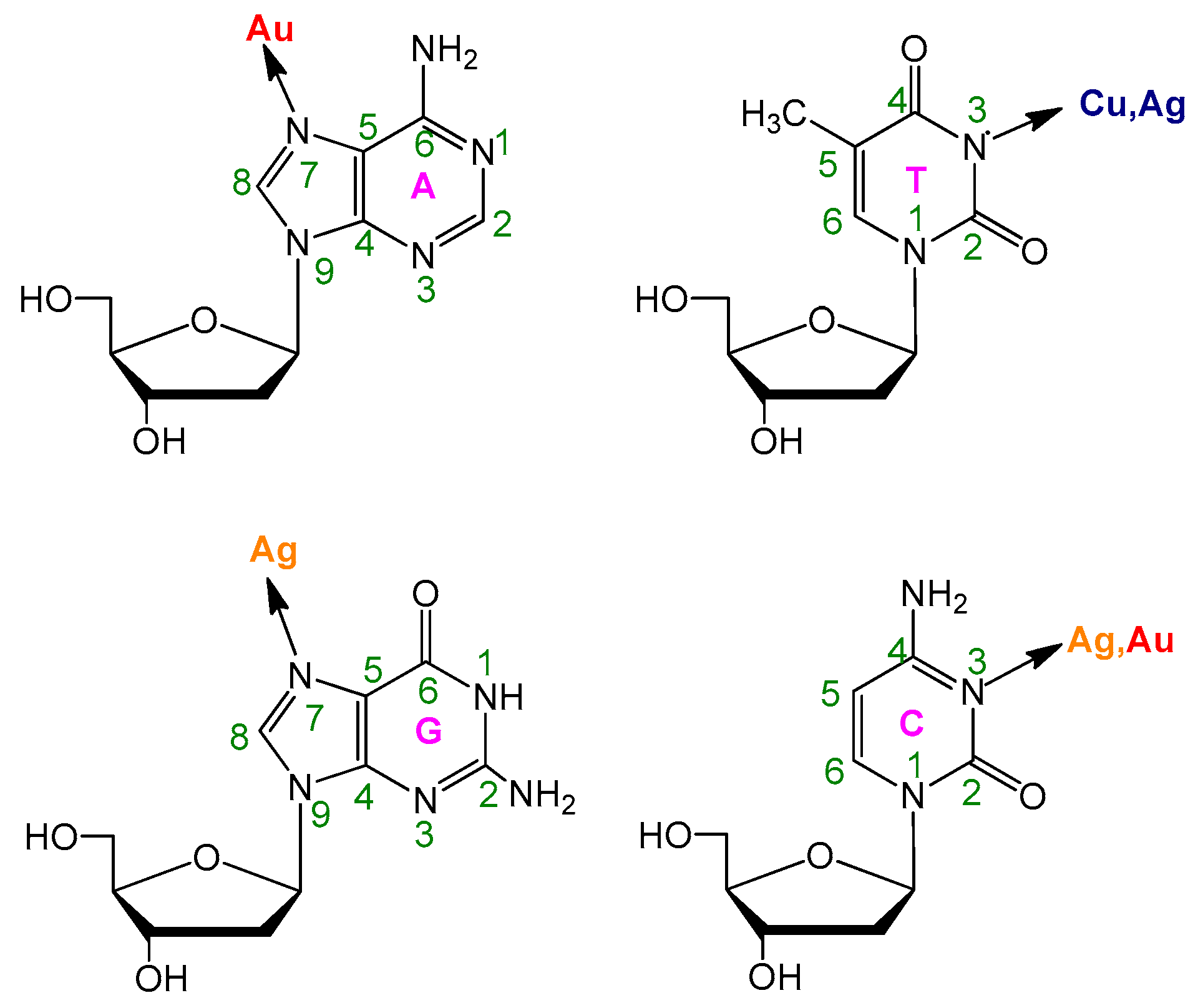
1. Introduction
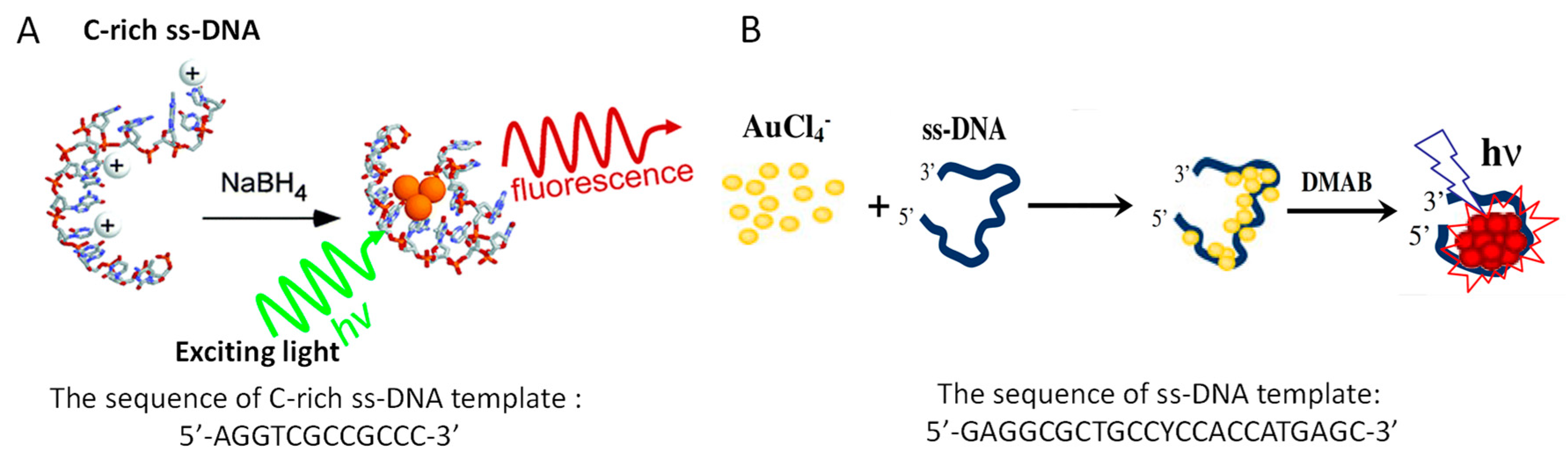
2. Synthesis of DNA-Templated Fluorescent Nanoclusters
3. DNA-Templated Fluorescent Nanoclusters for Sensing Metal Ions
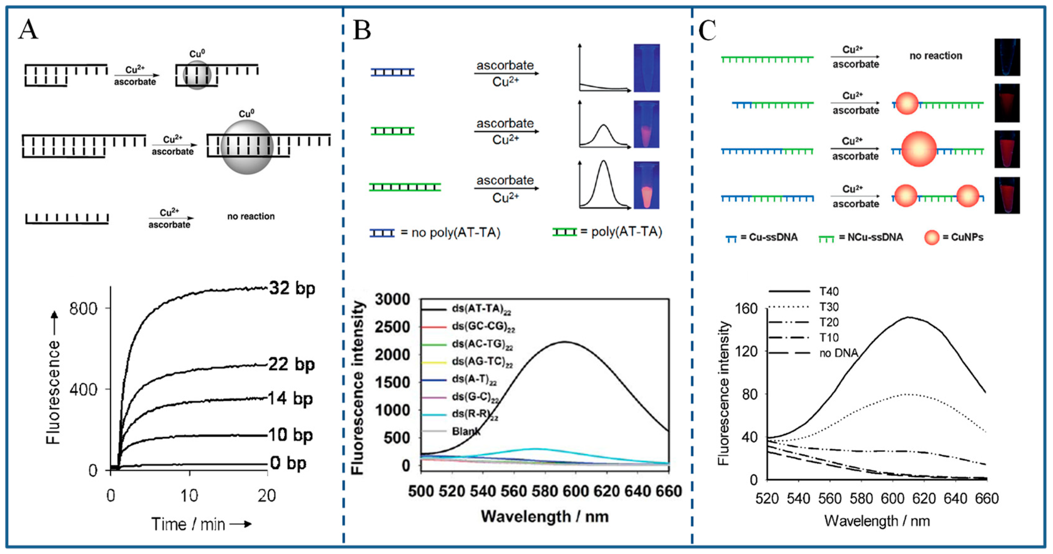
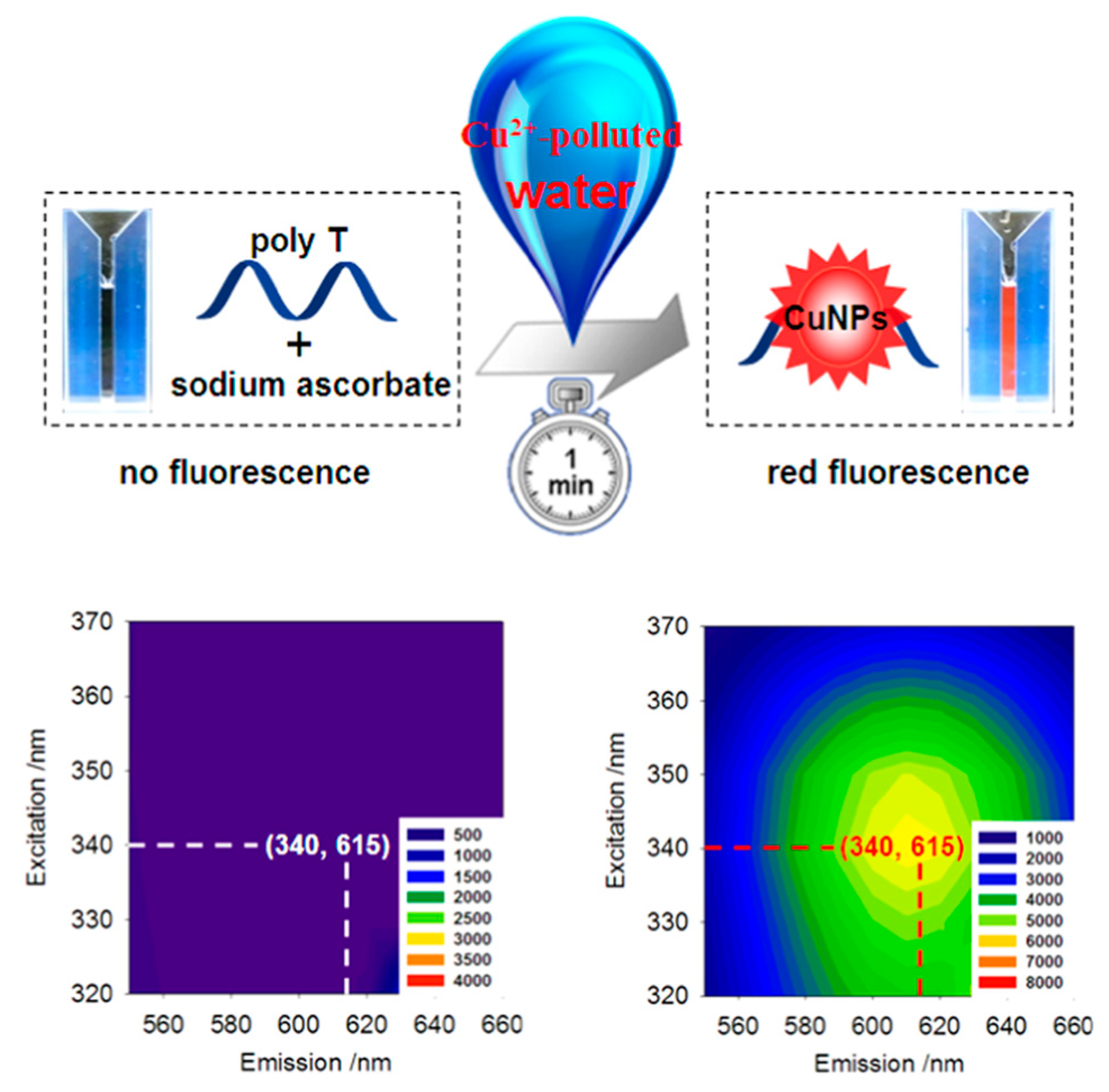
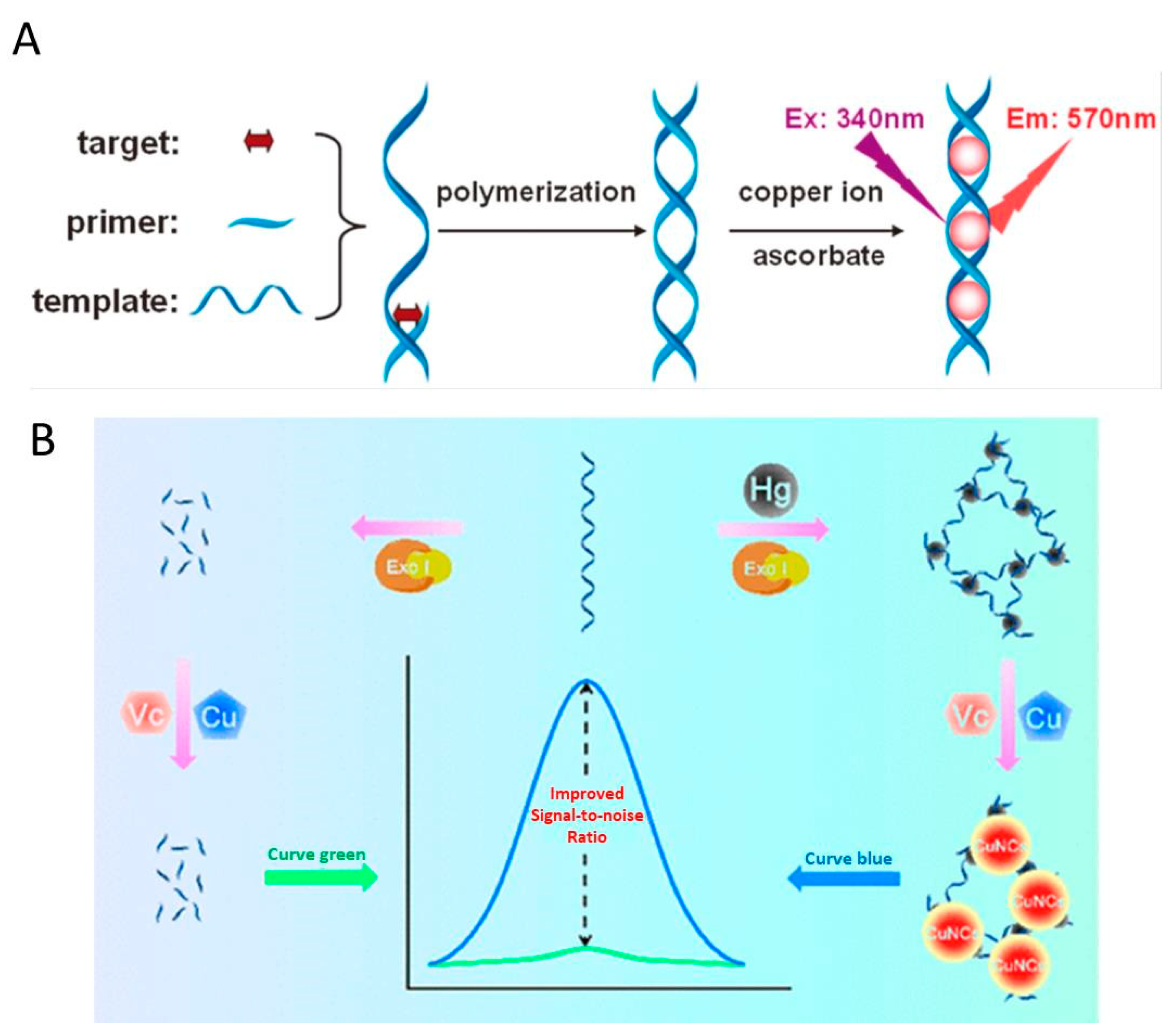
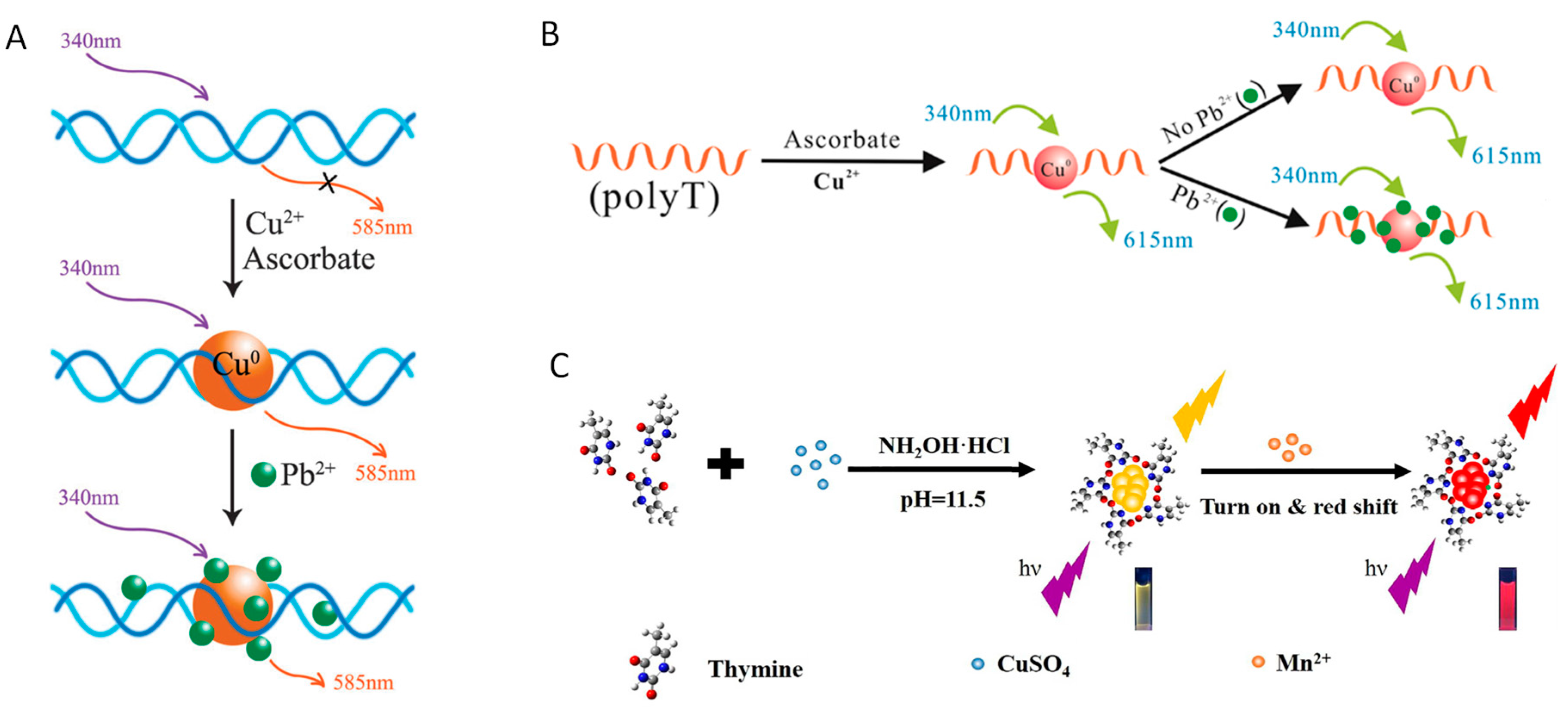
3.1. Copper Ions
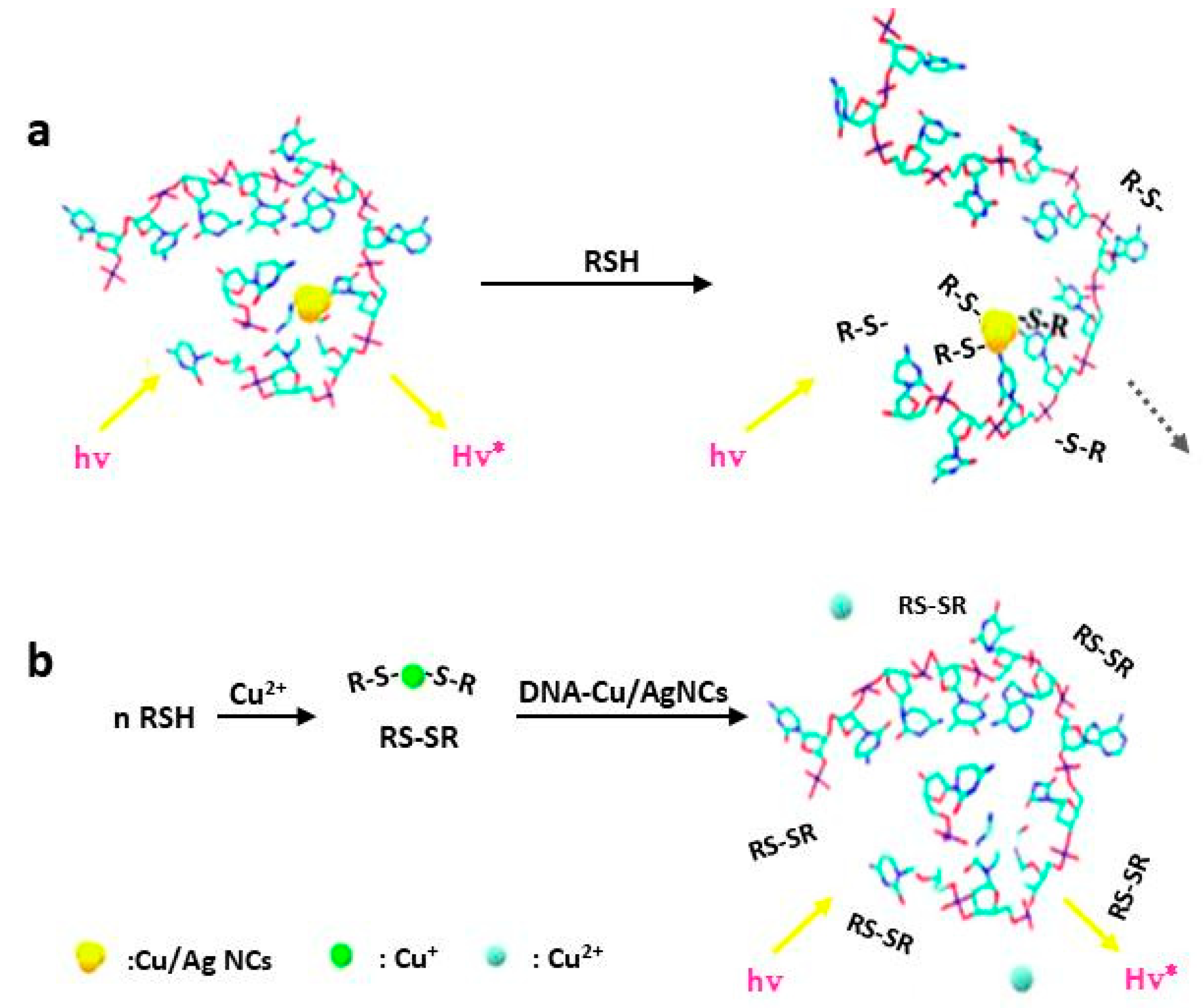
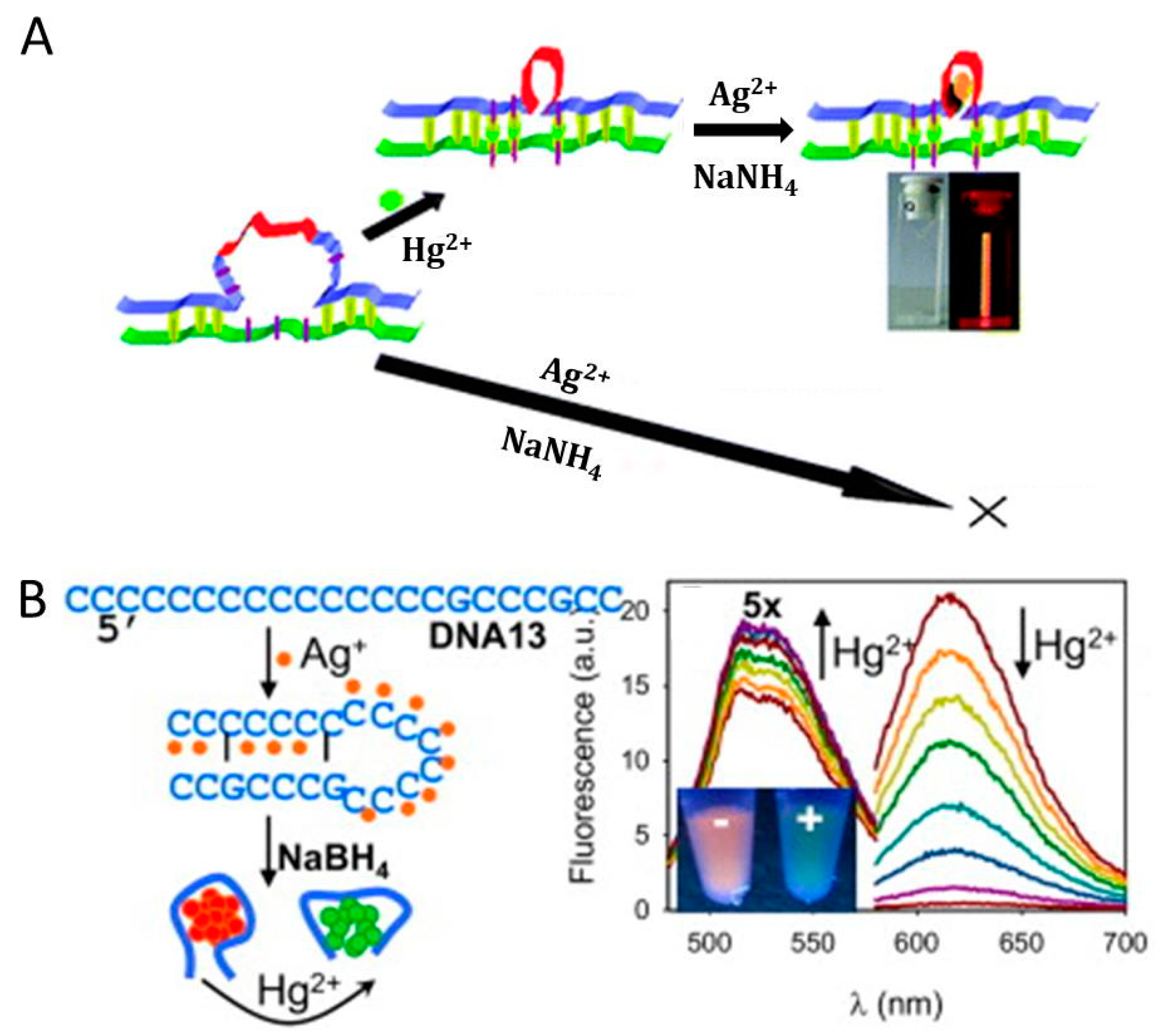
3.2. Mercury Ions
3.3. Lead Ions
3.4. Other Ions
4. Future Prospects
Author Contributions
Funding
Conflicts of Interest
References
- Qing, Z.; Bai, A.; Xing, S.; Zou, Z.; He, X.; Wang, K.; Yang, R. Progress in biosensor based on DNA-templated copper nanoparticles. Biosens. Bioelectron. 2019, 137, 96–109. [Google Scholar] [CrossRef] [PubMed]
- Zhang, L.; Wang, E. Metal nanoclusters: New fluorescent probes for sensors and bioimaging. Nano Today 2014, 9, 132–157. [Google Scholar] [CrossRef]
- Guo, Y.; Cao, F.; Lei, X.; Mang, L.; Cheng, S.; Song, J. Fluorescent copper nanoparticles: Recent advances in synthesis and applications for sensing metal ions. Nanoscale 2016, 8, 4852–4863. [Google Scholar] [CrossRef] [PubMed]
- Rui, L.; Wang, C.; Hu, J.; Su, Y.; Yi, L. DNA-templated copper nanoparticles: Versatile platform for label-free bioassays. Trends Anal. Chem. 2018, 105, 436–452. [Google Scholar]
- Liu, J. DNA-stabilized, fluorescent, metal nanoclusters for biosensor development. Trends Anal. Chem. 2014, 58, 99–111. [Google Scholar] [CrossRef]
- Qing, T.; Zhang, K.; Qing, Z.; Wang, X.; Long, C.; Zhang, P.; Hu, H.; Feng, B. Recent progress in copper nanocluster-based fluorescent probing: A review. Mikrochim. Acta 2019, 186, 670. [Google Scholar] [CrossRef]
- Liu, G.; Yong, S.; Cui, Q.; Fei, W.; Xu, S. Synthesis of DNA-templated fluorescent gold nanoclusters. Gold Bull. 2012, 45, 69–74. [Google Scholar] [CrossRef]
- Qing, Z.; Hou, L.; Yang, L.; Zhu, L.; Yang, S.; Zheng, J.; Yang, R. A reversible nanolamp for instantaneous monitoring of cyanide based on an elsner-like reaction. Anal. Chem. 2016, 88, 9759–9765. [Google Scholar] [CrossRef]
- Zhu, L.; Qing, Z. Direct detection of nucleic acid with minimizing background and improving sensitivity based on a conformation-discriminating indicator. Anal. Chem. 2017, 2, 1198–1204. [Google Scholar] [CrossRef]
- Yao, J.; Li, L.; Li, P.; Yang, M. Quantum dots: From fluorescence to chemiluminescence, bioluminescence, electrochemiluminescence, and electrochemistry. Nanoscale 2017, 9, 13364–13383. [Google Scholar] [CrossRef]
- Matea, C.T.; Mocan, T.; Tabaran, F.; Pop, T.; Mosteanu, O.; Puia, C.; Iancu, C.; Mocan, L. Quantum dots in imaging, drug delivery and sensor applications. Int. J. Nanomed. 2017, 12, 5421–5431. [Google Scholar] [CrossRef] [PubMed]
- Li, Y.; Luo, G.; Qing, Z. Colorimetric aminotriazole assay based on catalase deactivation-dependent longitudinal etching of gold nanorods. Microchim. Acta 2019, 186, 565. [Google Scholar] [CrossRef] [PubMed]
- Liu, C.; Chen, W.; Qing, Z.; Zheng, J.; Xiao, Y.; Yang, S.; Wang, L.; Li, Y.; Yang, R. In vivo lighted fluorescence via fenton reaction: Approach for imaging of hydrogen peroxide in living systems. Anal. Chem. 2016, 88, 3998–4003. [Google Scholar] [CrossRef] [PubMed]
- Petty, J.T.; Zheng, J.; Hud, N.V.; Dickson, R.M. DNA-templated Ag nanocluster formation. J. Am. Chem. Soc. 2004, 126, 5207–5212. [Google Scholar] [CrossRef] [PubMed]
- Hsu, Y.; Hung, M.; Chen, Y.; Wang, T.; Ou, Y.; Chen, S. Identifying reducing and capping sites of protein-encapsulated gold nanoclusters. Molecules 2019, 24, 1630. [Google Scholar] [CrossRef]
- Li, Y.; Jian, X.; Zhou, S.; Lu, Y.; Zhao, C.; Gao, Z.; Song, Y.-Y. Protein shell-encapsulated Pt clusters as continuous O2-supplied biocoats for photodynamic therapy in hypoxic cancer cells. ACS Appl. Mater. inter. 2019, 11, 17215–17225. [Google Scholar] [CrossRef]
- Sang, F.; Zhang, X.; Shen, F. Fluorescent methionine-capped gold nanoclusters for ultra-sensitive determination of copper(II) and cobalt(II), and their use in a test strip. Mikrochim. Acta 2019, 186, 373. [Google Scholar] [CrossRef]
- Xu, S.; Li, W.; Zhao, X.; Wu, T.; Cui, Y.; Fan, X.; Wang, W.; Luo, X. Ultra-highly efficient and stable fluorescent gold nanoclusters coated with screened peptides of unique sequences for effective protein and serum discrimination. Anal. Chem. 2019, 91, 13947–13952. [Google Scholar] [CrossRef]
- Wilner, O.I.; Willner, I. Functionalized DNA nanostructures. Chem. Rev. 2012, 112, 2528–2556. [Google Scholar] [CrossRef]
- Zhang, G.; Surwade, S.P.; Zhou, F.; Liu, H. DNA nanostructure meets nanofabrication. Chem. Soc. Rev. 2013, 42, 2488–2496. [Google Scholar] [CrossRef]
- Berti, L.; Burley, G.A. Nucleic acid and nucleotide-mediated synthesis of inorganic nanoparticles. Nat. Nanotechnol. 2008, 3, 81–87. [Google Scholar] [CrossRef] [PubMed]
- Ono, A.; Torigoe, H.; Tanaka, Y.; Okamoto, I. Binding of metal ions by pyrimidine base pairs in DNA duplexes. Chem. Soc. Rev. 2011, 40, 5855–5866. [Google Scholar] [CrossRef] [PubMed]
- Ono, A.; Cao, S.; Togashi, H.; Tashiro, M.; Fujimoto, T.; Machinami, T.; Oda, S.; Miyake, Y.; Okamoto, I.; Tanaka, Y. Specific interactions between silver(I) ions and cytosine–cytosine pairs in DNA duplexes. Chem. Commun. 2008, 39, 4825–4827. [Google Scholar] [CrossRef] [PubMed]
- Pompa, P.P.; Martiradonna, L.; Della Torre, A.; Della Sala, F.; Manna, L.; De Vittorio, M.; Calabi, F.; Cingolani, R.; Rinaldi, R. Metal-enhanced fluorescence of colloidal nanocrystals with nanoscale control. Nat. Nanotechnol. 2006, 1, 126–130. [Google Scholar] [CrossRef] [PubMed]
- Peng, Z.; Liu, H. Bottom-up nanofabrication using DNA nanostructures. Chem. Mater. 2016, 28, 1012–1021. [Google Scholar] [CrossRef]
- Qing, Z.; Zhu, L.; Hou, L.; Zou, Z.; Yang, S.; Yang, R. A dsDNA-lighted fluorophore for monitoring protein-ligand interaction through binding-mediated DNA protection. Sci. China Chem. 2018, 61, 1630–1636. [Google Scholar] [CrossRef]
- Qing, Z.; Xu, J.; Hu, J.; Zheng, J.; He, L.; Zou, Z.; Yang, S.; Tan, W.; Yang, R. In situ amplification-based imaging of RNA in living cells. Angew. Chem. Int. Ed. 2019, 58, 11574–11585. [Google Scholar] [CrossRef]
- Yang, L.; Qing, Z.; Liu, C.; Tang, Q.; Li, J.; Yang, S.; Zheng, J.; Yang, R.; Tan, W. Direct fluorescent detection of blood potassium by ion-selective formation of intermolecular G-Quadruplex and ligand binding. Anal. Chem. 2016, 88, 9285–9292. [Google Scholar] [CrossRef]
- Zhang, X.; Song, C.; Yang, K.; Hong, W.; Lu, Y.; Yu, P.; Mao, L. Photoinduced regeneration of an aptamer-based electrochemical sensor for sensitively detecting adenosine triphosphate. Anal. Chem. 2018, 90, 4968–4971. [Google Scholar] [CrossRef]
- Qing, Z.; Mao, Z.; Qing, T.; He, X.; Zou, Z.; He, D.; Shi, H.; Huang, J.; Liu, J.; Wang, K. Visual and portable strategy for copper(II) detection based on a striplike poly(thymine)-caged and microwell-printed hydrogel. Anal. Chem. 2014, 86, 11263–11268. [Google Scholar] [CrossRef]
- Qing, Z.; Zhu, L.; Yang, S.; Cao, Z.; He, X.; Wang, K.; Yang, R. In situ formation of fluorescent copper nanoparticles for ultrafast zero-background Cu2+ detection and its toxicides screening. Biosens. Bioelectron. 2016, 78, 471–476. [Google Scholar] [CrossRef] [PubMed]
- Qing, Z.; Zhu, L.; Li, X.; Yang, S.; Zou, Z.; Guo, J.; Cao, Z.; Yang, R. A target-lighted dsDNA-indicator for high-performance monitoring of mercury pollution and its antagonists screening. Environ. Sci. Technol. 2017, 51, 11884–11890. [Google Scholar] [CrossRef] [PubMed]
- Yaita, T. Trace copper(II) ions detection and removal from water using novel ligand modified composite adsorbent. Chem. Eng. J. 2013, 222, 67–76. [Google Scholar]
- Knecht, M.R.; Manish, S. Bio-inspired colorimetric detection of Hg2+ and Pb2+ heavy metal ions using Au nanoparticles. Anal. Bioanal. Chem. 2009, 394, 33–46. [Google Scholar] [CrossRef] [PubMed]
- Guo, Y.; Zhang, L.; Zhang, S.; Yang, Y.; Chen, X.; Zhang, M. Fluorescent carbon nanoparticles for the fluorescent detection of metal ions. Biosens. Bioelectron. 2015, 63, 61–71. [Google Scholar] [CrossRef] [PubMed]
- Guo, Y.; Wang, Z.; Qu, W.; Shao, H.; Jiang, X. Colorimetric detection of mercury, lead and copper ions simultaneously using protein-functionalized gold nanoparticles. Biosens. Bioelectron. 2011, 26, 4064–4069. [Google Scholar] [CrossRef]
- Bansod, B.; Kumar, T.; Thakur, R.; Rana, S.; Singh, I. A review on various electrochemical techniques for heavy metal ions detection with different sensing platforms. Biosens. Bioelectron. 2017, 94, 443–455. [Google Scholar] [CrossRef]
- Qian, X.; Xu, Z. Fluorescence imaging of metal ions implicated in diseases. Chem. Soc. Rev. 2015, 44, 4487–4493. [Google Scholar] [CrossRef]
- Huang, J.; Su, X.; Li, Z. Metal ion detection using functional nucleic acids and nanomaterials. Biosens. Bioelectron. 2017, 96, 127–139. [Google Scholar] [CrossRef]
- Chen, L.; Wang, C.; Yuan, Z.; Chang, H. Fluorescent gold nanoclusters: Recent advances in sensing and imaging. Anal. Chem. 2015, 87, 216–229. [Google Scholar] [CrossRef]
- Schaeffer, N.; Tan, B.; Dickinson, C.; Rosseinsky, M.J.; Laromaine, A.; Mccomb, D.W.; Stevens, M.M.; Wang, Y.; Petit, L.; Barentin, C. Fluorescent or not? Size-dependent fluorescence switching for polymer-stabilized gold clusters in the 1.1–1.7 nm size range. Chem. Commun. 2008, 34, 3986–3988. [Google Scholar] [CrossRef] [PubMed]
- Qing, Z.H.; Qing, T.P.; Mao, Z.G.; He, X.X.; Wang, K.M.; Zou, Z.; Shi, H.; He, D.G. dsDNA-specific fluorescent copper nanoparticles as a “green” nano-dye for polymerization-mediated biochemical analysis. Chem. Commun. 2014, 50, 12746–12748. [Google Scholar] [CrossRef] [PubMed]
- Song, C.; Yang, X.; Wang, K.; Wang, Q.; Huang, J.; Liu, J.; Liu, W.; Liu, P. Label-free and non-enzymatic detection of DNA based on hybridization chain reaction amplification and dsDNA-templated copper nanoparticles. Anal. Chim. Acta 2014, 827, 74–79. [Google Scholar] [CrossRef] [PubMed]
- Song, C.; Hong, W.; Zhang, X.; Lu, Y. Label-free and sensitive detection of ochratoxin A based on dsDNA-templated copper nanoparticles and exonuclease-catalyzed target recycling amplification. Analyst 2018, 143, 1829–1834. [Google Scholar] [CrossRef]
- Xu, F.; Shi, H.; He, X.; Wang, K.; He, D.; Guo, Q.; Qing, Z.; Yan, L.; Ye, X.; Li, D.; et al. Concatemeric dsDNA-templated copper nanoparticles strategy with improved sensitivity and stability based on rolling circle replication and its application in microRNA detection. Anal. Chem. 2014, 86, 6976–6982. [Google Scholar] [CrossRef]
- Zheng, J.; Dickson, R.M. Individual water-soluble dendrimer-encapsulated silver nanodot fluorescence. J. Am. Chem. Soc. 2002, 124, 13982–13983. [Google Scholar] [CrossRef]
- Richards, C.I.; Choi, S.; Hsiang, J.; Antoku, Y.; Vosch, T.; Bongiorno, A.; Tzeng, Y.; Dickson, R.M. Oligonucleotide-stabilized Ag nanocluster fluorophores. J. Am. Chem. Soc. 2008, 130, 5038–5039. [Google Scholar] [CrossRef]
- Sengupta, B.; Ritchie, C.M.; Buckman, J.G.; Johnsen, K.R.; Goodwin, P.M.; Petty, J.T. Base-directed formation of fluorescent silver clusters. J. Phys. Chem. C 2008, 112, 18776–18782. [Google Scholar] [CrossRef]
- Navarro, J.A.R.; Lippert, B. Molecular architecture with metal ions, nucleobases and other heterocycles. Coord. Chem. Rev. 1999, 185–186, 653–667. [Google Scholar] [CrossRef]
- Verma, S.; Mishra, A.K.; Kumar, J. The many facets of adenine: Coordination, crystal patterns, and catalysis. Acc. Chem. Res. 2010, 43, 79–91. [Google Scholar] [CrossRef]
- Ma, K.; Shao, Y.; Cui, Q.; Wu, F.; Xu, S.; Liu, G. Base-stacking-determined fluorescence emission of DNA abasic site-templated silver nanoclusters. Langmuir 2012, 28, 15313–15322. [Google Scholar] [CrossRef] [PubMed]
- Ma, K.; Cui, Q.; Liu, G.; Wu, F.; Xu, S.; Shao, Y. DNA abasic site-directed formation of fluorescent silver nanoclusters for selective nucleobase recognition. Nanotechnology 2011, 22, 305502. [Google Scholar] [CrossRef] [PubMed]
- Huang, Z.; Pu, F.; Hu, D.; Wang, C.; Ren, J.; Qu, X. Site-specific DNA-programmed growth of fluorescent and functional silver nanoclusters. Chemistry 2011, 17, 3774–3780. [Google Scholar] [CrossRef] [PubMed]
- Cui, Q.; Ma, K.; Shao, Y.; Xu, S.; Wu, F.; Liu, G.; Teramae, N.; Bao, H. Gap site-specific rapid formation of fluorescent silver nanoclusters for label-free DNA nucleobase recognition. Anal. Chim. Acta 2012, 724, 86–91. [Google Scholar] [CrossRef] [PubMed]
- Feng, L.; Huang, Z.; Ren, J.; Qu, X. Toward site-specific, homogeneous and highly stable fluorescent silver nanoclusters fabrication on triplex DNA scaffolds. Nucleic Acids Res. 2012, 40, e122. [Google Scholar] [CrossRef] [PubMed]
- Zhao, T.; Chen, Q.; Zeng, C.; Lan, Y.; Cai, J.; Liu, J.; Gao, J. Multi-DNA-Ag nanoclusters: Reassembly mechanism and sensing the change of HIF in cells. J. Mater. Chem. B 2013, 1, 4678–4683. [Google Scholar] [CrossRef]
- Konrad, K.; Korbinian, B. A highly charged Ag64+ core in a DNA-encapsulated silver nanocluster. Chemistry 2010, 16, 3285–3290. [Google Scholar]
- Yang, X.; Gan, L.; Han, L.; Wang, E.; Wang, J. High-yield synthesis of silver nanoclusters protected by DNA monomers and DFT prediction of their photoluminescence properties. Angew. Chem. Int. Ed. 2013, 52, 2022–2026. [Google Scholar] [CrossRef]
- Danielle, S.; Elisabeth, G. Stabilization of fluorescent silver clusters by RNA homopolymers and their DNA analogs: C,G versus A,T(U) dichotomy. Chem. Commun. 2011, 47, 4715–4717. [Google Scholar]
- Ai, J.; Guo, W.; Li, B.; Tao, L.; Dan, L.; Wang, E. DNA G-quadruplex-templated formation of the fluorescent silver nanocluster and its application to bioimaging. Talanta 2012, 88, 450–455. [Google Scholar] [CrossRef]
- Kennedy, T.A.; Maclean, J.L.; Liu, J. Blue emitting gold nanoclusters templated by poly-cytosine DNA at low pH and poly-adenine DNA at neutral pH. Chem. Commun. 2012, 48, 6845–6847. [Google Scholar] [CrossRef] [PubMed]
- Liu, G.; Shao, Y.; Wu, F.; Xu, S.; Peng, J.; Liu, L. DNA-hosted fluorescent gold nanoclusters: Sequence-dependent formation. Nanotechnology 2013, 24, 015503. [Google Scholar] [CrossRef]
- Rotaru, A.; Dutta, S.; Jentzsch, E.; Gothelf, K.; Mokhir, A. Selective dsDNA-templated formation of copper nanoparticles in solution. Angew. Chem. Int. Ed. 2010, 49, 5665–5667. [Google Scholar] [CrossRef] [PubMed]
- Song, Q.; Shi, Y.; He, D.; Xu, S.; Ouyang, J. Sequence-dependent dsDNA-templated formation of fluorescent copper nanoparticles. Chem. Eur. J. 2015, 21, 2417–2422. [Google Scholar] [CrossRef]
- Qing, T.; Qing, Z.; Mao, Z.; He, X.; Xu, F.; Wen, L.; He, D.; Shi, H.; Wang, K. dsDNA-templated fluorescent copper nanoparticles: Poly (AT-TA)-dependent formation. RSC Adv. 2014, 4, 61092–61095. [Google Scholar] [CrossRef]
- Liu, G.; Shao, Y.; Peng, J.; Dai, W.; Liu, L.; Xu, S.; Wu, F.; Wu, X. Highly thymine-dependent formation of fluorescent copper nanoparticles templated by ss-DNA. Nanotechnology 2013, 24, 345502. [Google Scholar] [CrossRef] [PubMed]
- Qing, Z.; He, X.; He, D.; Wang, K.; Xu, F.; Qing, T.; Yang, X. Poly(thymine)-templated selective formation of fluorescent copper nanoparticles. Angew. Chem. Int. Ed. 2013, 52, 9719–9722. [Google Scholar] [CrossRef]
- Li, J.; Fu, W.; Bao, J.; Wang, Z.; Dai, Z. Fluorescence regulation of copper nanoclusters via DNA template manipulation toward design of a high signal-to-noise ratio biosensor. ACS Appl. Mater. inter. 2018, 10, 6965–6971. [Google Scholar] [CrossRef]
- Xue, H.; Liu, T.; Zhuang, Y.; Wei, W.; Li, Y.; Fan, W.; Huang, Y. Recent advances in the analytical applications of copper nanoclusters. Trends Anal. Chem. 2016, 77, 66–75. [Google Scholar]
- Harris, E.D. Copper as a cofactor and regulator of copper, zinc superoxide dismutase. J. Nutr. 1992, 122, 636–640. [Google Scholar] [CrossRef]
- Plastino, J.; Green, E.L.; Sanders-Loehr, J.; Klinman, J.P. An unexpected role for the active site base in cofactor orientation and flexibility in the copper amine oxidase from Hansenula polymorpha. Biochemistry 1999, 38, 8204–8216. [Google Scholar] [CrossRef] [PubMed]
- Chen, P.; Solomon, E.I. Oxygen activation by the noncoupled binuclear copper site in peptidylglycine alpha-hydroxylating monooxygenase. Reaction mechanism and role of the noncoupled nature of the active site. J. Am. Chem. Soc. 2004, 126, 4991–5000. [Google Scholar] [CrossRef] [PubMed]
- Lan, G.Y.; Huang, C.C.; Chang, H.T. Silver nanoclusters as fluorescent probes for selective and sensitive detection of copper ions. Chem. Commun. 2010, 46, 1257–1259. [Google Scholar] [CrossRef] [PubMed]
- Su, Y.T.; Lan, G.Y.; Chen, W.Y.; Chang, H.T. Detection of copper ions through recovery of the fluorescence of DNA-templated copper/silver nanoclusters in the presence of mercaptopropionic acid. Anal. Chem. 2010, 82, 8566–8572. [Google Scholar] [CrossRef] [PubMed]
- Zhang, M.; Ye, B.C. Label-free fluorescent detection of copper(II) using DNA-templated highly luminescent silver nanoclusters. Analyst 2011, 136, 5139–5142. [Google Scholar] [CrossRef] [PubMed]
- Guo, W.; Yuan, J.; Wang, E. Oligonucleotide-stabilized Ag nanoclusters as novel fluorescence probes for the highly selective and sensitive detection of the Hg2+ ion. Chem. Commun. 2009, 23, 3395–3397. [Google Scholar] [CrossRef]
- Lan, G.-Y.; Chen, W.-Y.; Chang, H.-T. Control of synthesis and optical properties of DNA templated silver nanoclusters by varying DNA length and sequence. RSC Adv. 2011, 1, 802–807. [Google Scholar] [CrossRef]
- Deng, L.; Zhou, Z.; Li, J.; Li, T.; Dong, S. Fluorescent silver nanoclusters in hybridized DNA duplexes for the turn-on detection of Hg2+ ions. Chem. Commun. 2011, 47, 11065–11067. [Google Scholar] [CrossRef]
- Yin, J.; He, X.; Jia, X.; Wang, K.; Xu, F. Highly sensitive label-free fluorescent detection of Hg2+ ions by DNA molecular machine-based Ag nanoclusters. Analyst 2013, 138, 2350–2356. [Google Scholar] [CrossRef]
- MacLean, J.L.; Morishita, K.; Liu, J. DNA stabilized silver nanoclusters for ratiometric and visual detection of Hg2+ and its immobilization in hydrogels. Biosens. Bioelectron. 2013, 48, 82–86. [Google Scholar] [CrossRef]
- Ono, A.; Togashi, H. Highly selective oligonucleotide-based sensor for mercury(II) in aqueous solutions. Angew. Chem. Int. Ed. 2004, 43, 4300–4302. [Google Scholar] [CrossRef] [PubMed]
- Wang, H.; Chen, Y.; Li, Y.; Zhang, H.; Cao, J. A rapid, sensitive and label-free sensor for Hg(II) ion detection based on blocking of cysteine-quenching of fluorescent poly(thymine)-templated copper nanoparticles. RSC Adv. 2015, 5, 94099–94104. [Google Scholar] [CrossRef]
- Tipton, I.H. The human body burden of lead. Arch. Environ. Health 1968, 17, 965–978. [Google Scholar]
- Wasserman, G.; Graziano, J.H.; Factor-Litvak, P.; Md, D.P.; Morina, M.N.; Musabegovic, M.A.; Vrenezi, M.N.; Capuni-Paracka, M.S.; Lekic, M.V.; Preteni-Redjepi, M.E. Independent effects of lead exposure and iron deficiency anemia on developmental outcome at age 2 years. J. Pediatr. 1992, 121, 695–703. [Google Scholar] [CrossRef]
- Chen, J.; Liu, J.; Fang, Z.; Zeng, L. Random dsDNA-templated formation of copper nanoparticles as novel fluorescence probes for label-free lead ions detection. Chem. Commun. 2012, 48, 1057–1059. [Google Scholar] [CrossRef] [PubMed]
- Ou, L.; Li, X.; Liu, H.; Li, L.; Chu, X. Poly(thymine)-templated fluorescent copper nanoparticles for ultrasensitive label-free detection of Pb2+ ion. Anal. Sci. 2014, 30, 723–727. [Google Scholar] [CrossRef]
- Adams, L.F.; Ghiorse, W.C. Characterization of extracellular Mn2+-oxidizing activity and isolation of an Mn2+-oxidizing protein from Leptothrix discophora SS-1. J. Bacteriol. 1987, 169, 1279–1285. [Google Scholar] [CrossRef]
- Hu, S.; Zhang, J.; Tang, R.; Fan, J.; Liu, H.; Kang, W.; Lei, C.; Nie, Z.; Huang, Y.; Yao, S. Click-type protein-DNA conjugation for Mn2+ imaging in living cells. Anal. Chem. 2019, 91, 10180–10187. [Google Scholar] [CrossRef]
- Zhang, J.; Chen, X.; Yue, L.; Han, S.; Yao, D.; Liu, H. Nitrogen doped carbon quantum dots-enhanced chemiluminescence method for the determination of Mn2+. Anal. Methods 2018, 10, 541–547. [Google Scholar] [CrossRef]
- Han, B.; Xiang, R.; Hou, X.; Yu, M.; Peng, T.; Li, Y.; He, G. One-step rapid synthesis of single thymine-templated fluorescent copper nanoclusters for “turn on” detection of Mn2+. Anal. Methods 2017, 9, 2590–2595. [Google Scholar] [CrossRef]
- Wang, G.; Xu, G.; Zhu, Y.; Zhang, X. A “turn-on” carbon nanotube-Ag nanoclusters fluorescent sensor for sensitive and selective detection of Hg2+ with cyclic amplification of exonuclease III activity. Chem. Commun. 2013, 50, 747–750. [Google Scholar] [CrossRef] [PubMed][Green Version]
- Miao, P.; Zhang, T.; Xu, J.; Tang, Y. Electrochemical detection of miRNA combining T7 exonuclease-assisted cascade signal amplification and DNA-templated copper nanoparticles. Anal. Chem. 2018, 90, 11154–11160. [Google Scholar] [CrossRef] [PubMed]
- Sheng, F.; Zhang, X.; Wang, G. Novel ultrasensitive homogeneous electrochemical aptasensor based on dsDNA-templated copper nanoparticles for the detection of ractopamine. J. Mater. Chem. B 2017, 5, 53–61. [Google Scholar] [CrossRef]
- Liu, R.; Wang, C.; Xu, Y.; Hu, J.; Deng, D.; Lv, Y. Label-free DNA assay by metal stable isotope detection. Anal. Chem. 2017, 89, 13269–13274. [Google Scholar] [CrossRef] [PubMed]
- Yuan, P.-X.; Deng, S.-Y.; Zheng, C.-Y.; Cosnier, S.; Shan, D. In situ formed copper nanoparticles templated by TdT-mediated DNA for enhanced SPR sensor-based DNA assay. Biosens. Bioelectron. 2017, 97, 1–7. [Google Scholar] [CrossRef] [PubMed]
- Zhu, N.; Gu, L.; Wang, J.; Li, X.; Liang, G.; Zhou, J.; Zhang, Z. Novel and sensitive chemiluminescence sensors based on 2D-MOF nanosheets for one-step detection of glucose in human urine. J. Phys. Chem. C 2019, 123, 9388–9393. [Google Scholar] [CrossRef]
- Yang, N.; Huang, Y.; Ding, G.; Fan, A. In situ generation of prussian blue with potassium ferrocyanide to improve the sensitivity of chemiluminescence immunoassay using magnetic nanoparticles as label. Anal. Chem. 2019, 91, 4906–4912. [Google Scholar] [CrossRef]
- Zhang, B.; Zhang, F.; Zhang, P.; Shen, D.; Gao, X.; Zou, G. Ultrasensitive electrochemiluminescent sensor for MicroRNA with multinary Zn-Ag-In-S/ZnS nanocrystals as tags. Anal. Chem. 2019, 91, 3754–3758. [Google Scholar] [CrossRef]
- Li, S.; Liu, Z.; Li, J.; Chen, A.; Chai, Y.; Yuan, R.; Zhuo, Y. Enzyme-free target recycling and double-output amplification system for electrochemiluminescent assay of Mucin 1 with MoS2 nanoflowers as co-reaction accelerator. ACS Appl. Mater. inter. 2018, 10, 14483–14490. [Google Scholar] [CrossRef]
- Zhuang, Y.-T.; Chen, S.; Jiang, R.; Yu, Y.; Wang, J. Ultrasensitive colorimetric chromium chemosensor based on dye color switching under the Cr(VI)-stimulated Au NPs catalytic activity. Anal. Chem. 2019, 91, 5346–5353. [Google Scholar] [CrossRef]
- Wolfe, M.G.; Ali, M.M.; Brennan, J.D. Enzymatic litmus test for selective colorimetric detection of C-C single nucleotide polymorphisms. Anal. Chem. 2019, 91, 4735–4740. [Google Scholar] [CrossRef] [PubMed]
- Zhou, Q.; Zheng, J.; Qing, Z.; Zheng, M.; Yang, J.; Yang, S.; Ying, L.; Yang, R. Detection of circulating tumor DNA in human blood via DNA-mediated surface-enhanced Raman spectroscopy of single-walled carbon nanotubes. Anal. Chem. 2016, 88, 4759–4765. [Google Scholar] [CrossRef] [PubMed]


| Nanomaterial Used | Analytes | Strategy | LOD | Linear Range (nM) | Ref. |
|---|---|---|---|---|---|
| PolyT-templated F-CuNCs | Cu2+ | Turn-on | 20 µM | 20 µM–10 mM | [30] |
| PolyT-templated F-CuNCs | Cu2+ | Turn-on | 5.6 μM | 15–35 µM | [31] |
| DNA-templated F-AgNCs | Cu2+ | Turn-on | 8 nM | 25–250 nM | [73] |
| DNA-templated F-AgNCs | Cu2+ | Turn-on | 2.7 nM | 5–200 nM | [74] |
| DNA-templated F-AgNCs | Cu2+ | Turn-off | 10 nM | 10–1000 nM | [75] |
| dsDNA-templated F-CuNCs | Hg2+ | Turn-on | - | - | [42] |
| PolyT-templated F-CuNCs | Hg2+ | Turn-on | - | - | [62] |
| PolyT-templated F-CuNCs | Hg2+ | Turn-on | 16 pM | 50 pM–500 μM | [68] |
| DNA-templated F-AgNCs | Hg2+ | Turn-off | 5 nM | 5–1500 nM | [76] |
| DNA-templated F-AgNCs | Hg2+ | Turn-off | 0.9 nM | 2.5–50 nM | [77] |
| DNA-templated F-AgNCs | Hg2+ | Turn-on | 10 nM | 10–300 nM | [78] |
| DNA-templated F-AgNCs | Hg2+ | Turn-off | 4 nM | 10–200 nM | [80] |
| PolyT-templated F-CuNCs | Hg2+ | Turn-on | 0.1 nM | 0.5–30 nM | [82] |
| DNA-templated F-AgNCs | Hg2+ | Turn-on | 0.033 nM | 0.1–200 nM | [91] |
| dsDNA-templated F-CuNCs | Pb2+ | Turn-off | 5 nM, | 5–100 nM | [85] |
| PolyT-templated F-CuNCs | Pb2+ | Turn-off | 0.4 nM | 1.0–500 nM | [86] |
| SingleT-templated F-CuNCs | Mn2+ | Turn-on | 10 μM. | 100–250 μM | [90] |
© 2019 by the authors. Licensee MDPI, Basel, Switzerland. This article is an open access article distributed under the terms and conditions of the Creative Commons Attribution (CC BY) license (http://creativecommons.org/licenses/by/4.0/).
Share and Cite
Song, C.; Xu, J.; Chen, Y.; Zhang, L.; Lu, Y.; Qing, Z. DNA-Templated Fluorescent Nanoclusters for Metal Ions Detection. Molecules 2019, 24, 4189. https://doi.org/10.3390/molecules24224189
Song C, Xu J, Chen Y, Zhang L, Lu Y, Qing Z. DNA-Templated Fluorescent Nanoclusters for Metal Ions Detection. Molecules. 2019; 24(22):4189. https://doi.org/10.3390/molecules24224189
Chicago/Turabian StyleSong, Chunxia, Jingyuan Xu, Ying Chen, Liangliang Zhang, Ying Lu, and Zhihe Qing. 2019. "DNA-Templated Fluorescent Nanoclusters for Metal Ions Detection" Molecules 24, no. 22: 4189. https://doi.org/10.3390/molecules24224189
APA StyleSong, C., Xu, J., Chen, Y., Zhang, L., Lu, Y., & Qing, Z. (2019). DNA-Templated Fluorescent Nanoclusters for Metal Ions Detection. Molecules, 24(22), 4189. https://doi.org/10.3390/molecules24224189