Article

Isolation, Structure Elucidation, and Antiproliferative Activity of Butanolides and Lignan Glycosides from the Fruit of Hernandia nymphaeifolia

1
School of Pharmaceutical Sciences, College of Medical, Pharmaceutical and Health Sciences, Kanazawa University, Kanazawa 920-1192, Japan
2
Natural Products Research Laboratories, UNC Eshelman School of Pharmacy, University of North Carolina at Chapel Hill, Chapel Hill, NC 27599-7568, USA
3
Tokyo University of Pharmacy and Life Sciences, Hachioji, Tokyo 192-0392, Japan
4
NIH Special Volunteer, Wayne, PA 19087, USA
5
Natural Products Branch, Developmental Therapeutics Program, Division of Cancer Treatment and Diagnosis, National Cancer Institute, NCI at Frederick, Frederick, MD 21702-1201, USA
6
Molecular Targets Program, Center for Cancer Research, National Cancer Institute, NCI at Frederick, Frederick, MD 21702-1201, USA
7
Chinese Medicine Research and Development Center, China Medical University and Hospital, Taichung 40447, Taiwan
*
Author to whom correspondence should be addressed.
Molecules 2019, 24(21), 4005; https://doi.org/10.3390/molecules24214005
Submission received: 9 October 2019 / Revised: 24 October 2019 / Accepted: 31 October 2019 / Published: 5 November 2019
(This article belongs to the Special Issue Antitumor and Anti-HIV Agents from Natural Products)

Abstract

:
Seven new butanolides, peltanolides A–G (17), and two lignan glucosides, peltasides A (8) and B (9), along with eleven known compounds, 1020, were isolated from a crude CH3OH/CH2Cl2 (1:1) extract of the fruit of Hernandia nymphaeifolia (Hernandiaceae). The structures of 19 were characterized by extensive 1D and 2D NMR spectroscopic and HRMS analysis. The absolute configurations of newly isolated compounds 19 were determined from data obtained by optical rotation and electronic circular dichroism (ECD) exciton chirality methods. Butanolides and lignan glucosides have not been isolated previously from this genus. Several isolated compounds were evaluated for antiproliferative activity against human tumor cell lines. Lignans 15 and 16 were slightly active against chemosensitive tumor cell lines A549 and MCF-7, respectively. Furthermore, both compounds displayed significant activity (IC50 = 5 µM) against a P-glycoprotein overexpressing multidrug-resistant tumor cell line (KB-VIN) but were less active against its parent chemosensitive cell line (KB).

Graphical Abstract

1. Introduction

Plants in the genus Hernandia (Hernandiaceae) are found in subtropical and tropical areas [1]. They contain diverse bioactive secondary metabolites, especially lignans, including podophyllotoxin analogues [2,3], and benzylisoquinolines [4], including aporphines [5,6,7]. These compounds exhibit various biological activities, including significant cytotoxic [8,9], antiplasmodial [9,10], and antibacterial activities [2]. H. nymphaeifolia (C.Presl) Kubitzki (synonym: H. peltata Meisn.) is a common coastal tree and grows to 12–20 m in height. This plant has been used for the treatment of abdominal pains, boils, cough, diarrhea, eye problems, and convulsions as a traditional medicine in western Samoa [11]. A CH3OH/CH2Cl2 (1:1) extract of H. nymphaeifolia (N053499, originally described as H. peltata) provided by the U.S. National Cancer Institute Natural Products Branch (NCI, Frederick, MD, USA) exhibited broad cytotoxicity in the NCI-60 human tumor cell line (HTCL) assay, possibly due to the above or similar cytotoxic constituents. To supplement the reported phytochemical research on H. nymphaeifolia [2,4,9,12,13,14], we conducted a thorough study to identify new chemical compounds as part of our continuing investigation of rainforest plants. Accordingly, the extract of N053499 yielded seven new butanolides, peltanolides A–G (17), and two new lignan glycosides, peltasides A (8) and B (9), as well as eleven known compounds 1020 (Figure 1). Herein, we report the details of isolation, structure elucidation, and cytotoxicity of isolated compounds from H. nymphaeifolia.

2. Results and Discussion

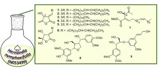
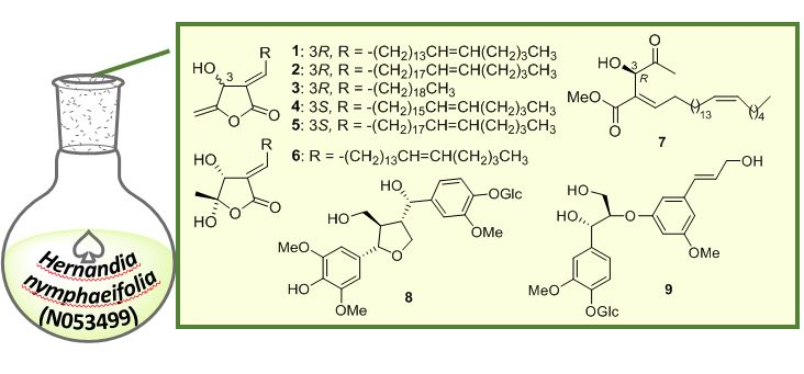
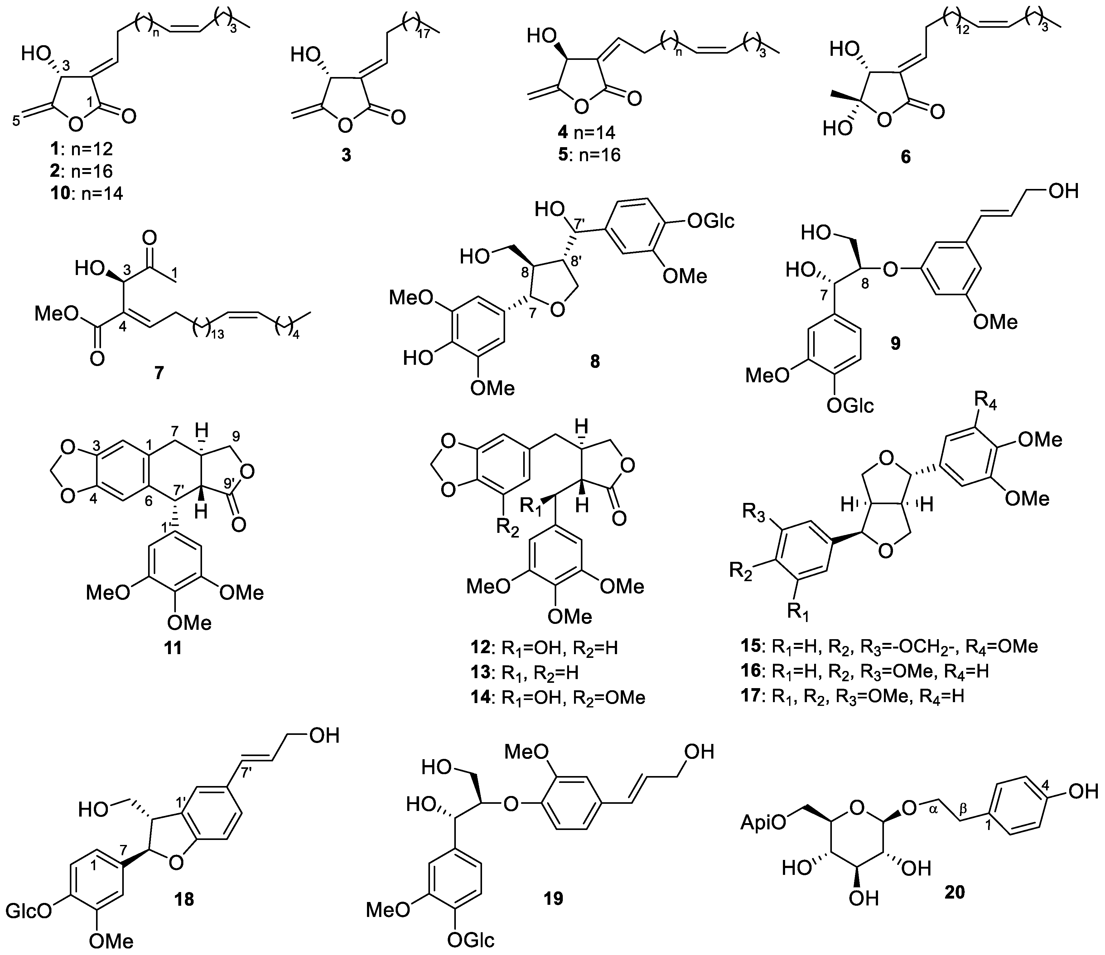
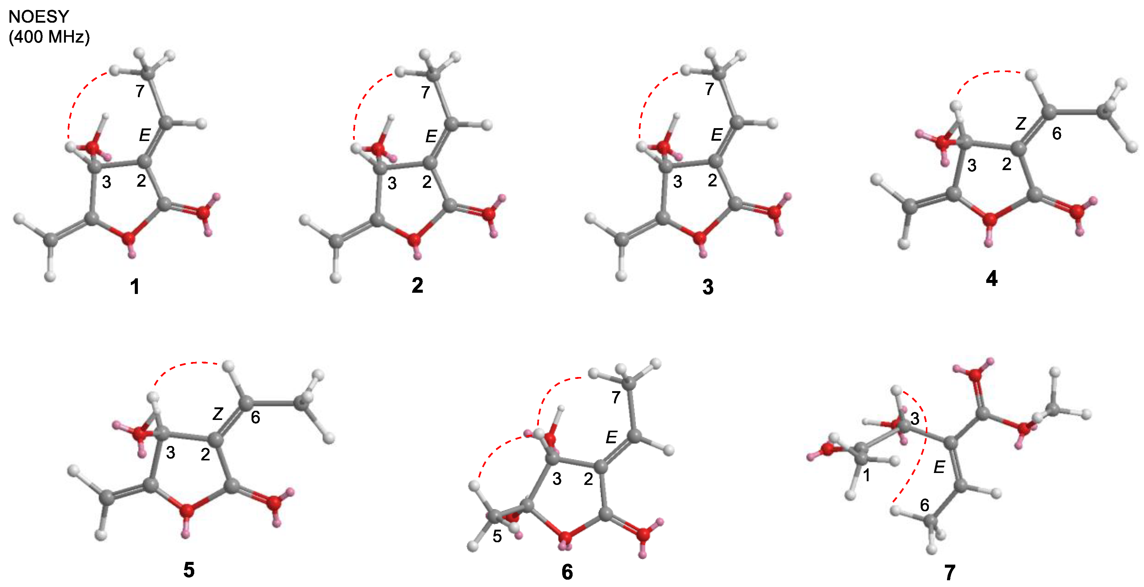
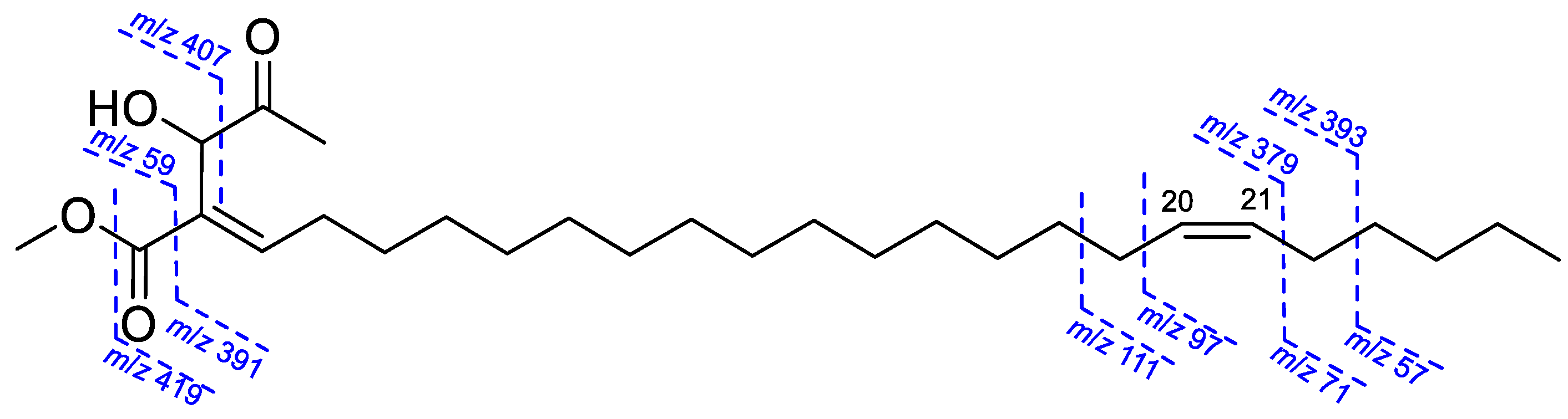
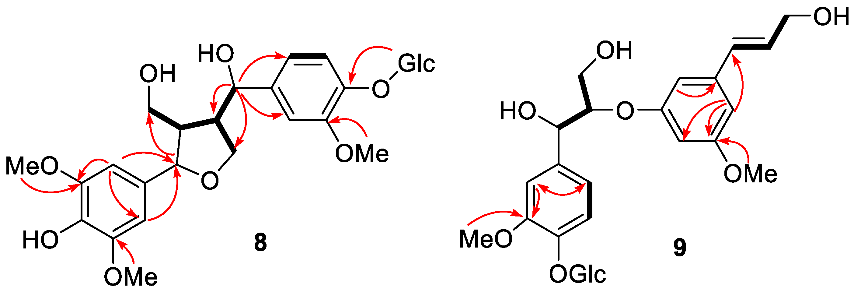
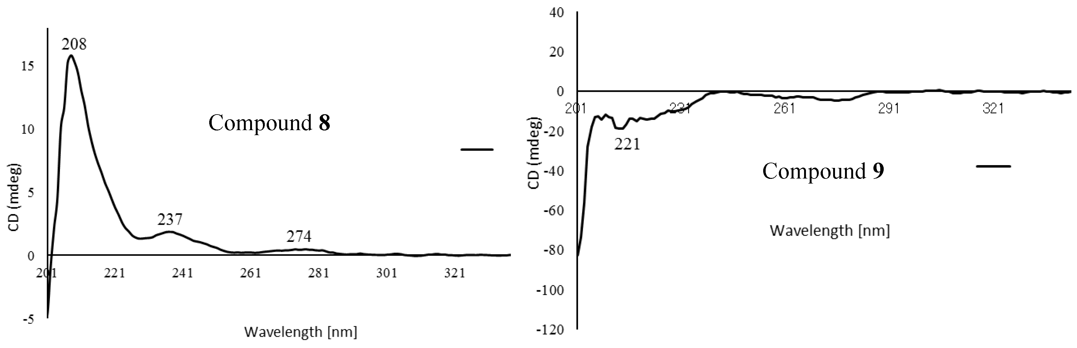
2.1. Structure Elucidation of Isolated Compounds from H. nymphaeifolia

The CH3OH/CH2Cl2 (1:1) extract of H. nymphaeifolia (fruit, N053499) was firstly partitioned with water and n-hexane. The water fraction was further partitioned with EtOAc and n-BuOH. All fractions were subjected to a combination of column chromatography, preparative HPLC, and preparative TLC using silica gel and octadecylsilyl (ODS) to give seven new butanolides, peltanolides A–G (17), and two new lignan glycosides, peltasides A (8) and B (9), as well as eleven known compounds, tambouranolide (10) [15], deoxypodophyllotoxin (11) [16], podorhizol (12) [17], bursehernin (13) [18], (2S,3S)-(+)-5′-methoxyyatein (14) [19], epiashantin (15) [20], epieudesmin (16) [21], (1S,3aR,4R,6aR)-1-(3,4-dimethoxyphenyl)-4-(3′,4′,5′-trimethoxyphenyl)tetrahydro-1H,3H-furo-[3–c]furan (17) [20], (7R,8S)-dehydrodiconiferyl alcohol-4-O-β-d-glucoside (18) [22], alaschanioside A (19) [23], and osmanthuside H (20) [24]. The structures of all known compounds were identified by comparison of their spectroscopic data with reported values.
Compound 1 was obtained as a yellow amorphous solid: [α]25D + 27.3 (c 0.075, CHCl3). It gave a [M]+ peak at m/z 390.3137, appropriate for a molecular formula of C25H42O3. The 1H and 13C NMR spectra (Table 1 and Table 2) contained signals attributed to oxymethine [δH 5.26 (1H, brs); δC 66.5, C-3], methylidene [δH 4.96 (1H, dd, J = 2.8, 1.4 Hz), 4.72 (1H, dd, J = 2.8, 1.4 Hz); δC 91.4, 157.6, C-4,5], vinyl [δH 7.09 (1H, td, J = 7.8, 2.2 Hz); δC 127.3, 150.3, C-2,6], and carbonyl (δC 166.5, C-1) groups, consistent with a β-hydroxy-γ-methylene-α,β-unsaturated-γ-lactone. The chemical shifts of H-6 (δH 7.09) and H-7 (δH 2.48) as well as allylic carbon C-3 (δC 66.5) and olefinic carbon C-6 (δC 150.3) were identical with those of tambouranolide (10) [15] and related linderanolides and isolinderanolides [25,26] with an E-configured double bond [Δ2(6)]. This assignment was also supported by a cross-peak between H-3 and H-7 in the NOESY spectrum (Figure 3). The presence of a long aliphatic chain containing a double bond was suggested by NMR resonances for olefinic and multiple methylene carbons. The allylic (δC 27.0, 27.2) and olefinic (δC 129.8, 129.9) carbon signals in the 13C NMR spectrum of 1 suggested that the internal olefin has the typical Z-configuration, comparable with those of 10 as well as the abovementioned linderanolides and isolinderanolides with Z-double bonds in the side chain. In a related E-isomer, the allylic and olefinic carbons appeared at 32.6 and 25.6 ppm and at 131.9 and 129.3 ppm, respectively [25]. The location of the olefinic bond at Δ20 was based on HMBC and COSY correlations (Figure 2). From the NMR and HREIMS data, compounds 10 and 1 differ only in the number of methylene groups (16 in 10, 14 in 1) in the long aliphatic chain. The absolute configuration of 1 was determined from its optical rotation, which was the same as that of 10. Furthermore, the total synthesis of peumusolide A analogues clearly proved that the optical rotation is positive for 3R compounds and negative for 3S [27,28]. Therefore, compound 1 (peltanolide A) was assigned as (2E,3R)-3-hydroxy-4-methylidene-2-[(15Z)-15-icosenylidene]butanolide.
Compound 2 was isolated as a yellow solid, [α]25D +26.1 (c 0.12, CHCl3). The HREIMS data supported a molecular formula of C29H51O3 from the peak at m/z 446.3743 [M]+. The MS data and NMR spectra indicated four additional methylene units compared with 1, and the optical rotation suggested the same configuration as that of 1. Thus, compound 2 (peltanolide B) was defined as (2E,3R)-3-hydroxy-4-methylidene-2-[(19Z)-19-tetracosenylidene]butanolide.
Compound 3 was obtained as a colorless oil: [α]25D +26.0 (c 0.07, CHCl3). The HREIMS data indicated a molecular formula of C25H44O3 from the peak at m/z 392.3302 [M]+, which was identical to that of miaolinolide [29]. One dimensional NMR spectra of 3 also displayed the similar signal pattern with one exception: the chemical shift of H-6 is δH 7.10 (1H, td, J = 7.8, 2.2 Hz) in 3 and δH 6.70 (1H, td, J = 8.0, 2.0 Hz) in miaolinolide. Thus, the Δ2(6) double bond has an E configuration in 3, rather than the Z configuration in miaolinolide [29]. This assignment was also proved that the chemical shift of H-6 in 3 was close to that of related butanolides with an E configuration of the Δ2(6) double bond [15,25,26,30], including compounds 1 and 2. A NOESY correlation between H-3 and H-7 (Figure 3) supported this conclusion. Based on their optical rotations, compound 3 and miaolinolide have the same absolute configuration. Hence, the structure of 3 (peltanolide C) was established as (2E,3S)-3-hydroxy-4-methylidene-2-icosylidenebutanolide.
HRFABMS of compound 4 showed a molecular formula C27H46O3 with a molecular ion at m/z 441.3357 [M + Na]+. The 1H and 13C NMR spectra of 4 (Table 1 and Table 2) were comparable to those of 10 but suggested different double bond [Δ2(6)] configurations and C-3 stereochemistries. For 4, the Δ2(6) configuration was determined as Z from a NOESY correlation between H-3 and H-6 (Figure 3) and the chemical shift of H-6 at 6.69 ppm rather than ca. 7.10 ppm for the E form. The C-3 stereochemistry was determined as S by comparison of optical rotations, [α]25D −29.7 (c 0.02, CHCl3) for 4 and [α]25D + 18.0 (c 0.03, CHCl3) for 10 with 3R. Thus, compound 4 (peltanolide D) was determined as (2Z,3S)-3-hydroxy-4-methylidene-2-[(17Z)-17-docosenylidene]butanolide.
Compound 5 was obtained as a yellow solid and displayed a peak at m/z 446.3749 [M]+ in the HREIMS spectrum, which agreed with a molecular formula of C29H50O3 and two additional methylene units (C2H4) compared with 4. This finding was also supported by the two NMR spectra. Both compounds also have the same absolute configurations based on their optical rotations, [α]25D −21.1 (c 0.015, CHCl3) for 5 and [α]25D −29.7 (c 0.02, CHCl3) for 4. Thus, compound 5 (peltanolide E) was defined as (2Z,3S)-3-hydroxy-4-methylidene-2-[(15Z)-15-icosenylidene]butanolid.
Compound 6 was isolated as a colorless oil. The HREIMS data indicated a molecular formula of C25H44O4 from the peak at m/z 408.3230 [M]+. Compared with 1, the 1H and 13C NMR spectra of 6 (Table 1 and Table 2) showed the absence of signals for a methylidene group and the presence of signals for a methyl group [δH 1.62 (3H, s)/δC 26.8] and a doubly oxygenated carbon (δC 100.1). The doubly oxygenated carbon was assigned as C-4 with an attached methyl group; these assignments were confirmed by HMBC correlations (Figure 2). A NOESY correlation between H-7 and H-3 as well as the chemical shift of H-6 at 7.04 ppm were consistent with Δ2(6) being the E-isomer. The stereochemistry of C-3 was determined as R based on the optical rotation [α]25D +116.0 (c 0.015, CHCl3) by comparison with related 4-hydroxybutanolides [31,32,33]. The NOESY correlation between H-3 and H-5 supported the 4S stereochemistry (Figure 3). TDDFT-ECD calculation was also sorted the (3R,4S) absolute configuration (Figure 4). Therefore, compound 6 (peltanolide F) was assigned as (2E,3R,4S)-3,4-dihydroxy-5-methyl-2-[(15Z)-15-icosenylidene]butanolide.
Compound 7 has the molecular formula, C28H50O4, based on the peak at m/z 450.3702 [M]+ in the HREIMS. All NMR data and EIMS fragment peaks (Figure 5) of 7 were identical to those of illigerone A [34]. However, the ECD spectrum of 7 exhibited a different Cotton effect from that of illigerone A, and TDDFT-ECD calculation was indicated the 3R absolute configuration (Figure 4). In addition, the experimental optical rotation, [α]25D −78.5 (c 0.03, acetonitrile), of 7 had a negative (levorotary) rather than positive (dextrorotary) value, as found with illigerone A [34]. We concluded that compound 7 (peltanolide G) is (3R,4E,20Z)-3-hydroxy-4-(2-methoxy-2-oxo)hexacosa-4,20-dien-2-one, the enantiomer of illigerone A.
Compound 8 was obtained as a yellow solid, and its molecular formula was determined to be C27H36O13 on the HRFABMS ion at m/z 591.2022 [M + Na]+. The 1H NMR data displayed five aromatic [δH 7.11 (1H, d, J = 8.2 Hz), 6.92 (1H, d, J = 1.8 Hz), 6.82 (1H, dd, J = 8.2, 1.8 Hz), and 6.52 (2H, s, overlap)], two oxymethine [δH 4.63 (1H, d, J = 7.3 Hz), 4.52 (1H, d, J = 8.2 Hz)], four oxymethylene protons [δH 4.26 (1H, dd (J = 9.0, 4.6 Hz), 3.92 (1H, m), 3.87 (1H, m), and 3.63 (1H, m)], three methoxy groups [δH 3.84 (6H, s), and 3.83 (3H, s)], and two methine protons [δH 2.53 (1H, m), 1.89 (1H, m)]. In addition, a glucopyranosyl anomeric proton was observed at δH 4.84 (1H, m). The 13C NMR spectrum showed 27 carbon signals, six from a glucose unit and three methoxy groups and the remaining 18 carbons from the lignan skeleton. The spectroscopic data of 8 resembled those of the known compound, (7S,8R,7′S,8′S)-4,9,7′-trihydroxy-3,3′-dimethoxy-7,9′-epoxylignan-4′-O-β-d-glucopyrano-side [35], except for the absence of the H-3 aromatic proton in the 1H NMR spectrum and the presence of an additional methoxy group in 8. The HMBC (Figure 6) and NOESY (Figure 7) spectra agreed with this structure, and the observed ROESY correlations between H-7/H-9, H-8/H-7′, and H-8′/H-9 (Figure 7) strongly suggested trans configurations of H-7/H-8 and H-8/H-8′. The CD spectrum of 8 showed positive Cotton effects at 237 nm and 274 nm (Figure 8), which were identical with those of the known compound [35]. Thus, the structure of 8 (peltaside A) was determined as (7S,8R,7′S,8′S)-4,9,7′-trihydroxy-3,5,3′-trimethoxy-7,9′-epoxylignan-4′-O-β-d-glucopyranoside.
Compound 9 was obtained as a yellow solid and its HRFABMS (m/z 561.1958 [M + Na]+) indicated the molecular formula C26H34O12. The 1H NMR spectrum of 9 displayed the signals for a trans-olefinic, two oxygenated methine, two oxygenated methylene, and six aromatic protons, as well as a β-glucose and two methoxy groups (Table 3). In addition, its 13C-NMR spectrum showed the signals for 26 carbons, including 12 aromatic, two methoxy, two olefinic, and six glucopyranosyl carbons (Table 3). The COSY, HMQC, HMBC, and NOESY spectra suggested that the glucopyranosyl moiety was attached to C-4 (Figure 5 and Figure 6). The relative configuration between C-7 and C-8 was assigned as erythro based on the small coupling constant (J = 4.8 Hz) in 1H NMR (Figure S57). The absolute configuration of 9, which showed a negative Cotton effect at 221 nm in the CD spectrum (Figure 8), was determined to be 7S,8R via a comparison with that of reported analogues [36,37,38]. Hence, compound 9 (peltaside B) is (7S,8R,7′E)-7,9,9′-trihydroxy-3,5′-dimethoxy-8-3′-oxyneolign-7′-ene-4-O-β-d-glucopyranoside.

2.2. Antiproliferative Activity of Isolated Compounds from H. nymphaeifolia

Compounds 1, 2, 8, 10, and 1318 were evaluated for antiproliferative effects against five human tumor cell lines, A549 (lung carcinoma), MCF-7 (estrogen receptor-positive and HER2-negative breast cancer), MDA-MB-231 (triple negative breast cancer), KB (cervical cancer cell line HeLa derivative), and P-glycoprotein (P-gp)-overexpressing multidrug-resistant (MDR) KB subline, KB-VIN (Table 4). The remaining compounds were not tested due to insufficient quantities. Butanolide 10 slightly inhibited MCF-7 and KB-VIN tumor cell growth with an IC50 value of 9 μM. Both lignans 15 and 16 showed antiproliferative activity against chemosensitive A549 and MCF-7 tumor cell lines, while 16 was also active against MDA-MB-231. Interestingly, compounds 15 and 16 also displayed moderate activity against the MDR cell line (KB-VIN) with an IC50 value of 5 μM but were less active against its parent chemosensitive cell line (KB). Compounds 1, 2, 8, 14 and 18 exhibited no activity against all tested cell lines. These results demonstrated that the CH3OH/CH2Cl2 (1:1) extract of H. nymphaeifolia contained antiproliferative natural products, which showed broad spectrum against HTCLs including MDR cells and could also work synergistically against MDR cells.

3. Materials and Methods

3.1. General Experimental Procedures

Infrared spectra (IR) were obtained with a Thermo Fisher Scientific (Waltham, MA, USA) NICOLET iS5 FT-TR spectrometer from samples in CHCl3 and MeOH. NMR spectra were measured on JEOL (Akishima, Tokyo, Japan) JNM-ECA600 and JNM-ECS400 spectrometers with tetramethylsilane as an internal standard, and chemical shifts are stated as δ values. HRMS data were recorded on a JEOL JMS-700 Mstation (FAB or EI) mass spectrometer. Analytical and preparative TLC were carried out on precoated silica gel 60F254 and RP-18F254 plates (0.25 or 0.50 mm thickness; Merck, Darmstadt, Germany). MPLC was performed on a Combiflash Rf (Teledyne Isco, Lincoln, NE, USA) with silica gel and C18 cartridges (Biotage, Uppsala Sweden). Preparative HPLC was carried out with a GL Science (Shinjuku, Tokyo, Japan) recycling system (PU714 pump and UV702 UV-Vis detector) using an InertSustain C18 column (5 µM, 20 × 250 mm).

3.2. Plant Material

The crude CH3OH/CH2Cl2 (1:1) extract (#N053499) from fruit of H. nymphaeifolia (Presl) Kubitzki (originally identified as H. peltata), collected in Java (Indonesia) was provided by NCI/NIH. The plant was collected on May 25, 1992 in a sandy habitat in the Ujung Kulon Reserve by A. McDonald. A voucher specimen for the plant collection was deposited at the Smithsonian Institution (Washington, WA, USA) and voucher extracts were deposited at the NCI (Frederick, MD, USA) and Kanazawa University (Kanazawa, Ishikawa, Japan).

3.3. Extraction and Isolation

The crude extract N053499 (25.0 g) was dissolved in CH3OH/H2O (9:1) then partitioned with n-hexane, EtOAc, and n-BuOH, yielding n-hexane (17.4 g), EtOAc (3.94 g), n-BuOH (1.72 g), and H2O (0.997 g) fractions. The EtOAc-soluble fraction was subjected to silica gel column chromatography (CC) with a gradient system [n-hexane/EtOAc 100:0 (500 mL)→90:10 (500 mL)→70:30 (1000 mL)→50:50 (1000 mL)→30:70 (1000 mL)→10:90 (1000 mL)→0:100 (500 mL)→EtOAc/MeOH 50:50 (500 mL)→MeOH (1000 mL)] to yield nine fractions, F1–F9. F3 (123 mg) was subjected to silica gel MPLC (RediSep Rf GOLD High Performance 4 g) eluted with n-hexane/EtOAc (9:1 to 0:1) to afford five subfractions 3a–e. Subfraction 3b (21.6 mg) was purified by repeated recycling reversed-phase preparative HPLC with H2O/MeOH (1:19) to provide compounds 2 (2.2 mg), 3 (1.4 mg), and 10 (1.2 mg). F4 (77.9 mg) was subjected to silica gel CC eluted with CH2Cl2 followed by MeOH to yield eight subfractions 4a–h. Subfraction 4d (1.0 mg) was further separated by preparative normal-phase TLC with CH2Cl2 to afford compound 5 (0.4 mg). Subfraction 4f (4.6 mg) was purified by repeated recycling preparative HPLC with H2O/MeOH (1:2) to afford compound 7 (0.6 mg). Subfraction 4h (54.0 mg) was purified by preparative normal-phase TLC with CH2Cl2/EtOAc (19:1) to afford compounds 13 (13.2 mg) and 15 (2.1 mg). F6 was subjected to silica gel CC eluted with CH2Cl2/EtOAc (19:1 to 0:1) followed by MeOH to obtain six subfractions, 6a–f. Subfraction 6b (37.5 mg) was purified by repeated recycling preparative HPLC with H2O/MeOH (1:2) to afford compounds 12 (7.1 mg) and 16 (20.3 mg). Subfraction 6c (15.9 mg) was purified by repeated recycling preparative HPLC with H2O/MeOH (1:2), to provide compound 17 (4.1 mg). The n-hexane fraction (12.0 g) was subjected to silica gel MPLC (RediSep Rf GOLD High Performance 120 g) with a gradient system [n-hexane/CH2Cl2 1:1 (600 mL)→2:3 (1400 mL)→3:7 (1200 mL)→4:1 (1400 mL)→CH2Cl2 (1200 mL)→CH2Cl2/EtOAc 1:1 (1000 mL)→EtOAc (1000 mL)→MeOH (1400 mL)] to yield 15 fractions, F1–F15. F6 (695 mg) was applied to silica gel MPLC (RediSep Rf GOLD High Performance 24 g) eluted with n-hexane/EtOAc (9:1 to 0:1) followed by MeOH to yield ten subfractions 6a–j. Subfraction 6e (197 mg) was subjected to silica gel CC eluted with n-hexane/EtOAc (2:3 to 0:1) followed by MeOH to yield 11 subfractions 6e1–11. Subfraction 6e5 (4.9 mg) was purified by preparative normal-phase TLC with n-hexane/CH2Cl2 (3:1) to afford compound 6 (0.4 mg). F11 (1.12 g) was applied to silica gel MPLC (RediSep Rf GOLD High Performance 24 g) with n-hexane/EtOAc (9:1 to 0:1) followed by MeOH to yield seven subfractions 11a–g. Subfraction 11f (535 mg) was purified by MPLC on ODS-25 (YMC-DispoPack AT 12 g) with H2O/CH3OH (1:3), followed by recycling preparative HPLC with H2O/MeOH (1:2) to afford compounds 13 (0.4 mg) and 14 (0.2 mg). F13 (1.35 g) was subjected to silica gel MPLC (RediSep Rf GOLD High Performance 24 g) with n-hexane/CH2Cl2/EtOAc (1:1:0 to 0:0:1) followed by MeOH to yield ten subfractions 13a–j. Subfraction 13e (48.2 mg) was purified by ODS preparative TLC eluted three times using MeOH to afford compounds 1 (3.2 mg), 2 (1.1 mg), and 10 (1.0 mg). The n-BuOH-soluble fraction (1.72 g) was subjected to silica gel MPLC (RediSep Rf GOLD High Performance 120 g) with a gradient system [CHCl3/MeOH 1:0 (1000 mL)→10:1 (1400 mL)→5:1 (1200 mL)→1:1 (1800 mL)→MeOH (1400 mL)] to yield nine fractions, F1–F9. F1 (147 mg) was subjected to silica gel CC eluted with CH2Cl2/EtOAc (1:0 to 0:1) followed by MeOH to obtain 14 subfractions, 1a–n. Compound 4 (0.3 mg) was obtained from subfraction 1e. Subfraction 1g (3.3 mg) was purified by preparative normal-phase TLC with CH2Cl2/EtOAc (95:5) to afford compound 10 (1.3 mg). Subfraction 1k was purified by recycling preparative HPLC with H2O/MeOH (1:3) to afford compounds 11 (1.8 mg), 14 (0.3 mg), and 17 (0.4 mg). F2 (44.3 mg) was subjected to silica gel CC eluted with CH2Cl2/EtOAc (9:0 to 0:1) followed by MeOH to obtain nine subfractions, 2a–i. Subfraction 2b (1.1 mg) was purified by ODS preparative TLC eluted three times using H2O/MeOH (1:8) to yield compound 13 (0.6 mg). F3 (33.8 mg) was purified by preparative normal-phase TLC with CHCl3/MeOH (9:1) to afford compound 8 (1.0 mg). F5 (149 mg) was subjected to silica gel CC eluted with CH2Cl2/MeOH (10:1 to 1:1) followed by MeOH to obtain seven subfractions, 5a–g. Subfraction 5d (75.6 mg) was purified by MPLC on ODS-25 (YMC-DispoPack AT 12 g) with H2O/MeOH (1:3), followed by recycling preparative HPLC with H2O/MeOH (2:3) to afford compounds 9 (1.3 mg), 18 (2.3 mg), 19 (1.0 mg), and 20 (1.2 mg).

3.3.1. Peltanolide A (1)

Yellow amorphous solid; [α]25D +27.3 (c 0.075, CHCl3); IR νmax (CHCl3) cm−1 2923, 2853, 2017, 1733, 1457, 1278, 1219; 1H and 13C NMR, Table 1 and Table 2; HREIMS m/z 390.3137 [M]+ (calcd for C25H42O3, 390.3134).

3.3.2. Peltanolide B (2)

Yellow amorphous solid; [α]25D +26.1 (c 0.12, CHCl3); IR νmax (CHCl3) cm−1 2923, 2852, 1731, 1464, 1265, 1074; 1H and 13C NMR, Table 1 and Table 2; HREIMS m/z 446.3743 [M]+ (calcd for C29H51O3, 446.3760).

3.3.3. Peltanolide C (3)

Colorless oil; [α]25D +26.0 (c 0.07, CHCl3); IR νmax (CHCl3) cm−1 2916, 2849, 2016, 1750, 1678, 1470, 1278, 1184; 1H and 13C NMR, Table 1 and Table 2; HREIMS m/z 392.3302 [M]+ (calcd for C25H44O3, 392.3290).

3.3.4. Peltanolide D (4)

Yellow amorphous solid; [α]25D −29.7 (c 0.02, CHCl3); IR νmax (CHCl3) cm−1 2923, 2852, 1783, 1733, 1465, 1373, 1287; 1H and 13C NMR, Table 1 and Table 2; HRFABMS m/z 441.3357 [M + Na]+ (calcd for C27H46O3Na, 441.3345).

3.3.5. Peltanolide E (5)

Yellow amorphous solid; [α]25D −21.1 (c 0.015, CHCl3); IR νmax (CHCl3) cm−1 2922, 2852, 2017, 1770, 1731, 1557, 1458, 1375, 1287; 1H and 13C NMR, Table 1 and Table 2; HREIMS m/z 446.3749 [M]+ (calcd for C29H50O3, 446.3760).

3.3.6. Peltanolide F (6)

Colorless oil; [α]25D +116.0 (c 0.015, CHCl3); IR νmax (CHCl3) cm−1 2923, 2852, 1733, 1558, 1540, 1456, 1287; 1H and 13C NMR, Table 1 and Table 2; HREIMS m/z 408.3230 [M]+ (calcd for C25H44O4, 408.3240).

3.3.7. Peltanolide G (7)

Colorless oil; [α]25D −78.5 (c 0.03, acetonitrile); IR νmax (CHCl3) cm-1 2922, 2852, 2016, 1717, 1669, 1558, 1456, 1436; 1H and 13C NMR, Table 1 and Table 2; HREIMS m/z 450.3702 [M]+ (calcd for C28H50O4, 450.3709).

3.3.8. Peltaside A (8)

Yellow solid; [α]25D +5.6 (c 0.055, MeOH); IR νmax (CHCl3) cm−1 3330, 2945, 2833, 1645, 1514, 1450, 1112; 1H and 13C NMR, Table 3; HRFABMS m/z 591.2022 [M + Na]+ (calcd for C27H36O13Na, 591.2054).

3.3.9. Peltaside B (9)

Yellow solid; [α]25D −71.8 (c 0.065, MeOH); IR νmax (CHCl3) cm−1 3386, 3293, 1657, 1511, 1265; 1H and 13C NMR, Table 3; HRFABMS m/z 561.1958 [M + Na]+ (calcd for C26H34O12Na, 561.1948).

3.4. Calculation of ECD Spectra

Preliminary conformational analysis for each compound was carried out by using CONFLEX8 with the MMFF94 force field. The conformers were further optimized in MeCN by density functional theory (DFT) method with the B3LYP functional and 6–31(d) basis set. The ECD spectrum was calculated by the time-dependent DFT (TDDFT) method with the CAM-B3LYP functional and TZVP basis set. The calculation was completed by the use of conformers within 2 kcal/mol predicted in MeCN. The solvent effect was introduced by the conductor-like polarizable continuum model (CPCM). The DFT optimization and TDDFT-ECD calculation were performed using Gaussian09 (Gaussian, Inc., Wallingford, CT, USA). The calculated spectrum was displayed by GaussView 5.0.920 with the peak half-width at half height being 0.333 eV. The Boltzmann-averaged spectrum at 298.15K was calculated using Excel 2016 (Microsoft Co., Redmond, WA, USA). The calculations were re-optimized according to the literature [39].

3.5. Assay for Antiproliferative Activity

Antiproliferative activity of the compounds was determined by the sulforhodamine B (SRB) assay as described previously [40]. Briefly, cell suspensions were seeded on 96-well microtiter plates at a density of 4000–12,000 cells per well and cultured for 72 h with test compound. The cells were fixed in 10% trichloroacetic acid and then stained with 0.04% SRB. The absorbance at 515 nm of 10 mM Tris base-solubilized protein-bound dye was measured using a microplate reader (ELx800, BioTek, Winooski, VT, U.S) operated by Gen5 software (BioTek). IC50 data were calculated statistically (MS Excel) from at least three independent experiments performed with duplication (n = 6). All human tumor cell lines, except KB-VIN, were obtained from the Lineberger Comprehensive Cancer Center (UNC-CH, Chapel Hill, NC, USA) or from ATCC (Manassas, VA, USA). KB-VIN was a generous gift from Professor Y.-C. Cheng of Yale University (New Haven, CT, USA).

4. Conclusions

As part of our continuing investigation of rainforest plants, we conducted a thorough study to identify new chemical compounds to supplement the reported phytochemical research on H. nymphaeifolia. Consequently, a CH3OH/CH2Cl2 (1:1) extract of H. nymphaeifolia (N053499) provided by NCI yielded seven new butanolides, peltanolides A–G (17), and two new lignan glycosides, peltasides A (8) and B (9), as well as eleven known compounds 1020. This is the first report to identify butanolides and lignan glucosides from this genus. The evaluation of antiproliferative activity against human tumor cell lines revealed that lignans 15 and 16 were slightly active against chemosensitive tumor cell lines A549 and MCF-7, respectively. Interestingly, both compounds displayed significant activity with IC50 valued of 5 µM against a P-glycoprotein overexpressing MDR tumor cell line (KB-VIN) although they were less active against its parent chemosensitive cell line (KB).

Supplementary Materials

NMR spectra and HRMS of new compounds are available online at https://www.mdpi.com/1420-3049/24/21/4005/s1.

Author Contributions

S.A., Y.S; investigation, performed the chemical experiments and data collection, S.F.; ECD calculation and analysis, M.G.; investigation, performed cytotoxic experiments using SRB assay, M.G., K.M., D.J.N, B.R.O. K.-H.L.; editing and guidance, K.N.-G; writing, conceptualization, and supervision.

Funding

This study was supported by JSPS KAKENHI Grant Number JP25293024, awarded to K.N.G. This work was also supported partially by NIH grant CA177584 from the National Cancer Institute, awarded to K.H.L., as well as the Eshelman Institute for Innovation, Chapel Hill, North Carolina, awarded to M.G. The content of this publication does not necessarily reflect the views or policies of the Department of Health and Human Services, nor does mention of trade names, commercial products, or organizations imply endorsement by the U.S. Government.

Acknowledgments

We appreciate critical comments, suggestions, and editing on the manuscript by Susan L. Morris-Natschke (UNC-CH).

Conflicts of Interest

The authors declare no conflict of interest.

References

  1. Lakshmi, V.; Pandey, K.; Mishra, S.K.; Srivastava, S.; Mishra, M.; Agarwa, S.K. An overview of family Hernandiaceae. Rec. Nat. Prod. 2009, 3, 1–22. [Google Scholar]
  2. Pettit, G.R.; Meng, Y.H.; Gearing, R.P.; Herald, D.L.; Pettit, R.K.; Doubek, D.L.; Chapuis, J.C.; Tackett, L.P. Antineoplastic Agents. 522. Hernandia peltata (Malaysia) and Hernandia nymphaeifolia (Republic of Maldives). J. Nat. Prod. 2004, 67, 214–220. [Google Scholar] [CrossRef] [PubMed]
  3. Udino, L.; Abaul, J.; Bourgeois, P.; Corrichon, L.; Duran, H.; Zedde, C. Lignans from the seeds of Hernandia sonora. Planta Med. 1999, 65, 279–281. [Google Scholar] [CrossRef] [PubMed]
  4. Chalandre, M.C.; Bruneton, J.; Cabalion, P.; Guinaudeau, H. Hernandiaceae. XII. Aporphine-benzylisoquinoline dimers isolated from Hernandia peltata. Can. J. Chem. 1986, 64, 123–126. [Google Scholar] [CrossRef]
  5. Wei, C.Y.; Wang, S.W.; Ye, J.W.; Hwang, T.L.; Cheng, M.J.; Sung, P.J.; Chang, T.H.; Chen, J.J. New anti-inflammatory aporphine and lignan derivatives from the root wood of Hernandia nymphaeifolia. Molecules 2018, 23, 2286. [Google Scholar] [CrossRef]
  6. Lavault, M.; Cabalion, P.; Bruneton, J. Study on Hernandiaceae. IV. Alkaloids of Hernandia peltata. Planta Med. 1982, 46, 119–121. [Google Scholar] [CrossRef]
  7. Chen, J.J.; Tsai, I.L.; Chen, I.S. New Oxoaporphine Alkaloids from Hernandia nymphaeifolia. J. Nat. Prod. 1996, 59, 156–158. [Google Scholar] [CrossRef]
  8. Chen, J.J.; Ishikawa, T.; Duh, C.Y.; Tsai, I.L.; Chen, I.S. New dimeric aporphine alkaloids and cytotoxic constituents of Hernandia nymphaefolia. Planta Med. 1996, 62, 528–533. [Google Scholar] [CrossRef]
  9. Angerhofer, C.K.; Guinaudeau, H.; Wongpanich, V.; Pezzuto, J.M.; Cordell, G.A. Antiplasmodial and cytotoxic activity of natural bisbenzylisoquinoline alkaloids. J. Nat. Prod. 1999, 62, 59–66. [Google Scholar] [CrossRef]
  10. Rasoanaivo, R.; Urverg, R.; Rafatro, H.; Ramanitrahasimbola, D.; Palazzino, G.; Galeffi, C.; Nicoletti, M. Alkaloids of Hernandia voyronii. Chloroquine-potentiating activity and structure elucidation of herveline D. Planta Med. 1998, 64, 58–62. [Google Scholar] [CrossRef]
  11. Dittmar, A. The effectiveness of Hernandia spp. (Hernandiaceae) in traditional Samoan medicine and according to scientific analyses. J. Ethnopharmacol. 1991, 33, 243–251. [Google Scholar] [CrossRef]
  12. Bruneton, J.; Shamma, M.; Minard, R.D.; Freyer, A.J.; Guinaudeau, H. Novel biogenetic pathways from (+)-reticuline. Three dimeric alkaloids: (+)-vanuatine, (+)-vateamine, and (+)-malekulatine. J. Org. Chem. 1983, 48, 3957–3960. [Google Scholar] [CrossRef]
  13. Chen, I.S.; Chen, J.J.; Duh, C.Y.; Tsai, I.L. Cytotoxic lignans from Formosan Hernandia nymphaeifolia. Phytochemistry 1997, 45, 991–996. [Google Scholar] [CrossRef]
  14. Chen, I.J.; Chang, Y.L.; Teng, C.M.; Chen, I.S. Anti-platelet aggregation alkaloids and lignans from Hernandia nymphaefolia. Planta Med. 2000, 66, 251–256. [Google Scholar] [CrossRef] [PubMed]
  15. Yoder, B.J.; Cao, S.; Norris, A.; Miller, J.S.; Ratovoson, F.; Andriantsiferana, R.; Rasamison, V.E.; Kingston, D.G.I. Tambouranolide, a new cytotoxic hydroxybutanolide from a Tambourissa sp. (Monimiaceae). Nat. Prod. Res. 2007, 21, 37–41. [Google Scholar] [CrossRef] [PubMed]
  16. Muto, N.; Tomokuni, T.; Haramoto, M.; Tatemoto, H.; Nakanishi, T.; Inatomi, Y.; Murata, H.; Inada, A. Isolation of apoptosis- and differentiation-inducing substances toward human promyelocytic leukemia HL-60 cells from leaves of Juniperus taxifolia. Biosci. Biotechnol. Biochem. 2008, 72, 477–484. [Google Scholar] [CrossRef] [PubMed]
  17. Trazzi, G.; André, M.F.; Coelho, F.J. Diastereoselective synthesis of β-piperonyl-γ-butyrolactones from Morita-Baylis-Hillman adducts. Highly efficient synthesis of (±)-yatein, (±)-podorhizol and (±)-epi-podorhizol. Braz. Chem. Soc. 2010, 21, 2327–2339. [Google Scholar] [CrossRef]
  18. Okunishi, T.; Umezawa, T.; Shimada, M. Enantiomeric compositions and biosynthesis of Wikstroemia sikokiana lignans. J. Wood Sci. 2000, 46, 234–242. [Google Scholar] [CrossRef]
  19. Li, N.; Wu, J.L.; Sakai, J.I.; Ando, M. Dibenzylbutyrolactone and Dibenzylbutanediol Lignans from Peperomia duclouxii. J. Nat. Prod. 2003, 66, 1421–1426. [Google Scholar] [CrossRef]
  20. Ahmed, A.A.; Mahmoud, A.A.; Ali, E.T.; Tzakou, O.; Couladis, M.; Mabry, T.J.; Gáti, T.; Tóth, G. Two highly oxygenated eudesmanes and ten lignans from Achillea holosericea. Phytochemistry 2002, 59, 851–856. [Google Scholar] [CrossRef]
  21. Iida, T.; Nakano, M.; Ito, K. Hydroperoxysesquiterpene and lignan constituents of Magnolia kobus. Phytochemistry 1982, 21, 673–675. [Google Scholar] [CrossRef]
  22. Asikin, Y.; Takahashi, M.; Mizu, M.; Takara, K.; Okua, H.; Wada, K. DNA damage protection against free radicals of two antioxidant neolignan glucosides from sugarcane molasses. J. Sci. Food Agric. 2016, 96, 1209–1215. [Google Scholar] [CrossRef] [PubMed]
  23. Calis, I.; Kirmizibekmez, H.; Beutler, J.A.; Donmez, A.A.; Yalc, F.N.; Kilic, I.E.; Ozlap, M.; Ruedi, P.; Tasdemir, D. Secondary metabolites of Phlomis viscosa and their biological activities. Turk. J. Chem. 2005, 29, 71–81. [Google Scholar]
  24. Sugiyama, M.; Kikuchi, M. Phenylethanoid glycosides from Osmanthus asiaticus. Phytochemistry 1993, 32, 1553–1555. [Google Scholar] [CrossRef]
  25. Seki, K.; Sasaki, T.; Wano, S.; Haga, K.; Kaneko, R. Linderanolides and isolinderanolides, ten butanolides from Lindera glauca. Phytochemistry 1995, 40, 1175–1181. [Google Scholar] [CrossRef]
  26. Anderson, J.E.; Ma, W.; Smith, D.L.; Chang, C.J.; Mclaughlin, J.L. Biologically active γ-lactones and methylketoalkenes from Lindera benzoin. J. Nat. Prod. 1992, 55, 71–83. [Google Scholar] [CrossRef]
  27. Tamura, S.; Doke, S.; Murakami, N. Total synthesis of peumusolide A, NES non-antagonistic inhibitor for nuclear export of MEK. Tetrahedron 2010, 66, 8476–8480. [Google Scholar] [CrossRef]
  28. Tamura, S.; Tonokawa, M.; Murakami, N. Stereo-controlled synthesis of analogs of peumusolide A, NES non-antagonistic inhibitor for nuclear export of MEK. Tetrahedron Lett. 2010, 51, 3134–3137. [Google Scholar] [CrossRef]
  29. Tsenga, M.; Su, Y.S.; Cheng, M.J.; Liu, T.W.; Chen, I.S.; Wu, M.D.; Chang, H.S.; Yuan, G.F. Chemical constituents from a soil-derived actinomycete, Actinomadura miaoliensis BCRC 16873, and their inhibitory activities on lipopolysaccharide-induced tumor necrosis factor production. Chem. Biodivers. 2013, 10, 303–312. [Google Scholar] [CrossRef]
  30. Tanaka, H.; Takaya, Y.; Toyoda, J.; Yasuda, T.; Sato, M.; Murata, J.; Murata, H.; Kaburagi, K.; Iida, O.; Sugiyama, K.; et al. Two new butanolides from the roots of Litsea acuminate. Phytochemistry Lett. 2015, 11, 32–36. [Google Scholar] [CrossRef]
  31. Cheng, H.I.; Lin, W.Y.; Duh, C.Y.; Lee, K.H.; Tsai, I.L.; Chen, I.S. New cytotoxic butanolides from Litsea acutivena. J. Nat. Prod. 2001, 64, 1502–1505. [Google Scholar] [CrossRef] [PubMed]
  32. Juan, C.; Martinez, V.; Yoshida, M.; Gottlieb, O.R. The chemistry of Brazilian Lauraceae. Part LXI. ω-Ethyl, ω-ethenyl and ω-ethynyl-α-alkylidene-γ-lactones from Clinostemon mahuba. Phytochemistry 1981, 20, 459–464. [Google Scholar] [CrossRef]
  33. Takeda, K.I.; Sakurawi, K.; Ishi, H. Components of the Lauracea family. I. New lactonic compounds from Litsea japonica. Tetrahedron 1972, 28, 3757–3766. [Google Scholar] [CrossRef]
  34. Li, X.J.; Dong, J.W.; Cai, L.; Wang, J.P.; Yu, N.X.; Ding, Z.T. Illigerones A and B, two new long-chain secobutanolides from Illigera henryi W. W. Sm. Phytochem. Lett. 2017, 19, 181–186. [Google Scholar] [CrossRef]
  35. Yang, Y.N.; Huang, X.Y.; Feng, Z.M.; Jiang, J.S.; Zhang, P.C. Hepatoprotective Activity of Twelve Novel 7′-Hydroxy Lignan Glucosides from Arctii Fructus. J. Agric. Food Chem. 2014, 62, 9095–9102. [Google Scholar] [CrossRef] [PubMed]
  36. Greca, M.D.; Molinaro, A.; Monaco, P.; Previtera, L. Neolignans from Arum italicum. Phytochemistry 1994, 35, 777–779. [Google Scholar] [CrossRef]
  37. Arnoldi, A.; Merlini, L. Asymmetric synthesis of 3-methyl-2-phenyl-1,4-benzodioxanes. Absolute configuration of the neolignans eusiderin and eusiderin C and D. J. Chem. Soc. Perkin Trans. I 1985, 2555–2557. [Google Scholar] [CrossRef]
  38. Gan, M.; Zhang, Y.L.; Lin, S.; Liu, M.T.; Song, W.X.; Zi, J.C.; Yang, Y.C.; Fan, X.N.; Shi, J.G.; Hu, J.F.; et al. Glycosides from the root of Iodes cirrhosa. J. Nat. Prod. 2008, 71, 647–654. [Google Scholar] [CrossRef]
  39. Pascitelli, G.; Bruhn, T. Good Computational Practice in the Assignment of Absolute Configurations by TDDFT Calculations of ECD Spectra. Chirality 2016, 28, 466–474. [Google Scholar] [CrossRef] [Green Version]
  40. Nakagawa-Goto, K.; Oda, A.; Hamel, E.; Ohkoshi, E.; Lee, K.H.; Goto, M. Development of a novel class of tubulin inhibitors from desmosdumotin B with a hydroxylated bicyclic B-ring. J. Med. Chem. 2015, 58, 2378–2389. [Google Scholar] [CrossRef]
Sample Availability: Not available.
Figure 1. Isolated compounds (120) from H. nymphaeifolia.
Molecules 24 04005 g001
Figure 2. Selected HMBC correlations (arrows in red), COSY connectivities (bold lines) for compounds 1−7.
Molecules 24 04005 g002
Figure 3. Key NOESY (red dashed lines) correlations for compounds 17.
Molecules 24 04005 g003
Figure 4. Experimental and calculated ECD spectra of compounds 6 and 7.
Molecules 24 04005 g004
Figure 5. EIMS fragmentation of 7.
Molecules 24 04005 g005
Figure 6. Selected HMBC correlations (arrows in red), COSY connectivities (bold lines) for 8 and 9.
Molecules 24 04005 g006
Figure 7. Key NOESY correlations (red lines) and Key ROESY correlations (blue lines) for 8 and 9.
Molecules 24 04005 g007
Figure 8. Experimental ECD spectra of compounds 8 and 9.
Molecules 24 04005 g008
Table 1. 1H NMR Spectroscopic data of compounds 17.
1a (CDCl3)2b (CDCl3)3b (CDCl3)4b (CDCl3)5b (CDCl3)6a (CDCl3)7b (CDCl3)
PositionδH (J in Hz)δH (J in Hz)δH (J in Hz)δH (J in Hz)δH (J in Hz)δH (J in Hz)δH (J in Hz)
1 2.15 s
35.26 brs5.26 brs5.26 brd (5.6)5.11 m5.10 brd (7.3)4.82 brs4.89 d (4.2)
5a4.72 dd (2.8, 1.4)4.72 dd (2.8, 1.4)4.72 dd (2.8, 1.4)4.67 dd (2.8, 1.4)4.66 dd (2.8, 1.4)1.62 s7.07 t (8.0)
5b4.96 dd (2.8, 1.4)4.96 dd (2.8, 1.4)4.96 dd (2.8, 1.4)4.89 dd (2.8, 1.4)4.89 dd (2.8, 1.4)
67.09 td (7.8, 2.2)7.09 td (7.8, 2.2)7.10 td (7.8, 2.2)6.69 td (7.8, 2.2)6.67 td (7.8, 2.2)7.04 td (7.8, 2.2)2.34 td (14.8, 8.0)
72.48 m2.48 m2.48 m, 2.43 m2.78 m2.76 m2.38 m1.50 m
81.52 m1.52 m1.53 m1.46 m1.45 m1.52 m1.25 mp
9–181.26 mc1.26 me1.25 mg1.25 mh1.25 mj1.25 mm1.25 mp
192.00 m1.26 me1.25 mg1.25 mh1.25 mj2.01 mn2.00 mq
205.36 md1.26 me1.25 mg1.25 mh1.25 mj5.34 t (4.8)o5.34 t (4.8)r
215.36 md1.26 me1.25 mg2.02 m1.25 mj5.34 t (4.8)o5.34 t (4.8)r
222.02 m1.26 me1.25 mg5.35 mi1.25 mj2.01 mn2.00 mq
231.26 mc2.00 m1.25 mg5.35 mi2.01 mk1.25 mm1.25 mp
241.32 m5.36 mf1.25 mg2.025.33 ml1.311.25 mp
250.89 t (6.9)5.36 mf0.89 br t (7.3)1.25 mh5.33 ml0.89 t (6.0)1.31 m
26 2.02 m 1.33 m2.01 mk 0.89 t (6.0)
27 1.26 me 0.88 t (6.9)1.25 mj
28 1.32 m 1.32 m
29 0.89 t (7.3) 0.87 t (6.9)
2-OH 2.26 m
3-OH 4.00 brs
OCH3 3.72 s
a 600 MHz, b 400 MHz, c–r Overlapping signals.
Table 2. 13C NMR Spectroscopic data in CDCl3 of compounds 1−7.
1a2b3b4b5b6a7b
Positionδcδcδcδcδcδcδc
1166.5166.5166.3163.1 166.424.8
2127.3127.3127.2127.4127.3125.2206.3
366.566.566.668.968.970.973.4
4157.6157.6157.5160.1160.2100.1129.8
591.491.491.590.390.426.8149.0
6150.3150.3150.3151.4151.4151.928.7
729.829.829.829.829.830.129.3
828.428.428.4–29.6d28.428.429.5–29.9g29.4–30.0h
9–1829.4–30.029.4–30.0c28.4–29.6d29.4–30.0e29.4–30.0f29.5–29.9g29.4–30.0h
1927.029.4–30.0c28.4–29.6d29.4–30.0e29.4–30.0f27.027.2
20129.829.4–30.0c28.4–29.6d29.4–30.0e29.4–30.0f129.8129.8
21129.929.4–30.0c28.4–29.6d27.029.4–30.0f129.9129.9
2227.229.4–30.0c28.4–29.6d129.829.4–30.0f27.727.2
2332.027.032.0129.927.032.129.8
2422.4129.822.827.2129.822.531.9
2514.0129.914.132.0129.914.122.7
26 27.2 22.327.2 14.1
27 32.0 14.032.0
28 22.4 22.4
29 14.0 14.0
COO 166.5
OCH3 52.0
a 150 MHz, b 100 MHz, c–h Overlapping signals.
Table 3. 1H and 13C NMR Spectroscopic Data of Compounds 8 and 9.
8 (CD3OD)9 (CD3OD)
PositionδCaδH (J in Hz)bδCaδH (J in Hz)b
1134.1 131.5
2104.6c6.52 sd112.37.09 brs
3149.3 147.4
4135.9 150.5
5149.3 120.67.06 d (8.2)
6104.6c6.52 sd121.16.96 dd (2.2, 8.2)
785.14.63 d (7.3)74.94.85 overlap
853.71.89 m85.94.36 m
962.33.87 m 62.23.82 m
3.63 m 3.47 m
1′139.5 137.8
2′112.26.92 d (1.8)118.7g6.86 brsg
3′150.7 149.2
4′147.5 118.7g6.86 brsg
5′117.57.11 d (8.2)151.2
6′120.96.82 dd (8.2, 1.8)111.36.97 d (2.2)
7′74..84.52 d (8.2)132.96.52 d (15.4)
8′50.72.53 m111.36.27 dd (5.7, 15.4)
9′76.24.26 dd (9.0, 4.6)63.84.19 d (5.9)
3.92 m
3-OMe56.93.84 se56.73.81 s
5-OMe56.83.84 se56.53.79 s
3′-OMe56.73.83 s
Glc-1102.74.84 m103.14.81 d (6.9)
Glc-274.93.4–3.8 mf73.93.4–3.8 mh
Glc-378.23.4–3.8 mf78.23.4–3.8 mh
Glc-471.43.4–3.8 mf71.43.4–3.8 mh
Glc-577.93.4–3.8 mf77.93.4–3.8 mh
Glc-662.53.4–3.8 mf62.53.4–3.8 mh
a 100 Hz, b 400 Hz, ch Overlapping signals.
Table 4. Antiproliferative Activity of the Isolated Compounds.
CompoundsCell Linesa (IC50 μM)b
A549MDA-MB-231MCF-7KBKB-VIN
1>4022.5>4025.731.7
221.924.624.622.621.4
835.135.335.732.721.7
1012.510.88.818.68.8
1332.837.733.5>408.7
1423.232.932.823.219.9
158.120.86.820.35.4
165.78.28.112.65.3
1737.8>4038.0>408.2
18>40>40>40>40>40
Paclitaxel (nM)6.58.412.17.12213
a A549 (lung carcinoma), MDA-MB-231 (triple-negative breast cancer), MCF-7 (estrogen receptor-positive & HER2-negative breast cancer), KB (cervical cancer cell line HeLa derivative), KB-VIN (P-gp-overexpressing multidrug-resistant (MDR) subline of KB). b Antiproliferative activity expressed as IC50 values for each cell line cultured with compound for 72 h, the concentration of compound that caused 50% reduction relative to untreated cells determined by the SRB assay. IC50 of all compounds were calculated.

