Article

Photon-Induced Superior Antibacterial Activity of Palladium-Decorated, Magnetically Separable Fe3O4/Pd/mpg-C3N4 Nanocomposites

Department of Nano-Physics, Gachon University, 1342 Seongnamdaero, Sujeong-gu, Seongnam-si, Gyeonggi-do 13120, Korea
*
Author to whom correspondence should be addressed.
Molecules 2019, 24(21), 3888; https://doi.org/10.3390/molecules24213888
Submission received: 18 September 2019 / Revised: 19 October 2019 / Accepted: 24 October 2019 / Published: 29 October 2019
(This article belongs to the Section Photochemistry)

Abstract

:
Three-component nanocomposites (Fe3O4/Pd/mpg-C3N4) have been systematically synthesized using a three-step solution method for the photocatalytic bacterial decontamination. The mesoporous g-C3N4 nanosheets (mpg-C3N4), which were prepared by the acid treatment, showed a great improvement in photocatalytic performance. The photoluminescence intensity of the mpg-C3N4 nanosheets was disclosed to drop about 60% from the value of normal g-C3N4 nanosheets. Decoration of mpg-C3N4 with palladium (Pd) nanoparticles led to the effective suppression of carrier recombination and the carrier migration to Fe3O4 nanoparticles. It was revealed that the three-component nanocomposites degraded 99.9% of E. coli and 99.8% of S. aureus bacterial strains within 2 h of solar light irradiation at a 100 μg/mL concentration, demonstrating their superb photocatalytic antibacterial activity. In addition, the nanocomposites could be easily separated from the bacterial cells and repeatedly used for photocatalytic bacterial degradation with good recyclability. The strong photon-induced antibacterial activity and good recyclability of the three-component nanocomposites may represent their potential as a promising antibacterial photocatalyst.

Graphical Abstract

1. Introduction

Environmental protection and wastewater treatment using photocatalysts have been investigated in depth. Particularly, graphitic carbon nitride (g-C3N4) and its nanocomposites have been adopted for sensing and removal of various toxic pollutants in air and wastewater [1,2]. Despite this advancement, however, there are limited number of photocatalysts that can be applied to inactivate the microbial contaminants by solar light illumination. It is known that the performance of photocatalysts is greatly influenced by their surface area [3], separation of photo-induced charge carriers [4], and exposed reactive sites on the surface [5]. These situations signal the need for a new generation of photocatalysts possessing both highly efficient photoactivity and superior antibacterial activity. To this aim, we have synthesized three-component nanocomposites that can efficiently interact with microbial cells and weaken the cell activity. There have been reported various g-C3N4-based photocatalysts employing nanoscale heterojunctions with other noble metals such as Au, Ag, Pt, and Pd with differing morphologies [6,7,8,9]. Such noble metals were revealed to increase the lifespan of photogenerated carriers by acting as an electron sink, thus reducing the carrier recombination rate. Among all noble metals, palladium (Pd) is the most attractive, owing to its pronounced synergy effect with g-C3N4 for enhanced photocatalytic performance [10]. Although those hybrid nanostructures have been commercially utilized, they are not adequate for photokilling of bacterial contaminants, partly due to the limited surface area of the photocatalysts [11]. For this reason, pure g-C3N4 nanosheets with relatively small surface area and wide band gap may not be an ideal framework for hybridization with noble metals. Morphology-modifying techniques like etching can be utilized to synthesize mesoporous g-C3N4 nanosheets (mpg-C3N4), thereby increasing total surface area [12,13]. Furthermore, incorporating Fe3O4 nanoparticles (Fe3O4 NPs) as the third structural component, which are magnetically recyclable after use, can allow another positive effects. The Fe3O4 NPs can play as a supplementary sink for electrons that are migrated from Pd NPs or g-C3N4 nanosheets. Prior reports on Fe3O4 NPs-decorated nanocomposites such as g-C3N4/Fe3O4/TiO2 nanocomposites [14], Fe3O4/g-C3N4 nanocomposites [15], and magnetically recyclable ZnO/Fe3O4/g-C3N4 photocatalysts [16] demonstrated their photocatalytic performance for removal of toxic dyes and pollutants. However, they have never been explored for removal of microbial contaminants, and their synthesis methods were complex.
In this work, we synthesized Fe3O4/Pd/mpg-C3N4 three-component nanocomposites by a continuous three-step solution method. The nanocomposites were revealed to have strong photoinduced antibacterial activity and magnetic separation capability of microbial contaminants. The as-synthesized nanocomposites showed superior photokilling performance of bacterial strains, proving their potential as a new generation of photocatalysts with low cost, high efficiency, high recovery, and superior antibacterial activity.

2. Results and Discussion

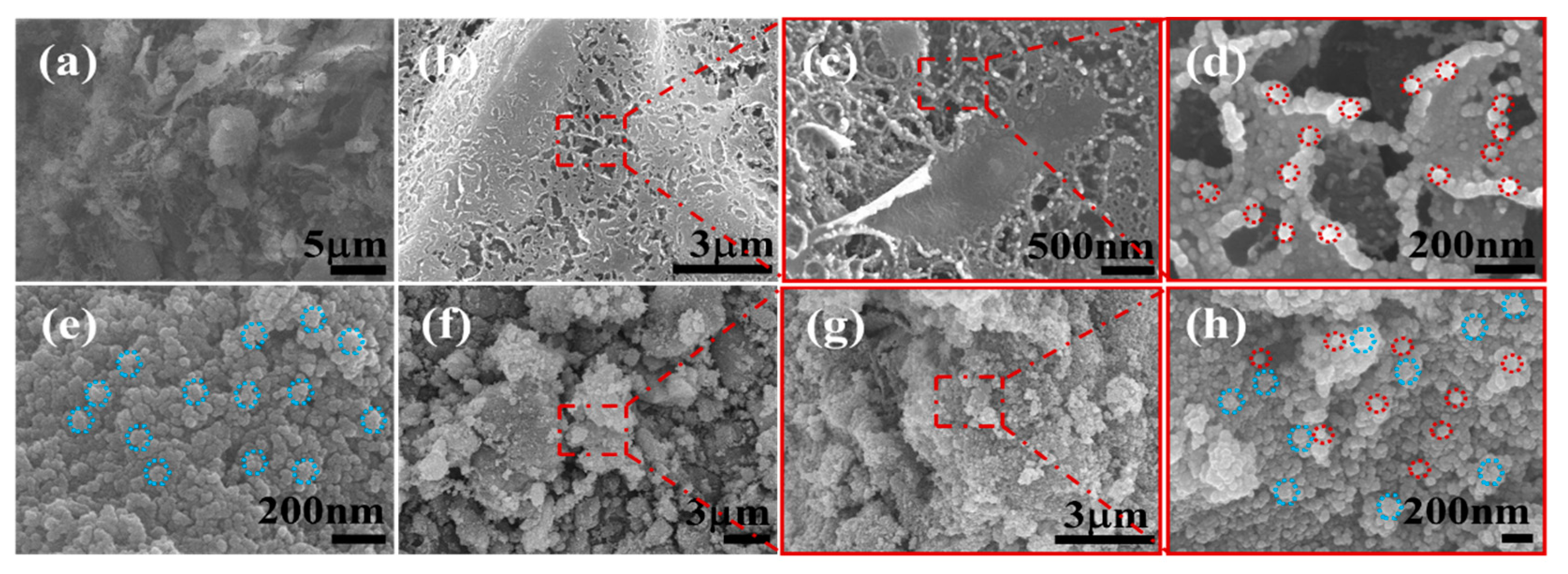
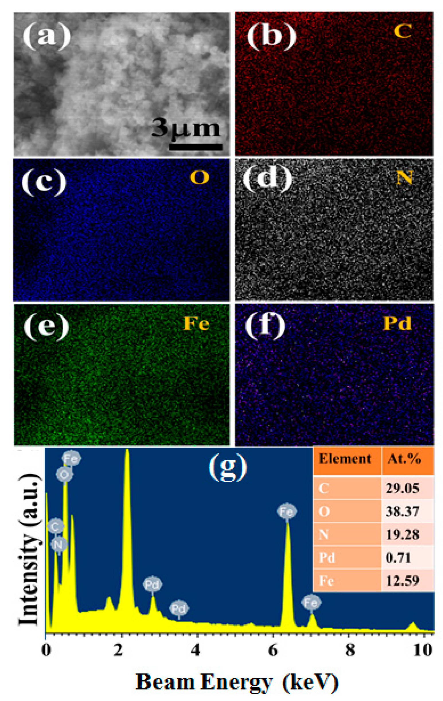
2.1. Morphological Characterization of Nanocomposites

The SEM images of mpg-C3N4, Fe3O4 NPs, Pd NPs/mpg-C3N4 hybrid, Fe3O4 NPs/mpg-C3N4 hybrid, and Fe3O4 NPs/Pd NPs/mpg-C3N4 nanocomposite are shown in Figure 1. Here, the Fe3O4 NPs/Pd NPs/mpg-C3N4 nanocomposite was synthesized by a sequential three-step solution method (see Section 3.2 for more details). At the first step, normal g-C3N4 nanosheets were transformed to mpg-C3N4 nanosheets by chemical oxidation with K2CrO7 solution in H2SO4. At the second step, Pd NPs were decorated over the mpg-C3N4 nanosheets using a simple polyol method. Finally, magnetic Fe3O4 NPs covered the Pd NPs/mpg-C3N4 hybrid powder by a co-precipitation method. In Figure 1a, the mpg-C3N4 nanosheets obtained from the acid etching look like flakes with high porosity. These porous g-C3N4 nanosheets are known to have increased hydrophilicity and large surface area [12]. The increase in hydrophilicity becomes possible by adding hydrophilic functional groups such as –COOH, –OH, and –C=O to the open ends of bulk g-C3N4 during etching. It was reported that the enhanced specific surface area of mesoporous g-C3N4 could improve its photocatalytic activity [17]. However, the wide band gap and fast carrier recombination limits the possible use of mpg-C3N4 nanosheets as novel photocatalysts. Hence, Pd NPs were introduced as a secondary component to retard the carrier recombination. It is found from Figure 1b that the mpg-C3N4 nanosheets are well decorated with Pd NPs to form Pd NPs/mpg-C3N4 hybrid nanostructure. From the magnified images in Figure 1c,d, it is confirmed that the small and monosized Pd NPs are coated like a porous film keeping the high porosity of mpg-C3N4. Although the photocatalytic performance of Pd NPs/mpg-C3N4 hybrid nanostructure may be improved, its recovery characteristics are poor, limiting its practical applications. Thus, we incorporated magnetic nanoparticles (Fe3O4 NPs) as the third component. To compare with Pd NPs, the morphology of pure Fe3O4 NPs is provided in Figure 1e. This image reveals that Fe3O4 NPs are more rugged and more agglomerated than Pd NPs. The Fe3O4 NPs are nicely decorated on mpg-C3N4, as can be seen in Figure 1f. Finally, Figure 1g,h shows that both Pd NPs and Fe3O4 NPs are evenly distributed over the mpg-C3N4 nanosheets in a Fe3O4/Pd/mpg-C3N4 nanocomposite sample, maintaining the high porosity. To the best of our knowledge, the material design and realization of the Fe3O4/Pd/mpg-C3N4 nanocomposite is the first demonstration.
To further assess the elemental distribution and composition of the nanocomposite, SEM-EDX analysis was performed and its result is shown in Figure 2. From the comparison of real SEM image (Figure 2a) with EDX element maps (Figure 2b–f), it is noted that all the elements are uniformly distributed throughout the nanocomposite sample and no elements other than constituents are observed. This result manifests the high compositional purity and homogeniety of Fe3O4/Pd/mpg-C3N4 three-component nanocomposites. Interestingly, the content of Pd NPs is estimated to be only 0.71% for this sample (see the SEM-EDX spectrum in Figure 2g).

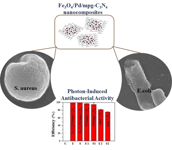
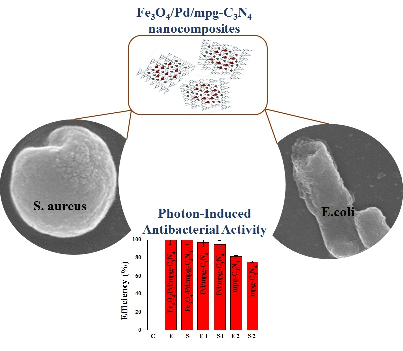
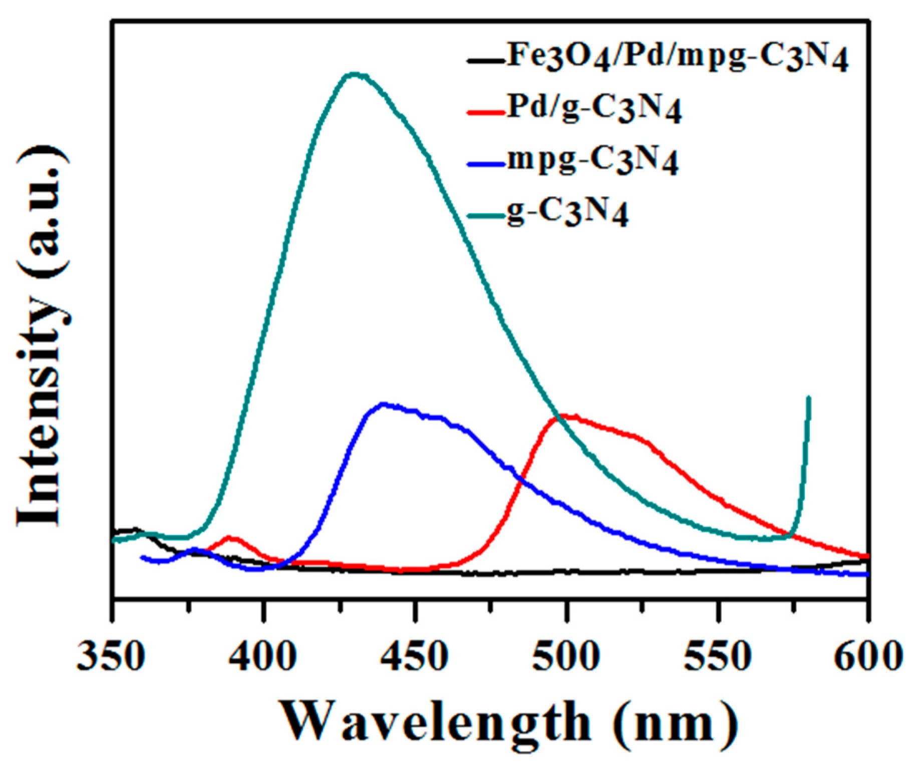
2.2. Carrier Separation and Bacterial Degradation

Suppression of carrier recombination is critically required to enhance the photocatalytic activity of a photocatalyst. As a technique to estimate the carrier separation and recombination rate, photoluminescence (PL) measurements were conducted with excitation wavelength at 320 nm. Figure 3 exhibits PL spectra of normal g-C3N4, mpg-C3N4, Pd/mpg-C3N4 hybrid, and Fe3O4/Pd/mpg-C3N4 nanocomposite. The emission peaks of the samples are positioned in the wavelength range of 350–600 nm. The normal g-C3N4 shows a very strong emission peak centered at 430 nm. The intense PL peak indicates that photogenerated charge carriers massively recombine to disappear in this material. From the peak position, the band gap of normal g-C3N4 is calculated at 2.88 eV, which is close to its ideal value (2.7 eV). This moderately large band gap is known to lower its light-absorbing capability [18]. It is noteworthy that the PL intensity of mpg-C3N4 nanosheets is about 60% lower as compared to normal C3N4, implicating that the high porosity and increased surface area facilitate charge separation. This may be realized because more electrons of photogenerated charge pairs can easily move to larger carrier-depleted surface area. Moreover, a slight red shift of peak position from 430 nm to 440 nm is observed. When the mpg-C3N4 nanosheets are decorated with Pd NPs, the PL intensity decreases more by ~5% and the red shift becomes noticeable. The additional intensity drop is attributed to electron transfer to Pd NPs and the red shift of the peak position may originate from effective band gap reduction by Pd band formation in mpg-C3N4 band. Surprisingly, the PL peak intensity seems negligible in Fe3O4/Pd/mpg-C3N4 nanocomposite. This substantial intensity reduction suggests that photogenerated electrons in mpg-C3N4 nanosheets transfer ultimately to Fe3O4 NPs via Pd NPs and this electron transfer chain works efficiently.
Photodegradation tests were performed on two bacterial strains (E. coli and S. aureus) for treatment of bacteria-contaminated water. For the tests, the bacteria were diluted to 1 × 10⁸ colony forming unit per milliliter (CFU/mL), and different concentrations of photocatalysts were added and irradiated by solar light (refer to Section 3.3). Figure 4 shows the photon-induced bacterial degradation efficiency of mpg-C3N4, Pd/mpg-C3N4 hybrids, and Fe3O4/Pd/mpg-C3N4 nanocomposites at different concentrations (25, 50, 75, and 100 μg/mL), employing the standard plate count method. The data were collected after 2 h of irradiation and a control sample was also tested by keeping the sample at room temperature in the dark environment. The bacterial degradation efficiency of E. coli and S. aureus was calculated by counting the number of colonies using the formula given below:
Degradation efficiency (%) = (NcNt)/Nc × 100
where Nc is the colony count of control sample and Nt is the colony count of test sample with E. coli or S. aureus. In the dark, the bacterial degradation efficiency was estimated below 10% for both E. Coli and S. aureus. In Figure 4a, the degradation efficiencies of three photocatalysts for E. coli are presented as a function of photocatalyst concentration. It is found that the degradation efficiency monotonically increases with increasing the concentration. However, a clear difference is observed in the efficiencies of different photocatalysts at the respective concentrations. The difference is more pronounced at lower concentrations. For instance, at a concetration of 25 μg/mL, the degradation efficiency was estimated at 84.1%, 62.2%, and 41.3% in the order of Fe3O4/Pd/mpg-C3N4 nanocomposites, Pd/mpg-C3N4 hybrids, and mpg-C3N4 nanosheets. When the conentration was increased to 50 μg/mL, the degradation efficiency was improved to 93.8%, 74.3%, and 47.3%. The rather large difference in efficiency (~20%) between Fe3O4/Pd/mpg-C3N4 nanocomposites and Pd/mpg-C3N4 hybrids indicates the importance of magnetic separation of bacterial species at relatively low photocatalyst concentrations. Finally, the degradation efficiency of Fe3O4/Pd/mpg-C3N4 nanocomposites, Pd/mpg-C3N4 hybrids, and mpg-C3N4 nanosheets reached 99.9%, 97%, and 81.7% at a 100 μg/mL concentration. Compared with a previous report, in which the photokilling efficiency of pure Fe3O4 NPs for E. coli at the same concentration of 100 μg/mL was 65.6% after 2 h of solar light illumination [19], this degradation efficiency of the nanocomposites represents remarkable progress. Similar trend can be found for S. aureus, as shown in Figure 4b. The Fe3O4/Pd/mpg-C3N4 nanocomposites, Pd/mpg-C3N4 hybrids, and mpg-C3N4 nanosheets exhibited the bacterial degradation efficiency of 99.8%, 94.9%, and 75.8% at a 100 μg/mL concentration. The S. aureus cell membrane is thick and its composition also varies depending on the cell wall thickness. For this reason, the comparative efficiency for S. aureus is lower than for E. coli at every concentration. At lower concentration of 25 μg/mL of catalysts, the degradation efficiency was estimated at 82.7%, 60.9%, and 39.8%, which is slightly lower than the efficiency for E. coli. However, the relatively large efficiency difference (~20%) between Fe3O4/Pd/mpg-C3N4 nanocomposites and Pd/mpg-C3N4 hybrids is also found at lower concentrations for this case.

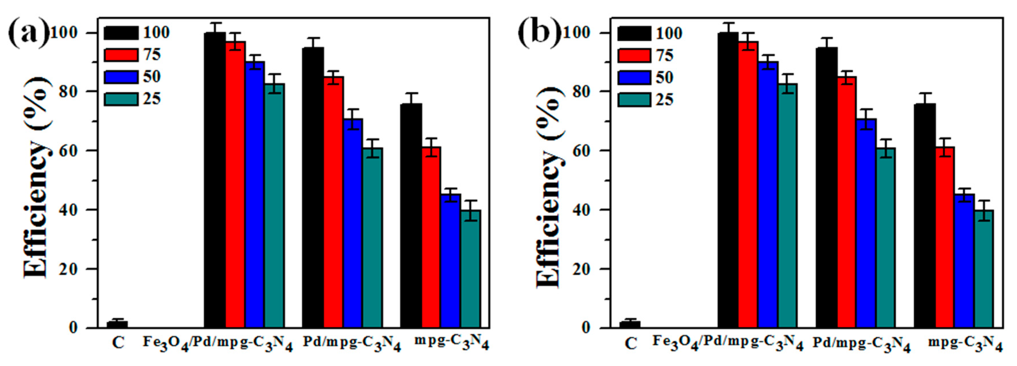
2.3. Photocatalytic Cell Destruction Analysis

The morphological disintegration mechanism of bacteria induced by photocatalysis of Fe3O4/Pd/mpg-C3N4 nanocomposites was also studied by SEM. The concentration of the nanocomposites was fixed at 100 μg/mL and solar light was illuminated over a time span of 2 h. The illumination time-dependent cell images are shown in Figure 5a–h for E. coli and in Figure 5i–p for S. aureus. It is observed that the two bacteria undergo different disintegration mechanisms. The bacterial cell wall is made of lipopolysaccharides, peptidoglycan multilayer, and phospholipid layer, and the wall of E. coli cell is thinner (15–20 nm) than S. aureus (20–80 nm) cell [20,21]. E. coli cell has smooth membrane and rod-shaped morphology before irradiation, as shown in Figure 5a. After 15 min of illumination, the cell shows slight deformation due to the formation and interaction of active species including ·O2, h+, and H2O2 (Figure 5b). In 30 min, the cell deformation progresses due to the attachment of nanocomposites and attack of more reactive species generated by photocatalysis (Figure 5c). After 45 min of exposure, the E. coli cell is mostly covered with nanocomposites and surface interaction goes further (Figure 5d). In 60 min, the cell membrane appears highly damaged, which is responsible for cell death due to the leakage of intracellular materials (Figure 5e). After 75 min of illumination, the ruptured cell membrane provides entrance to the reactive species, which leads to more damage (Figure 5f). From the inset of Figure 5f, a clear hole in the cell wall is found, through which cell materials leak out. The cell is decomposed into its components in 90 min (Figure 5g) and entirely consumed in 120 min (Figure 5h). There has been a report that Fe3O4/TiO2 hybrid nanosheets showed antibacterial activity by solar light irradiation [21]. Considering its result that 4 h of exposure was necessary to completely distort E. coli, our complete disintegration time less than 2 h is noticeable.
S. aureus bacteria show no photon-induced serious damage in morphology, as shown in Figure 5i–p. The S. aureus cell shape is round and its diameter is smaller than E. coli. At the beginning (Figure 5i), the cells appear clustered with smooth surface, until the nanocomposites start to interact with the cell surface in 30 min (Figure 5j,k). In Figure 5l,m, curved areas that are pointed by red arrows are found, which may result from the attack of reactive species. The cell damage is not apparent because the nanocomposite has difficulty penetrating the thick cell wall of S. aureus. However, the cell cluster appears to be fully dumped into the nanocomposites in 60 min (Figure 5n). Some decomposition of cellular components is noticed in further light exposure, which might be caused by nanocomposite-induced stress to the cell wall (Figure 5o,p).

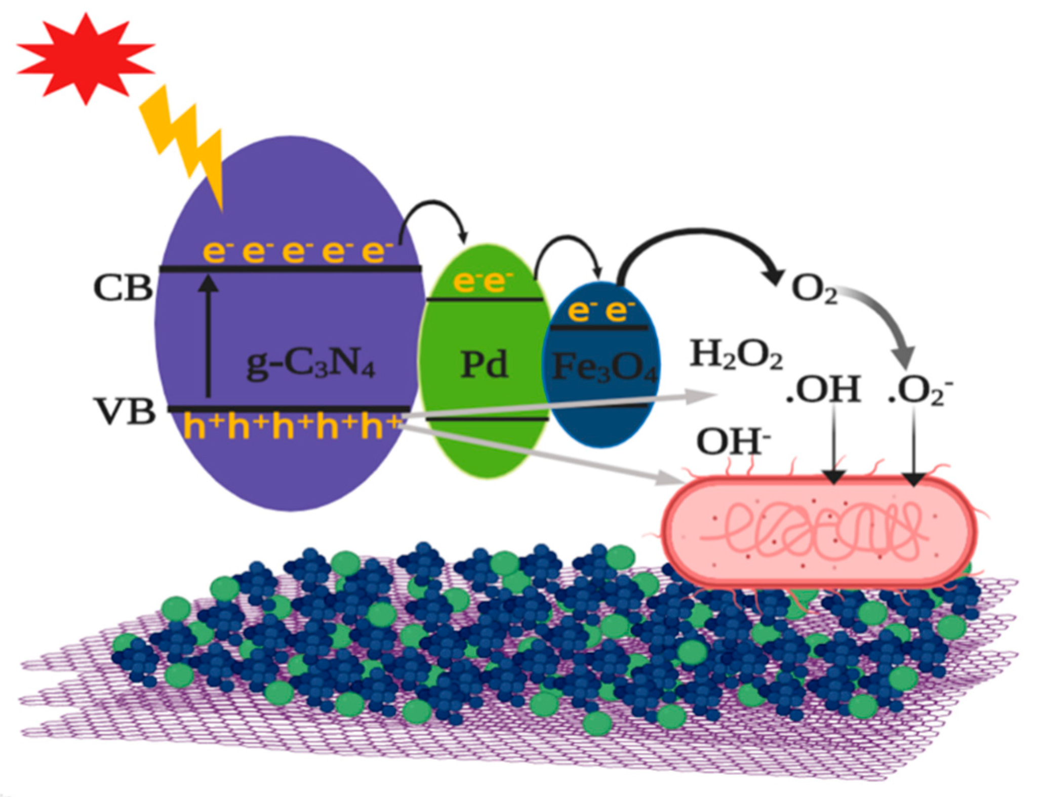
2.4. Mechanism of Photocatalytic Antibacterial Activity

A schematic illustration of photocatalytic antibacterial mechanism of three-component nanocomposites is presented in Figure 6. When solar light is irradiated, a lot of electron–hole pairs are generated in mpg-C3N4 nanosheets [22]. Immediately, the photogenerated electrons are transferred to Pd NPs, which facilitate charge separation and also act as stepping stones for further electron transfer. Most of electrons coming into the Pd NPs again move to the conduction band of Fe3O4 NPs [23]. For this reason, the conduction band of Fe3O4 NPs would be a main supplier of energetic electrons. On the other hand, photogenerated holes are left behind in the valence band of mpg-C3N4, due to the energy barrier at interface with Pd NPs. Through these processes, photogenerated charge carriers are well separated, leading to the effective suppression of carrier recombination. Those separated electrons and holes interact with some reactive species such as O2, H2O2, H2O, and OH. As a result, superoxide anion radicals (·O2) and hydroxyl radicals (·OH), which have hyper-reactivity and strong oxidation capability, are formed and they attack bacteria. Consequently, the oxidative stress at bacterial surface increases and cell materials are damaged, leading to the cell wall disintegration and malfunction, and ultimately to cell death. Through these correlated mechanisms, the three-component nanocomposites can show enhanced antibacterial activity.

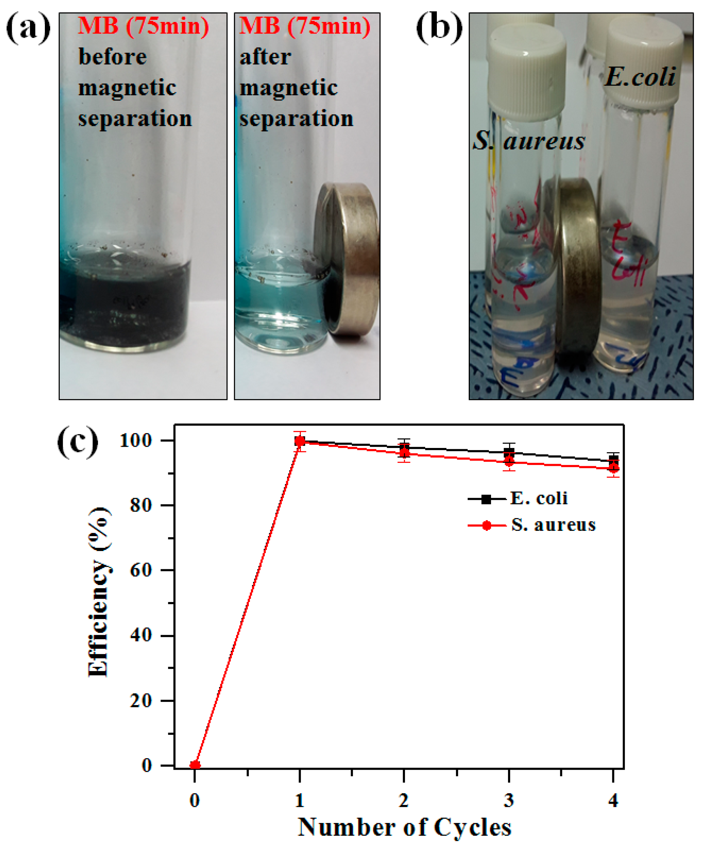
2.5. Magnetic Separation and Recyclability of Photocatalysts

The recovery characteristics and re-usability of photocatalysts were also examined. Due to the strong magnetic property of Fe3O4 NPs, the Fe3O4/Pd/mpg-C3N4 nanocomposites could be easily collected after use, as demonstrated in Figure 7a,b. Figure 7a compares photo images taken before and after magnetic separation of the nanocomposites dispersed in methylene blue solution (10 μM). The images were obtained after irradiating solar light for 75 min. The near-transparent color of the solution confirms that the nanocomposites were completely separated from the solution. The similar and complete magnetic separation of the nanocomposites can also be noticed from bacterial cells, as shown in Figure 7b. To check the recyclability of the nanocomposites, cyclic tests were performed, in which each cycle consisted of photocatalytic bacterial degradation and post-collection of the used nanocomposites. For the tests, the Fe3O4/Pd/mpg-C3N4 nanocomposites were repeatedly used while bacterial suspension was refreshed in each cycle. The nanocomposites recovered after each cycle were first sonicated in 75% ethanol solution to get rid of bound bacterial cells, then washed in sterile 0.85% (wt/vol) saline solution for re-use. Figure 7c displays the cyclic degradation efficiency of the nanocomposites for E. coli and S. aureus. Here, the bacterial cells containing the nanocomposites were illuminated with solar light for 2 h in each cycle. For both bacteria, the change in the degradation efficiency appears to be small. For example, the efficiency drops only by 6% for E. coli after four cycles. This small efficiency change along with the quick and easy magnetic separation of the nanocomposites may ensure good recyclability.

3. Materials and Methods

3.1. Chemicals and Reagents

Iron(III) chloride hexahydrate (FeCl3·6H2O), iron(II) chloride tetrahydrate (FeCl4·4H2O), polyvinylpyrrolidone (PVP), melamine (C6H6N6), ethylene glycol (EG, C2H6O2), sodium tetrachloropalladate (Na2PdCl4), and ammonia (NH3) were all supplied from Sigma Aldrich, Ethyl alcohol (C2H5OH) and acetone were purchased from Daejung Chem. All the chemicals were used as purchased without further treatment.

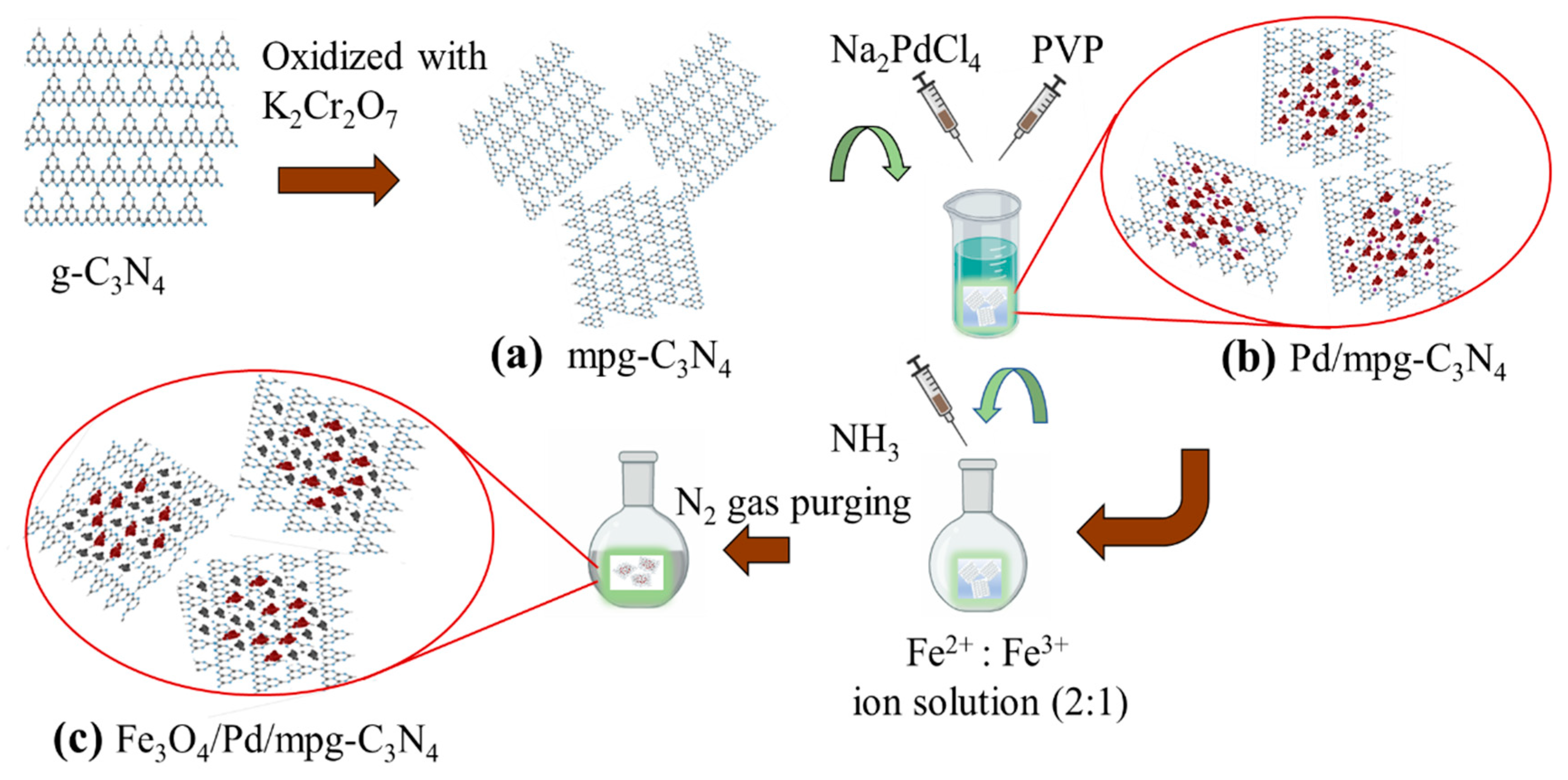
3.2. Nanocomposite Preparation

The nanocomposites were synthesized using an easy, reliable, and continuous three-step solution method. As presented in Figure 8, the first step of nanocomposite preparation was to synthesize mpg-C3N4 (see Figure 8a). For this, normal g-C3N4 nanosheets were first prepared by heating melamine at 550 °C for 2 h and ground into fine powder. Then, 500 mg of this powder was chemically oxidized with 8 g of K2CrO7 solution in H2SO4 for 2 h at room temperature under continuous stirring. Next, the solution was diluted with deionized (DI) water and residual acids were removed through washing, centrifuging, and drying at 80 °C. At the second step, Pd NPs were decorated to make heterojunctions with mpg-C3N4 nanosheets by a simple polyol method [24], as shown in Figure 8b. In detail, 10 mL of EG was stabilized at 161 °C for 2 h and then 50 mg of mpg-C3N4 was added into EG. Subsequently, 0.432 g of Na2PdCl4 and 0.0832 g of PVP solution in 2 ml of EG was added dropwise over 30 min of time period. The hybrid nanostructure powder was obtained after washing with acetone and drying at 70 °C. At the last step, magnetic Fe3O4 NPs were decorated on this powder using co-precipitation method in inert gas atmosphere (see Figure 8c), following the previously reported procedure [25]. In order to make Fe3O4/Pd/mpg-C3N4, FeCl2·4H2O and FeCl3·6H2O (molar ratio = 2:1) were dissolved into 100 mL of DI water and heated at 90 °C for 1 h under nitrogen gas flow. Then, 10 mL of 25% ammonia solution was added slowly. Finally, the black product was magnetically separated, washed, and dried to obtain nanocomposite powder.

3.3. Antibacterial Activity

The photon-induced antibacterial activity of the three-component nanocomposites at different concentrations (25, 50, 75, and 100 μg/mL) was evaluated by testing two bacterial strains (E. coli and S. aureus). The bacteria were precultured in LB broth at 37 °C for 15 h and then resuspended in 0.85% saline solution to further dilute to 1 × 10⁸ CFU/mL. Here, varying concentrations of photocatalysts in 0.85% saline solution were tested for 3 mL of bacterial suspension and they were agitated while irradiated by 280 W xenon lamp for 2 h. Simultaneously, another test was also performed in dark circumstance to compare the effect of photocatalysts without light illumination. After this, the photocatalysts were magnetically separated from irradiated suspension simply by placing a magnet nearby for about 10 min. Then, the solution was carefully taken out to investigate bacterial viability after incubating the agar plates at 37 °C for 24 h. The viable colonies were counted by visual inspection, following the standard plate count method.

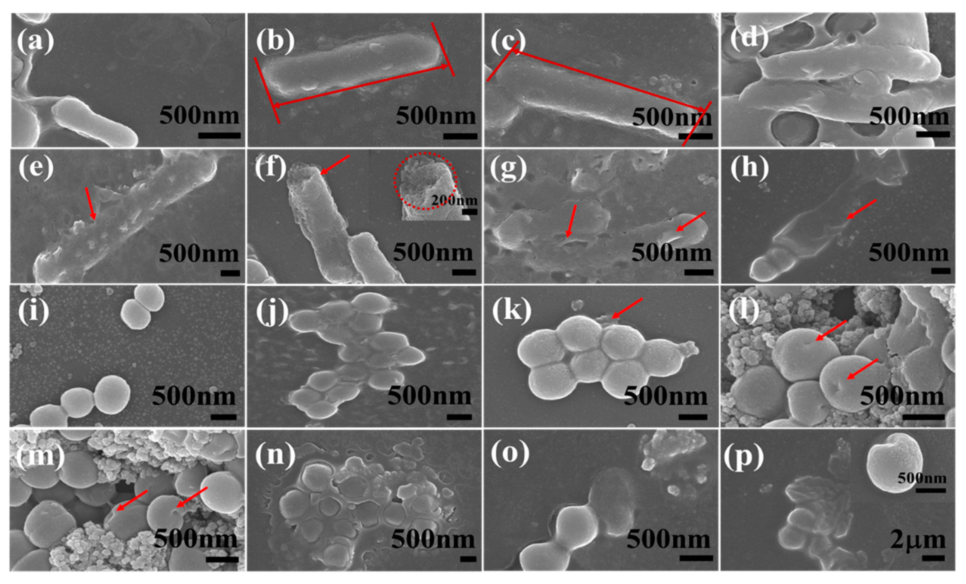
3.4. SEM Observation of Bacterial Morphological Changes

The solar light-irradiated bacterial solution was evaluated for the morphological change caused by photocatalysis with Fe3O4/Pd/mpg-C3N4 nanocomposites (100 μg/mL) to better understand photon-induced damage of the bacterial cells. For SEM observation, the bacterial sample was first fixed on the silica glass using 4% of formaldehyde and 2.5% of glutaraldehyde. Then, it was washed by dehydrating the samples into ethanol with the purity of 20%, 40%, 60%, 80%, 95%, and absolute ethanol in a progressive manner for 10 min each. Last, the sample was gold-coated and mounted on the copper grid to observe the morphological changes.

4. Conclusions

In this work, Fe3O4/Pd/mpg-C3N4 three-component nanocomposites were synthesized by a continous three-step solution method. The mpg-C3N4 nanosheets were highly porous and both Pd NPs and Fe3O4 NPs were evenly distributed over the nanosheets. PL measurements confirmed excellent charge carrier separation in the nanocomposites. Notably, the bacterial degradation efficiency of Fe3O4/Pd/mpg-C3N4 nanocomposites was predominant over mpg-C3N4 nanosheets and Pd/mpg-C3N4 hybrids. In particular, the efficiency reached more than 99% for both E. coli and S. aureus after 2 h of solar light illumination. Time-dependent photodegradation study of E. coli and S. aureus demonstrated sequential membrane degradation and cell activity lowering by the attack of highly reactive species. On the basis of roles of respective structural components, the superb photocatalytic and antibacterial machanism of the three-component nanocomposites was proposed. In addition, the nanocomposites showed good recyclability due to the magnetic Fe3O4 NPs.

Author Contributions

D.T. performed major experimental works, analyzed the data, and prepared the initial manuscript. Q.T.H.T. contributed to nanomaterial synthesis. J.-S.N. guided whole research and edited the manuscript. All authors approved the final manuscript.

Funding

This research was supported by Basic Science Research Program through the National Research Foundation of Korea (NRF) funded by the Ministry of Education (2016R1D1A1B03932515). This research was also supported by The National Research Foundation of Korea (NRF) grant funded by the Korea government (MSIT) (No. 2019R1A2C1008746).

Acknowledgments

Jin-Seo Noh would like to acknowledge Kyusik Yoon for his permission to use some research tools.

Conflicts of Interest

The authors declare no conflict of interest.

References

  1. Wen, J.; Xie, J.; Chen, X.; Li, X. A review on g-C3N4-based photocatalysts. Appl. Surf. Sci. 2017, 391, 72–123. [Google Scholar] [CrossRef]
  2. Fakhrul Ridhwan Samsudin, M.; Bacho, N.; Sufian, S. Recent Development of Graphitic Carbon Nitride-Based Photocatalyst for Environmental Pollution Remediation. In Nanocatalysts; IntechOpen: London, UK, 2019. [Google Scholar] [Green Version]
  3. Oliveira, M.M.; Ugarte, D.; Zanchet, D.; Zarbin, A.J.G. Influence of synthetic parameters on the size, structure, and stability of dodecanethiol-stabilized silver nanoparticles. J. Colloid Interface Sci. 2005, 292, 429–435. [Google Scholar] [CrossRef]
  4. Azeez, F.; Al-Hetlani, E.; Arafa, M.; Abdelmonem, Y.; Nazeer, A.A.; Amin, M.O.; Madkour, M. The effect of surface charge on photocatalytic degradation of methylene blue dye using chargeable titania nanoparticles. Sci. Rep. 2018, 8, 7104. [Google Scholar] [CrossRef]
  5. Wu, M.; Gong, Y.; Nie, T.; Zhang, J.; Wang, R.; Wang, H.; He, B. Template-free synthesis of nanocage-like g-C3N4 with high surface area and nitrogen defects for enhanced photocatalytic H2 activity. J. Mater. Chem. A 2019, 7, 5324–5332. [Google Scholar] [CrossRef]
  6. Jiang, L.; Yuan, X.; Pan, Y.; Liang, J.; Zeng, G.; Wu, Z.; Wang, H. Doping of graphitic carbon nitride for photocatalysis: A reveiw. Appl. Catal. B Environ. 2017, 217, 388–406. [Google Scholar] [CrossRef]
  7. Samanta, S.; Martha, S.; Parida, K. Facile Synthesis of Au/g-C3N4 Nanocomposites: An Inorganic/Organic Hybrid Plasmonic Photocatalyst with Enhanced Hydrogen Gas Evolution Under Visible-Light Irradiation. ChemCatChem 2014, 6, 1453–1462. [Google Scholar]
  8. Li, Y.; Yan, Y.; Li, Y.; Zhang, H.; Li, D.; Yang, D. Size-controlled synthesis of Pd nanosheets for tunable plasmonic properties. CrystEngComm 2015, 17, 1833–1838. [Google Scholar] [CrossRef]
  9. Le, S.; Jiang, T.; Zhao, Q.; Liu, X.; Li, Y.; Fang, B.; Gong, M. Cu-doped mesoporous graphitic carbon nitride for enhanced visible-light driven photocatalysis. RSC Adv. 2016, 6, 38811–38819. [Google Scholar] [CrossRef]
  10. Wang, N.; Wang, J.; Hu, J.; Lu, X.; Sun, J.; Shi, F.; Liu, Z.-H.; Lei, Z.; Jiang, R. Design of Palladium-Doped g-C3N4 for Enhanced Photocatalytic Activity toward Hydrogen Evolution Reaction. ACS Appl. Energy Mater. 2018, 1, 2866–2873. [Google Scholar] [CrossRef]
  11. Cai, J.; Wu, M.; Wang, Y.; Zhang, H.; Meng, M.; Tian, Y.; Li, X.; Zhang, J.; Zheng, L.; Gong, J. Synergetic Enhancement of Light Harvesting and Charge Separation over Surface-Disorder-Engineered TiO2 Photonic Crystals. Chem 2017, 2, 877–892. [Google Scholar] [CrossRef]
  12. Sun, S.; Liang, S. Recent advances in functional mesoporous graphitic carbon nitride (mpg-C3N4) polymers. Nanoscale 2017, 9, 10544–10578. [Google Scholar] [CrossRef] [PubMed]
  13. Li, H.-J.; Sun, B.-W.; Sui, L.; Qian, D.-J.; Chen, M. Preparation of water-dispersible porous g-C3N4 with improved photocatalytic activity by chemical oxidation. Phys. Chem. Chem. Phys. 2015, 17, 3309–3315. [Google Scholar] [CrossRef] [PubMed]
  14. Wei, X.-N.; Wang, H.-L. Preparation of magnetic g-C3N4/Fe3O4/TiO2 photocatalyst for visible light photocatalytic application. J. Alloys Compd. 2018, 763, 844–853. [Google Scholar] [CrossRef]
  15. Yang, J.; Chen, H.; Gao, J.; Yan, T.; Zhou, F.; Cui, S.; Bi, W. Synthesis of Fe3O4/g-C3N4 nanocomposites and their application in the photodegradation of 2,4,6-trichlorophenol under visible light. Mater. Lett. 2016, 164, 183–189. [Google Scholar] [CrossRef]
  16. Wu, Z.; Chen, X.; Liu, X.; Yang, X.; Yang, Y. A Ternary Magnetic Recyclable ZnO/Fe3O4/g-C3N4 Composite Photocatalyst for Efficient Photodegradation of Monoazo Dye. Nanoscale Res. Lett. 2019, 14, 147. [Google Scholar] [CrossRef]
  17. Fei, B.; Tang, Y.; Wang, X.; Dong, X.; Liang, J.; Fei, X.; Xu, L.; Song, Y.; Zhang, F. One-pot synthesis of porous g-C3N4 nanomaterials with different morphologies and their superior photocatalytic performance. Mater. Res. Bull. 2018, 102, 209–217. [Google Scholar] [CrossRef]
  18. Zou, H.; Yan, X.; Ren, J.; Wu, X.; Dai, Y.; Sha, D.; Pan, J.; Liu, J. Photocatalytic activity enhancement of modified g-C3N4 by ionothermal copolymerization. J. Mater. 2015, 1, 340–347. [Google Scholar] [CrossRef]
  19. Ma, S.; Zhan, S.; Jia, Y.; Zhou, Q. Superior Antibacterial Activity of Fe3O4-TiO2 Nanosheets under Solar Light. ACS Appl. Mater. Interfaces 2015, 7, 21875–21883. [Google Scholar] [CrossRef]
  20. Dakal, T.C.; Kumar, A.; Majumdar, R.S.; Yadav, V. Mechanistic basis of antimicrobial actions of silver nanoparticles. Front. Microbiol. 2016, 7, 1831. [Google Scholar] [CrossRef]
  21. Cegelski, L.; Romaniuk, J.A.H. Bacterial cell wall composition and the influence of antibiotics by cell-wall and whole-cell NMR. Philos. Trans. R. Soc. B 2015, 370, 20150024. [Google Scholar]
  22. Zhao, H.; Yu, H.; Quan, X.; Chen, S.; Zhang, Y.; Zhao, H.; Wang, H. Fabrication of atomic single layer graphitic-C3N4 and its high performance of photocatalytic disinfection under visible light irradiation. Appl. Catal. B Environ. 2014, 152–153, 46–50. [Google Scholar] [CrossRef]
  23. Sadjadi, S.; Malmir, M.; Heravi, M.M.; Kahangi, F.G. Magnetic covalent hybrid of graphitic carbon nitride and graphene oxide as an efficient catalyst support for immobilization of Pd nanoparticles. Inorg. Chim. Acta 2019, 488, 62–70. [Google Scholar] [CrossRef]
  24. Long, N.V.; Hien, T.D.; Asaka, T.; Ohtaki, M.; Nogami, M. Synthesis and characterization of Pt–Pd nanoparticles with core-shell morphology: Nucleation and overgrowth of the Pd shells on the as-prepared and defined Pt seeds. J. Alloys Compd. 2011, 509, 7702–7709. [Google Scholar] [CrossRef]
  25. Nemati, F.; Heravi, M.M.; Saeedi Rad, R. Nano-Fe3O4 Encapsulated-Silica Particles Bearing Sulfonic Acid Groups as a Magnetically Separable Catalyst for Highly Efficient Knoevenagel Condensation and Michael Addition Reactions of Aromatic Aldehydes with 1,3-Cyclic Diketones. Chin. J. Catal. 2012, 33, 1825–1831. [Google Scholar] [CrossRef]
Sample Availability: Samples of the compounds are available from the authors.
Figure 1. SEM images of (a) mpg-C3N4 nanosheets, (bd) Pd/mpg-C3N4 nanosheets, (e) pure Fe3O4 nanoparticles, and (fh) Fe3O4/Pd/mpg-C3N4 nanocomposites. Red circles and blue circles represent individual Pd nanoparticles and Fe3O4 nanoparticles, respectively.
Molecules 24 03888 g001
Figure 2. (a) SEM image of a Fe3O4/Pd/mpg-C3N4 nanocomposite sample. (bf) EDX element maps of the sample. The maps are displayed in the order of C, O, N, Fe, and Pd. (g) EDX spectrum of the sample.
Molecules 24 03888 g002
Figure 3. Photoluminescence (PL) spectra of normal g-C3N4, mpg-C3N4, Pd/mpg-C3N4 hybrid, and Fe3O4/Pd/mpg-C3N4 nanocomposite.
Molecules 24 03888 g003
Figure 4. Bacterial degradation efficiency of (a) E. coli and (b) S. aureus with 108 CFU/mL. The efficiency of mpg-C3N4, Pd/mpg-C3N4 hybrids, and Fe3O4/Pd/mpg-C3N4 nanocomposites was estimated at different concentrations (25, 50, 75, and 100 μg/mL) after 2 h of solar light irradiation and magnetic separation. The symbol C represents control sample.
Molecules 24 03888 g004
Figure 5. Time-dependent SEM images of E. coli (ah) and S. aureus (ip) treated with Fe3O4/Pd/mpg-C3N4 nanocomposites (100 μg/mL) under solar light illumination. (a,i) 0 min, (b,j) 15 min, (c,k) 30 min, (d,l) 45 min, (e,m) 60 min, (f,n) 75 min, (g,o) 90 min, (h,p) 120 min. The red arrows and a circle point damaged parts of bacterial cells.
Molecules 24 03888 g005
Figure 6. Schematic illustration of photocatalytic antibacterial mechanism of Fe3O4/Pd/mpg-C3N4 nanocomposites under solar light illumination.
Molecules 24 03888 g006
Figure 7. (a) Comparison of methylene blue solutions before and after magnetic separation of the nanocomposites after 75 min of light exposure. (b) Photo images of bacterial cells (E. coli and S. aureus) after 10-min-long magnetic separation of the nanocomposites. (c) Degradation efficiency of bacterial cells (E. coli and S. aureus) in the presence of Fe3O4/Pd/mpg-C3N4 nanocomposites (100 μg/mL) under solar light irradiation over four cycles of tests. (b) Recovery mechanism of nanocomposite from bacterial cells with magnetic separation for 10 min.
Molecules 24 03888 g007
Figure 8. Stepwise synthesis process of Fe3O4/Pd/mpg-C3N4 nanocomposites: (a) mpg-C3N4 nanosheets, (b) Pd-decorated mpg-C3N4 nanosheets, (c) Fe3O4/Pd/mpg-C3N4 nanocomposites.
Molecules 24 03888 g008

