Sesquiterpenoids from Artemisia vestita and Their Antifeedant and Antifungal Activities

Abstract
:1. Introduction
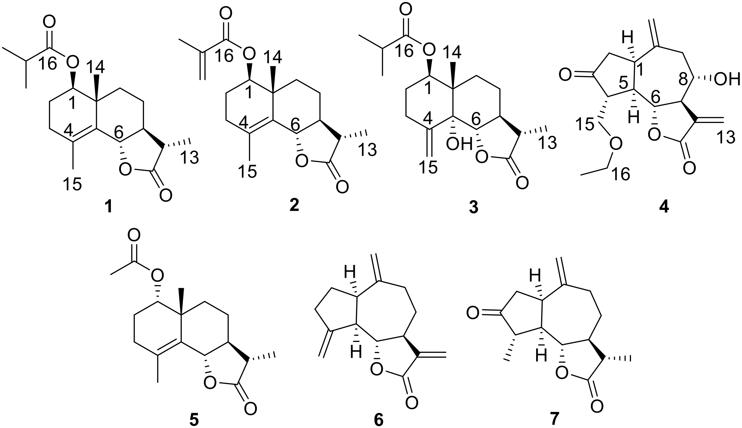
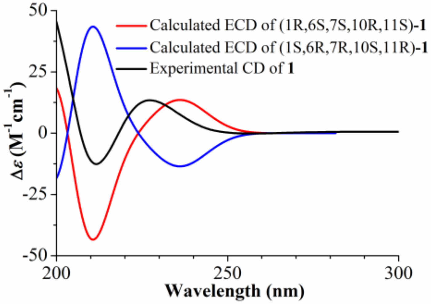
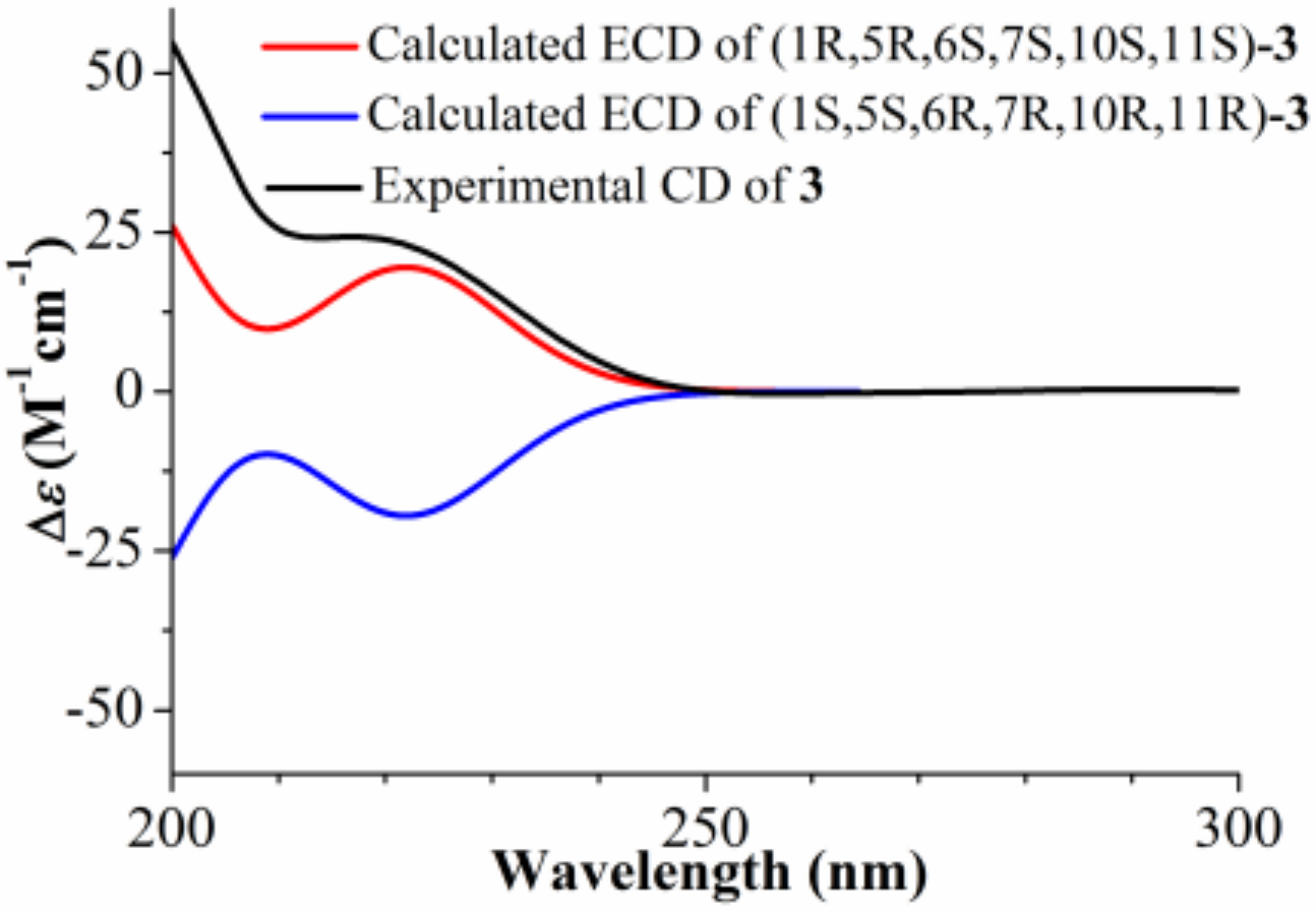
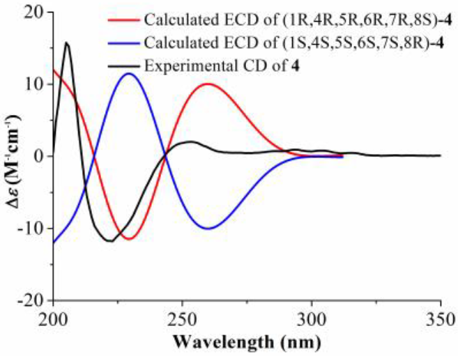
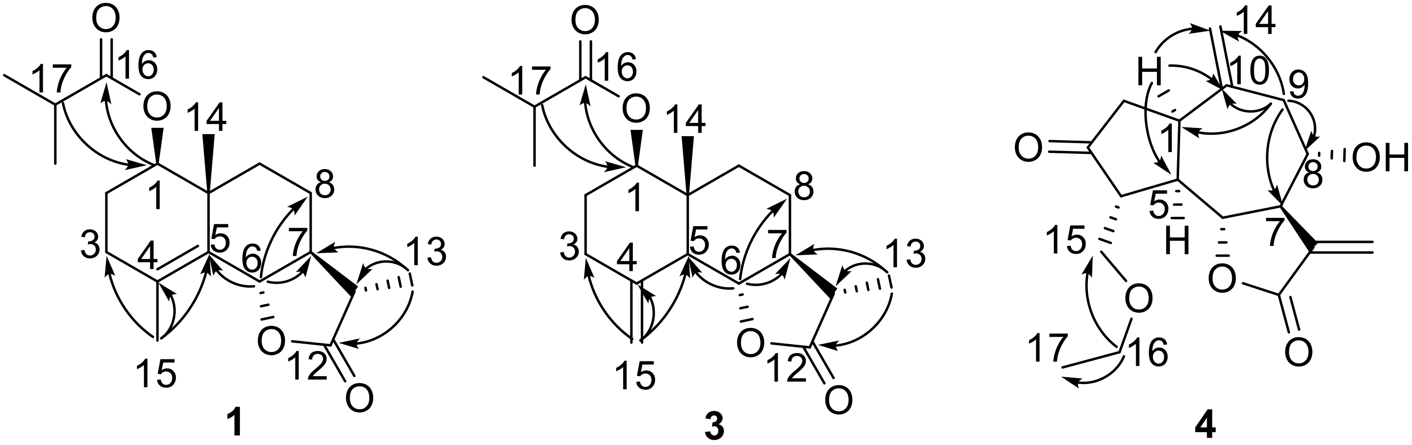
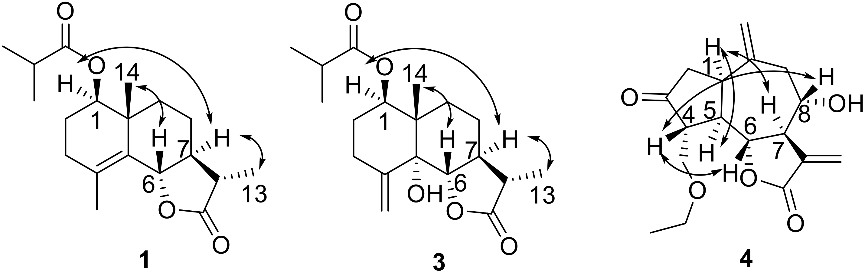
2. Results
3. Materials and Methods
3.1. General Experimental Material
3.2. Plant Material
3.3. Extraction and Isolation
3.4. Spectroscopic Data
3.5. Antifeedant Bioassay
3.6. Antifungal Bioassay
4. Conclusions
Supplementary Materials
Author Contributions
Funding
Conflicts of Interest
References
- Bora, K.S.; Sharma, A. The genus Artemisia: A comprehensive review. Pharm. Biol. 2011, 49, 101–109. [Google Scholar] [CrossRef] [PubMed]
- Tan, R.X.; Zheng, W.F.; Tang, H.Q. Biologically active substances from the genus Artemisia. Planta Med. 1998, 64, 295–302. [Google Scholar] [CrossRef] [PubMed]
- Abad, M.J.; Bedoya, L.M.; Apaza, L.; Bermejo, P. The Artemisia L. genus: A review of bioactive essential oils. Molecules 2012, 17, 2542–2566. [Google Scholar] [CrossRef] [PubMed]
- Yin, Y.; Gong, F.Y.; Wu, X.X.; Sun, Y.; Li, Y.H.; Chen, T.; Xu, Q. Anti-inflammatory and immunosuppressive effect of flavones isolated from Artemisia vestita. J. Ethnopharmacol. 2008, 120, 1–6. [Google Scholar] [CrossRef] [PubMed]
- Sun, Y.; Li, Y.H.; Wu, X.X.; Zheng, W.; Guo, Z.H.; Li, Y.; Chen, T.; Hua, Z.C.; Xu, Q. Ethanol extract from Artemisia vestita, a traditional Tibetan medicine, exerts anti-sepsis action through down-regulating the MAPK and NF-κB pathways. Int. J. Mol. Med. 2006, 17, 957–962. [Google Scholar] [CrossRef] [PubMed]
- Tian, S.H.; Chai, X.Y.; Zan, K.; Zeng, K.W.; Tu, P.F. Three new eudesmane sesquiterpenes from Artemisia vestita. Chin. Chem. Lett. 2013, 24, 797–800. [Google Scholar] [CrossRef]
- Tian, S.H.; Chai, X.Y.; Zan, K.; Zeng, K.W.; Guo, X.; Jiang, Y.; Tu, P.F. Arvestolides A–C, new rare sesquiterpenes from the aerial parts of Artemisia vestita. Tetrahedron Lett. 2013, 54, 5035–5038. [Google Scholar] [CrossRef]
- Chu, S.S.; Liu, Q.R.; Liu, Z.L. Insecticidal activity and chemical composition of the essential oil of Artemisia vestita from China against Sitophilus zeamais. Biochem. Syst. Ecol. 2010, 38, 489–492. [Google Scholar] [CrossRef]
- Irum, S.; Ahmed, H.; Mukhtar, M.; Mushtaq, M.; Mirza, B.; Donskow-Łysoniewska, K.; Qayyum, M.; Simsek, S. Anthelmintic activity of Artemisia vestita Wall ex DC. and Artemisia maritima L. against Haemonchus contortus from sheep. Vet. Parasitol. 2015, 212, 451–455. [Google Scholar] [CrossRef] [PubMed]
- Mann, R.S.; Kaufman, P.E. Natural product pesticides: Their development, delivery and use against insect vectors. Mini Rev. Org. Chem. 2012, 9, 185–202. [Google Scholar] [CrossRef]
- Wu, H.B.; Wu, H.B.; Wang, W.S.; Liu, T.T.; Qi, M.G.; Feng, J.C.; Li, X.Y.; Liu, Y. Insecticidal activity of sesquiterpene lactones and monoterpenoid from the fruits of Carpesium abrotanoides. Ind. Crops Prod 2016, 92, 77–83. [Google Scholar] [CrossRef]
- Neves, M.; Morais, R.; Gafner, S.; Stoeckli-Evans, H.; Hostettmann, K. New sesquiterpene lactones from the Portuguese liverwort Targionia lorbeeriana. Phytochemistry 1999, 50, 967–972. [Google Scholar] [CrossRef]
- Wu, X.L.; Xiong, X.J.; Lu, W.Q.; Huang, H.; Shen, Y.H.; Wu, Z.J.; Chen, W.S. New sesquiterpenenoids from Ainsliaea yunnanensis. Molecules 2016, 21, 1031. [Google Scholar] [CrossRef] [PubMed]
- Uchida, I.; Kuriyama, K. The π-π circular dichroism of α,β-unsaturated γ-lactones. Tetrahedron Lett. 1974, 15, 3761–3764. [Google Scholar] [CrossRef]
- Miyase, T.; Fukushima, S. Studies on sesquiterpene glycosides from Sonchus oleraceus L. Chem. Pharm. Bull. 1987, 35, 2869–2874. [Google Scholar] [CrossRef]
- Barbetti, P.; Fardella, G.; Chiappini, I.; Scarcia, V.; Candiani, A.F. New cytotoxic selenoderivatives of guaianolides. Eur. J. Med. Chem. 1989, 24, 299–305. [Google Scholar] [CrossRef]
- Bucar, F.; Wube, A.; Schmid, M. Natural product isolation—How to get from biological material to pure compounds. Nat. Prod. Rep. 2013, 30, 525–545. [Google Scholar] [CrossRef] [PubMed]
- Wu, H.B.; Liu, T.T.; Lian, Y.X.; Wang, W.S. Two new eremophilenolides from the roots of Ligulariopsis shichuana and their anti-phytopathogenic fungal and antifeedant activities. Nat. Prod. Res. 2019, 33, 1442–1448. [Google Scholar] [CrossRef] [PubMed]
Sample Availability: Not available. |


| Position | 1 | 2 | 3 | 4 | ||||
|---|---|---|---|---|---|---|---|---|
| δH (J in Hz) | δC | δH (J in Hz) | δC | δH (J in Hz) | δC | δH (J in Hz) | δC | |
| 1 | 4.75, dd (11.5, 2.8) | 78.9 | 4.80, t (7.8) | 79.7 | 5.34, dd (11.0, 4.5) | 74.1 | 3.23, dt (8.8, 4.0) | 40.9 |
| 2a | 1.76, m | 23.8 | 1.82, m | 23.7 | 1.90, m | 26.8 | 2.56, dd (18.2, 8.8) | 44.7 |
| 2b | 1.71, overlapped | 1.80, m | 1.61, m | 2.46, dd (18.2, 4.0) | ||||
| 3a | 2.27, overlapped | 32.8 | 2.27, overlapped | 32.9 | 2.71, m | 29.3 | 216.8 | |
| 3b | 2.01, m | 2.03, m | 2.16, m | |||||
| 4 | 126.4 | 126.5 | 144.6 | 2.38, m | 52.7 | |||
| 5 | 128.6 | 128.5 | 77.1 | 2.90, dd (17.6, 8.3) | 45.9 | |||
| 6 | 4.55, d (11.2) | 82.6 | 4.57, d (11.2) | 82.6 | 4.25, d (9.8) | 81.4 | 4.02, t (8.3) | 81.8 |
| 7 | 1.70, overlapped | 52.8 | 1.68, m | 52.8 | 2.39, overlapped | 45.3 | 3.08, m | 49.7 |
| 8a | 1.90, m | 24.3 | 1.91, m | 24.3 | 1.82, m | 22.7 | 3.94, m | 73.1 |
| 8b | 1.49, m | 1.51, m | 1.47, overlapped | |||||
| 9a | 1.73, overlapped | 38.0 | 1.76, overlapped | 38.1 | 1.72, overlapped | 29.8 | 2.86, dd (12.8, 5.6) | 47.1 |
| 9b | 1.28, m | 1.28, m | 1.46, overlapped | 2.34, dd (12.8, 8.3) | ||||
| 10 | 41.0 | 41.1 | 43.9 | 143.4 | ||||
| 11 | 2.28, overlapped | 41.0 | 2.27, overlapped | 41.1 | 2.31, overlapped | 41.2 | 136.9 | |
| 12 | 178.7 | 178.7 | 179.1 | 169.6 | ||||
| 13a | 1.23, d (7.2) | 12.4 | 1.22, d (7.2) | 12.4 | 1.23, d (7.2) | 12.4 | 6.39, s | 125.2 |
| 13b | 6.31, s | |||||||
| 14a | 1.19, s | 19.8 | 1.20, s | 19.8 | 1.00, s | 14.6 | 5.08, s | 115.9 |
| 14b | 4.87, s | |||||||
| 15a | 1.85, s | 19.8 | 1.82, s | 19.8 | 5.55, s | 112.7 | 3.91, dd (9.1, 2.7) | 68.9 |
| 15b | 5.00, s | 3.70, dd (9.1, 2.7) | ||||||
| 16 | 176.6 | 168.9 | 176.3 | 3.47, m | 66.7 | |||
| 17 | 2.55, m | 34.5 | 136.8 | 2.51, m | 34.4 | 1.16, t (7.0) | 14.9 | |
| 18 | 1.17, d (7.0) | 18.9 | 6.10, s; 5.57, s | 125.3 | 1.16, s | 19.0 | ||
| 19 | 1.18, d (7.0) | 19.1 | 1.96, s | 18.3 | 1.17, s | 19.1 | ||
| Compounds | EC50 (μg/cm2) | Compounds | EC50 (μg/cm2) |
|---|---|---|---|
| 1 | 32.4 | 5 | 33.0 |
| 2 | 37.9 | 6 | 31.9 |
| 3 | 42.1 | 7 | 25.3 |
| 4 | 27.6 | Neem oil | 4.89 |
© 2019 by the authors. Licensee MDPI, Basel, Switzerland. This article is an open access article distributed under the terms and conditions of the Creative Commons Attribution (CC BY) license (http://creativecommons.org/licenses/by/4.0/).
Share and Cite
Ding, Y.-H.; Wang, H.-T.; Shi, S.; Meng, Y.; Feng, J.-C.; Wu, H.-B. Sesquiterpenoids from Artemisia vestita and Their Antifeedant and Antifungal Activities. Molecules 2019, 24, 3671. https://doi.org/10.3390/molecules24203671
Ding Y-H, Wang H-T, Shi S, Meng Y, Feng J-C, Wu H-B. Sesquiterpenoids from Artemisia vestita and Their Antifeedant and Antifungal Activities. Molecules. 2019; 24(20):3671. https://doi.org/10.3390/molecules24203671
Chicago/Turabian StyleDing, Ying-Hui, Hui-Ting Wang, Sha Shi, Yao Meng, Jin-Chao Feng, and Hai-Bo Wu. 2019. "Sesquiterpenoids from Artemisia vestita and Their Antifeedant and Antifungal Activities" Molecules 24, no. 20: 3671. https://doi.org/10.3390/molecules24203671
APA StyleDing, Y.-H., Wang, H.-T., Shi, S., Meng, Y., Feng, J.-C., & Wu, H.-B. (2019). Sesquiterpenoids from Artemisia vestita and Their Antifeedant and Antifungal Activities. Molecules, 24(20), 3671. https://doi.org/10.3390/molecules24203671