
Modulatory Effect of Guinep (Melicoccus bijugatus Jacq) Fruit Pulp Extract on Isoproterenol-Induced Myocardial Damage in Rats. Identification of Major Metabolites Using High Resolution UHPLC Q-Orbitrap Mass Spectrometry

 ,
,  ,
,  , ,
, ,
Abstract
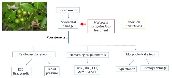
1. Introduction
2. Results
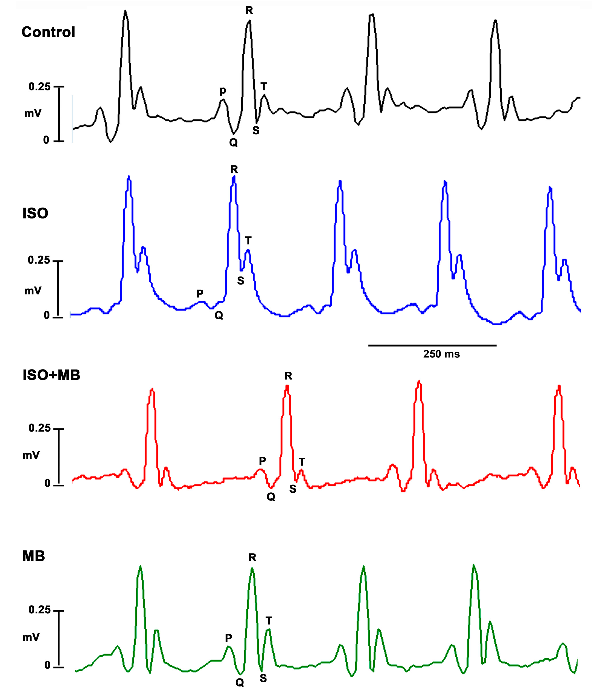
2.1. Blood Pressure Changes and Electrocardiogram (ECG)
2.2. Hematological Parameters
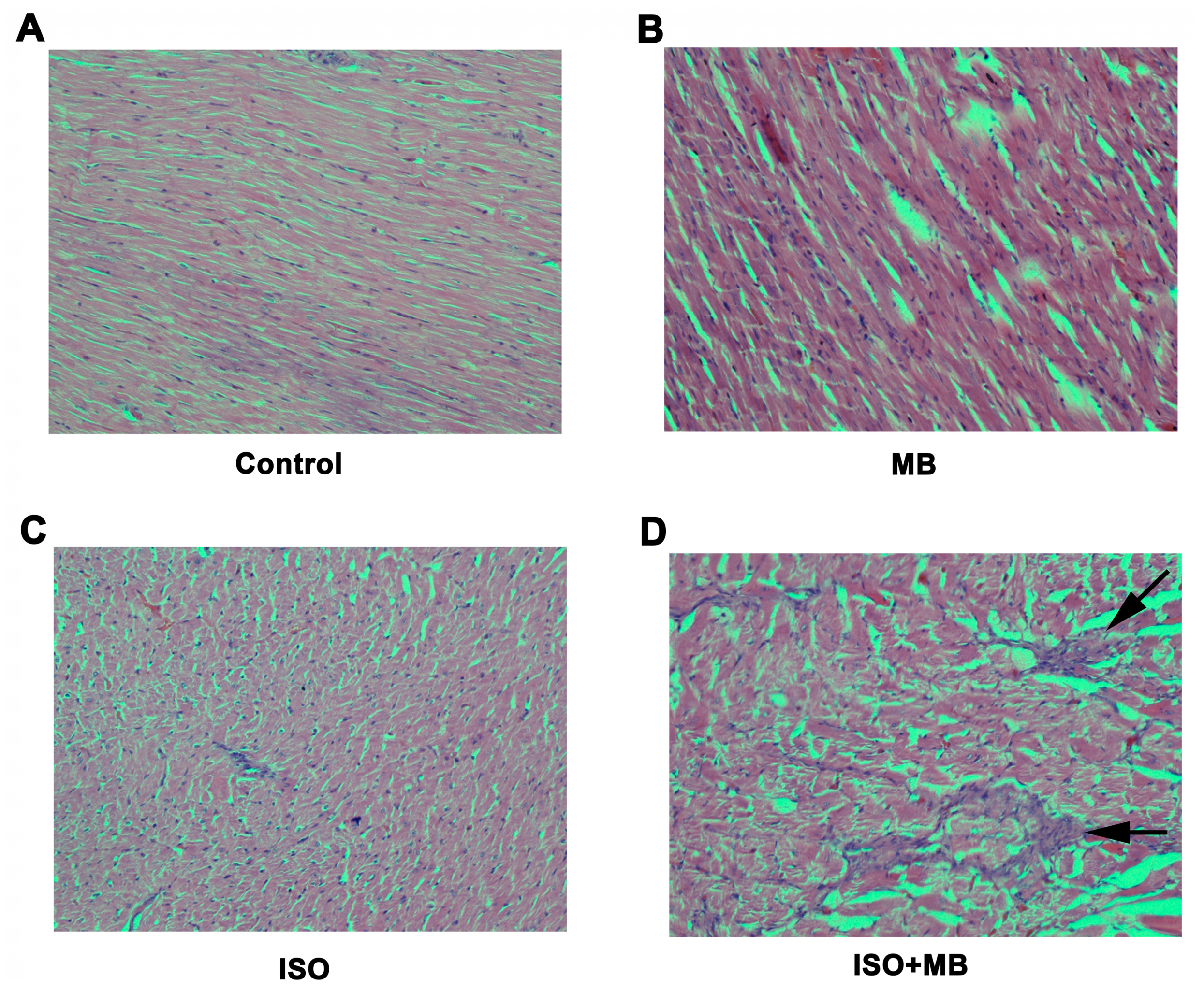
2.3. Histo-Morphological Analysis
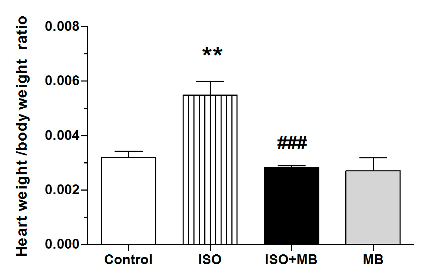
2.4. Ratio of Heart Weight to Body Weight
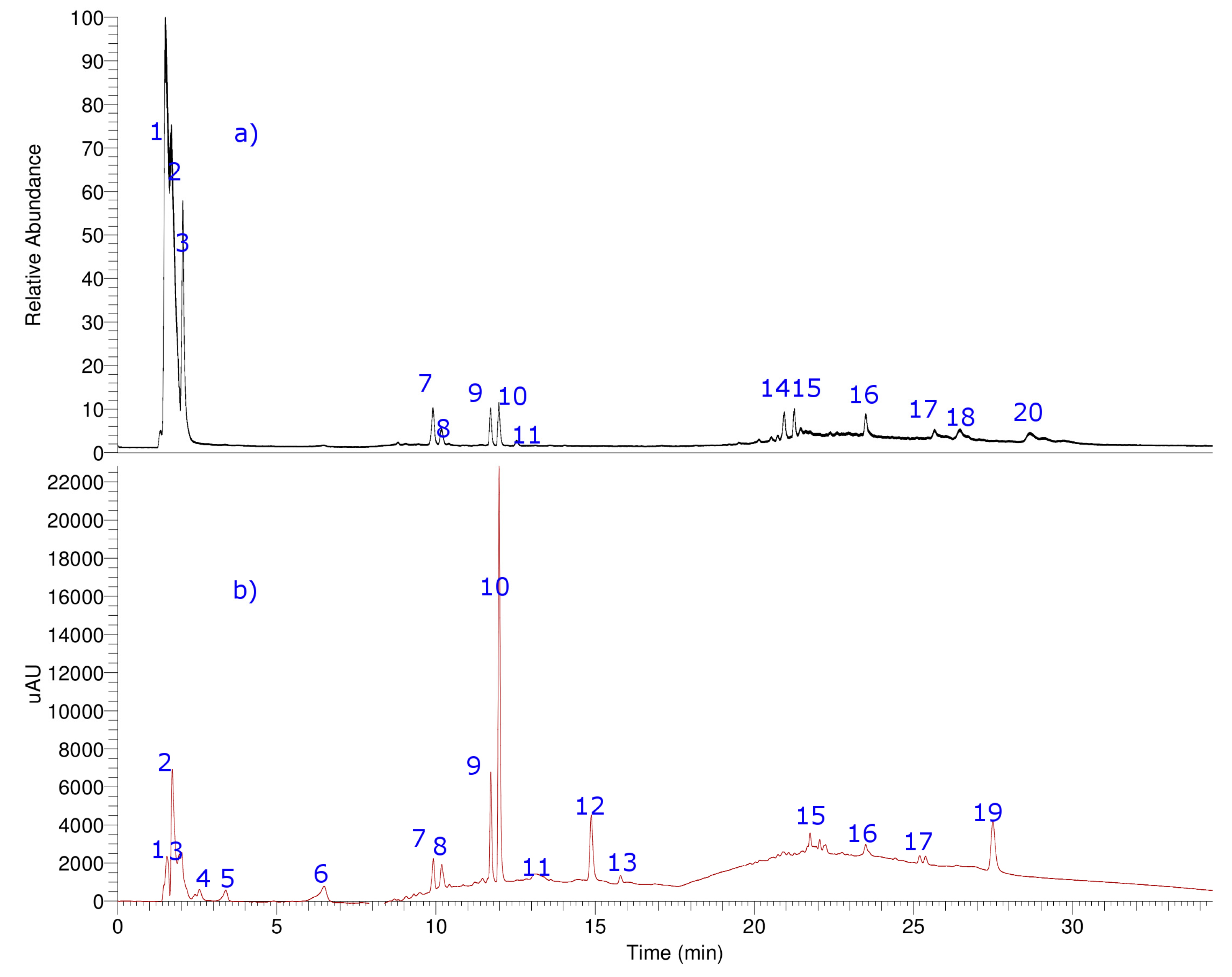
2.5. Identification of the Compounds
2.5.1. Simple Organic Acids and Sugars
2.5.2. Flavonoids
2.5.3. Phenolic Compounds
2.5.4. Fatty Acids
2.5.5. Terpenes and Related Compounds
3. Discussion
4. Materials and Methods
4.1. Plant Material and Extraction
4.2. Experimental Animals
4.3. Blood Pressure Recordings and ECG
4.4. Biochemical Estimation
4.5. Histo-Morphological Appraisal
4.6. UHPLC-DAD-MS Instrument
4.7. LC Parameters and MS Parameters
4.8. Statistical Analysis
5. Conclusions
Supplementary Materials
Author Contributions
Funding
Acknowledgments
Conflicts of Interest
References
- Lobo Filho, H.G.; Ferreira, N.L.; Sousa, R.B.; Carvalho, E.R.; Lobo, P.L.; Lobo Filho, J.G. Experimental model of myocardial infarction induced by isoproterenol in rats. Rev. Bras. Cir. Cardiovasc. 2011, 26, 469–476. [Google Scholar] [CrossRef] [PubMed]
- Górecki, A.; Bednarz, B.; Jaxa-Chamiec, T.; Maciejewski, P.; Łukaszewicz, R.; Ceremuzyński, L.; Dyduszyński, A. Lipid profile during the first 24 hours after myocardial infarction has significant prognostic value. Kardiol. Pol. 2004, 60, 229–236. [Google Scholar] [PubMed]
- Thippeswamy, B.; Thakker, S.; Tubachi, S.; Kalyani, G.; Netra, M.; Patil, U.; Desai, S.; Gavimath, C.; Veerapur, V. Cardioprotective effect of Cucumis trigonus Roxb on Isoproterenol-induced myocardial infarction in rat. Am. J. Pharmaco. Toxico. 2009, 4, 29–37. [Google Scholar] [CrossRef]
- Nwokocha, C.; Palacios, J.; Simirgiotis, M.J.; Thomas, J.; Nwokocha, M.; Young, L.; Thompson, R.; Cifuentes, F.; Paredes, A.; Delgoda, R. Aqueous extract from leaf of Artocarpus altilis provides cardio-protection from isoproterenol induced myocardial damage in rats: Negative chronotropic and inotropic effects. J. Ethnopharmacol. 2017, 203, 163–170. [Google Scholar] [CrossRef] [PubMed]
- Singal, P.K.; Beamish, R.E.; Dhalla, N.S. Potential oxidative pathways of catecholamines in the formation of lipid peroxides and genesis of heart disease. Adv. Exp. Med. Biol. 1983, 161, 391–401. [Google Scholar] [PubMed]
- Haenen, G.R.; Veerman, M.; Bast, A. Reduction of beta-adrenoceptor function by oxidative stress in the heart. Free Radic Biol. Med. 1990, 9, 279–288. [Google Scholar] [CrossRef]
- Tappia, P.S.; Hata, T.; Hozaima, L.; Sandhu, M.S.; Panagia, V.; Dhalla, N.S. Role of oxidative stress in catecholamine-induced changes in cardiac sarcolemmal Ca2+ transport. Arch. Biochem. Biophys. 2001, 387, 85–92. [Google Scholar] [CrossRef]
- Jagadeesh, G.S.; Nagoor Meeran, M.F.; Selvaraj, P. Activation of β1-adrenoceptor triggers oxidative stress mediated myocardial membrane destabilization in isoproterenol induced myocardial infarcted rats: 7-hydroxycoumarin and its counter action. Eur. J. Pharmacol. 2016, 777, 70–77. [Google Scholar] [CrossRef]
- Bystrom, L.M.; Lewis, B.A.; Brown, D.L.; Rodriguez, E.; Obendorf, R.L. Phenolics, Sugars, Antimicrobial and Free-Radical-Scavenging Activities of Melicoccus bijugatus Jacq. Fruits from the Dominican Republic and Florida. Plant Food. Hum. Nutr. 2009, 64, 160–166. [Google Scholar] [CrossRef]
- Bystrom, L.M. The potential health effects of Melicoccus bijugatus Jacq. fruits: Phytochemical, chemotaxonomic and ethnobotanical investigations. Fitoterapia 2012, 83, 266–271. [Google Scholar] [CrossRef]
- Padilla, F.C.; Rincon, A.M.; Bou-Rached, L. Polyphenol content and antioxidant activity of several seeds and nuts. Arch. Latinoam. Nutr. 2008, 58, 303–308. [Google Scholar] [PubMed]
- Bystrom, L.M.; Lewis, B.A.; Brown, D.L.; Rodriguez, E.; Obendorf, R.L. Characterisation of phenolics by LC–UV/Vis, LC–MS/MS and sugars by GC in Melicoccus bijugatus Jacq. ‘Montgomery’ fruits. Food Chem. 2008, 111, 1017–1024. [Google Scholar] [CrossRef] [PubMed]
- Brooks, W.W.; Conrad, C.H. Isoproterenol-induced myocardial injury and diastolic dysfunction in mice: Structural and functional correlates. Comp. Med. 2009, 59, 339–343. [Google Scholar] [PubMed]
- Cifuentes, F.; Bravo, J.; Norambuena, M.; Stegen, S.; Ayavire, A.; Palacios, J. Chronic exposure to arsenic in tap water reduces acetylcholine-induced relaxation in the aorta and increases oxidative stress in female rats. Int. J. Toxicol. 2009, 28, 534–541. [Google Scholar] [CrossRef] [PubMed]
- Arora, R.; Deshmukh, R. Embelin Attenuates Intracerebroventricular Streptozotocin-Induced Behavioral, Biochemical, and Neurochemical Abnormalities in Rats. Mol. Neurobiol. 2017, 54, 6670–6680. [Google Scholar] [CrossRef] [PubMed]
- Simirgiotis, M.J.; Quispe, C.; Mocan, A.; Villatoro, J.M.; Areche, C.; Bórquez, J.; Sepúlveda, B.; Echiburu-Chau, C. UHPLC high resolution orbitrap metabolomic fingerprinting of the unique species Ophryosporus triangularis Meyen from the Atacama Desert, Northern Chile. Rev. Bras. Farmacogn. 2017. [Google Scholar] [CrossRef]
- Vuong, Q.V.; Hirun, S.; Phillips, P.A.; Chuen, T.L.K.; Bowyer, M.C.; Goldsmith, C.D.; Scarlett, C.J. Fruit-derived phenolic compounds and pancreatic cancer: Perspectives from Australian native fruits. J. Ethnopharmacol. 2014, 152, 227–242. [Google Scholar] [CrossRef]
- Simirgiotis, M.J.; Quispe, C.; Bórquez, J.; Areche, C.; Sepúlveda, B.X. Fast Detection of Phenolic Compounds in Extracts of Easter Pears (Pyrus communis) from the Atacama Desert by Ultrahigh-Performance Liquid Chromatography and Mass Spectrometry (UHPLC-Q/Orbitrap/MS/MS). Molecules 2016, 21, 92. [Google Scholar] [CrossRef]
- Jiménez-Sánchez, C.; Lozano-Sánchez, J.; Rodríguez-Pérez, C.; Segura-Carretero, A.; Fernández-Gutiérrez, A. Comprehensive, untargeted, and qualitative RP-HPLC-ESI-QTOF/MS2 metabolite profiling of green asparagus (Asparagus officinalis). J. Food Compos. Anal. 2016, 46, 78–87. [Google Scholar] [CrossRef]
- Simirgiotis, M.J.; Ramirez, J.E.; Hirschmann, G.S.; Kennelly, E.J. Bioactive coumarins and HPLC-PDA-ESI-ToF-MS metabolic profiling of edible queule fruits (Gomortega keule), an endangered endemic Chilean species. Food Res. Int. 2013, 54, 532–543. [Google Scholar] [CrossRef]
- Dong, M.; Oda, Y.; Hirota, M. (10E,12Z,15Z)-9-hydroxy-10,12,15-octadecatrienoic acid methyl ester as an anti-inflammatory compound from Ehretia dicksonii. Biosci. Biotechnol. Biochem. 2000, 64, 882–886. [Google Scholar] [CrossRef]
- Oguro, D.; Watanabe, H. Asymmetric Synthesis and Sensory Evaluation of Sedanenolide. Biosci. Biotech. Bioch. 2011, 75, 1502–1505. [Google Scholar] [CrossRef]
- Torkamani, M.R.D.; Abbaspour, N.; Jafari, M.; Samadi, A. Elicitation of Valerenic Acid in the Hairy Root Cultures of Valeriana officinalis L (Valerianaceae). Trop. J. Pharm. Res. 2014, 13, 943–949. [Google Scholar] [CrossRef]
- Ishizaka, N.; Tomiyama, K.; Katsui, N.; Murai, A.; Masamune, T. Biological activities of rishitin, an antifungal compound isolated from diseased potato tubers, and its derivatives1. Plant Cell Physiol. 1969, 10, 183–192. [Google Scholar] [CrossRef]
- Zhang, H.Y.; Gao, Y.; Lai, P.X. Chemical Composition, Antioxidant, Antimicrobial and Cytotoxic Activities of Essential Oil from Premna microphylla Turczaninow. Molecules 2017, 22, 381. [Google Scholar] [CrossRef]
- Subash, D.; Kapoor, N.; Nityanand, S. Effect of isoprenaline on lipid profil and cardiac enzymes in rats. Ind. J. Exp. Biol. 1978, 16, 376–378. [Google Scholar]
- Nirmala, C.; Puvanakrishnan, R. Protective role of curcumin against isoproterenol induced myocardial infarction in rats. Mol. Cell Biochem. 1996, 159, 85–93. [Google Scholar] [CrossRef] [PubMed]
- Montessuit, C.; Thorburn, A. Transcriptional activation of the glucose transporter GLUT1 in ventricular cardiac myocytes by hypertrophic agonists. J. Biol. Chem. 1999, 274, 9006–9012. [Google Scholar] [CrossRef]
- Patel, V.; Upaganlawar, A.; Zalawadia, R.; Balaraman, R. Cardioprotective effect of melatonin against isoproterenol induced myocardial infarction in rats: A biochemical, electrocardiographic and histoarchitectural evaluation. Eur. J. Pharmacol. 2010, 644, 160–168. [Google Scholar] [CrossRef]
- Li, P.G.; Xu, J.W.; Ikeda, K.; Kobayakawa, A.; Kayano, Y.; Mitani, T.; Ikami, T.; Yamori, Y. Caffeic acid inhibits vascular smooth muscle cell proliferation induced by angiotensin II in stroke-prone spontaneously hypertensive rats. Hypertens. Res. 2005, 28, 369–377. [Google Scholar] [CrossRef] [PubMed]
- Luceri, C.; Giannini, L.; Lodovici, M.; Antonucci, E.; Abbate, R.; Masini, E.; Dolara, P. p-Coumaric acid, a common dietary phenol, inhibits platelet activity in vitro and in vivo. Br. J. Nutr. 2007, 97, 458–463. [Google Scholar] [CrossRef] [PubMed]
- Ghoneim, M.A.; Hassan, A.I.; Mahmoud, M.G.; Asker, M.S. Protective Effect of Adansonia digitata against Isoproterenol-Induced Myocardial Injury in Rats. Anim. Biotechnol. 2016, 27, 84–95. [Google Scholar] [CrossRef] [PubMed]
- Dianita, R.; Jantan, I.; Amran, A.Z.; Jalil, J. Protective effects of Labisia pumila var. alata on biochemical and histopathological alterations of cardiac muscle cells in isoproterenol-induced myocardial infarction rats. Molecules 2015, 20, 4746–4763. [Google Scholar] [CrossRef] [PubMed]
- Sabeena Farvin, K.H.; Anandan, R.; Kumar, S.H.; Shiny, K.S.; Sankar, T.V.; Thankappan, T.K. Effect of squalene on tissue defense system in isoproterenol-induced myocardial infarction in rats. Pharmacol. Res. 2004, 50, 231–236. [Google Scholar] [CrossRef] [PubMed]
- Zhang, J.; Knapton, A.; Lipshultz, S.E.; Weaver, J.L.; Herman, E.H. Isoproterenol-induced cardiotoxicity in sprague-dawley rats: Correlation of reversible and irreversible myocardial injury with release of cardiac troponin T and roles of iNOS in myocardial injury. Toxicol. Pathol. 2008, 36, 277–278. [Google Scholar] [CrossRef] [PubMed]
- Ismail, Z.; Mahmoud, A.; Khaled, R.; Sami, A.; Karim, A.; Iyad, A.; Mohamed, K.; Samir, K.; Moaath, J.; Ahmad, A. Effects of experimental acute myocardial infarction on blood cell counts and plasma biochemical values in a nude rat model (Crl:NIH-Fox1RNU). Comp. Clin. Pathol. 2009, 18, 433–437. [Google Scholar] [CrossRef]
- Sangeetha, T.; Quine, S.D. Protective effect of S-allyl cysteine sulphoxide (alliin) on glycoproteins and hematology in isoproterenol induced myocardial infarction in male Wistar rats. J. Appl. Toxicol. 2008, 28, 710–716. [Google Scholar] [CrossRef]
- Nyonseu Nzebang, D.C.; Ngaha Njila, M.I.; Bend, E.F.; Oundoum Oundoum, P.C.; Koloko, B.L.; Bogning Zangueu, C.; Belle Ekedi, P.; Sameza, M.; Massoma Lembè, D. Evaluation of the toxicity of Colocasia esculenta (Aracaceae): Preliminary study of leaves infected by Phytophthora colocasiae on wistar albinos rats. Biomed. Pharmacother. 2018, 99, 1009–1013. [Google Scholar] [CrossRef] [PubMed]
- Zunjar, V.; Dash, R.P.; Jivrajani, M.; Trivedi, B.; Nivsarkar, M. Antithrombocytopenic activity of carpaine and alkaloidal extract of Carica papaya Linn. leaves in busulfan induced thrombocytopenic Wistar rats. J. Ethnopharmacol. 2016, 181, 20–25. [Google Scholar] [CrossRef]
- Cifuentes, F.; Paredes, A.; Palacios, J.; Muñoz, F.; Carvajal, L.; Nwokocha, C.R.; Morales, G. Hypotensive and antihypertensive effects of a hydroalcoholic extract from Senecio nutans Sch. Bip. (Compositae) in mice: Chronotropic and negative inotropic effect, a nifedipine-like action. J. Ethnopharmacol. 2016, 179, 367–374. [Google Scholar] [CrossRef]
- Cifuentes, F.; Palacios, J.; Nwokocha, C.R. Synchronization in the Heart Rate and the Vasomotion in Rat Aorta: Effect of Arsenic Trioxide. Cardiovasc. Toxicol. 2016, 16, 79–88. [Google Scholar] [CrossRef] [PubMed]
- Simirgiotis, M.J.; Quispe, C.; Bórquez, J.; Schmeda-Hirschmann, G.; Avendaño, M.; Sepúlveda, B.; Winterhalter, P. Fast high resolution Orbitrap MS fingerprinting of the resin of Heliotropium taltalense Phil. from the Atacama Desert. Ind. Crops Prod. 2016, 85, 159–166. [Google Scholar] [CrossRef]
- Garneau, F.X.; Collin, G.J.; Jean, F.I.; Gagnon, H.; Lopez Arze, J.B. Essential oils from Bolivia. XII. Asteraceae: Ophryosporus piquerioides (DC) Benth. ex Baker. J. Essent. Oil Res. 2013, 25, 388–393. [Google Scholar] [CrossRef]
- Cifuentes, F.; Palacios, J.; Paredes, A.; Nwokocha, C.R.; Paz, C. 8-Oxo-9-Dihydromakomakine Isolated from Aristotelia chilensis Induces Vasodilation in Rat Aorta: Role of the Extracellular Calcium Influx. Molecules 2018, 23, 3050. [Google Scholar] [CrossRef] [PubMed]
Sample Availability: Samples of Melicoccus bijugatus is available from the corresponding author. |


| Normotensive | Myocardial Damage | |||
|---|---|---|---|---|
| Control | MB | ISO | ISO + MB | |
| MAP, mmHg | 101 ± 3 | 69 ± 2 *** | 113 ± 5 | 83 ± 6 *,### |
| SBP, mmHg | 131 ± 3 | 85 ± 2 *** | 133 ± 4 | 106 ± 6 ***,### |
| DBP, mmHg | 87 ± 5 | 60 ± 3 *** | 108 ± 8 * | 72 ± 8 ### |
| PP, mmHg | 44 ± 5 | 27 ± 2 *** | 28 ± 2 ** | 27 ± 3 ** |
| HR, bpm | 246 ± 25 | 146 ± 11 ** | 307 ± 20 | 181 ± 18 ### |
| Control | MB | ISO | ISO + MB | |
|---|---|---|---|---|
| WBC (103/µL) | 9.5 ± 1.2 | 8.6 ± 1.3 | 4.1 ± 1.0 * | 7.1 ± 1.2 |
| LYM (103/µL) | 5.8 ± 0.6 | 5.9 ± 1.0 | 3.0 ± 0.8 | 4.9 ± 0.8 |
| MID (103/µL) | 1.5 ± 0.2 | 1.1 ± 0.2 | 0.62 ± 0.2 * | 1.1 ± 0.1 |
| GRA (103/µL) | 2.4 ± 0.5 | 1.5 ± 0.3 | 0.5 ± 0.1 ** | 1.2 ± 0.3 |
| LYM (%) | 61.4 ± 2.9 | 66.8 ± 2.7 | 73.9 ± 0.9 ** | 69.6 ± 1.1 |
| MID (%) | 14.5 ± 0.6 | 13.9± 1.0 | 14.5 ± 0.8 | 13.2 ± 1.5 |
| GRA (%) | 27.3 ± 0.9 | 19.3 ± 2.4 | 11.6 ± 1.0 *** | 17.3 ± 1.4 * |
| Control | MB | ISO | ISO + MB | |
|---|---|---|---|---|
| RBC (106/µL) | 6.8 ± 0.2 | 5.9 ± 0.4 | 5.2 ± 0.2 ** | 6.3 ± 0.42 |
| HGB (g/dL) | 14.8 ± 0.3 | 13.6 ± 0.6 | 12.5 ± 0.6 | 14.1 ± 1.0 |
| HCT (%) | 40.0 ± 0.8 | 36.2 ± 1.2 | 32.6 ± 1.5 ** | 34.8 ± 1.4 |
| MCV (fL) | 2.4 ± 0.5 | 1.5 ± 0.3 | 0.5 ± 0.1 ** | 1.2 ± 0.3 |
| MCH (pg) | 21.7 ± 0.2 | 22.2 ± 0.5 | 24 ± 0.6 ** | 22.2 ± 0.4 |
| MCHC (g/dL) | 37.1 ± 0.4 | 37.4 ± 0.6 | 38.6 ± 0.4 | 37.7 ± 0.2 |
| RDW | 15.0 ± 0.3 | 15.7 ± 0.5 | 15.3 ± 1.2 | 16.6 ± 0.9 |
| PLT (103/µL) | 680.3 ± 31.0 | 840.0 ± 21.1 * | 668.8 ± 49.0 | 918.8 ± 42.7 **,## |
| MPV | 6.6 ± 0.2 | 7.7 ± 0.4 | 7.0 ± 0.2 | 7.4 ± 0.5 |
| Peak | Retention Time (min) | UV Max | Tentative Identification | Elemental Composition [M − H]− | Theoretical Mass (m/z) | Measured Mass (m/z) | Accuracy (dppm) | MSn Ions (dppm) |
|---|---|---|---|---|---|---|---|---|
| 1 | 1.72 | 220 | Citric acid | C6H7O7− | 191.01863 | 191.01933 | 3.68 | |
| 2 | 1.82 | 222 | Isocitric acid | C6H7O7− | 191.01863 | 191.01955 | 3.04 | |
| 3 | 1.65 | - | Saccharose | C12H21O11− | 341.10784 | 341.10783 | 2.44 | |
| 4 | 2.55 | - | Glucose | C6H11O6− | 179.05501 | 179.05550 | 2.69 | |
| 5 | 3.37 | 230 | Furoic acid | C5H3O3− | 111.00767 | 111.00787 | 3.25 | |
| 6 | 6.35 | 245 | Salicilic acid glucoside | C13H15O8− | 299.07614 | 299.07712 | 3.25 | 137.02440 |
| 7 | 9.80 | 245–325 | Aflavarin | C24H21O9− | 453.11801 | 453.11703 | −2.1 | |
| 8 | 10.24 | - | Trihydroxyoctadecatrienoic acid | C18H29O5− | 325.20205 | 325.18443 | −54.2 | |
| 9 | 11.98 | 275–339 | Coumaric acid glucoside | C15H17O8− | 325.09179 | 325.09277 | 3.01 | 163.0291, 145.02870, 117.03368 |
| 10 | 12.24 | 275–339 | Coumaric acid galactoside | C15H17O8− | 325.09179 | 325.09271 | 2.82 | 163.0291, 145.02870, 117.03368 |
| 11 | 13.35 | 275–339 | Feruloyl glucoside | C16H19O9− | 355.10346 | 355.10336 | 3.34 | 147.04449, 193.05058 |
| 12 | 15.02 | 254–354 | Isorhamnetin-3-O-gglucoside | C22H21O11− | 477.11679 | 477.11670 | −27.12 | 314.04370 |
| 13 | 15.83 | 275–339 | Feruloyl galactoside | C16H19O9− | 355.10346 | 355.10355 | 3.34 | 147.04449, 193.05056 |
| 14 | 21.57 | 236–329 | 3-O-Caffeoylquinic acid | C16H17O9− | 353.0878 | 353.0878 | 0.53 | 191.05608 |
| 15 | 21.87 | - | Rishitin | C14H22O2− | 221.15361 | 221.15488 | 5.74 | |
| 16 | 23.54 | 300 | Embelin | C17H26O4− | 293.17474 | 293.17587 | 3.87 | |
| 17 | 25.38 | 235 | Sedanenolide | C12H15O2− | 191.10666 | 191.10741 | 3.92 | |
| 18 | 26.45 | 225 | Valerenic acid | C15H21O2− | 233.15470 | 233.15455 | 3.87 | 149.13301 |
| 19 | 27.78 | 214 | Blumenol C | C13H22O2− | 209.15631 | 209.15430 | 3.30 | |
| 20 | 28.32 | 220 | Hydroxyheptadecatrienoic acid | C17H27O7− | 311.18640 | 311.16876 | −56.6 |
© 2019 by the authors. Licensee MDPI, Basel, Switzerland. This article is an open access article distributed under the terms and conditions of the Creative Commons Attribution (CC BY) license (http://creativecommons.org/licenses/by/4.0/).
Share and Cite
Nwokocha, C.R.; Warren, I.; Palacios, J.; Simirgiotis, M.; Nwokocha, M.; Harrison, S.; Thompson, R.; Paredes, A.; Bórquez, J.; Lavado, A.; et al. Modulatory Effect of Guinep (Melicoccus bijugatus Jacq) Fruit Pulp Extract on Isoproterenol-Induced Myocardial Damage in Rats. Identification of Major Metabolites Using High Resolution UHPLC Q-Orbitrap Mass Spectrometry. Molecules 2019, 24, 235. https://doi.org/10.3390/molecules24020235
Nwokocha CR, Warren I, Palacios J, Simirgiotis M, Nwokocha M, Harrison S, Thompson R, Paredes A, Bórquez J, Lavado A, et al. Modulatory Effect of Guinep (Melicoccus bijugatus Jacq) Fruit Pulp Extract on Isoproterenol-Induced Myocardial Damage in Rats. Identification of Major Metabolites Using High Resolution UHPLC Q-Orbitrap Mass Spectrometry. Molecules. 2019; 24(2):235. https://doi.org/10.3390/molecules24020235
Chicago/Turabian StyleNwokocha, Chukwuemeka R., Isheba Warren, Javier Palacios, Mario Simirgiotis, Magdalene Nwokocha, Sharon Harrison, Rory Thompson, Adrian Paredes, Jorge Bórquez, Astrid Lavado, and et al. 2019. "Modulatory Effect of Guinep (Melicoccus bijugatus Jacq) Fruit Pulp Extract on Isoproterenol-Induced Myocardial Damage in Rats. Identification of Major Metabolites Using High Resolution UHPLC Q-Orbitrap Mass Spectrometry" Molecules 24, no. 2: 235. https://doi.org/10.3390/molecules24020235
APA StyleNwokocha, C. R., Warren, I., Palacios, J., Simirgiotis, M., Nwokocha, M., Harrison, S., Thompson, R., Paredes, A., Bórquez, J., Lavado, A., & Cifuentes, F. (2019). Modulatory Effect of Guinep (Melicoccus bijugatus Jacq) Fruit Pulp Extract on Isoproterenol-Induced Myocardial Damage in Rats. Identification of Major Metabolites Using High Resolution UHPLC Q-Orbitrap Mass Spectrometry. Molecules, 24(2), 235. https://doi.org/10.3390/molecules24020235