Article

Bioactive Constituents from the Aerial Parts of Pluchea indica Less

1
Tianjin State Key Laboratory of Modern Chinese Medicine, Tianjin University of Traditional Chinese Medicine, 312 Anshanxi Road, Nankai District, Tianjin 300193, China
2
Tianjin Key Laboratory of TCM Chemistry and Analysis, Institute of Traditional Chinese Medicine, Tianjin University of Traditional Chinese Medicine, 312 Anshanxi Road, Nankai District, Tianjin 300193, China
*
Authors to whom correspondence should be addressed.
Molecules 2018, 23(9), 2104; https://doi.org/10.3390/molecules23092104
Submission received: 8 August 2018 / Revised: 15 August 2018 / Accepted: 21 August 2018 / Published: 21 August 2018

Abstract

:
Four new thiophenes, (3′′R)-pluthiophenol (1), (3′′R)-pluthiophenol-4′′-acetate (2), 3′′-ethoxy-(3′′S)-pluthiophenol (3), 3′′-ethoxy-(3′′S)-pluthiophenol-4′′-acetate (4), together with twenty-five known compounds were obtained from the 70% ethanol-water extract of the aerial parts of Pluchea indica Less. Their structures were elucidated by spectroscopic methods. Among the known isolates, compounds 7, 8, 11, 14, 15, 18, 20, 23, 2527 were isolated from Asteraceae family firstly, while compounds 6, 9, 10, 12, 13, 16, 19, 21, 28 were isolated from Pluchea genus for the first time. Meanwhile, compounds 1, 2, 10, 13, 18, 23 displayed significant inhibitory activities on LPS-induced NO production at 40 µM from RAW 264.7 macrophages, while compounds 3, 4, 2629 possessed moderate inhibitory effects.

1. Introduction

As one of the largest families, the Asteraceae (Compositae) family contains about 1600–1700 genera and 24,000–30,000 species. Most of the Asteraceae family plants are herbs and shrubs, and have been widely used as herbal medicines since ancient times all over the world [1]. Pluchea indica Less., belongs to Pluchea genus, Asteraceae family, is a 1 to 2 meters high shrub. It mainly distributes in the tropical and subtropical regions of Africa, Asia, America, Australia, and China’s southern provinces. As an amphibious woody semi-mangrove plant, it plays an important role in maintaining the ecological balance in the coastal areas of Southeast Asia in China [2]. As a folk medicine in Guangxi, it exhibits the function of softening hardness and dissolving lump [3]. As a type of food, it possesses the activity of warming the stomach [4]. Its main chemical compositions are thiophenes, quinic acids, sesquiterpenes, lignans, flavonoids, and sterols [2]. Pharmacological studies have shown that the plant exhibits many pharmacological functions such as anti-inflammatory [5], anti-cancer [6], anti-oxidant [7], anti-microbial [8], and insecticidal activities [9].
Through the summary of relevant literature, it is found that the pharmacodynamic material basis is not yet clear for the lack of systematic research on the plant. In the course of studying the anti-inflammatory activity of various medicinal plants, 70% EtOH extract of P. indica was found to possess significant in vitro anti-inflammatory bioactivity. Based on the anti-inflammatory activity on LPS-induced NO production from RAW 264.7 macrophages, a systematic chemical component study of P. indica aerial parts was carried out. In this paper, the isolation and identification of constituents were described as well as their inhibitory effect on the production of NO in RAW 264.7 cells induced by LPS.

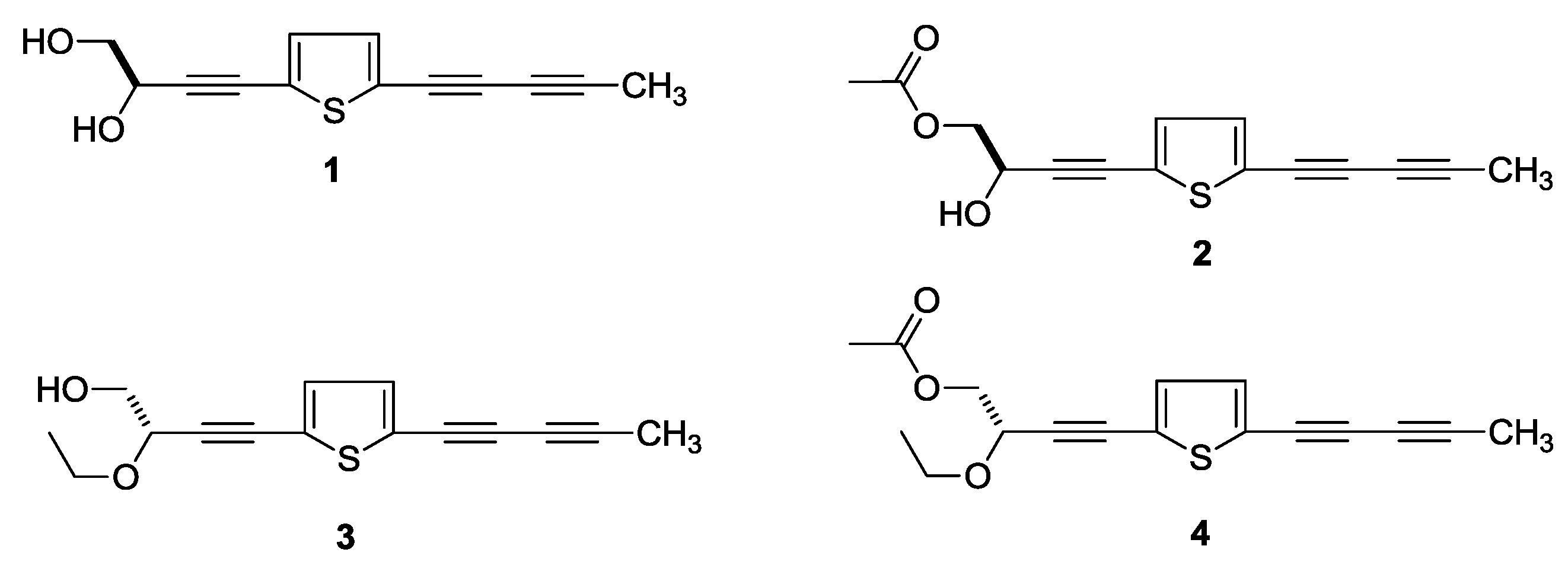
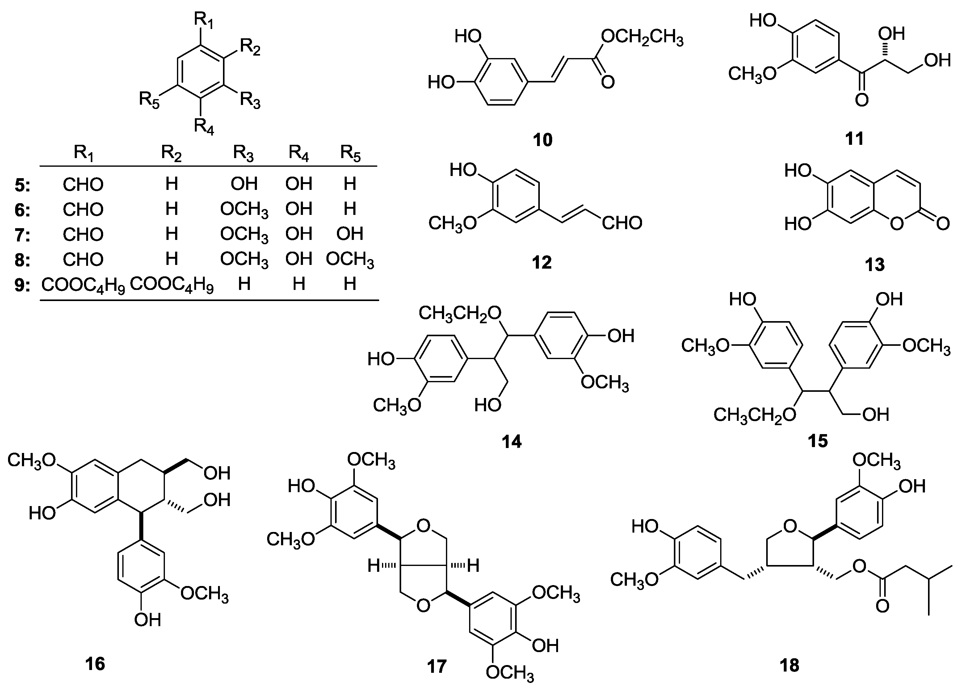
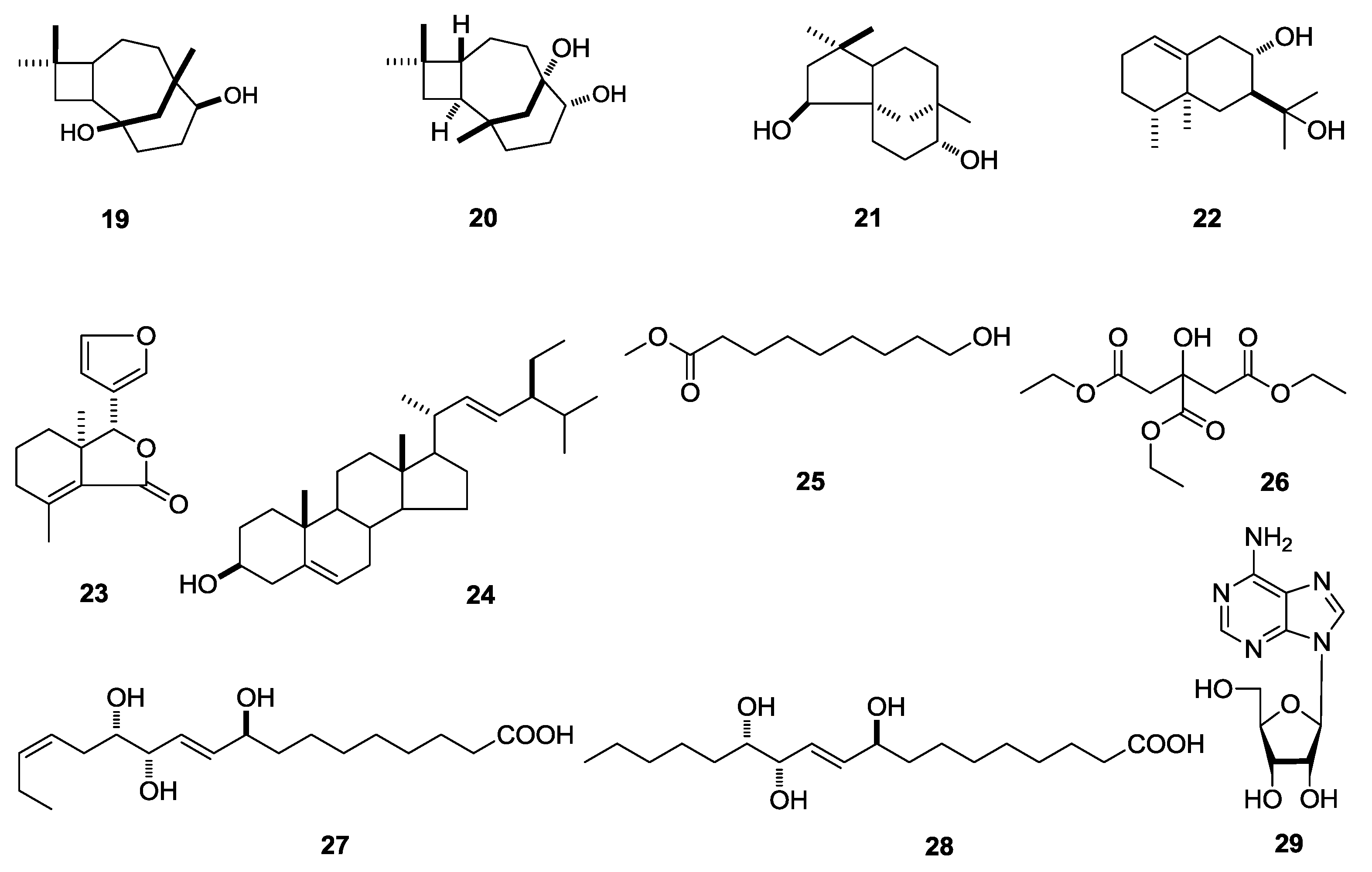
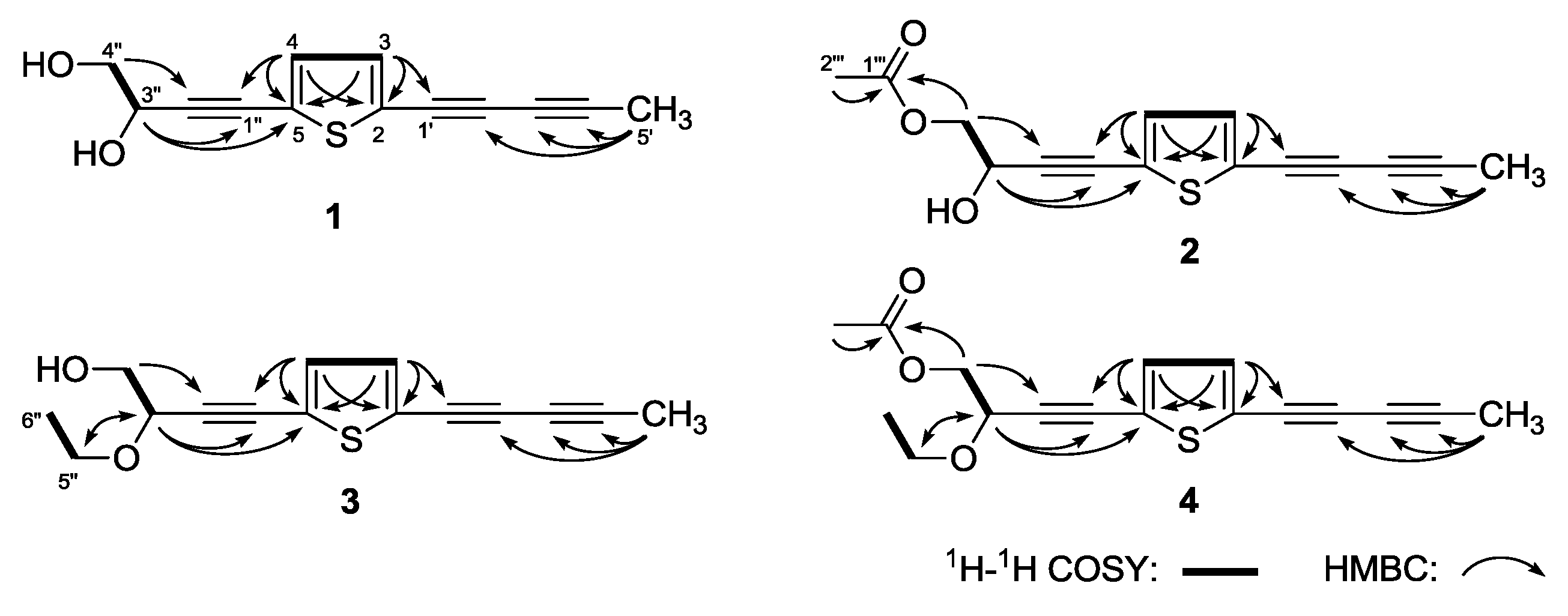
2. Results and Discussion

In the course of our investigation of the bioactive constituents from the 70% ethanol-water (EtOH) extract of the aerial parts of P. indica, four new thiophenes, named as (3′′R)-pluthiophenol (1), (3′′R)-pluthiophenol-4′′-acetate (2), 3′′-ethoxy-(3′′S)-pluthiophenol (3), 3′′-ethoxy-(3′′S)-pluthiophenol-4′′-acetate (4) (Figure 1) as well as twenty-five known compounds, 3,4-dihydroxy benzaldehyde (5) [10], vanillin (6) [11], 3,4-dihydroxy-5-methoxybenzaldehyde (7) [12], syringicaldehyde (8) [13], dibutylphthalate (9) [14], ethyl caffeate (10) [15], 2,3-dihydroxy-1-(4-hydroxy-3-methoxyphenyl)-propan-1-one (11) [16], trans-coniferyl aldehyde (12) [17], esculetin (13) [18], threo-2,3-bis(4-hydroxy-3-methoxyphenyl)-3-ethoxypropan-1-ol (14) [19], erythro-2,3-bis(4-hydroxy-3-methoxyphenyl)-3-ethoxypropan-1-ol (15) [20], (+)-isolariciresinol (16) [16,21], (–)-(7S,7′S,8R,8′R)-4,4′-dihydroxy-3,3′,5,5′-pentamethoxy-7,9′:7′,9-diepoxylignane (17) [22,23], (+)-9′-isovaleryllariciresinol (18) [24,25,26], caryolane-1,9β-diol (19) [27], (8R,9R)-isocaryolane-8,9-diol (20) [28], clovane-2α,9β-diol (21) [27], valenc-1(10)-ene-8,11-diol (22) [2], fraxinellone (23) [29], stigmasterol (24) [30], methyl 9-hydroxynonanoate (25) [31], triethyl citrate (26) [32], 9,12,13-trihydroxyoctadeca-10(E),15(Z)-dienoic acid (27) [33], pinellic acid (28) [34], adenosine (29) [35] (Figure 2) were obtained.
(3′′R)-Pluthiophenol (1) was isolated as yellow oil. Its molecular formula was revealed to be C13H10O2S by positive ESI-Q-Orbitrap MS analysis (m/z 231.04726 [M + H]+, calcd for C13H11O2S, 231.04743). The characteristic absorptions in its IR spectrum suggested the presences of hydroxyl (3312 cm−1), thiophene ring (3105, 1448 cm−1), and alkynyl (2222 cm−1). Its 1H-NMR (CD3OD, 500 MHz) (Table 1) spectrum indicated the presence of one methyl [δ 2.02 (3H, s, H3-5′)], one hydroxymethyl [δ 3.64 (1H, dd, J = 7.0, 11.5 Hz), 3.68 (1H, dd, J = 5.0, 11.5 Hz), H2-4′′], one oxygenated methine [δ 4.55 (1H, dd, J = 5.0, 7.0 Hz, H-3′′)], and a couple of olefinic protons [δ 7.08 (1H, d, J = 4.0 Hz, H-4), 7.15 (1H, d, J = 4.0 Hz, H-3)]. The four carbon signals [δ 124.6 (C-2), 125.9 (C-5), 133.3 (C-4), 134.9 (C-3)] in the low field area of 13C-NMR (CD3OD, 125 MHz) spectrum, combining with the special coupling constant (JH-3,4 = 4.0 Hz) and MS data confirmed the existence of the thiophene ring. The 1H-1H COSY spectrum of 1 indicated the presence of two partial structures written in bold lines as shown in Figure 3. Furthermore, in the HMBC experiment, the long-range correlations were observed from δH 7.15 (H-3) to δC 66.8 (C-1′), 124.9 (C-2), 125.9 (C-5); δH 7.08 (H-4) to δC 78.1 (C-1′′), 124.9 (C-2), 125.9 (C-5); δH 2.02 (H3-5′) to δC 64.6 (C-3′), 80.1 (C-2′), 84.5 (C-4′); δH 4.55 (H-3′′) to δC 78.1 (C-1′′), 94.5 (C-2′′), 125.9 (C-5); δH 3.64, 3.68 (H2-4′′) to δC 64.6 (C-3′′), 94.5 (C-2′′). Consequently, the planar structure of 1 was determined. Finally, through the comparison of the optical rotation { [ α ] D 25 + 11.4° (MeOH)} of 1 with those of (R)- and (S)-(3E)-2-hydroxy-2-methyl-4-[1,8:4,5-bis(methylenedioxy)-2-naphthyl]but-3-enyl acetate, {R: [ α ] D 20 + 22.6° (MeOH); S: [ α ] D 20 − 20.0° (MeOH)], respectively} [36], its absolute configuration was elucidated to be 3′′R.
(3′′R)-Pluthiophenol-4′′-acetate (2) was obtained as yellow oil with positive optical rotation [ α ] D 25 + 7.3° (MeOH)}. The molecular formula, C15H12O3S of 2 was determined from ESI-Q-Orbitrap MS (m/z 273.05781 [M + H]+, calcd for C15H13O3S, 273.05799) analysis, which was 42 Da more than that of 1, suggesting that there was one more acetyl group in 2. Meanwhile, the 1H-, 13C- (Table 2, CD3OD) and 2D- (1H-1H COSY, HSQC) NMR spectra verified the existence of the acetyl group [δH 2.08 (3H, s, H3-2′′′), δC 172.5 (C-1′′′)]. The acetyl group was elucidated to substitute in C-4′′ by the long-range correlations observed from H-4′′ to C-1′′′ in the HMBC experiment. Similarly, according to the optical rotation, the absolute configuration of 2 was determined to be 3′′R [36], and its structure was determined to be (3′′R)-pluthiophenol-4′′-acetate.
The planar structures of 1 and 2 had been reported by Bitew et al. [37], while their absolute configurations were not being determined. Here, they were clarified by the comparison of optical rotation with those of known compounds [36] for the first time.
3′′-Ethoxy-(3′′S)-pluthiophenol (3), yellow oil, the molecular formula, C15H14O2S (m/z 259.07875 [M + H]+, calcd for C15H15O2S, 259.07873) was determined by ESI-Q-Orbitrap MS. Except for the similar aglycone with 1 indicated by its 1H- and 13C-NMR (Table 3) spectra, there was one more ethoxy signal [δ 1.24 (3H, t like, ca. J = 7 Hz, H3-6′′), 3.55, 3.83 (1H each, both dq, J = 7.0, 9.0 Hz, H2-5′′)] in 3. The ethoxy was clarified to link to C-3′′ position by the long-range correlation observed from δH 3.55, 3.83 (H2-5′′) to δC 15.5 (C-6′′), 72.7 (C-3′′). At last, its absolute configuration was elucidated to be 3′′S by the optical rotation { [ α ] D 25 − 16.7° (MeOH)} determination [36].
3′′-Ethoxy-(3′′S)-pluthiophenol-4′′-acetate (4) was isolated as yellow oil. The ESI-Q-Orbitrap MS {m/z 301.08969 [M + H]+ (calcd for C17H17O3S, 301.08929)} and 1H-, 13C- (Table 4, CD3OD), 2D- (1H-1H COSY, HSQC, HMBC) NMR experiments suggested that there was one more acetyl group [δH 2.07 (3H, s, H3-2′′′), δC 172.3 (C-1′′′)] at C-4′′ of aglycone than 3. Finally, comparing the optical rotation { [ α ] D 25 − 8.9° (MeOH)} with reference [36], the absolute configuration of 4 was revealed to be 3′′S. Thus, its structure was determined as 3′′-ethoxy-(3′′S)-pluthiophenol-4′′-acetate.
The structures of known compounds 529 were identified by comparing their 1H-, 13C-NMR data with references.
The potential in vitro anti-inflammatory effects of 70% EtOH extract (PI) and 95% EtOH eluent (PIE) and compounds 129 obtained from the aerial parts of P. indica on LPS-stimulated NO production were accomplished by pretreating RAW 264.7 macrophages cells with them for 1 h before stimulating with LPS (500 ng/mL) for 24 h, respectively. Griess reagent (St. Louise, MO, USA) was used to measure NO concentrations in the culture medium. Comparing to unstimulated normal (negative control), NO production in LPS-stimulated RAW 264.7 macrophages was markedly induced (Table 5). PI and PIE displayed potential inhibitory activities on LPS-induced NO production at 100 µg/mL. Further, using the same activity screening assay, the compounds isolated from active fractions were tested at a final concentration of 40 µM. Under this concentration, cells showed no significant influence on cell viability by dimethyl thiazolyl diphenyl tetrazolium (MTT) assay. Compared with untreated cells, the changes in cell viability were less than 10% (data not shown). As results, compounds 1, 2, 10, 13, 18, 23 showed significant inhibitory effects at 40 µM, while 3, 4, 2629 possessed moderate in vitro anti-inflammatory activity. These results suggested that compounds 1, 2, 10, 13, 18, 23 may exhibit potent anti-inflammatory activity.

3. Experimental

3.1. General

NMR spectra were tested on a Bruker 500 MR NMR spectrometer (Bruker BioSpin AG Industriestrasse 26 CH-8117, Fällanden, Switzerland) at 500 MHz for 1H- and 125 MHz for 13C-NMR (internal standard: TMS). Positive and negative -ion HRESI-TOF/Orbitrap-MS were determined on Thermo UHPLC-ESI-Q-Orbitrap MS spectrometer (Thermo, Waltham, MA, USA) and Agilent Technologies 6520 Accurate-Mass Q-Tof LC/MS spectrometer (Agilent Corp., Santa Clara, CA, USA). Optical rotations, UV and IR spectra were run on a Rudolph Autopol® IV automatic polarimeter (l = 50 mm) (Rudolph Research Analytical, Hackettstown NJ, USA), Varian Cary 50 UV-Vis (Varian, Inc., Hubbardsdon, MA, USA) and Varian 640-IR FT-IR spectrophotometer (Varian Australia Pty Ltd., Mulgrave, Australia), respectively.
CC were performed on macroporous resin D101 (Haiguang Chemical Co., Ltd., Tianjin, China), Silica gel (48–75 μm, Qingdao Haiyang Chemical Co., Ltd., Qingdao, China), ODS (50 μm, YMC Co., Ltd., Tokyo, Japan), and Sephadex LH-20 (Ge Healthcare Bio-Sciences, Uppsala, Sweden). Preparative high-performance liquid chromatography (Prep-HPLC) column: Cosmosil 5C18-MS-II (4.6 mm × 250 mm) and (20 mm i.d. × 250 mm, Nakalai Tesque, Inc., Tokyo, Japan); Wacopak Navi C30-5 (4.6 mm × 250 mm) and (7.5 mm × 250 mm, Wako Pure Chemical Industries) were used to separate the constituents.

3.2. Plant Material

The aerial parts of Pluchea indica Less. were collected from Hepu city, Guangxi province, China and identified by Dr. Wei Songji (Zhuang Medical College, Guanxi University of Chinese Medicine). The voucher specimen was deposited at the Academy of traditional Chinese Medicine of Tianjin University of TCM.

3.3. Extraction and Isolation

The dried aerial parts of P. indica (10.0 kg) were refluxed three times with 70% EtOH. A 70% EtOH extract (1851.0 g) was provided by evaporating the solvent under pressure. Dissolved the residue in H2O, and the residue was then subjected to D101 CC (H2O → 95% EtOH), H2O (1110.2 g) and 95% EtOH (224.7 g) eluent were afforded, respectively.
The 95% EtOH eluent (160.7 g) was subjected to silica gel CC [CHCl3-MeOH (100:1 → 100:5, v/v) → CHCl3-MeOH-H2O (10:3:1 → 7:3:1 → 6:4:1 → 5:5:1, v/v/v, lower layer) → MeOH] to yield nine fractions (Fraction 1–Fraction 9).
Fraction 2 (0.6 g) was separated by silica gel CC [Hexane → Hexane-EtOAc (25:1 → 100:7 → 10:1, v/v) → EtOAc], and eight fractions (Fraction 2-1–Fraction 2-8) were obtained. Fraction 2-4 (89.2 mg) was purified by pHPLC [CH3CN-H2O (73:27, v/v) + 1% HAc, Cosmosil 5C18-MS-II column] to yield 3′′-ethoxy-(3′′S)-pluthiophenol-4′′-acetate (4, 22.8 mg) and fraxinellone (23, 3.5 mg).
Fraction 3 (4.2 g) was subjected to SiO2 gel CC [Hexane → Hexane-EtOAc (100:1 → 100:3 → 25:1 → 20:1 → 100:7 → 10:1 → 5:1, v/v) → EtOAc], eleven fractions (Frction 3-1–Fraction 3-11) were obtained. Fraction 3-4 (219.6 mg) was separated by pHPLC [CH3CN-H2O (32:68, v/v) + 1% HAc, Cosmosil 5C18-MS-II column] to afford stigmasterol (24, 27.7 mg). Fraction 3-5 (253.9 mg) was isolated by pHPLC [MeOH-H2O (85:15, v/v) + 1% HAc, Cosmosil 5C18-MS-II column] to yield (3′′R)-pluthiophenol-4′′-acetate (2, 12.3 mg) and dibutylphthalate (9, 18.1 mg). Fraction 3-6 (139.7 mg) was purified by pHPLC [CH3CN-H2O (20:80, v/v) + 1% HAc, Cosmosil 5C18-MS-II column] to obtain vanillin (6, 7.4 mg). Fraction 3-7 (195.8 mg) was separated by pHPLC [CH3CN-H2O (23:77, v/v) + 1% HAc, Cosmosil 5C18-MS-II column] to afford triethyl citrate (26, 9.6 mg). Fraction 3-8 (342.9 mg) was isolated by pHPLC [CH3CN-H2O (23:77, v/v) + 1% HAc, Cosmosil 5C18-MS-II column] to obtain trans-coniferyl aldehyde (12, 12.3 mg). Fraction 3-10 (133.8 mg) was purified by pHPLC [CH3CN-H2O (50:50, v/v) + 1% HAc, Cosmosil 5C18-MS-II column] to yield syringicaldehyde (8, 7.4 mg).
Fraction 4 (5.3 g) was isolated by ODS CC [MeOH-H2O (30% → 40% → 50% → 58% → 60% → 70% → 80% → 100%, v/v)] to afford thirteen fractions (Fraction 4-1–Fraction 4-13). Fraction 4-5 (244.9 mg) was subjected to pHPLC [MeOH-H2O (43:57, v/v) + 1% HAc, Cosmosil 5C18-MS-II column], eight fractions (Fraction 4-5-1–Fraction 4-5-8) were obtained. Fraction 4-5-4 (40.6 mg) was further separated by pHPLC [CH3CN-H2O (25:75, v/v) + 1% HAc, Cosmosil 5C18-MS-II column] to afford seven fractions (Fraction 4-5-4-1–Fraction 4-5-4-7). Among them, Fraction 4-5-4-5 (11.9 mg) was identified as (–)-(7S,7′S,8R,8′R)-4,4′-dihydroxy-3,3′,5,5′-pentamethoxy-7,9′:7′,9-diepoxylignane (17, 11.9 mg). Fraction 4-5-4-2 (6.1 mg) was purified by [CHCl3-MeOH (100:3, v/v) → MeOH] to yield threo-2,3-bis(4-hydroxy-3-methoxyphenyl)-3-ethoxypropan-1-ol (14, 2.5 mg) and erythro-2,3-bis(4-hydroxy-3-methoxyphenyl)-3-ethoxypropan-1-ol (15, 2.1 mg). Fraction 4-6 (405.9 mg) was isolated by pHPLC [CH3CN-H2O (25:75, v/v) + 1% HAc, Cosmosil 5C18-MS-II column] to obtain ethyl caffeate (10, 41.5 mg). Fraction 4-10 (536.4 mg) was purified by pHPLC [CH3CN-H2O (41:59, v/v) + 1% HAc, Cosmosil 5C18-MS-II column] to yield (3′′R)-pluthiophenol (1, 69.6 mg) and caryolane-1,9β-diol (19, 13.3 mg). Fraction 4-11 (414.0 mg) was subjected to pHPLC [CH3CN-H2O (41:59, v/v) + 1% HAc, Cosmosil 5C18-MS-II column], six fractions were obtained (Fraction 4-11-1–Fraction 4-11-6). Among them, fractions 4-11-3 and 4-11-4 were elucidated as (8R,9R)-isocaryolane-8,9-diol (20, 16.7 mg) and (+)-9′-isovaleryllariciresinol (18, 14.2 mg), respectively. Fraction 4-11-2 (14.7 mg) was purified by pHPLC [MeOH-H2O (75:25, v/v) + 1% HAc, Cosmosil 5C18-MS-II column] to afford clovane-2α,9β-diol (21, 4.8 mg). Fraction 4-11-5 (66.3 mg) was further isolated by pHPLC [MeOH-H2O (65:35, v/v) + 1% HAc, Wacopak Navi C30-5 column], and 3′′-ethoxy-(3′′S)-pluthiophenol (3, 9.3 mg) was yield. Fraction 4-12 (314.8 mg) was separated by pHPLC [CH3CN-H2O (38:62, v/v) + 1% HAc, Cosmosil 5C18-MS-II column] to obtain valenc-1(10)-ene-8,11-diol (22, 19.8 mg).
Fraction 5 (8.0 g) was separated by Sephadex LH-20 CC [CHCl3-MeOH (1:1, v/v)] to afford four fractions (Fraction 5-1–Fraction 5-4). Fraction 5-2 (3.3 g) was isolated by ODS CC [MeOH-H2O (30% → 42% → 57% → 100%, v/v)], and ten fractions (Fraction 5-2-1–Fraction 5-2-10) were yielded. Fraction 5-2-1 (394.5 mg) was isolated by pHPLC [MeOH-H2O (23:77, v/v) + 1% HAc, Cosmosil 5C18-MS-II column] to afford 3,4-dihydroxy benzaldehyde (5, 44.2 mg), 3,4-dihydroxy-5-methoxybenzaldehyde (7, 9.1 mg), 2,3-dihydroxy-1-(4-hydroxy-3-methoxyphenyl)-propan-1-one (11, 6.6 mg), and esculetin (13, 12.0 mg). Fraction 5-2-8 (207.7 mg) was subjected to pHPLC [CH3CN-H2O (40:60, v/v) + 1% HAc, Cosmosil 5C18-MS-II column], methyl 9-hydroxynonanoate (25, 6.9 mg) was obtained. Fraction 5-2-9 (143.3 mg) was further purified by pHPLC [CH3CN-H2O (41:59, v/v) + 1% HAc, Cosmosil 5C18-MS-II column] to yield (3′′R)-pluthiophenol (1, 5.5 mg), 9,12,13-trihydroxyoctadeca-10(E),15(Z)-dienoic acid (27, 14.1 mg) and pinellic acid (28, 3.6 mg).
Fraction 7 (46.1 g) was isolated by Sephadex LH-20 CC [CHCl3-MeOH (1:1, v/v)] to obtained three fractions (Fraction 7-1–Fraction 7-3). Fraction 7-2 (15.5 g) was further separated by ODS CC [MeOH-H2O (30% → 40% → 50% → 60% → 70% → 100%, v/v)], and ten fractions (Fraction 7-2-1–Fraction 7-2-10) were given. Fraction 7-2-3 (1500.0 mg) was subjected to pHPLC [CH3CN-H2O (18:82, v/v) + 1% HAc, Cosmosil 5C18-MS-II column] to obtain eleven fractions (Fraction 7-2-3-1–Fraction 7-2-3-11). Fraction 7-2-3-1 (156.2 mg) was purified by pHPLC [MeOH-H2O (15:85, v/v) + 1% HAc, Cosmosil 5C18-MS-II column] to yield adenosine (29, 5.3 mg). Fraction 7-2-3-7 (178.3 mg) was further isolated by pHPLC [CH3CN-H2O (20:80, v/v) + 1% HAc, Cosmosil 5C18-MS-II column] to obtain (+)-isolariciresinol (16, 7.4 mg).
(3′′R)-Pluthiophenol (1): Yellow oil; [ α ] D 25 + 11.4° (c = 0.04, MeOH); UV λmax (MeOH) nm (log ε): 209 (4.45), 235 (3.92), 246 (4.07), 319 (4.49), 340 (4.47); IR νmax (KBr) cm−1: 3312, 3105, 2955, 2919, 2871, 2467, 2222, 2148, 1448, 1077, 1022, 804; 1H- and 13C-NMR data, see Table 1; ESI-Q-Orbitrap MS: Positive-ion mode m/z 231.04726 [M + H]+ (calcd for C13H11O2S, 231.04743).
(3′′R)-Pluthiophenol-4′′-acetate (2): Yellow oil; [ α ] D 25 + 7.3° (c = 0.06, MeOH); UV λmax (MeOH) nm (log ε): 208 (4.54), 235 (4.01), 246 (4.16), 319 (4.58), 340 (4.56); IR νmax (KBr) cm−1: 3099, 2977, 2233, 1745, 1520, 1448, 1381, 1326, 1229, 1106, 1046, 807; 1H- and 13C-NMR data, see Table 2; ESI-Q-Orbitrap MS: Positive-ion mode m/z 273.05781 [M + H]+ (calcd for C15H13O3S, 273.05799).
3′′-Ethoxy-(3′′S)-pluthiophenol (3): Yellow oil; [ α ] D 25 – 16.7° (c = 0.06, MeOH); UV λmax (MeOH) nm (log ε): 208 (4.47), 235 (3.99), 246 (4.11), 319 (4.50), 340 (4.47); IR νmax (KBr) cm–1: 3439, 3097, 2975, 2931, 2876, 2231, 1447, 1376, 1327, 1118, 807; 1H- and 13C-NMR data, see Table 3; ESI-Q-Orbitrap MS: Positive-ion mode m/z 259.07875 [M + H]+ (calcd for C15H15O2S, 259.07873).
3′′-Ethoxy-(3′′S)-pluthiophenol-4′′-acetate (4): Yellow oil; [ α ] D 25 – 8.9° (c = 0.04, MeOH); UV λmax (MeOH) nm (log ε): 208 (4.72), 235 (4.18), 246 (4.34), 319 (4.76), 340 (4.73); IR νmax (KBr) cm−1: 3098, 2976, 2228, 1745, 1445, 1377, 1330, 1231, 1105, 1048, 808; 1H- and 13C-NMR data, see Table 4; ESI-Q-Orbitrap MS: Positive-ion mode m/z 301.08969 [M + H]+ (calcd for C17H17O3S, 301.08929).

3.4. In Vitro Anti-Inflammatory Assay

3.4.1. Materials

Lipopolysaccharides (LPS) and dexamethasone (Dex) were purchased from Sigma Chemical (St. Louise, MO, USA); penicillin and streptomycin were purchased from Thermo Fisher Scientific (Waltham, MA, USA); dulbecco’s modified eagle medium (DMEM) medium was purchased from HyClone (Marlborough, MA, USA); fetal bovine serum (FBS) were purchased from Biological Industries (Beit Haemek, Israel); nitric oxide fluorometric assay kit was purchased from Beyotime Biotechnology (Shanghai, China).

3.4.2. Cell Culture

RAW 264.7 macrophages (IBMS, CAMS/PUMC, Beijing China) were maintained in DMEM supplemented with 10% heat-inactivated FBS, 100 U/mL penicillin, and 100 µg/mL streptomycin in a humidified atmosphere containing 5% CO2 at 37 °C.

3.4.3. Measurement of NO Levels

Nitrite, as a major stable product of NO, the level of it measured by Griess reagent was considered to reflect the concentration of NO in culture supernatants. Extract, eluent and compounds obtained from the aerial parts of P. indica were used to pretreat the cells for 1 h before stimulating with LPS (500 ng/mL) for 24 h. After incubation, each culture medium (50 µL) was mixed with an equal volume of Griess reagent. An ELISA plate reader was used to determine the nitrite levels at 540 nm, and the concentrations were calculated by referring to a NaNO2 standard calibration curve [38].

3.5. Statistical Analysis

Values are expressed as mean ± S.D. SPSS 11.0 was used to conduct the statistics of all the grouped data. p < 0.05 was considered to indicate statistical significance. One-way analysis of variance (ANOVA) and Tukey’s Studentized range test were used for the evaluation of the significant differences between means and post hoc, respectively.

4. Conclusions

In summary, during the investigation of the chemical compositions from the aerial parts of P. indica, twenty-nine compounds, including four new ones, (3′′R)-pluthiophenol (1), (3′′R)-pluthiophenol-4′′-acetate (2), 3′′-ethoxy-(3′′S)-pluthiophenol (3), 3′′-ethoxy-(3′′S)-pluthiophenol-4′′-acetate (4), along with twenty-five known ones (529) were obtained. The structures of them were determined by means of spectroscopic methods.
Meanwhile, the potential anti-inflammatory effects of compounds 129 on LPS-stimulated NO production were examined. As a result, compounds 1, 2, 10, 13, 18, 23 displayed significant inhibitory activities on LPS-induced NO production at 40 μM, while 3, 4, 2629 possessed moderate inhibitory effects. These results suggested that compounds 1, 2, 10, 13, 18, 23 may have potent anti-inflammatory activity.

Author Contributions

Y.Z. and T.W. designed and guided the research; J.R. and Z.L. performed the experimental work and wrote the manuscript; J.Y. and P.H. helped to check the accurate of NMR data analysis; H.Y. and L.H. perfected language. All authors discussed, edited and approved the final version.

Funding

Part of this research was supported by Programs for National Natural Science Foundation of China (81673688) and Programs for New Century Excellent Talents in University (NCET-12-1069).

Conflicts of Interest

The authors declare no conflict of interest.

References

  1. Panda, S.K.; Luyten, W. Antiparasitic activity in Asteraceae with special attention to ethnobotanical use by the tribes of Odisha, India. Parasite 2018, 25, 10–25. [Google Scholar] [CrossRef] [PubMed]
  2. Wang, J.; Pei, Y.H.; Lin, W.H.; Deng, Z.W.; Qiao, L. Chemical constituents from the stems and leaves of marine mangrove plant Pluchea indica (L.) Less. Shenyang Yaoke Daxue Xuebao 2008, 25, 960–963. [Google Scholar]
  3. Tan, H.; Shen, Z.; Lin, W.; Yang, A.; Xing, J.; Cao, Y. Study on chemical constituents from roots of Pluchea indica. Shanghai Zhongyiyao Daxue Xuebao 2010, 24, 83–86. [Google Scholar]
  4. Zhou, J.S.; Wang, F.G.; Xing, F.W. Pluchea sagitallis, a naturalized medical plant in mainland China. Guangxi Zhiwu 2010, 30, 455–457. [Google Scholar]
  5. Buapool, D.; Mongkol, N.; Chantimal, J.; Roytrakul, S.; Srisook, E.; Srisook, K. Molecular mechanism of anti-inflammatory activity of Pluchea indica leaves in macrophages RAW 264.7 and its action in animal models of inflammation. J. Ethnopharm. 2013, 146, 495–504. [Google Scholar] [CrossRef] [PubMed]
  6. Cho, J.J.; Cho, C.L.; Kao, C.L.; Chen, C.M.; Tseng, C.N.; Lee, Y.Z.; Liao, L.J.; Hong, Y.R. Crude aqueous extracts of Pluchea indica (L.) Less. inhibit proliferation and migration of cancer cells through induction of p53-dependent cell death. BMC Complem. Altern. Med. 2012, 12, 265. [Google Scholar] [CrossRef] [PubMed]
  7. Noridayu, A.R.; Hii, Y.F.; Faridah, A.; Khozirah, S.; Lajis, N. Antioxidant and antiacetylcholinesterase activities of Pluchea indica Less. Inter. Food Res. J. 2011, 18, 925–929. [Google Scholar]
  8. Qiu, Y.Q.; Qi, S.H.; Zhang, S.; Tian, X.P.; Xiao, Z.H.; Li, M.Y.; Li, Q.X. Thiophene derivatives from the aerial part of Pluchea indica. Heterocycles 2008, 75, 1757–1764. [Google Scholar]
  9. Biswas, R.; Dutta, P.K.; Achari, B.; Bandyopadhyay, D.; Mishra, M.; Pramanik, K.C.; Chatterjee, T.K. Isolation of pure compound R/J/3 from Pluchea indica (L.) Less. and its anti-amoebic activities against Entamoeba histolytica. Phytomedicine 2007, 14, 534–537. [Google Scholar] [CrossRef] [PubMed]
  10. Imashiro, F.; Maeda, S.; Takegoshi, K.; Terao, T.; Saika, A. Hydrogen bonding and conformational effects on carbon-13-NMR chemical shifts of hydroxybenzaldehydes in the solid state. Chem. Phys. Lett. 1983, 99, 189–192. [Google Scholar] [CrossRef]
  11. Sun, B.H.; Yashikawa, M.; Chen, Y.J.; Wu, L.J. Chemistry study of Stellaria dichatoma L. var. lanceolata Bge. Shenyang Yaoke Daxue Xuebao 2006, 23, 84–87. [Google Scholar]
  12. Qiu, Y.Q.; Qi, S.H.; Zhang, C.; Li, Q.X. Chemical constituents of Pluchea indica (II). Zhongcaoyao 2010, 41, 24–27. [Google Scholar]
  13. Li, B.; Zhang, Y.; Du, W.P.; Liu, H.D.; Liu, B.; Lai, X.W.; Xu, P. Chemical constituents from shoot of Phyllostachys edulis (II). Zhongyaocai 2015, 38, 2535–2537. [Google Scholar] [PubMed]
  14. Chang, R.J.; Wang, C.H.; Zeng, Q.; Guan, B.; Zhang, W.D.; Jin, H.Z. Chemical constituents of the stems of Celastrus rugosus. Arch. Pharm. Res. 2013, 36, 1291–1301. [Google Scholar] [CrossRef] [PubMed]
  15. Lee, S.J.; Jang, H.J.; Kim, Y.; Oh, H.M.; Lee, S.; Jung, K.; Kim, Y.H.; Lee, W.S.; Lee, S.W.; Rho, M.C. Inhibitory effects of IL-6-induced STAT3 activation of bioactive compounds derived from Salvia plebeia R. Br. Process Biochem. 2016, 51, 2222–2229. [Google Scholar] [CrossRef]
  16. Baderschneider, B.; Winterhalter, P. Isolation and characterization of novel benzoates, cinnamates, flavonoids, and lignans from riesling wine and screening for antioxidant activity. J. Agric. Food Chem. 2001, 49, 2788–2798. [Google Scholar] [CrossRef] [PubMed]
  17. He, K.; Cao, T.W.; Wang, H.L.; Geng, C.A.; Zhang, X.M.; Chen, J.J. Chemical constituents of Swertia kouitchensis Franch. Zhongguo Zhongyao Zazhi 2015, 40, 3811–3817. [Google Scholar] [PubMed]
  18. Huo, L.N.; Wang, W.; Liu, Y.; Liu, X.H.; Zhang, L.; Cheng, K.; Liu, K.; Gao, H. Chemical constituents from leaves of Perilla frutescens (II). Zhongcaoyao 2016, 47, 26–31. [Google Scholar]
  19. Lee, T.H.; Kuo, Y.C.; Wang, G.J.; Kuo, Y.H.; Chang, C.I.; Lu, C.K.; Lee, C.K. Five new phenolics from the roots of Ficus beecheyana. J. Nat. Prod. 2002, 65, 1497–1500. [Google Scholar] [CrossRef] [PubMed]
  20. Jang, D.S.; Park, E.J.; Kang, Y.H.; Vigo, J.S.; Graham, J.G.; Cabieses, F.; Fong, H.H.S.; Pezzuto, J.M.; Kinghorn, A.D. Phenolic compounds obtained from stems of Couepia ulei with the potential to induce quinone reductase. Arch. Pharm. Res. 2004, 27, 169–172. [Google Scholar] [CrossRef] [PubMed]
  21. Aranya, J.; Hongjie, Z.; Ghee, T.T.; Cuiying, M.; Nguyen, V.H.; Nguyen, M.C.; Nuntavan, B.D.; Doel, S.; Harry, H.S.F. Bioactive constituents from roots of Bursera tonkinensis. Phytochemistry 2005, 66, 2745–2751. [Google Scholar]
  22. Kwon, H.C.; Choi, S.U.; Lee, J.O.; Bae, K.H.; Zee, O.P.; Lee, K.R. Two new lignans from Lindera obtusiloba Blume. Arch. Pharm. Res. 1999, 22, 777–781. [Google Scholar] [CrossRef]
  23. Xiong, L.; Zhu, C.; Li, Y.; Tian, Y.; Lin, S.; Yuan, S.; Hu, J.; Hou, Q.; Chen, N.; Yang, Y.; Shi, J. Lignans and neolignans from Sinocalamus affinis and their absolute configurations. J. Nat. Prod. 2011, 74, 1188–1200. [Google Scholar] [CrossRef] [PubMed]
  24. Lin, S.; Chen, T.; Liu, X.H.; Shen, Y.H.; Li, H.L.; Shan, L.; Liu, R.H.; Xu, X.K.; Zhang, W.D.; Wang, H. Iridoids and lignans from Valeriana jatamansi. J. Nat. Prod. 2010, 73, 632–638. [Google Scholar] [CrossRef] [PubMed]
  25. Yang, Y.N.; Huang, X.Y.; Feng, Z.M.; Jiang, J.S.; Zhang, P.C. Hepatoprotective activity of twelve novel 7′-hydroxy lignan glucosides from Arctii Fructus. J. Agric. Food Chem. 2014, 62, 9095–9102. [Google Scholar] [CrossRef] [PubMed]
  26. Lee, J.; Lee, D.; Jang, D.S.; Nam, J.W.; Kim, J.P.; Park, K.H.; Yang, M.S.; Seo, E.K. Two new stereoisomers of tetrahydrofuranoid lignans from the flower buds of Magnolia fargesii. Chem. Pharm. Bull. 2007, 55, 137–139. [Google Scholar] [CrossRef] [PubMed]
  27. Heymann, H.; Tezuka, Y.; Kikuchi, T.; Supriyatna, S. Constituents of Sindora sumatrana Miq. I. Isolation and NMR spectral analysis of sesquiterpenes from the dried pods. Chem. Pharm. Bull. 1994, 42, 138–146. [Google Scholar] [CrossRef]
  28. Ascari, J.; Boaventura, M.A.D.; Takahashi, J.A.; Duran-Patron, R.; Hernandez-Galan, R.; Macias-Sanchez, A.J.; Collado, I.G. Biotransformation of bioactive isocaryolanes by Botrytis cinerea. J. Nat. Prod. 2011, 74, 1707–1712. [Google Scholar] [CrossRef] [PubMed]
  29. Du, C.F.; Tu, P.F.; Chang, H.T. Studies on the chemical references of Cortex dietamni. Drug Stand. Chin. 2006, 7, 49. [Google Scholar]
  30. Somsak, N.; Peerawit, P.; Chusri, T. Hypoglycemic activity in diabetic rats of stigmasterol and sitosterol-3-O-β-d-glucopyranoside isolated from Pseuderanthemum palatiferum (Nees) Radlk. leaf extract. J. Med. Plants Res. 2015, 9, 629–635. [Google Scholar] [CrossRef]
  31. Marc von, C.; Michael, A.R.M. Catalytic oxyfunctionalization of methyl 10-undecenoate for the synthesis of step-growth polymers. Macromol. Chem. Phys. 2017, 218, 1700153. [Google Scholar]
  32. Holecek, J.; Lycka, A.; Micak, D.; Nagy, L.; Vanko, G.; Brus, J.; Raj, S.S.S.; Fun, H.K.; Ng, S.W. Infrared, 119Sn, 13C and 1H-NMR, 119Sn and 13C-CP/MAS-NMR and moessbauer spectral study of some tributylstannyl citrates and propane-1,2,3-tricarboxylates. Collect. Czech. Chem. Commun. 1999, 64, 1028–1048. [Google Scholar] [CrossRef]
  33. Shin, J.S.; Hong, Y.; Lee, H.H.; Ryu, B.; Cho, Y.W.; Kim, N.J.; Jang, D.S.; Lee, K.T. Fulgidic acid isolated from the rhizomes of Cyperus rotundus suppresses LPS-induced iNOS, COX-2, TNF-α, and IL-6 expression by AP-1 inactivation in RAW 264.7 macrophages. Biol. Pharm. Bull. 2015, 38, 1081–1086. [Google Scholar] [CrossRef] [PubMed]
  34. Miura, A.; Kuwahara, S. A concise synthesis of pinellic acid using a cross-metathesis approach. Tetrahedron 2009, 65, 3364–3368. [Google Scholar] [CrossRef]
  35. Chen, X.M.; Zhou, W.W.; Wang, C.L.; Guo, S.X. Study on chemical constituents of mycelium of Polyporus umbellatus (pers.) Fries. Zhongguo Xiandai Zhongyao 2014, 3, 187–191. [Google Scholar]
  36. Guerrero-Vasquez, G.A.; Galarza, F.A.D.; Molinillo, J.M.G.; Andrade, C.K.Z.; Macias, F.A. Enantioselective total syntheses of (R)- and (S)-naphthotectone, and stereochemical assignment of the natural product. Eur. J. Org. Chem. 2016, 2016, 1599–1605. [Google Scholar] [CrossRef]
  37. Bitew, H.; Mammo, W.; Hymete, A.; Yeshak, M.Y. Antimalarial activity of acetylenic thiophenes from Echinops hoehnelii Schweinf. Molecules 2017, 22, 1965/1–1965/10. [Google Scholar] [CrossRef] [PubMed]
  38. Zhang, Y.; Chao, L.; Ruan, J.; Zheng, C.; Yu, H.; Qu, L.; Han, L.; Wang, T. Bioactive constituents from the rhizomes of Dioscorea septemloba Thunb. Fitoterapia 2016, 115, 165–172. [Google Scholar] [CrossRef] [PubMed]
Sample Availability: Samples of all the compounds are available from the authors.
Figure 1. The new compounds 14 obtained from the aerial parts of P. indica.
Molecules 23 02104 g001
Figure 2. The known compounds 529 obtained from the aerial parts of P. indica.
Molecules 23 02104 g002aMolecules 23 02104 g002b
Figure 3. The main 1H-1H COSY and HMBC correlations of 14.
Molecules 23 02104 g003
Table 1. 1H- and 13C-NMR data for 1 in CD3OD and CDCl3.
No.in CD3ODin CDCl3
δCδH (J in Hz)δCδH (J in Hz)
2124.6124.2
3134.97.15 (d, 4.0)133.67.10 (d, 4.0)
4133.37.08 (d, 4.0)132.47.04 (d, 4.0)
5125.9123.8
1′66.866.4
2′80.179.6
3′64.664.1
4′84.583.6
5′4.22.02 (s)4.82.04 (s)
1′′78.179.0
2′′94.591.4
3′′64.64.55 (dd, 5.0, 7.0)63.84.68 (dd, 4.0, 6.0)
4′′66.93.64 (dd, 7.0, 11.5)66.23.77 (dd, 6.0, 11.5)
3.68 (dd, 5.0, 11.5) 3.83 (dd, 4.0, 11.5)
Table 2. 1H- and 13C-NMR data for 2 in CD3OD.
No.δCδH (J in Hz)No.δCδH (J in Hz)
2125.01′′78.5
3135.07.17 (d, 4.0)2′′93.3
4133.67.10 (d, 4.0)3′′61.84.76 (dd, 5.0, 6.5)
5125.54′′68.14.19 (dd, 6.5, 11.0)
1′67.0 4.21 (dd, 5.0, 11.0)
2′80.11′′′172.5
3′64.72′′′20.72.08 (s)
4′84.6
5′4.12.03 (s)
Table 3. 1H- and 13C-NMR data for 3 in CD3OD.
No.δCδH (J in Hz)No.δCδH (J in Hz)
2124.85′4.12.03 (s)
3135.07.16 (d, 4.0)1′′79.5
4133.67.10 (d, 4.0)2′′92.5
5125.63′′72.74.34 (t, 5.5)
1′66.74′′65.63.69 (d, 5.5)
2′80.25′′66.13.55 (dq, 7.0, 9.0)
3′64.5 3.83 (dq, 7.0, 9.0)
4′84.66′′15.51.24 (t like, ca. 7)
Table 4. 1H- and 13C-NMR data 4 in CD3OD.
No.δCδH (J in Hz)No.δCδH (J in Hz)
2125.21′′79.9
3135.17.17 (d, 4.0)2′′91.2
4133.97.12 (d, 4.0)3′′69.54.57 (dd, 4.5, 6.0)
5125.24′′66.54.23 (dd, 4.5, 11.5)
1′66.6 4.26 (dd, 6.0, 11.5)
2′80.35′′66.13.55 (dq, 7.0, 9.0)
3′64.5 3.81 (dq, 7.0, 9.0)
4′84.76′′15.41.24 (t like, ca. 7)
5′4.12.03 (s)1′′′172.4
2′′′20.72.07 (s)
Table 5. Inhibitory effects of positive control, PI, PIE and compounds 129 obtained from the aerial parts of P. indica on NO production in RAW 264.7 macrophages.
No.NRC (%)No.NRC (%)No.NRC (%)
Normal0.6 ± 0.4892.6 ± 5.119104.8 ± 1.5
Control100.0 ± 3.19101.1 ± 2.22095.1 ± 0.6
Dex62.2 ± 2.6 ***1077.9 ± 1.5 **21101.6 ± 2.0
PI87.8 ± 2.0 **11100.9 ± 2.822103.8 ± 1.9
PIE77.9 ± 1.2 ***1294.2 ± 3.92352.1 ± 2.3 ***
184.5 ± 0.9 **1388.5 ± 1.2 **2492.5 ± 0.8
283.4 ± 0.8 **14101.7 ± 3.22593.6 ± 1.2
386.9 ± 1.9 *1599.7 ± 2.32691.1 ± 0.9 *
490.1 ± 0.6 *16101.9 ± 1.42790.3 ± 0.8 *
592.8 ± 0.417101.7 ± 0.12889.5 ± 0.9 *
699.6 ± 1.21877.6 ± 1.0 ***2988.7 ± 2.2 *
7103.9 ± 6.7
Positive control: Dexamethasone (Dex). Nitrite relative concentration (NRC): percentage of control group, which set as 100%. Values represent the mean ± SD of three determinations. * p < 0.05; ** p < 0.01; *** p < 0.001 (Differences between compound-treated group and control group). N = 4. Final concentration was 100 μg/mL for PI and PIE, 40 μM for 129, and 1 μg/mL (2.6 μM) for positive control (Dex), respectively.

