Measurement of Cyanine Dye Photobleaching in Photosensitizer Cyanine Dye Conjugates Could Help in Optimizing Light Dosimetry for Improved Photodynamic Therapy of Cancer

Abstract
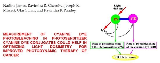
1. Introduction
2. Results and Discussion
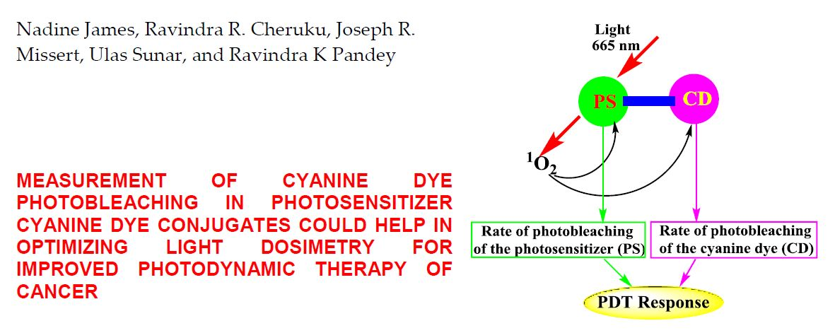
2.1. Mechanism of Photo-Induced Bleaching
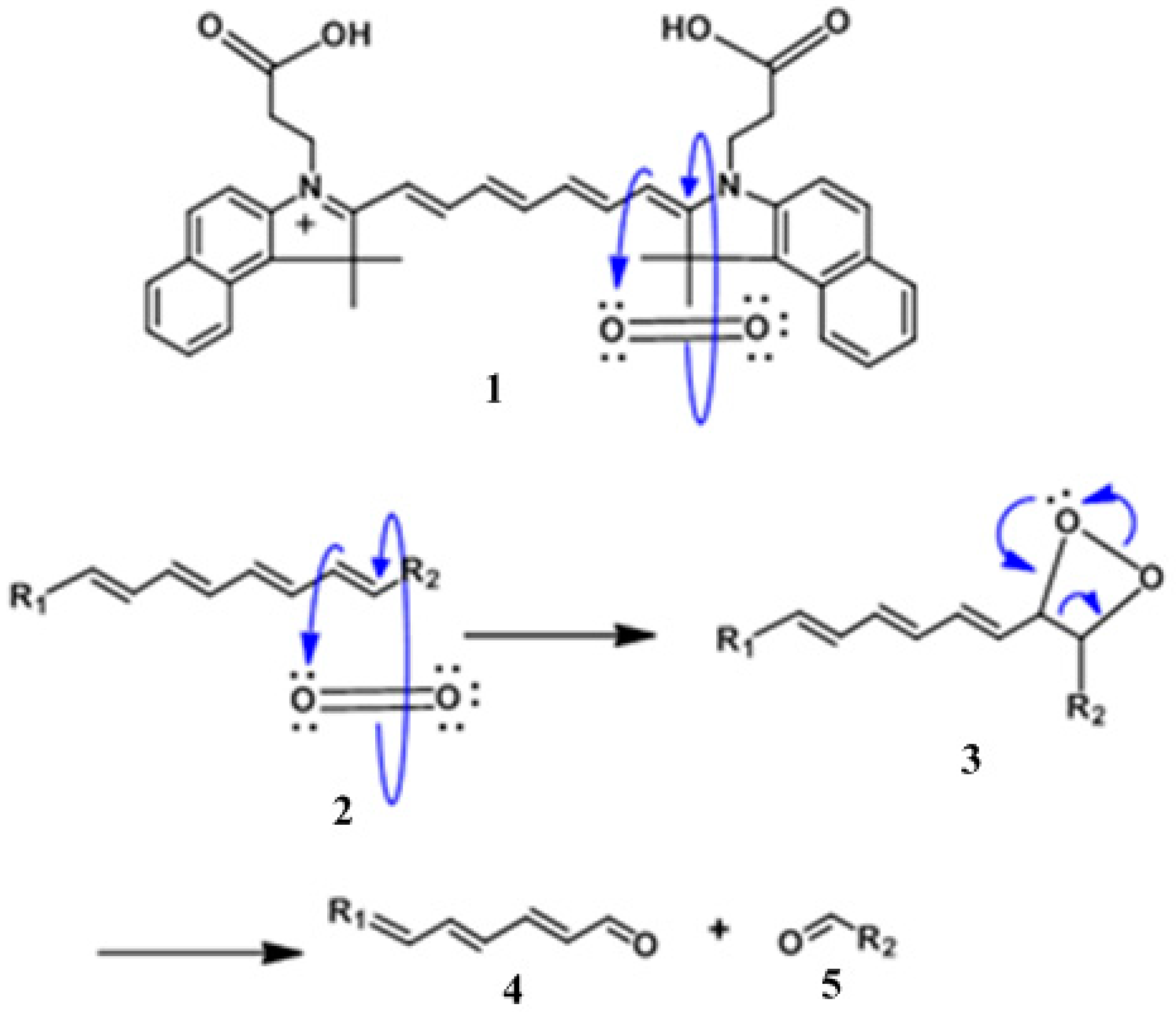
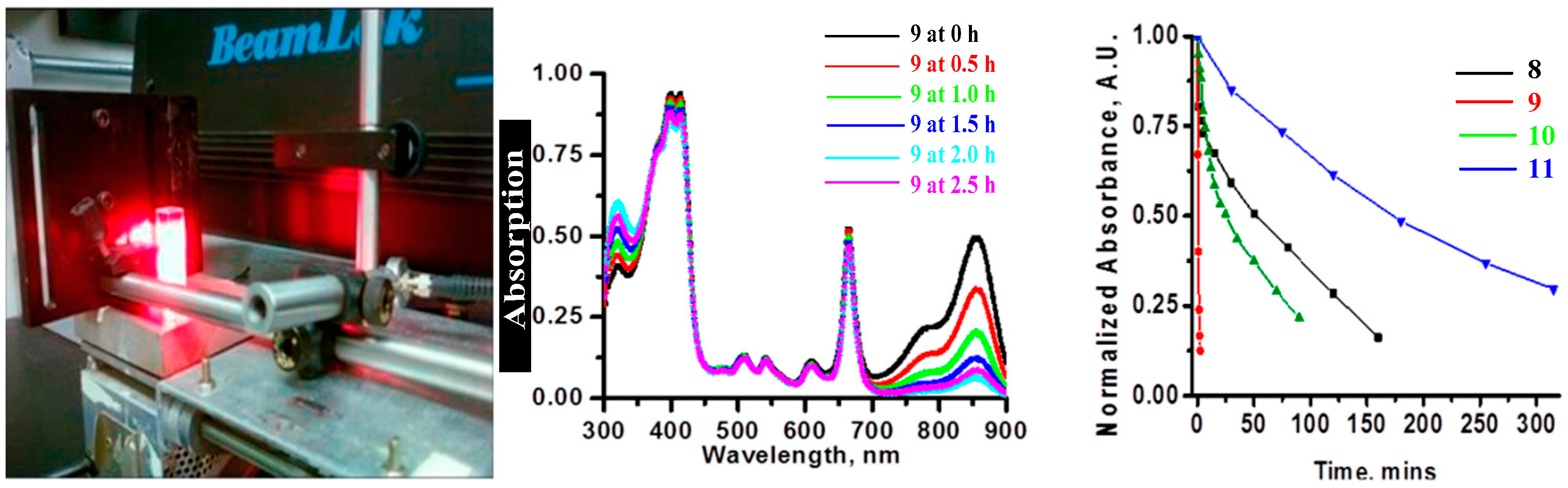
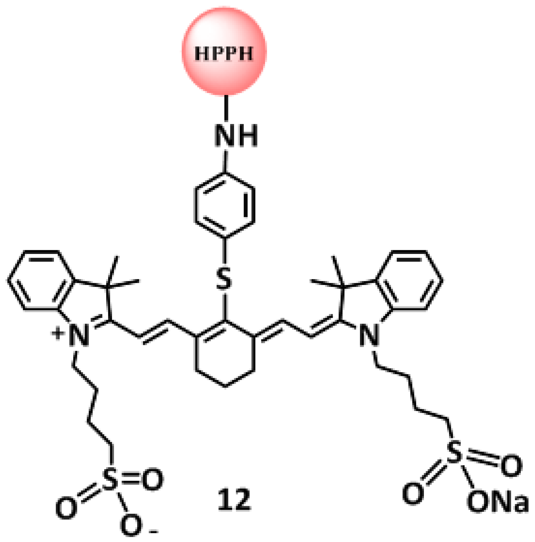
2.2. In Vitro Photobleaching of HPPH Cyanine Dye and CD Conjugates
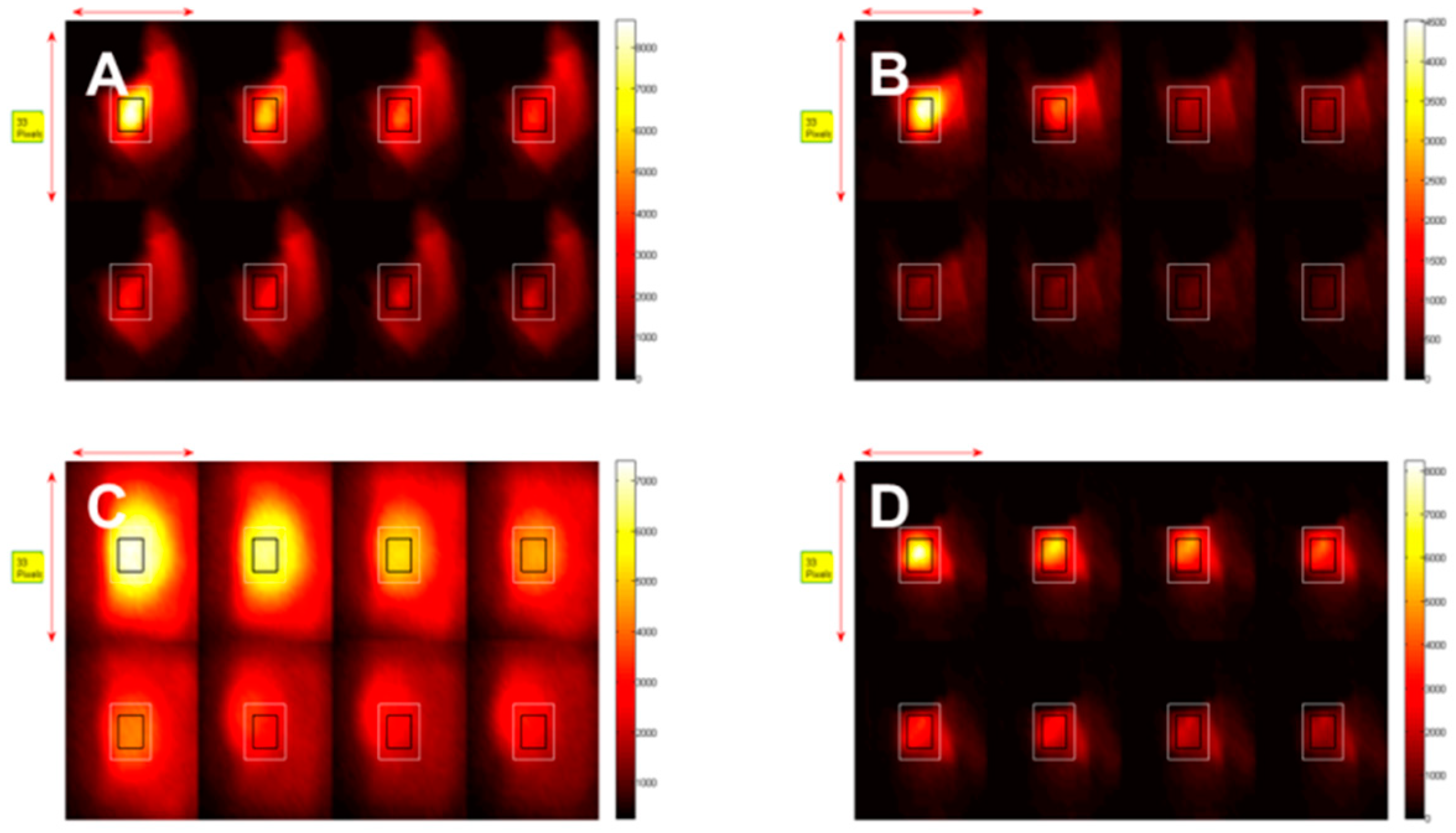
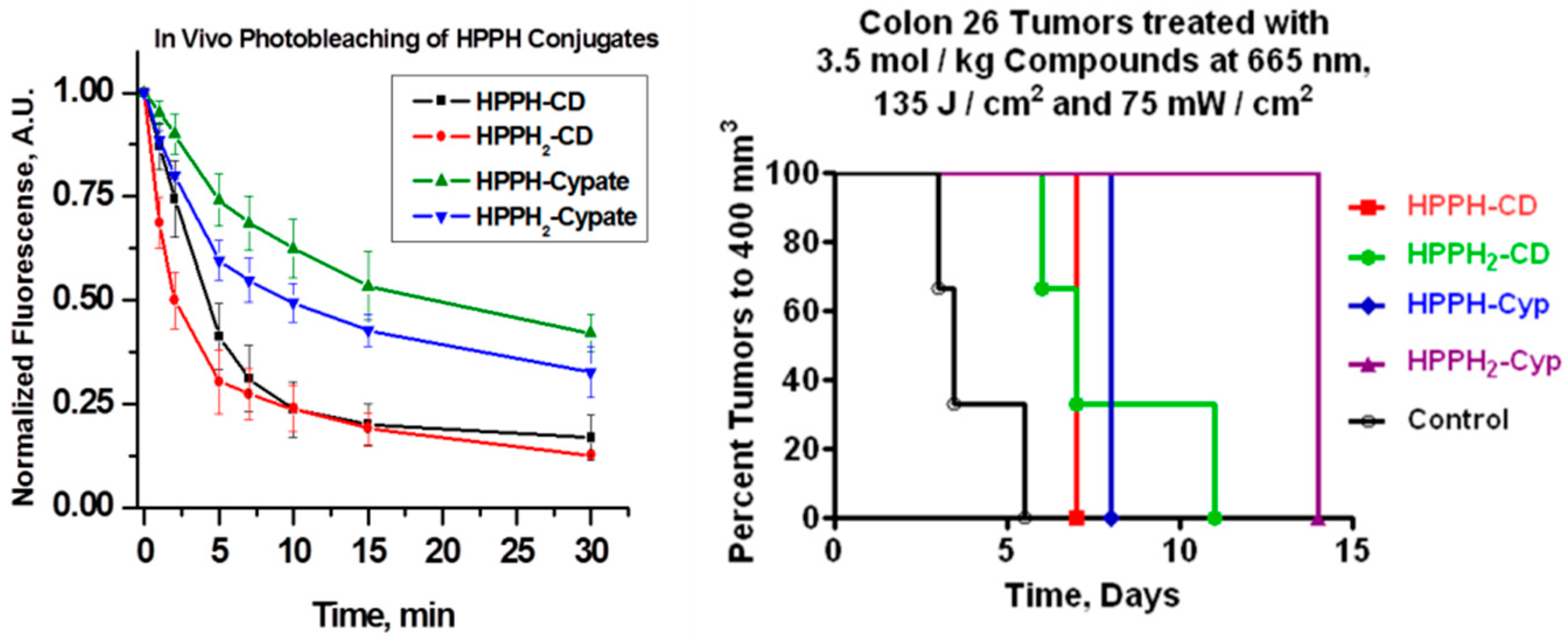
2.3. In Vivo Photo-Induced Bleaching Kinetics
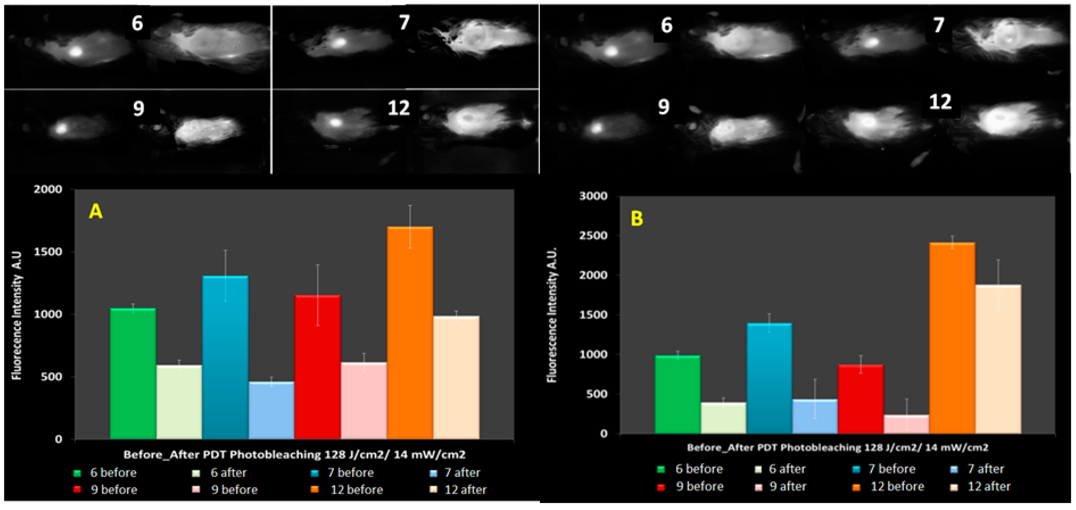
2.4. In Vivo Photobleaching before and after Low Fluence Light Treatments
- (a)
- (b)
- Photophysical characterization: UV-vis absorption spectra were acquired using a Shimadzu UV-3600 spectrophotometer. Fluorescence spectra were recorded using a Fluorolog-3-spectrofluorometer or a SPEX 270M Spectrometer (Jobin Yvon, Longjumeau, France). The SPEX 270 M Spectrometer was utilized for measurement in NIR range; laser lines from Argon ion laser (Spectra Physics) or laser diodes emitting at 630 nm and 785 nm was used as excitation wavelength and the emission was recorded (Table 1, Table 2 and Table 3).
- (c)
- In vitro and in vivo photobleaching: In vitro photobleaching was conducted by observing the UV-vis spectra of conjugates at 5 μM concentrations dissolved either in methanol or in 1% Tween80 formulation diluted with methanol (30-fold, for complete disaggregation of the product). The solution of conjugate in cuvette was then irradiated with light (therapeutic light dose) at various time points for 30 min. At these time intervals, the UV-vis spectrum was taken and the rate of photobleaching (decrease of absorption intensity) at the longest wavelength absorptions of the PS and CD moieties present in PS-CD conjugates were measured and plotted against time.To determine the rate of in vivo photobleaching of the PS and CD moieties during the PDT BALB/c mice (5 mice/group) bearing Colon26 tumors were injected (i.v.) with the conjugate(s) and at 24 h post injection (the time point for maximal uptake of the compound). Three sets of therapeutic light doses (light fluence and fluence rates) used for this study were: (135 J/cm2, 128 J/cm2), 128 J/cm2, 14 mW/cm2) and (48 J/cm2, 7 mW/cm2). The fluorescence intensity was measured using the RED (615–665 nm; 750 nm long-pass) and NIR (710–760 nm; 800 nm long pass) excitation and emission before and after PDT for the PS and CD moieties respectively. These experiments were performed using the drug dose of 1.5 μmol /kg for all the conjugates.
- (d)
- In vivo PDT efficacy: Prior to commencement of in vivo studies, all procedures or protocols were approved by the institutional animal care committee (IACUC). In brief, BALB/c mice 5–8 weeks of age were obtained from NCI Jackson Laboratory. The mice were inoculated subcutaneously (S. C.) on the right posterior shoulder with Colon-26 (1 × 10⁶ cells in 50 μL medium) between 7 and 14 weeks of age. The tumors reaching the appropriate treatment size (4–5 mm diameter), the mice were injected with the conjugate intravenously (i.v.) via tail vein injection. At 24 h post injection, the mice were restrained in plexiglass holders without anesthesia, treated with a 1.1 cm diameter area of drug-activating laser light at 665 nm at different light fluence and fluence rates (Table 1 and Table 2). The mice were observed daily for tumor re-growth and tumor cure. Upon tumor recurrence measurements were taken using two orthogonal measurements length and width (perpendicular to L); volumes of tumors were calculated using the Microsoft Excel formula V = L*W2/2 and recorded. Mice were considered cured if there was no palpable tumor by day 60; however, if the tumor reached 400 mm in size they were euthanized.
- (e)
- Tumor imaging: BALB/c mice bearing Colon-26 tumors (3 mice/group) were injected (i.v.) with the conjugate (s) and imaged at three time points 24, 48 and 72 h after being anesthetized with Ketamine/Xylazine, delivered intraperitoneally or anesthetized with isoflurane. Compounds were imaged using a Maestro GNIR Flex in vivo imaging system using a broadband excitation at 710–740 nm and 800 nm long pass emission.
3. Conclusions
Author Contributions
Funding
Acknowledgments
Conflicts of Interest
References
- Li, B.; Lin, L.; Lin, H.; Wilson, B.C. Photosensitized singlet oxygen generation and detection: Recent advances and future perspectives in cancer photodynamic therapy. J. Biophotonics 2016, 9, 1314–1325. [Google Scholar] [CrossRef] [PubMed]
- Sanchez-Barcelo, E.J.; Mediavilla, M.D. Recent patents on light based therapies: Photodynamic therapy, photothermal therapy and photoimmunotherapy. Phys. Med. Biol. 2014, 8, 1–8. [Google Scholar] [CrossRef]
- Ozog, D.M.; Rkein, A.M.; Fabi, S.G.; Gold, M.H.; Goldman, M.P.; Lowe, N.J.; Martin, G.M.; Munavalli, G.S. Photodynamic Therapy: A Clinical Consensus Guide. Dermatol. Surg. 2016, 42, 804–827. [Google Scholar] [CrossRef] [PubMed]
- Francisco, J.C.; Barış, K.M.; Biel, C.E.; Alessandra, S.R.F.; Saba, R.P. Takes Vincent Vander PoortenAlfio Ferlito. A Review of Photodynamic Therapy for Neoplasms of the Head and Neck. Adv. Ther. 2018, 35, 324–340. [Google Scholar]
- Shafirstein, G.; Battoo, A.; Harris, K.; Baumann, H.; Gollnick, S.O.; Lindenmann, J.; Nwogu, C.E. Photodynamic Therapy of Non-Small Cell Lung Cancer. Narrative Review and Future Directions. Ann. Am. Thorac. Soc. 2016, 13, 265–275. [Google Scholar] [CrossRef] [PubMed]
- Nwogu, C.; Pera, P.; Bshara, W.; Attwood, K.; Pandey, R. Photodynamic therapy of human lung cancer xenografts in mice. J. Surg. Res. 2016, 200, 8–12. [Google Scholar] [CrossRef] [PubMed]
- Davila, M.L. Photodynamic therapy. Gastrointest. Endosc. Clin. N. Am. 2011, 21, 67–79. [Google Scholar] [CrossRef] [PubMed]
- Duvanskii, V.A.; Kniazev, M.V.; Pravednikov, P.V. Modern aspects of photodynamic therapy of esophageal. Eksp. Klin. Gastroenterol. 2011, 10, 111–116. [Google Scholar]
- Fiorelli, A.; Prencipe, A.; Santini, M. The renaissance of photodynamic therapy for early esophageal cancer: Is it the time? J. Thorac. Dis. 2018, 10 (Suppl. 9), S1013–S1015. [Google Scholar] [CrossRef] [PubMed]
- Straten, V.D.; Mashayekhi, V.; de Bruijn, H.S.; Oliveira, S.; Robinson, D.J. Oncologic Photodynamic Therapy: Basic principles, current clinical status and future directions. Cancers 2017, 18, 19. [Google Scholar] [CrossRef] [PubMed]
- Shishkova, N.; Kuznetsova, O.; Berezov, T. Photodynamic therapy in gastroenterology. J. Gastrointest. Cancer 2013, 44, 251–259. [Google Scholar] [CrossRef] [PubMed]
- Nelke, K.H.; Pawlak, W.; Leszczyszyn, J.; Gerber, H. Photodynamic therapy in head and neck cancer. Postepy Hig. Med. Dosw. (Online) 2014, 68, 119–128. [Google Scholar] [CrossRef] [PubMed]
- Dobson, J.; de Queiroz, G.F.; Golding, J.P. Photodynamic therapy and diagnosis: Principles and comparative aspects. Vet. J. 2018, 233, 8–18. [Google Scholar] [CrossRef] [PubMed]
- Wen, P.Y.; Omuro, A.; Ahluwalia, M.S.; Fathallah-Shaykh, H.M.; Mohile, N.; Lager, J.J.; Laird, A.D.; Tang, J.; Jiang, J.; Egile, C.; et al. Phase I dose-escalation study of the PI3K/mTOR inhibitor voxtalisib (SAR245409, XL765) plus temozolomide with or without radiotherapy in patients with high-grade glioma. Neuro-Oncology 2015, 17, 1275–1283. [Google Scholar] [CrossRef] [PubMed]
- Arvind, S.; Arivazhagan, A.; Santosh, V.; Chandramouli, B.A. Differential expression of a novel voltage gated potassium channel–Kv 1.5 in astrocytomas and its impact on prognosis in glioblastoma. Br. J. Neurosurg. 2012, 26, 16–20. [Google Scholar] [CrossRef] [PubMed]
- Gheewala, T.; Skwor, T.; Munirathinam, G. Photosensitizers in Prostate cancer therapy. Oncotarget 2017, 8, 30524–30538. [Google Scholar] [CrossRef] [PubMed]
- Shafirstein, G.; Bellnier, D.; Oakley, E.; Hamilton, S.; Potasek, M.; Beeson, K.; Parilov, E. Interstitial Photodynamic Therapy-A focused Review. Cancers 2017, 9, 12. [Google Scholar] [CrossRef] [PubMed]
- Kawczyk-Krupka, A.; Wawrzyniec, K.; Musiol, S.K.; Potempa, M.; Bugaj, A.M.; Sieroń, A. Treatment of localized prostate cancer using WST-09 and WST-11 mediated vascular targeted photodynamic therapy-A review. Photodiagn. Photodyn. Ther. 2015, 12, 567–574. [Google Scholar] [CrossRef] [PubMed]
- Moore, C.M.; Azzouzi, A.R.; Barret, E.; Villers, A.; Muir, G.H.; Barber, N.J.; Bott, S.; Trachtenberg, J.; Arumainayagam, N.; Gaillac, B.; et al. Determination of optimal drug dose and light dose index to achieve minimally invasive focal ablation of localised prostate cancer using WST11-vascular-targeted photodynamic (VTP) therapy. BJU Int. 2015, 116, 888–896. [Google Scholar] [CrossRef] [PubMed]
- Bugaj, A.M. Vascular targeted photochemotherapy using padoporfin and padeliporfin as a method of the focal treatment of localised prostate cancer-clinician’s insight. J. Photochem. Photobiol. World J. Methodol. 2016, 6, 65–76. [Google Scholar] [CrossRef] [PubMed]
- Spring, B.Q.; Rizvi, I.; Xu, N.; Hasan, T. The role of photodynamic therapy in overcoming cancer drug resistance. Photochem. Photobiol. Sci. 2015, 14, 1476–1491. [Google Scholar] [CrossRef] [PubMed]
- Pandey, R.K.; Kessel, D.; Dougherty, T.J. Handbook of Photodynamic Therapy: Updates on Recent Applications of Porphyrin-Based Compounds; World Scientific: Hackensack, NJ, USA, 2016. [Google Scholar]
- Ascencio, M.; Collinet, P.; Farine, M.O.; Mordon, S. Protoporphyrin IX fluorescence photobleaching is a useful tool to predict the response of rat ovarian cancer following hexaminolevulinate photodynamic therapy. Lasers Surg. Med. 2008, 40, 332–341. [Google Scholar] [CrossRef] [PubMed]
- McIlroy, B.W.; Mann, T.S.; Dysart, J.S.; Wilson, B.C. The effects of oxygenation and photosensitizer substrate binding on the use of fluorescence photobleaching as a dose metric for photodynamic therapy. Vib. Spectrosc. 2002, 28, 25–35. [Google Scholar] [CrossRef]
- Wilson, B.C.; Patterson, M.S.; Lilge, L. Implicit and explicit dosimetry in photodynamic therapy: A New paradigm. Lasers Med. Sci. 1997, 12, 182–199. [Google Scholar] [CrossRef] [PubMed]
- Zhu, T.C.; Finlay, J.C. The role of photodynamic therapy (PDT) physics. Med. Phys. 2008, 35, 3127–3136. [Google Scholar] [CrossRef] [PubMed]
- Patterson, M.S.; Madsen, S.J.; Wilson, B.C. Experimental tests of the feasibility of singlet oxygen luminescence monitoring in vivo during photodynamic therapy. J. Photochem. Photobiol. B Biol. 1990, 5, 69–84. [Google Scholar] [CrossRef]
- Strekowski, L. (Ed.) Synthesis, Properties and Applications. In Heterocyclic Polymethine Dyes; Springer: Berlin, Germany, 2008. [Google Scholar]
- James, N.S.; Chen, Y.; Joshi, P.; Ohulchanskyy, T.Y.; Ethirajan, M.; Henary, M.; Strekowsk, L.; Pandey, R.K. Evaluation of polymethine dyes as potential probes for near infrared fluorescence imaging of tumors: Part-I. Theranostics 2013, 3, 692–702. [Google Scholar] [CrossRef] [PubMed]
- James, N.S.; Ohulchanskyy, T.Y.; Chen, Y.; Joshi, P.; Zheng, X.; Goswami, L.N.; Pandey, R.K. Comparative tumor imaging and PDT efficacy of HPPHconjugated in the mono- and di-forms to various polymethine cyanine dyes: Part-2. Theranostics 2013, 3, 703–718. [Google Scholar] [CrossRef] [PubMed]
- Henderson, B.W.; Busch, T.M.; Snyder, J.W. Fluence rate as a modulator of PDT mechanisms. Lasers Surg. Med. 2006, 38, 489–493. [Google Scholar] [CrossRef] [PubMed]
- Achilefu, S., Bornhop, D.J., Raghavachari, R., Eds.; Molecular Probes for Biomedical Applications II. In Proceedings of the SPIE, San Jose, CA, USA, 21–22 January 2008. [Google Scholar]
- Henderson, B.W.; Gollnick, S.O.; Snyder, J.W.; Busch, T.M.; Kousis, P.C.; Cheney, R.T.; Morgan, J. Choice of Oxygen-Conserving Treatment Regimen Determines the Inflammatory Response and Outcome of Photodynamic Therapy of Tumors. Cancer Res. 2004, 64, 2120–2126. [Google Scholar] [CrossRef] [PubMed]
- Pogue, B.W.; Sheng, C.; Benevides, J.; Forcione, D.; Puricelli, B.; Nishioka, N.; Hasan, T. Protoporphyrin IX fluorescence photobleaching increases with the use of fractionated irradiation in the esophagus. J. Biomed. Opt. 2008, 13, 034009. [Google Scholar] [CrossRef] [PubMed]
- Robinson, D.J.; de Bruijn, H.S.; van der Veen, N.; Stringer, M.R.; Brown, S.B.; Star, W.M. Fluorescence photobleaching of ALA-induced protoporphyrin IX during photodynamic therapy of normal hairless mouse skin: The effect of light dose and irradiance and the resulting biological effect. Photochem. Photobiol. 1998, 67, 140–149. [Google Scholar] [CrossRef] [PubMed]
- Robinson, D.J.; de Bruijn, H.S.; van der Veen, N.; Stringer, M.R.; Brown, S.B.; Star, W.M. Protoporphyrin IX fluorescence photobleaching during ALA-mediated photodynamic therapy of UVB-induced tumors in hairless mouse skin. Photochem. Photobiol. 1999, 69, 61–70. [Google Scholar] [CrossRef] [PubMed]
- Sheng, C.; Hoopes, P.J.; Hasan, T.; Pogue, B.W. Photobleaching-based dosimetry predicts deposited dose in ALA-PpIX PDT of rodent esophagus. Photochem. Photobiol. 2007, 83, 738–748. [Google Scholar] [CrossRef] [PubMed]
- Van Veen, R.L.; Aalders, M.C.; Pasma, K.L.; Siersema, P.D.; Haringsma, J.; van de Vire, W.; Gabeler, E.E.; Robinson, D.J.; Sterenborg, H.J. In situ light dosimetry during photodynamic therapy of Barrett’s esophagus with 5-aminolevulinic acid. Lasers Surg. Med. 2002, 31, 299–304. [Google Scholar] [CrossRef] [PubMed]
- Patterson, M.S.; Wilson, B.C. Theoretical study of the influence of sensitizer photobleaching on depth of necrosis in photodynamic therapy. In Proceedings of the SPIE, Los Angeles, CA, USA, 3 November 1994; pp. 208–219. [Google Scholar]
- Patterson, M.S.; Wilson, B.C.; Graff, R. In vivo tests of the concept of photodynamic threshold dose in normal rat liver photosensitized by aluminum chlorosulfonated phthalocyanine. Photochem. Photobiol. 1990, 51, 343–349. [Google Scholar] [CrossRef] [PubMed]
- Wilson, B.C.; Weersink, R.A.; Lilge, L. Fluorescence in Photodynamic Therapy Dosimetry; Marcel Dekker, Inc.: New York, NY, USA, 2003; pp. 529–561. [Google Scholar]
Sample Availability: Samples of the compounds are not available from the authors. |


| Compounds | Fluorescence (Not an Absolute Value) | Photobleached 128 J/cm2 14 mW/cm2 | % Tumor Cured 128 J/cm2 14 mW/cm2 | |||||
|---|---|---|---|---|---|---|---|---|
| Drug# | Dose (μmol/kg) | Pre PDT HPPH | Pre PDT CD | Post PDT HPPH | Post PDT CD | % HPPH Photo-Bleached | % CD Photo-Bleached | PDT ONLY |
| 6 | 1.5 | 1048 | 989 | 592 | 397 | 43 | 60 | 80 |
| 7 | 1.5 | 1308 | 1396 | 460 | 439 | 65 | 69 | 80 |
| 9 | 1.5 | 1154 | 874 | 616 | 239 | 47 | 73 | 80 |
| 12 | 1.5 | 1701 | 2413 | 986 | 1881 | 42 | 22 | 40 |
| Compounds | Fluorescence (Not an Absolute Value) | Photobleached 48 J/cm2 7 mW/cm2 | % Tumor Cured 48 J/cm2 7 mW/cm2 | |||||
|---|---|---|---|---|---|---|---|---|
| Drug# | Dose (μmol/kg) | Pre PDT HPPH | Pre PDT CD | Post PDT HPPH | Post PDT CD | % HPPH Photo-Bleached | % CD Photo-Bleached | PDT ONLY |
| 6 | 1.5 | 1060 | 976 | 825 | 492 | 22 | 50 | ND |
| 7 | 1.5 | 1485 | 1319 | 1170 | 1019 | 21 | 23 | ND |
| 9 | 1.5 | 986 | 809 | 808 | 459 | 18 | 43 | ND |
| 12 | 1.5 | 1428 | 2538 | 1238 | 1783 | 13 | 30 | ND |
| Compounds | Fluorescence (Not an Absolute Value) | Photobleached 135 J/cm2 75 mW/cm2 | % Tumor Cured 135 J/cm2 75 mW/cm2 | |||||
|---|---|---|---|---|---|---|---|---|
| Drug# | Dose (μmol/kg) | Pre PDT HPPH | Pre PDT CD | Post PDT HPPH | Post PDT CD | % HPPH Photo-Bleached | % CD Photo-Bleached | % Tumor Cure |
| 6 | 1.5 | 1136 | 1025 | 670 | 416 | 41 | 60 | 0 |
| 7 | 1.5 | 1380 | 1319 | 655 | 560 | 53 | 58 | 0 |
| 9 | 1.5 | 996 | 904 | 683 | 430 | 31 | 52 | 33 |
| 12 | 1.5 | 1392 | 2543 | 1390 | 2143 | 8 | 16 | 66 |
© 2018 by the authors. Licensee MDPI, Basel, Switzerland. This article is an open access article distributed under the terms and conditions of the Creative Commons Attribution (CC BY) license (http://creativecommons.org/licenses/by/4.0/).
Share and Cite
James, N.S.; Cheruku, R.R.; Missert, J.R.; Sunar, U.; Pandey, R.K. Measurement of Cyanine Dye Photobleaching in Photosensitizer Cyanine Dye Conjugates Could Help in Optimizing Light Dosimetry for Improved Photodynamic Therapy of Cancer. Molecules 2018, 23, 1842. https://doi.org/10.3390/molecules23081842
James NS, Cheruku RR, Missert JR, Sunar U, Pandey RK. Measurement of Cyanine Dye Photobleaching in Photosensitizer Cyanine Dye Conjugates Could Help in Optimizing Light Dosimetry for Improved Photodynamic Therapy of Cancer. Molecules. 2018; 23(8):1842. https://doi.org/10.3390/molecules23081842
Chicago/Turabian StyleJames, Nadine S., Ravindra R. Cheruku, Joseph R. Missert, Ulas Sunar, and Ravindra K. Pandey. 2018. "Measurement of Cyanine Dye Photobleaching in Photosensitizer Cyanine Dye Conjugates Could Help in Optimizing Light Dosimetry for Improved Photodynamic Therapy of Cancer" Molecules 23, no. 8: 1842. https://doi.org/10.3390/molecules23081842
APA StyleJames, N. S., Cheruku, R. R., Missert, J. R., Sunar, U., & Pandey, R. K. (2018). Measurement of Cyanine Dye Photobleaching in Photosensitizer Cyanine Dye Conjugates Could Help in Optimizing Light Dosimetry for Improved Photodynamic Therapy of Cancer. Molecules, 23(8), 1842. https://doi.org/10.3390/molecules23081842