Stability of Antibacterial Silver Carboxylate Complexes against Staphylococcus epidermidis and Their Cytotoxic Effects

 , ,
, , 
Abstract

1. Introduction
2. Results
2.1. Stability of Silver Complexes
2.2. Antibacterial Experiments
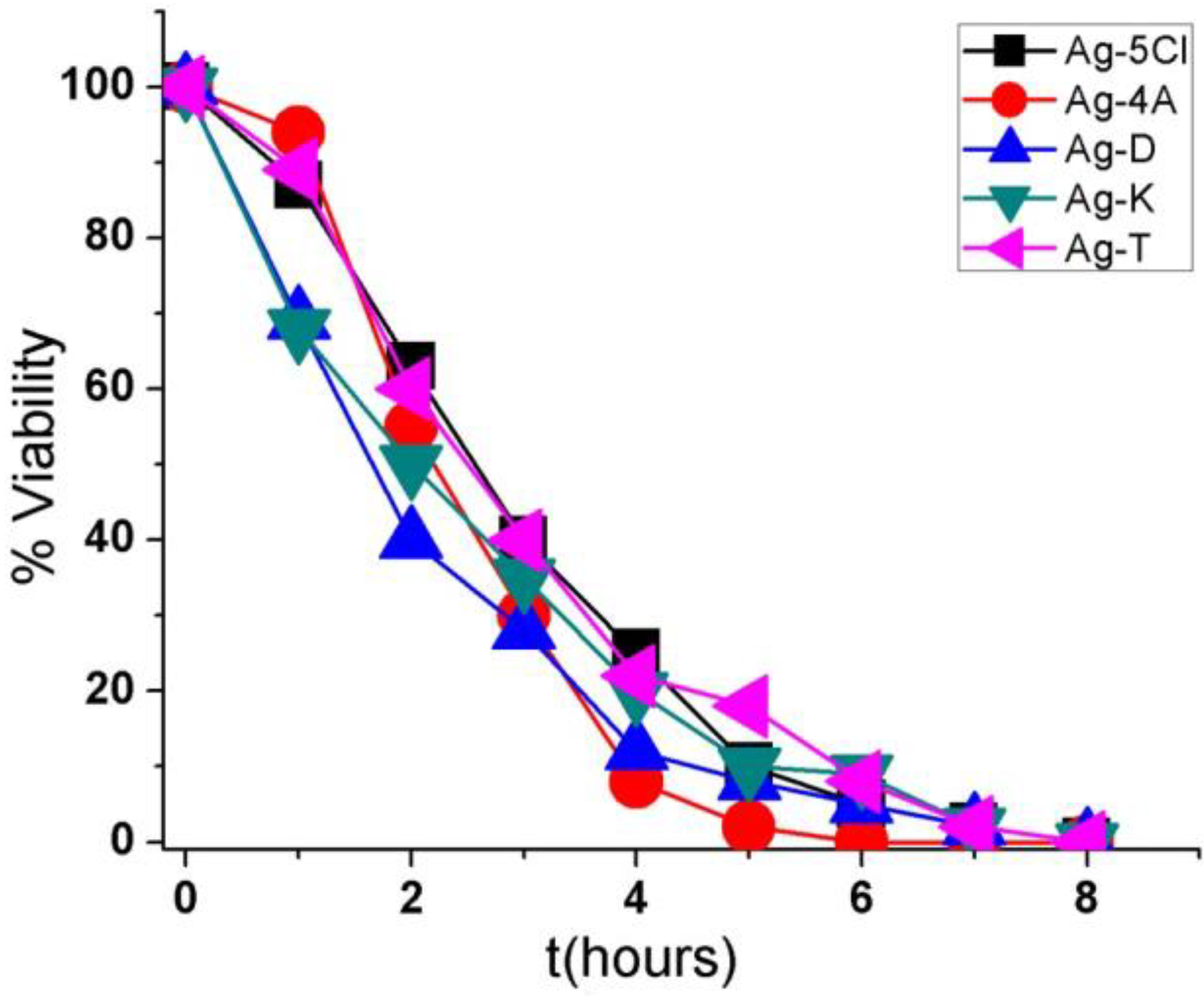
2.3. Viability Test
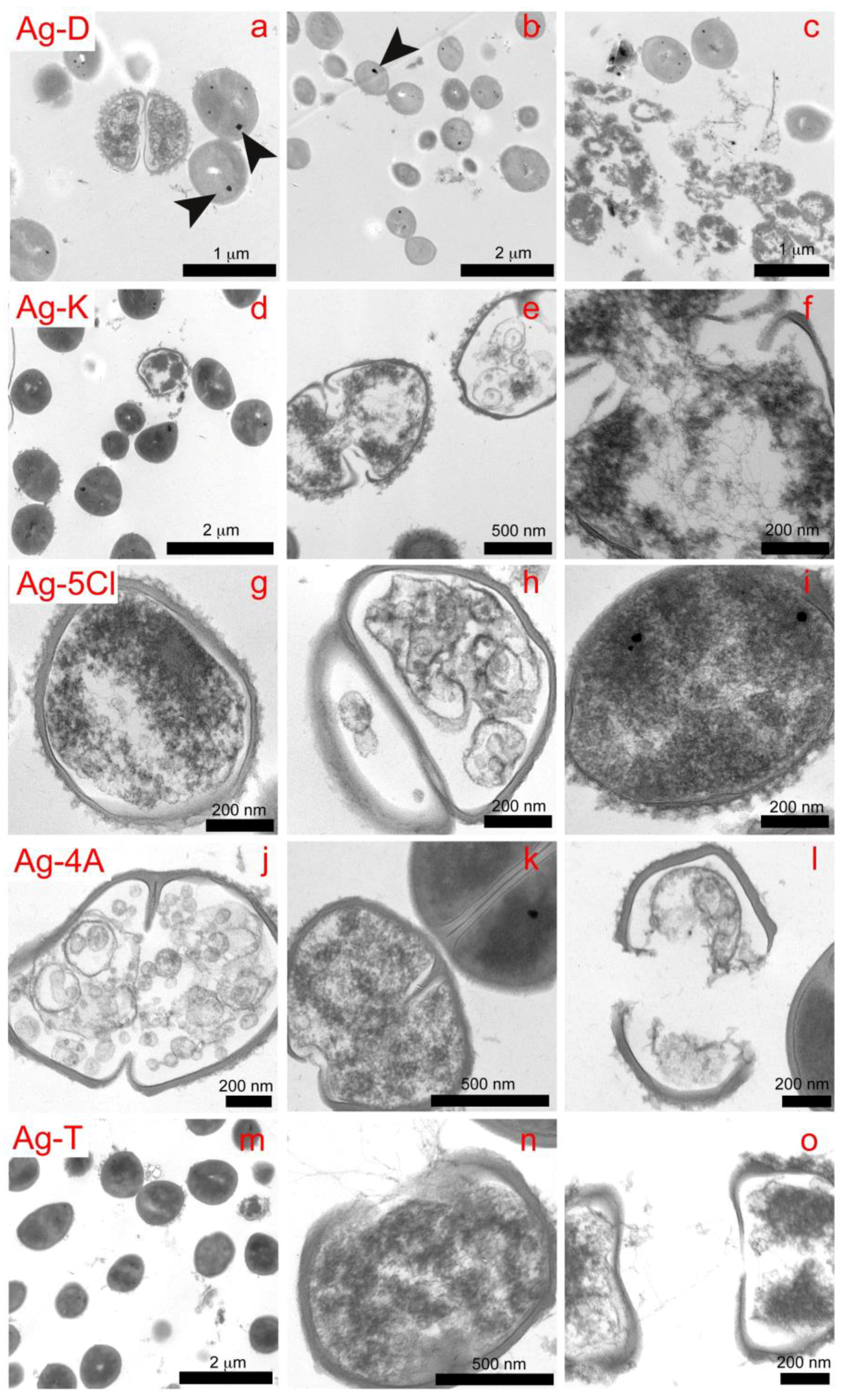
2.4. TEM
2.5. Scanning Electron Microscopy (SEM)
2.6. Cell Viability Assay
3. Materials and Methods
3.1. Synthesis and Stability of Silver Complexes
3.2. Antibacterial Activity
3.3. Morphological Changes by TEM/SEM Images
3.4. Cell Viability Assay
4. Conclusions
Author Contributions
Funding
Acknowledgments
Conflicts of Interest
References
- Serenella, M.; Massimiliano, P.; Guido, C.; Valeria, M.N.; Joanna, I.L.; Maurizio, R.; Maria, A.Z.; Zoroddu, A.M.; Zoroddu, A.M. Silver coordination compounds: A new horizon in medicine. Coord. Chem. Rev. 2016, 327–328, 349–359. [Google Scholar]
- Azócar, M.I.; Gómez, G.; Levín, P.; Paez, M.; Muñoz, H.; Dinamarca, N. Review: Antibacterial behavior of carboxylate silver(I) complexes. J. Coord. Chem. 2014, 67, 23–24. [Google Scholar] [CrossRef]
- Nomiya, K.; Yoshizawa, A.; Tsukagoshi, K.; Kasuga, N.C.; Hirakawa, S.; Watanabe, J. Synthesis and structural characterization of silver(I), aluminium(III) and cobalt(II) complexes with 4-isopropyltropolone (hinokitiol) showing noteworthy biological activities. Action of silver(I)-oxygen bonding complexes on the antimicrobial activities. J. Inorg. Biochem. 2004, 98, 46–60. [Google Scholar] [CrossRef] [PubMed]
- Gerasimchuk, N. Synthesis, Properties, and Applications of Light-Insensitive Silver(I) Cyanoximates. Eur. J. Inorg. Chem. 2014, 2014, 4518–4531. [Google Scholar] [CrossRef]
- Gerasimchuk, N.; Gamian, A.; Glover, G.; Szponar, B. Light Insensitive Silver(I) Cyanoximates As Antimicrobial Agents for Indwelling Medical Devices. Inorg. Chem. 2010, 49, 9863–9874. [Google Scholar] [CrossRef] [PubMed]
- Soliman, S.M.; Elsilk, S.E. Synthesis, structural analyses and antimicrobial activity of the water soluble 1D coordination polymer [Ag(3-aminopyridine)]ClO4. J. Mol. Struct. 2017, 1149, 58–68. [Google Scholar] [CrossRef]
- Azocar, M.I.; Muñoz, H.; Levin, P.; Dinamarca, N.; Gomez, G.; Ibañez, A.; Garland, M.T.; Paez, M.A. Synthesis and characterization of silver(I) complexes with ligands having anti-inflammatory properties. Commun. Inorg. Synth. 2013, 1, 19–21. [Google Scholar]
- Otto, M. Staphylococcus epidermidis pathogenesis. In Staphylococcus Epidermidis: Methods in Molecular Biology (Methods and Protocols); Fey, P., Ed.; Springer: New York, NY, USA, 2014; Volume 1106, pp. 17–31. [Google Scholar]
- Schaeffer, C.; Woods, K.M.; Longo, G.M.; Kiedrowski, M.R.; Paharik, A.E.; Büttner, H.; Christner, M.; Boissy, R.J.; Horswill, A.R.; Rohde, H.; et al. Accumulation-associated protein enhances Staphylococcus epidermidis biofilm formation under dynamic conditions and is required for infection in a rat catheter model. Infect. Immun. 2015, 83, 211–226. [Google Scholar] [CrossRef] [PubMed]
- Azócar, M.I.; Aldabaldetrecu, M.; Levin, P.; Tamayo, L.; Guerrero, J.; Páez, M.A. Correlating light and thermal stability of silver carboxylate complexes by infrared and 13C NMR spectroscopy. J. Coord. Chem. 2016, 69, 3472–3479. [Google Scholar] [CrossRef]
- Marchetti, F.; Palmucci, J.; Pettinari, C.; Pettinari, R.; Scuri, S.; Grappasonni, I.; Cocchioni, M.; Amati, M.; Lelj, F.; Crispini, A. Linkage Isomerism in Silver Acylpyrazolonato Complexes and Correlation with Their Antibacterial Activity. Inorg. Chem. 2016, 55, 5453–5466. [Google Scholar] [CrossRef] [PubMed]
- Sevim, H.A.; Bahtiyar, S.; Sema, C.; Orhan, B. Synthesis, characterization, photoluminescent properties and antimicrobial activities of two novel polymeric silver(I) complexes with diclofenac. J. Mol. Struct. 2017, 1130, 156–164. [Google Scholar]
- Braga, D.; Grepioni, F.; Andre, V.; Duarte, M.T. Drug-containing coordination and hydrogen bonding networks obtained mechanochemically. CrystEngComm 2009, 11, 2618–2621. [Google Scholar] [CrossRef]
- Wang, R.; Hong, M.; Zhao, Y.; Weng, J.; Cao, R. Synthesis and crystal structure of a novel two-dimensional corrugated coordination polymer. Inorg. Chem. Commun. 2002, 5, 487–489. [Google Scholar] [CrossRef]
- Glemser, O.; Sauer, H. Silver Oxide. In Handbook of Preparative Inorganic Chemistry; Brauer, G., Ed.; Academic Press: Cambridge, MA, USA, 1963; p. 1037. [Google Scholar]
- Antigoni, A.; Evroula, H.; Nikolaos, P.X.; Dionissios, M.; Despo, F.K. Factors affecting diclofenac decomposition in water by UV-A/TiO2 photocatalysis. Chem. Eng. J. 2010, 161, 53–59. [Google Scholar]
- Jivani, S.G.; Stella, V.J. Mechanism of Decarboxylation of p-Aminosalicylic Acid. J. Pharm. Sci. 1985, 74, 1274–1282. [Google Scholar] [CrossRef] [PubMed]
- Leo, G.; Hi-Shi, C.; Becker, A. Kinetics and mechanisms of the autoxidation of ketorolac tromethamine in aqueous solution. Int. J. Pharm. 1988, 41, 95–104. [Google Scholar] [CrossRef]
- Leo, G.; Hi-Shi, C.; Johnson, D. Light degradation of ketorolac tromethamine. Int. J. Pharm. 1988, 41, 105–113. [Google Scholar] [CrossRef]
- Foye, W.; Turcotte, J. Stability of metal complexes of nuclear-substituted salicylic acids: Correlation with biological effects. J. Pharm. Sci. 1962, 51, 329–332. [Google Scholar] [CrossRef] [PubMed]
- Kantouch, A.; El-Sayed, A.A.; Salama, M.; El-Kheir, A.A.; Mowafi, S. Salicylic acid and some of its derivatives as antibacterial agents for viscose fabric. Int. J. Biol. Macromol. 2013, 62, 603–607. [Google Scholar] [CrossRef] [PubMed]
- Liu, J.; Hurt, R.H. Ion Release Kinetics and Particle Persistence in Aqueous Nano-Silver Colloids. Environ. Sci. Technol. 2010, 44, 2169–2175. [Google Scholar] [CrossRef] [PubMed]
- Fox, C.L.; Modak, S. Mechanism of Silver Sulfadiazine Action on Burn Wound Infections. Antimicrob. Agents Chemother. 1974, 5, 582–588. [Google Scholar] [CrossRef] [PubMed]
- Azocar, M.I.; Gómez, G.; Velasquez, C.; Abarca, R.; Kogan, M.J.; Paez, M. Antibacterial, kinetics and bacteriolytic properties of silver(I) pyridinedicarboxylate compounds. Mater. Sci. Eng. C 2014, 37, 356–362. [Google Scholar] [CrossRef] [PubMed]
- Abarca, R.; Gómez, G.; Velásquez, C.; Páez, M.A.; Gulppi, M.; Arrieta, A.; Azócar, M.I. Antibacterial behavior of Pyridinecarboxylatesilver(I) complexes. Chin. J. Chem. 2012, 30, 1631–1635. [Google Scholar] [CrossRef]
- Abu-Youssef, M.A.M.; Soliman, S.M.; Langer, V.; Gohar, Y.M.; Hasanen, A.A.; Makhyoun, M.A.; Zaky, A.H.; Öhrström, L.R. Synthesis, Crystal Structure, Quantum Chemical Calculations, DNA Interactions, and Antimicrobial Activity of [Ag(2-amino-3-methylpyridine)2]NO3 and [Ag(pyridine-2-carboxaldoxime)NO3]. Inorg. Chem. 2010, 49, 9788–9797. [Google Scholar] [CrossRef] [PubMed]
- Abu-Youssef, M.A.M.; Langer, V.; Ohrstrom, L. Synthesis, a case of isostructural packing, and antimicrobial activity of silver(I)quinoxaline nitrate, silver(I)(2,5-dimethylpyrazine) nitrate and two related silver aminopyridine compounds. Dalton Trans. 2006, 2542–2550. [Google Scholar] [CrossRef] [PubMed]
- Li, G.Y.; Guan, R.L.; Ji, L.N.; Chao, H. DNA condensation induced by metal complexes. Coord. Chem. Rev. 2014, 281, 100–113. [Google Scholar] [CrossRef]
- Azócar, M.I.A.; Tamayo, L.; Vejar, N.; Gomez, G.; Zhou, X.; Thompsom, G.; Cerda, E.; Kogan, M.J.; Salas, E.; Paez, M.A. A systematic study of antibacterial silver nanoparticles: Efficiency, enhanced permeability, and cytotoxic effects. J. Nanopart. Res. 2014, 16, 2465. [Google Scholar] [CrossRef]
- Allison, D.G.; Maira-Litran, T.; Gilbert, P. Antimicrobial Resistance of Biofilms; Harwood Academic: Amsterdam, The Netherlands, 2000; pp. 149–166. [Google Scholar]
- Mah, T.F.; O’Toole, G.A. Mechanisms of biofilm resistance to antimicrobial agents. Trends Microbiol. 2001, 9, 34–39. [Google Scholar] [CrossRef]
- Chaw, K.C.; Manimaran, M.; Tay, F.E. Role of silver ions in destabilization of intermolecular adhesion forces measured by atomic force microscopy in Staphylococcus epidermidis biofilms. Antimicrob. Agents Chemother. 2005, 49, 4853–4859. [Google Scholar] [CrossRef] [PubMed]
- Lewis, K. Riddle of Biofilm Resistance. Antimicrob. Agents Chemother. 2001, 45, 999–1007. [Google Scholar] [CrossRef] [PubMed]
- Klasen, H.J. A historical review of the use of silver in the treatment of burns. II. Renewed interest for silver. Burns 2000, 26, 131–138. [Google Scholar] [PubMed]
- Schierholz, J.M.; Beuth, J.; Pulverer, G. Role of Silver Ions in Destabilization of Intermolecular Adhesion Forces Measured by Atomic Force Microscopy in Staphylococcus epidermidis Biofilms. J. Antimicrob. Chemother. 1999, 43, 2819–2821. [Google Scholar]
- Mazumder, M.U.; Sukul, A.; Saha, S.K.; Chowdhury, A.A.; Mamun, Y. A comprehensive in vitro biological investigation of metal complexes of tolfenamic acid. Alex. J. Med. 2017, 54, 23–26. [Google Scholar] [CrossRef]
- Jittreetat, T.; Shin, Y.S.; Hwang, H.S.; Lee, B.S.; Kim, Y.S.; Sannikorn, P.; Kim, C.H. Tolfenamic Acid Inhibits the Proliferation, Migration, and Invasion of Nasopharyngeal Carcinoma: Involvement of p38-Mediated Down-Regulation of Slug. Yonsei Med. J. 2016, 57, 588–598. [Google Scholar] [CrossRef] [PubMed]
- Tamayo, L.A.; Zapata, P.A.; Vejar, N.D.; Azócar, M.I.; Gulppi, M.A.; Zhou, X.; Thompson, G.E.; Rabagliati, F.M.; Páez, M.A. Release of silver and copper nanoparticles from polyethylene nanocomposites and their penetration into Listeria monocytogenes. Mater. Sci. Eng. C 2014, 40, 24–31. [Google Scholar] [CrossRef] [PubMed]
Sample Availability: Samples of the compounds are available from the authors. |


| Points | Elements | ||||
|---|---|---|---|---|---|
| C | O | S | Cu | Ag | |
| 2356 | 80.15 | 16.36 | 0.24 | 2.21 | 1.04 |
| 2357 | 80.86 | 15.58 | 0.56 | 1.69 | 0.31 |
| 2358 | 80.96 | 16.99 | 0.34 | 1.53 | 0.19 |
| 2359 | 81.59 | 16.00 | 0.41 | 1.72 | 0.27 |
© 2018 by the authors. Licensee MDPI, Basel, Switzerland. This article is an open access article distributed under the terms and conditions of the Creative Commons Attribution (CC BY) license (http://creativecommons.org/licenses/by/4.0/).
Share and Cite
Aldabaldetrecu, M.; Tamayo, L.; Alarcon, R.; Walter, M.; Salas-Huenuleo, E.; Kogan, M.J.; Guerrero, J.; Paez, M.; Azócar, M.I. Stability of Antibacterial Silver Carboxylate Complexes against Staphylococcus epidermidis and Their Cytotoxic Effects. Molecules 2018, 23, 1629. https://doi.org/10.3390/molecules23071629
Aldabaldetrecu M, Tamayo L, Alarcon R, Walter M, Salas-Huenuleo E, Kogan MJ, Guerrero J, Paez M, Azócar MI. Stability of Antibacterial Silver Carboxylate Complexes against Staphylococcus epidermidis and Their Cytotoxic Effects. Molecules. 2018; 23(7):1629. https://doi.org/10.3390/molecules23071629
Chicago/Turabian StyleAldabaldetrecu, Maialen, Laura Tamayo, Romina Alarcon, Mariana Walter, Edison Salas-Huenuleo, Marcelo J. Kogan, Juan Guerrero, Maritza Paez, and Manuel I. Azócar. 2018. "Stability of Antibacterial Silver Carboxylate Complexes against Staphylococcus epidermidis and Their Cytotoxic Effects" Molecules 23, no. 7: 1629. https://doi.org/10.3390/molecules23071629
APA StyleAldabaldetrecu, M., Tamayo, L., Alarcon, R., Walter, M., Salas-Huenuleo, E., Kogan, M. J., Guerrero, J., Paez, M., & Azócar, M. I. (2018). Stability of Antibacterial Silver Carboxylate Complexes against Staphylococcus epidermidis and Their Cytotoxic Effects. Molecules, 23(7), 1629. https://doi.org/10.3390/molecules23071629