
Circular Dichroism in Fluorescence Emission Following the C 1s→π* Excitation and Resonant Auger Decay of Carbon Monoxide

 , , and
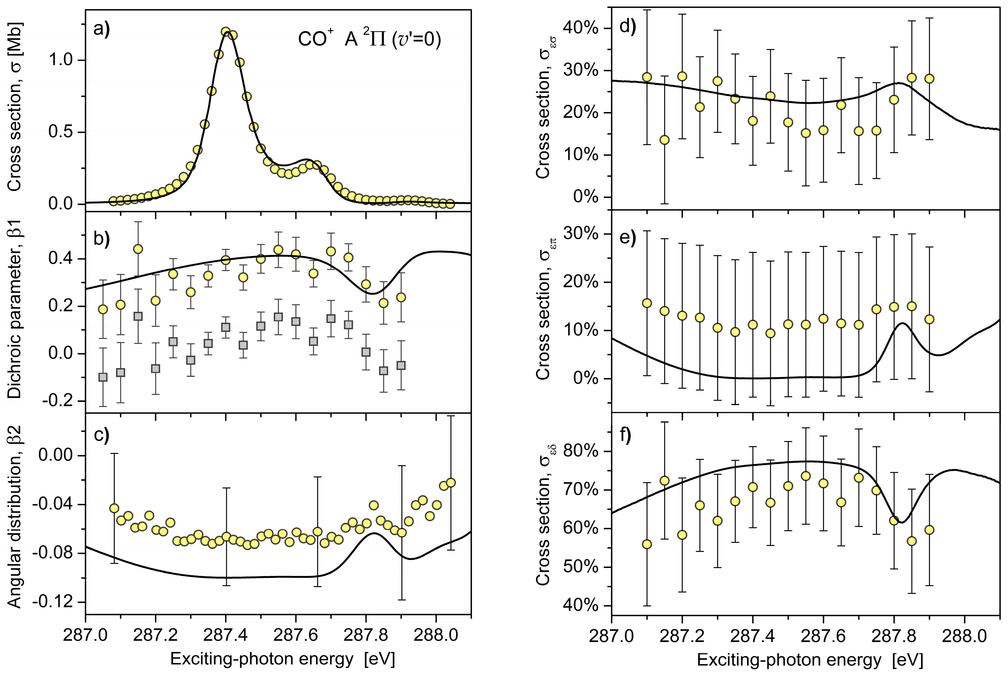
Abstract
1. Introduction
2. Results and Discussion
3. Materials and Methods
3.1. Theory
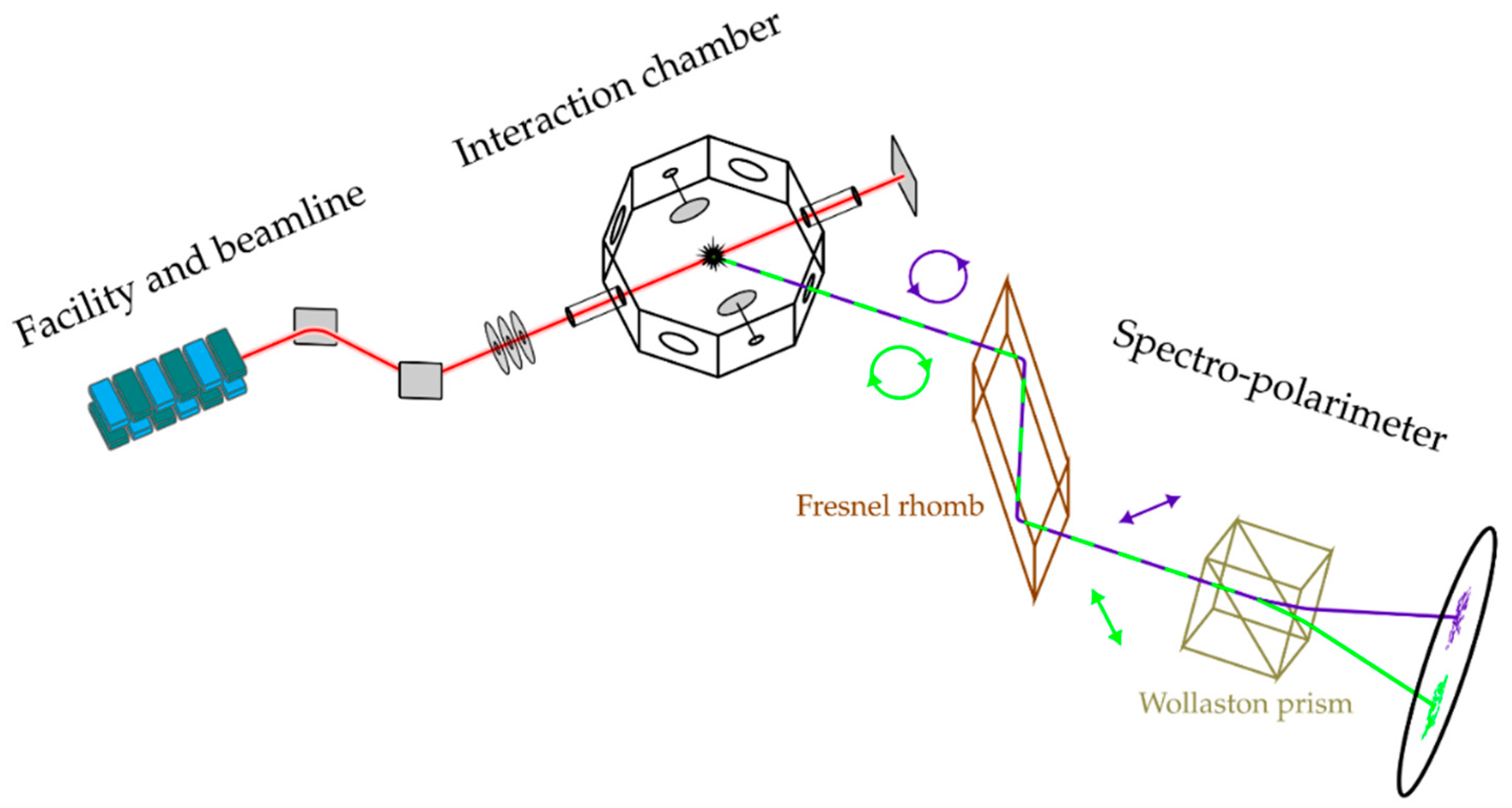
3.2. Experiment
3.3. Data Analysis
4. Conclusions and Outlook
Author Contributions
Funding
Acknowledgments
Conflicts of Interest
References
- Bethe, H.A.; Salpeter, E.E. Quantum Mechanics of One- and Two-Electron Atoms; Springer: Berlin, Germany, 1957; ISBN 978-3-662-12869-5. [Google Scholar]
- Schmidt, V. Photoionization of atoms using synchrotron radiation. Rep. Prog. Phys. 1992, 55, 1483–1659. [Google Scholar] [CrossRef]
- Cherepkov, N.A. Theory of spin polarisation phenomena in molecular photoionisation processes. J. Phys. B 1981, 14, 2165–2177. [Google Scholar] [CrossRef]
- Cherepkov, N.A. Circular dichroism of molecules in the continuous absorption region. Chem. Phys. Lett. 1982, 87, 344–348. [Google Scholar] [CrossRef]
- Snell, G.; Langer, B.; Drescher, M.; Müller, N.; Zimmermann, B.; Hergenhahn, U.; Viefhaus, J.; Heinzmann, U.; Becker, U. Complete Description of the Xe 4d Photoionization by Spin-Resolved Photoelectron and Auger Spectroscopy. Phys. Rev. Lett. 1999, 82, 2480–2483. [Google Scholar] [CrossRef]
- Schmidtke, B.; Khalil, T.; Drescher, M.; Müller, N.; Kabachnik, N.M.; Heinzmann, U. Testing the feasibility of a complete Auger decay experiment by spin- and angle-resolved electron spectroscopy on Xe N4O2,3O2,3 3P1. J. Phys. B 2000, 33, 5225–5242. [Google Scholar] [CrossRef]
- Schmidtke, B.; Khalil, T.; Drescher, M.; Müller, N.; Kabachnik, N.M.; Heinzmann, U. The Kr M4,5N1N2,3 1P1 Auger decay: Measurement of the transferred spin polarization and analysis of Auger amplitudes. J. Phys. B 2001, 34, 4293–4310. [Google Scholar] [CrossRef]
- Lagutin, B.M.; Petrov, I.D.; Sukhorukov, V.L.; Kammer, S.; Mickat, S.; Schill, R.; Schartner, K.-H.; Ehresmann, A.; Shutov, Y.A.; Schmoranzer, H. Raman regime energy dependence of alignment and orientation of kr ii states populated by resonant auger effect. Phys. Rev. Lett. 2003, 90, 073001. [Google Scholar] [CrossRef] [PubMed]
- Lagutin, B.M.; Petrov, I.D.; Sukhorukov, V.L.; Demekhin, P.V.; Zimmermann, B.; Mickat, S.; Kammer, S.; Schartner, K.-H.; Ehresmann, A.; Shutov, Y.A.; et al. The interference effects in the alignment and orientation of the Kr II 4p45p states following Kr I 3d9np resonance excitation. J. Phys. B 2003, 36, 3251–3268. [Google Scholar] [CrossRef]
- Schill, R.; Hasselkamp, D.; Kammer, S.; Mickat, S.; Zimmermann, B.; Schartner, K.-H.; Ehresmann, A.; Schmoranzer, H.; Schlüter, M.; Shutov, Y.A.; et al. Partial wave analysis of the Kr I 3d95/25p3/2→Kr II 4p4(1D)5p 2F7/2 decay, based on orientation and alignment transfer. J. Phys. B 2003, 36, L57–L61. [Google Scholar] [CrossRef]
- Schartner, K.-H.; Schill, R.; Hasselkamp, D.; Mickat, S.; Kammer, S.; Werner, L.; Klumpp, S.; Ehresmann, A.; Schmoranzer, H.; Lagutin, B.M.; et al. Partial wave analysis of interfering Kr 3d95p resonant Raman Auger transitions based on measurements of alignment and orientation parameters within the natural line width. J. Phys. B 2005, 38, 4155–4170. [Google Scholar] [CrossRef]
- Schartner, K.-H.; Schill, R.; Hasselkamp, D.; Mickat, S.; Kammer, S.; Werner, L.; Klumpp, S.; Ehresmann, A.; Schmoranzer, H.; Lagutin, B.M.; et al. Interference between resonant Raman Auger decay and direct excitation manifested in orientation and alignment of KrII 4p4(1D)5p 2P3/2 ions. J. Phys. B 2007, 40, 1443–1450. [Google Scholar] [CrossRef]
- Demekhin, P.V.; Petrov, I.D.; Ehresmann, A. Partial-photoelectron-wave analysis in diatomic molecule photoionization by fluorescence polarization experiments. Phys. Rev. A 2010, 82, 041401. [Google Scholar] [CrossRef]
- Demekhin, P.V.; Petrov, I.D.; Sukhorukov, V.L.; Kielich, W.; Reiß, P.; Hentges, R.; Haar, I.; Schmoranzer, H.; Ehresmann, A. Interference effects during the Auger decay of the C*O(1s−1π*) resonance studied by angular distribution of the CO+(A) photoelectrons and polarization analysis of the CO+(A→X) fluorescence. Phys. Rev. A 2009, 80, 063425. [Google Scholar] [CrossRef]
- Cherepkov, N.A.; Semenov, S.K. On a complete experiment on photoionization of atoms. J. Phys. B 2004, 37, 1267–1272. [Google Scholar] [CrossRef]
- Krupenie, P.H. The band spectrum of carbon monoxide. Natl. Stand. Ref. Data Ser. 1966, 5, 1–87. [Google Scholar]
- Blum, K. Density Matrix Theory and Applications, 2nd ed.; Plenum: New York, NY, USA, 1996; ISBN 978-1-4757-4931-1. [Google Scholar]
- Demekhin, P.V.; Ehresmann, A.; Sukhorukov, V.L. Single center method: A computational tool for ionization and electronic excitation studies of molecules. J. Chem. Phys. 2011, 134, 024113. [Google Scholar] [CrossRef] [PubMed]
- Demekhin, P.V.; Omel’yanenko, D.V.; Lagutin, B.M.; Sukhorukov, V.L.; Werner, L.; Ehresmann, A.; Schartner, K.-H.; Schmoranzer, H. Investigation of photoionization and photodissociation of an oxygen molecule by the method of coupled differential equations. Opt. Spektrosc. 2007, 102, 318–329. [Google Scholar] [CrossRef]
- Galitskiy, S.A.; Artemyev, A.N.; Jänkälä, K.; Lagutin, B.M.; Demekhin, P.V. Hartree-Fock calculation of the differential photoionization cross sections of small Li clusters. J. Chem. Phys. 2015, 142, 034306. [Google Scholar] [CrossRef] [PubMed]
- Cesar, A.; Ågren, H. State interference in resonance Auger and x-ray emission. Phys. Rev. A 1992, 45, 2833–2841. [Google Scholar] [CrossRef] [PubMed]
- Gel’mukhanov, F.K.; Mazalov, L.N.; Kondratenko, A.V. A theory of vibrational structure in the X-ray spectra of molecules. Chem. Phys. Lett. 1977, 46, 133–137. [Google Scholar] [CrossRef]
- Schmoranzer, H.; Liebel, H.; Vollweiler, F.; Müller-Albrecht, R.; Ehresmann, A.; Schartner, K.-H.; Zimmermann, B. Photon-induced fluorescence spectroscopy (PIFS). Nucl. Instrum. Methods Phys. Res. A 2001, 467–468, 1526–1528. [Google Scholar] [CrossRef]
- Ozga, C.; Reiss, P.; Kielich, W.; Klumpp, S.; Knie, A.; Ehresmann, A. Fluorescence cascades after excitation of XeII 5p46p satellite states by synchrotron radiation. J. Phys. B 2015, 48, 015004. [Google Scholar] [CrossRef]
- Hans, A.; Knie, A.; Schmidt, P.; Ben Ltaief, L.; Ozga, C.; Reiß, P.; Huckfeldt, H.; Förstel, M.; Hergenhahn, U.; Ehresmann, A. Lyman-series emission after valence and core excitation of water vapor. Phys. Rev. A 2015, 92, 032511. [Google Scholar] [CrossRef]
- Hans, A.; Schmidt, P.; Ozga, C.; Hartmann, G.; Holzapfel, X.; Ehresmann, A.; Knie, A. Extreme ultraviolet to visible dispersed single photon detection for highly sensitive sensing of fundamental processes in diverse samples. Materials 2018, 11, 869. [Google Scholar] [CrossRef] [PubMed]
- Gaupp, A.; Schäfers, F.; MacDonald, M.; Uschakow, S.; Salashchenko, N.N.; Gaykovich, P.K. Carbon K-edge polarimetry with Cr/Sc multilayers. J. Phys. Conf. Ser. 2013, 425, 122013. [Google Scholar] [CrossRef]
- Ritchie, B. Theory of the angular distribution of photoelectrons ejected from optically active molecules and molecular negative ions. Phys. Rev. A 1976, 13, 1411–1415. [Google Scholar] [CrossRef]


© 2018 by the authors. Licensee MDPI, Basel, Switzerland. This article is an open access article distributed under the terms and conditions of the Creative Commons Attribution (CC BY) license (http://creativecommons.org/licenses/by/4.0/).
Share and Cite
Pitzer, M.; Schmidt, P.; Ozga, C.; Hans, A.; Reiß, P.; Petrov, I.D.; Artemyev, A.N.; Ehresmann, A.; Knie, A.; Demekhin, P.V. Circular Dichroism in Fluorescence Emission Following the C 1s→π* Excitation and Resonant Auger Decay of Carbon Monoxide. Molecules 2018, 23, 1534. https://doi.org/10.3390/molecules23071534
Pitzer M, Schmidt P, Ozga C, Hans A, Reiß P, Petrov ID, Artemyev AN, Ehresmann A, Knie A, Demekhin PV. Circular Dichroism in Fluorescence Emission Following the C 1s→π* Excitation and Resonant Auger Decay of Carbon Monoxide. Molecules. 2018; 23(7):1534. https://doi.org/10.3390/molecules23071534
Chicago/Turabian StylePitzer, Martin, Philipp Schmidt, Christian Ozga, Andreas Hans, Philipp Reiß, Ivan D. Petrov, Anton N. Artemyev, Arno Ehresmann, André Knie, and Philipp V. Demekhin. 2018. "Circular Dichroism in Fluorescence Emission Following the C 1s→π* Excitation and Resonant Auger Decay of Carbon Monoxide" Molecules 23, no. 7: 1534. https://doi.org/10.3390/molecules23071534
APA StylePitzer, M., Schmidt, P., Ozga, C., Hans, A., Reiß, P., Petrov, I. D., Artemyev, A. N., Ehresmann, A., Knie, A., & Demekhin, P. V. (2018). Circular Dichroism in Fluorescence Emission Following the C 1s→π* Excitation and Resonant Auger Decay of Carbon Monoxide. Molecules, 23(7), 1534. https://doi.org/10.3390/molecules23071534