Synthesis and Fluorescent Property Study of Novel 1,8-Naphthalimide-Based Chemosensors


Abstract
:
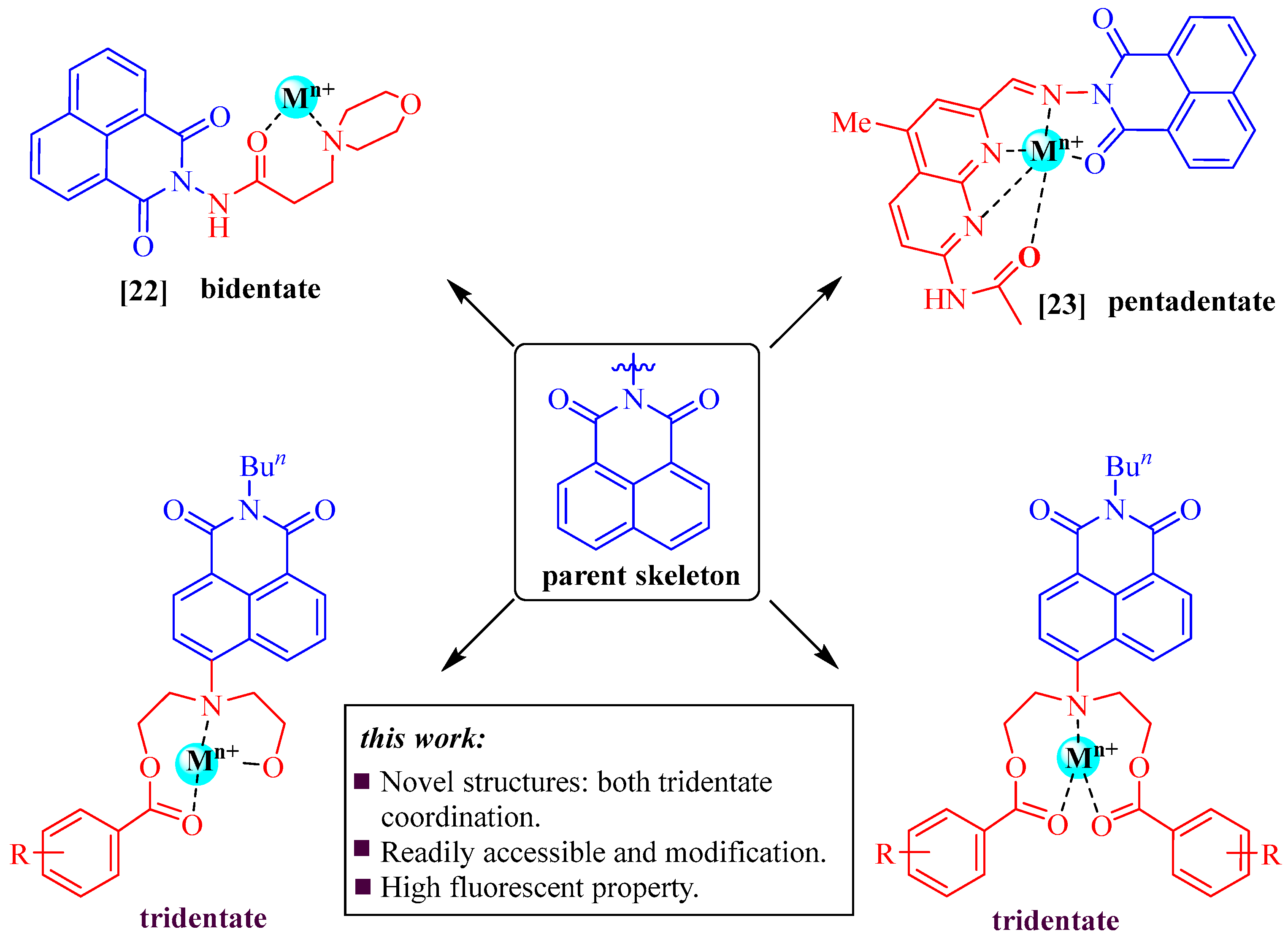
1. Introduction
2. Results and Discussion
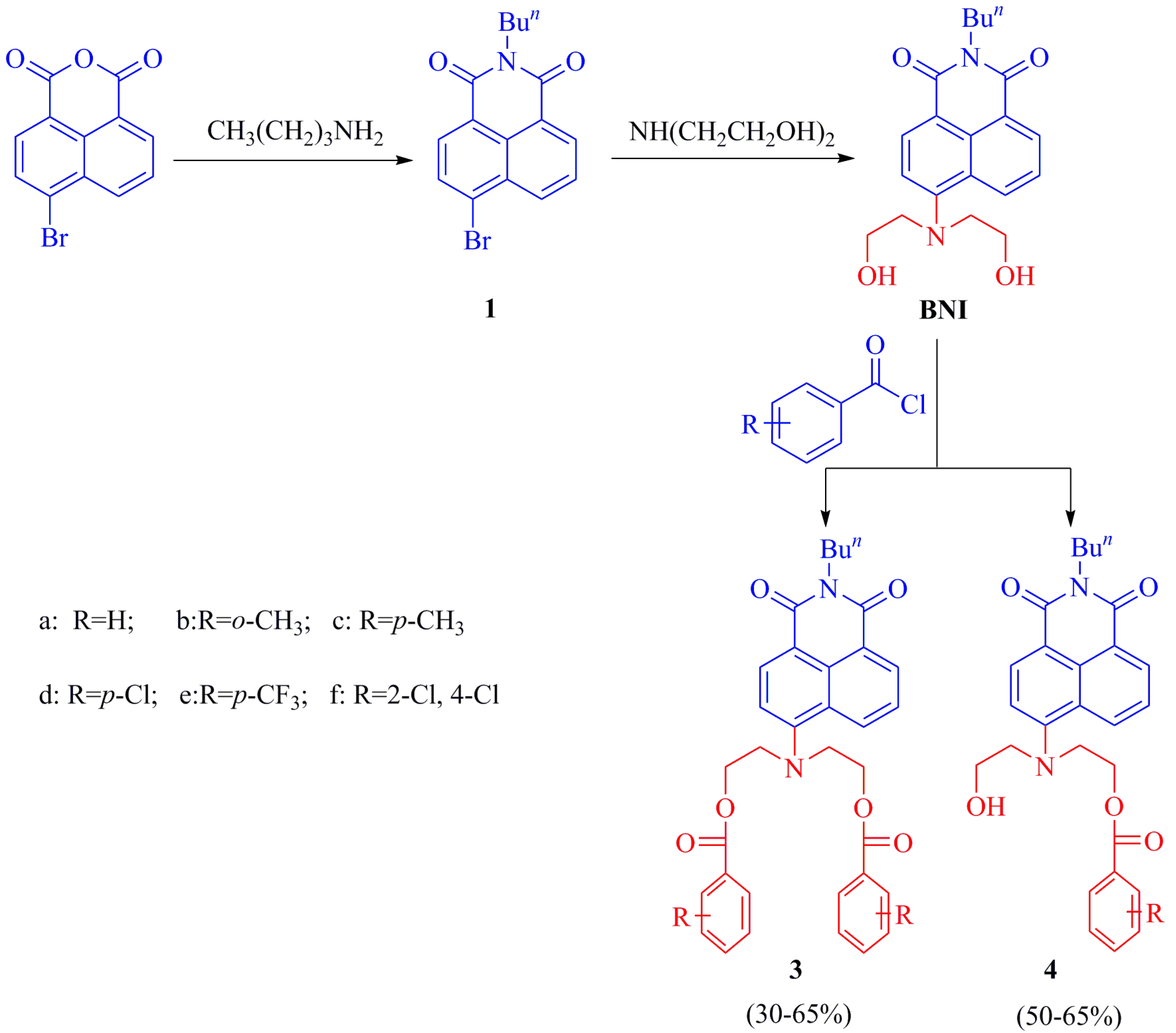
2.1. Synthesis and Characterization
2.2. Absorption Spectra and Fluorescence
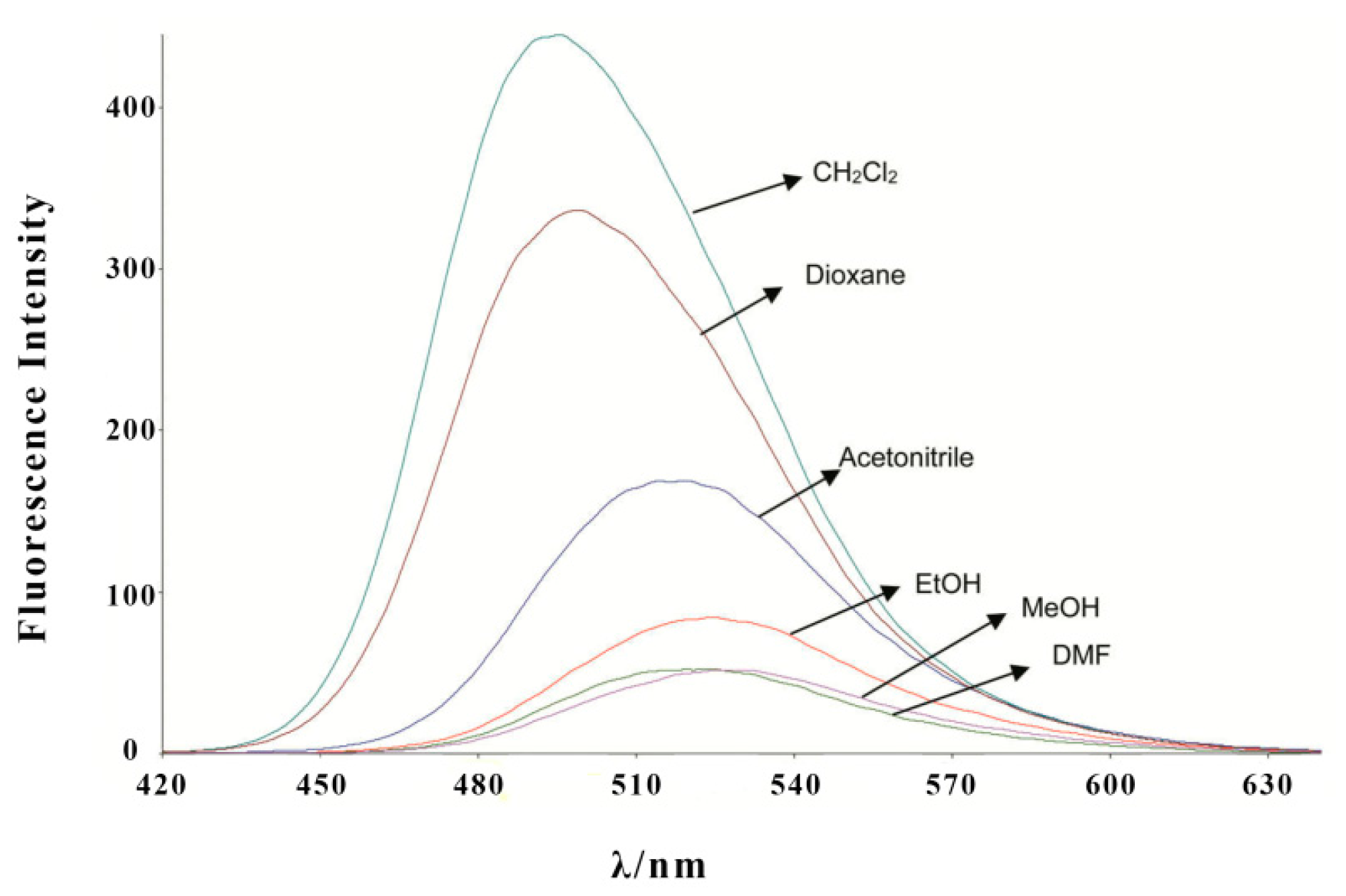
2.3. Solvent Effect
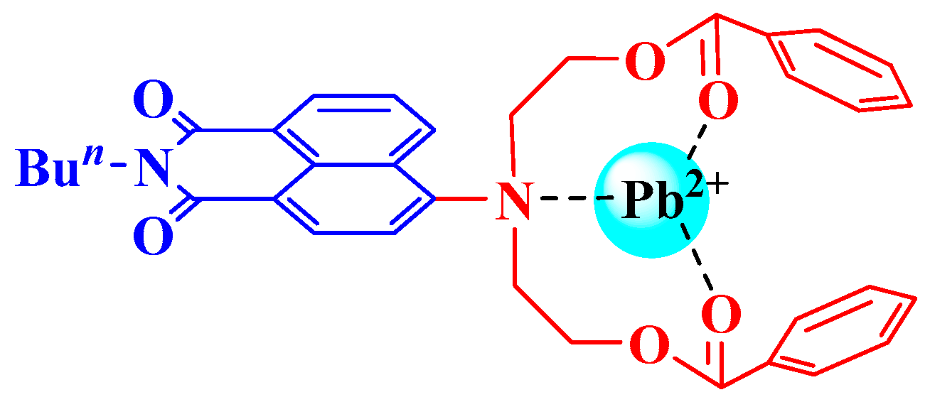
2.4. Selectivity of Probe 3f
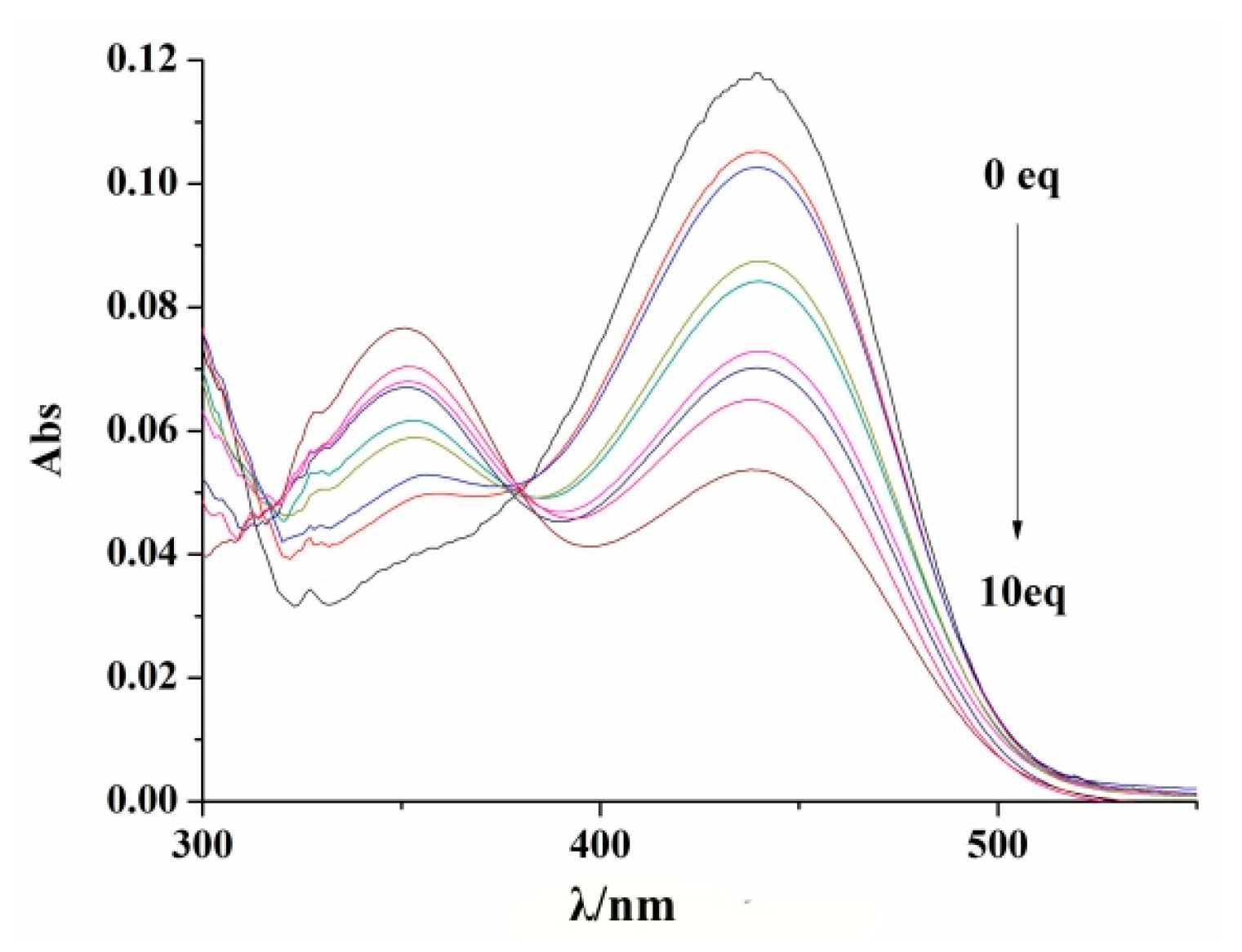
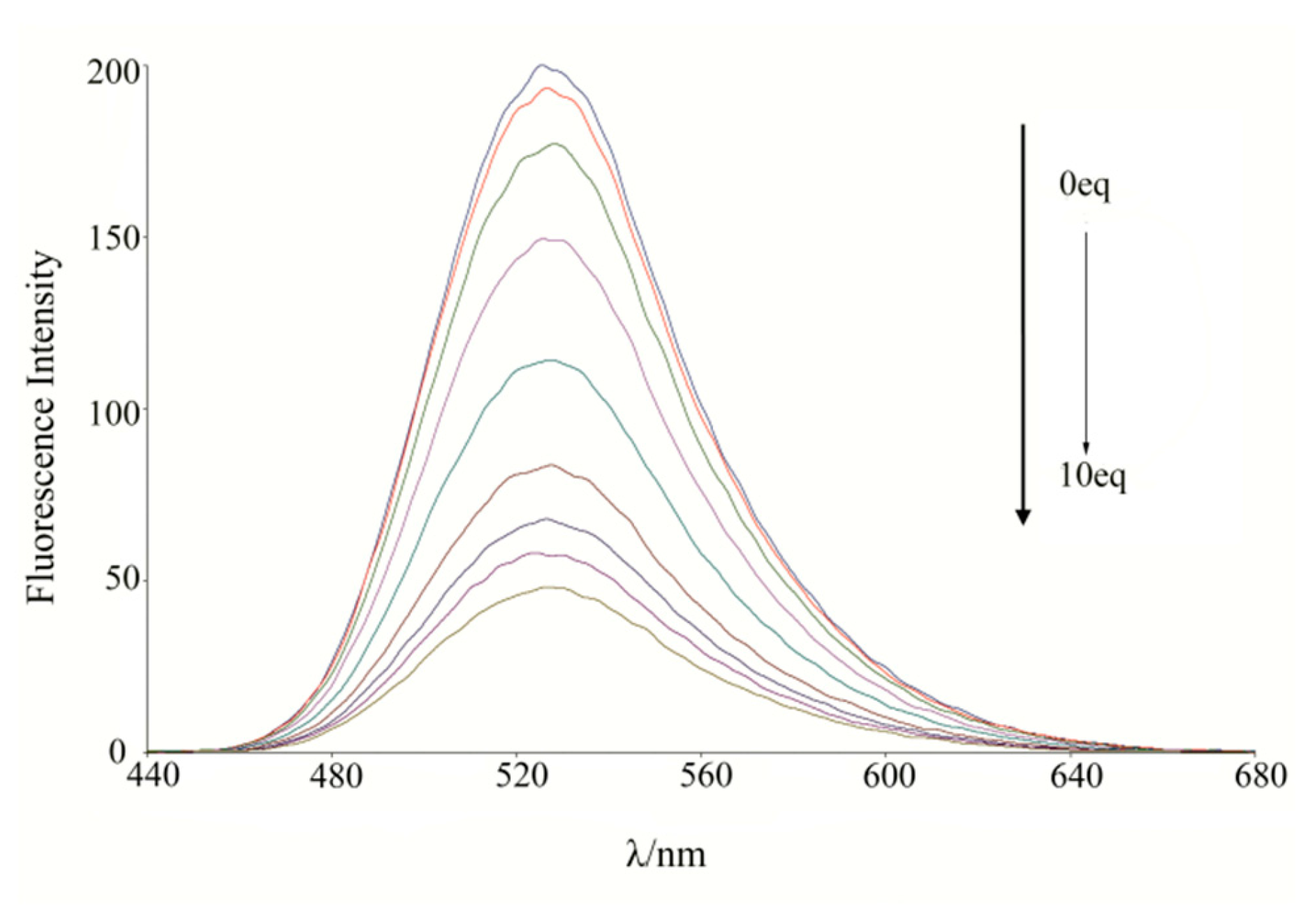
2.5. Titration Experiments
2.6. Effect of pH
3. Experimental
3.1. Chemicals and Instruments
3.2. Testing Methods
3.3. Synthesis of N-n-Butyl-4-bromo-1,8-naphthalimide (1)
3.4. Synthesis of N-n-Butyl-4-N′,N′-dihydroxyethyl-1,8-naphthalimide (BNI)
3.5. General Procedure for the Synthesis of N-n-Butyl-4-[N′,N′-dihydroxyethyl]-1,8-naphthalene Imide
3.5.1. [(2-Butyl-1,3-dioxo-2,3-dihydro-1H-benzo[de]isoquinolin-6-yl)imino]diethane-2,1-diyl dibenzoate (3a)
3.5.2. [(2-Butyl-1,3-dioxo-2,3-dihydro-1H-benzo[de]isoquinolin-6-yl)imino]diethane-2,1-diyl bis(2-methylbenzoate) (3b)
3.5.3. [(2-Butyl-1,3-dioxo-2,3-dihydro-1H-benzo[de]isoquinolin-6-yl)imino]diethane-2,1-diyl bis(4-methylbenzoate) (3c)
3.5.4. [(2-Butyl-1,3-dioxo-2,3-dihydro-1H-benzo[de]isoquinolin-6-yl)imino]diethane-2,1-diyl bis(4-chlorobenzoate) (3d)
3.5.5. [(2-Butyl-1,3-dioxo-2,3-dihydro-1H-benzo[de]isoquinolin-6-yl)imino]diethane-2,1-diyl bis[4-(trifluoromethyl)benzoate] (3e)
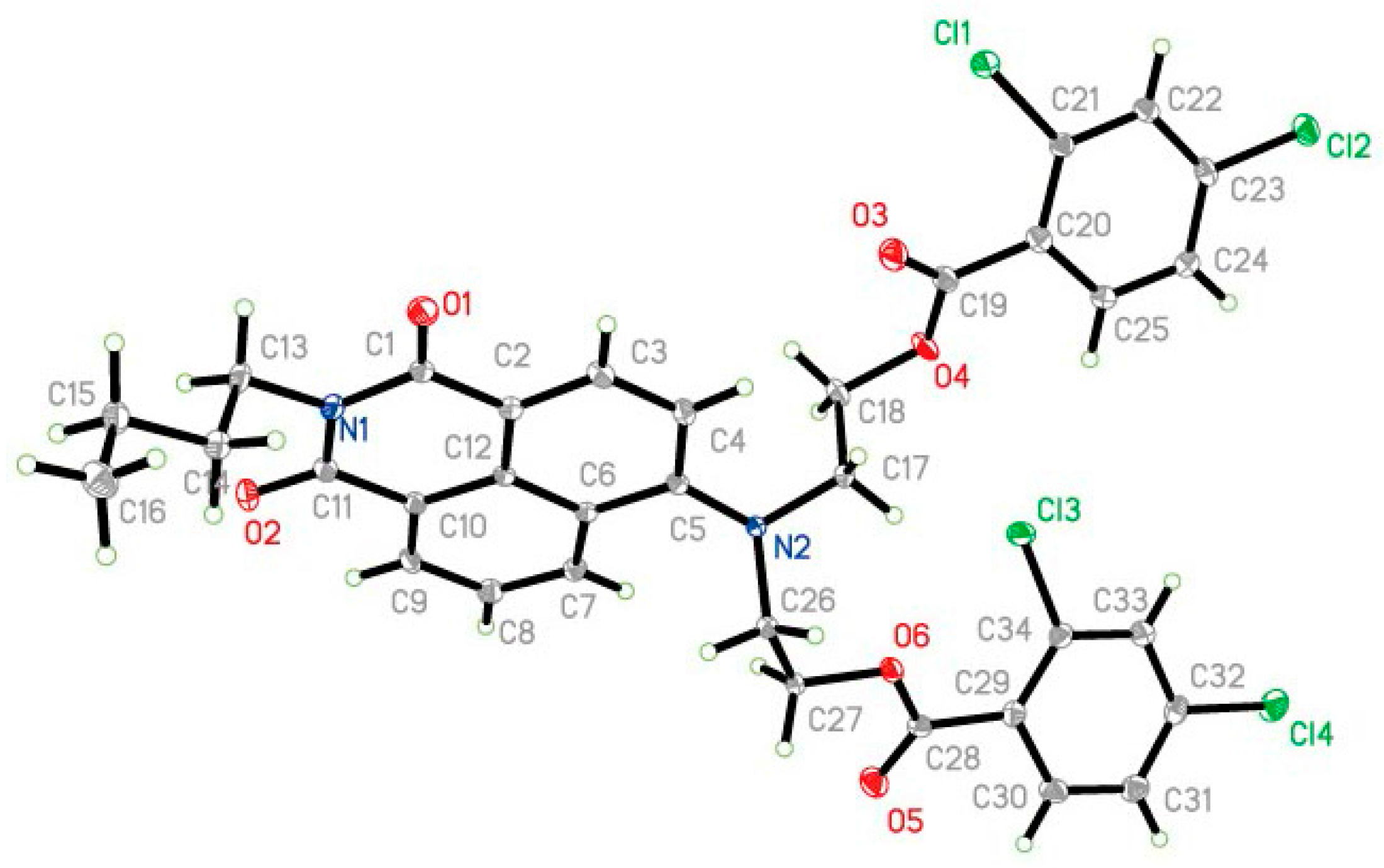
3.5.6. [(2-Butyl-1,3-dioxo-2,3-dihydro-1H-benzo[de]isoquinolin-6-yl)imino]diethane-2,1-diyl bis(2,4-dichlorobenzoate) (3f)
3.5.7. 2-[(2-Butyl-1,3-dioxo-2,3-dihydro-1H-benzo[de]isoquinolin-6-yl)-(2-hydroxy-ethyl)-amino]-ethyl Ester (4a)
3.5.8. 2-[(2-Butyl-1,3-dioxo-2,3-dihydro-1H-benzo[de]isoquinolin-6-yl)(2-hydroxyethyl)amino]ethyl-2-methylbenzoate (4b)
3.5.9. 2-[(2-Butyl-1,3-dioxo-2,3-dihydro-1H-benzo[de]isoquinolin-6-yl)(2-hydroxyethyl)amino]ethyl-4-methylbenzoate (4c)
3.5.10. 2-[(2-Butyl-1,3-dioxo-2,3-dihydro-1H-benzo[de]isoquinolin-6-yl)(2-hydroxyethyl)amino]ethyl-4-chlorobenzoate (4d)
3.5.11. 2-[(2-Butyl-1,3-dioxo-2,3-dihydro-1H-benzo[de]isoquinolin-6-yl)(2-hydroxyethyl)amino]ethyl-4-(trifluoromethyl)benzoate (4e)
3.5.12. 2-[(2-Butyl-1,3-dioxo-2,3-dihydro-1H-benzo[de]isoquinolin-6-yl)(2-hydroxyethyl)amino]ethyl-2,4-dichlorobenzoate (4f)
4. Conclusions
Supplementary Materials
Acknowledgments
Author Contributions
Conflicts of Interest
References
- Lu, D.Q.; Zhou, L.Y.; Wang, R.W.; Zhang, X.B.; He, L.; Zhang, J.; Hu, X.X.; Tan, W.H. A two-photon fluorescent probe for endogenous superoxide anion radical detection and imaging in living cells and tissues. Sens. Actuators B Chem. 2017, 250, 259–266. [Google Scholar] [CrossRef]
- Sun, J.Q.; Ye, B.F.; Xia, G.M.; Zhao, X.H.; Wang, H.M. A colorimetric and fluorescent chemosens or for the highly sensitive detection of CO2 gas: Experiment and DFT calculation. Sens. Actuators B Chem. 2016, 233, 76–82. [Google Scholar] [CrossRef]
- Hu, X.X.; Zheng, X.L.; Fan, X.X.; Su, Y.T.; Zhan, X.Q.; Zheng, H. Semicarbazide-based naphthalimide as a highly selective and sensitive colorimetric and turn-on fluorescent chemodosimeter for Cu2+. Sens. Actuators B Chem. 2016, 227, 191–197. [Google Scholar] [CrossRef]
- Ge, J.; Geng, X.; Du, Y.H.; Chen, J.J.; Zhang, L.; Bai, D.M.; Ji, D.Y.; Hu, Y.L.; Li, Z.H. Highly sensitive fluorescence detection of mercury (II) ions based on WS2, nanosheets and T7 exonuclease assisted cyclic enzymatic amplification. Sens. Actuators B Chem. 2017, 249, 189–194. [Google Scholar] [CrossRef]
- Sun, J.Q.; Ye, B.F.; Xia, G.M.; Wang, H.M. A multi-responsive squaraine-based “turn on” fluorescent chemosensor for highly sensitive detection of Al3+, Zn2+, and Cd2+, in aqueous media and its biological application. Sens. Actuators B Chem. 2017, 249, 386–394. [Google Scholar] [CrossRef]
- Pak, Y.L.; Swamy, K.M.; Yoon, J.Y. Recent progress in fluorescent imaging probes. Sensors 2015, 15, 24374–24396. [Google Scholar] [CrossRef] [PubMed]
- Yao, S.K.; Qian, Y. A naphthalimide-rhodamine two-photon fluorescent turn-on probe for hypochlorous acid by desulfurization-cyclization and fluorescence resonance energy transfer. Sens. Actuators B Chem. 2017, 252, 877–885. [Google Scholar] [CrossRef]
- Wang, Y.; Mao, P.D.; Wu, W.N.; Mao, X.J.; Zhao, X.L.; Xu, Z.Q.; Fan, Y.C.; Xu, Z.H. A novel colorimetric and ratiometric fluorescent Cu2+ sensor based on hydrazone bearing 1,8-naphthalimide and pyrrole moieties. Sens. Actuators B Chem. 2017, 251, 813–820. [Google Scholar] [CrossRef]
- Balakrishnan, C.; Subha, L.; Neelakantan, M.A.; Mariappan, S.S. Synthesis, spectroscopy, X-ray crystallography, DFT calculations, DNA binding and molecular docking of a propargyl arms containing Schiff base. Spectrochim. Acta Part A 2015, 150, 671–681. [Google Scholar] [CrossRef] [PubMed]
- Liu, D.Y.; Qi, J.; Liu, X.; Cui, Z.; Chang, H.; Chen, J.; Yang, G. 4-Amino-1,8-naphthalimide-based fluorescent Cd2+ sensor with high selectivity against Zn2+ and its imaging in living cells. Sens. Actuators B Chem. 2014, 204, 655–658. [Google Scholar] [CrossRef]
- Xu, Y.L.; Mao, S.S.; Peng, H.P.; Wang, F.; Zhang, H.; Aderinto, S.O.; Wu, H.L. A fluorescent sensor for selective recognition of Al3+ based on naphthalimide Schiff-base in aqueous media. J. Lumin. 2017, 192, 56–63. [Google Scholar] [CrossRef]
- Lv, F.; Chen, Y.F.; Tang, T.X.; Chen, Y.H.; Xu, D.M. A new reactive 1,8-naphthalimide derivative for highly selective and sensitive detection of Hg2+. J. Fluoresc. 2017, 27, 1285–1292. [Google Scholar] [CrossRef] [PubMed]
- Yuan, X.; Xu, X.J.; Zhao, C.X.; Zhang, F.; Lu, Y.X.; Shen, Y.J.; Wang, C.Y. A novel colorimetric and fluorometric fluoride ion probe based on photoinduced electron transfer signaling mechanism. Sens. Actuators B Chem. 2017, 253, 1096–1105. [Google Scholar] [CrossRef]
- Li, Z.; Zhou, Y.; Yin, K.; Yu, Z.; Li, Y.; Ren, J. A new fluorescence “turn-on” type chemosensor for Fe3+ based on naphthalimide and coumarin. Dyes Pigments 2014, 105, 7–11. [Google Scholar] [CrossRef]
- Zhang, H.F.; Yin, C.X.; Lin, T.; Chao, J.B.; Zhang, Y.B.; Huo, F.J. Selective “off-on” detection of magnesium (II) ions using a naphthalimide-derived fluorescent probe. Dyes Pigments 2017, 146, 344–351. [Google Scholar] [CrossRef]
- Ren, J.; Wu, Z.; Zhou, Y.; Li, Y.; Xu, Z. Colorimetric fluoride sensor based on 1,8-naphthalimide derivatives. Dyes Pigments 2011, 91, 442–445. [Google Scholar] [CrossRef]
- Georgiev, N.I.; Bojinov, V.B.; Marinova, N. Novel PAMAM Light-harvesting antennae based on 1,8-naphthalimide: Synthesis, energy transfer, photophysical and pH sensing properties. Sens. Actuators B Chem. 2010, 150, 655–666. [Google Scholar] [CrossRef]
- Ozdemir, M. A rhodamine-based colorimetric and fluorescent probe for dual sensing of Cu2+ and Hg2+, ions. J. Photochem. Photobiol. A 2016, 318, 7–13. [Google Scholar] [CrossRef]
- Ott, I.; Qian, X.; Xu, Y.; Vlecken, D.H.; Marques, I.J.; Kubutat, D.; Will, J.; Sheldrick, W.S.; Jesse, P.; Prokop, A.; et al. A gold (I) phosphine complex containing a naphthalimide ligand functions as a TrxR inhibiting antiproliferative agent and angiogenesis inhibitor. J. Med. Chem. 2009, 52, 763–770. [Google Scholar] [CrossRef] [PubMed]
- Yang, J.; Wang, X.; Xu, L. Studies on the synthesis and spectral properties of novel 4-benzofuranyl-1,8-naphthalimide derivatives. Dyes Pigments 2005, 67, 27–33. [Google Scholar] [CrossRef]
- Jin, Z.N.; Li, N.J.; Wang, C.F.; Jiang, H.J.; Lu, J.M.; Zhou, Q.Z. Synthesis and fluorescence property of some novel 1,8-naphthalimide derivatives containing a thiophene ring at the C-4 position. Dyes Pigments 2013, 96, 204–210. [Google Scholar] [CrossRef]
- Georgiev, N.I.; Dimitrova, M.D.; Mavrova, A.T.; Bojinov, V.B. Synthesis, fluorescence-sensing and molecular logic of two water-soluble 1,8-naphthalimides. Spectrochim. Acta Part A 2017, 183, 7–16. [Google Scholar] [CrossRef] [PubMed]
- Yue, X.L.; Li, C.R.; Yang, Z.Y. A novel Schiff-base fluorescent probe based on 1,8-naphthyridine and naphthalimide for Al3+. Inorg. Chim. Acta 2017, 464, 167–171. [Google Scholar] [CrossRef]
- Ye, F.; Chai, Q.; Liang, X.M.; Li, M.Q.; Wang, Z.Q.; Ying, Y. A highly selective and sensitive fluorescent turn-off probe for Cu2+ based on a guanidine derivative. Molecules 2017, 22, 1741. [Google Scholar] [CrossRef] [PubMed]
- Guo, X.F.; Zhu, B.C.; Liu, Y.Y.; Zhang, Y.; Jia, L.H.; Qian, X.H. Synthesis and properties of N-Butyl-4-(aza-15-crown-5)-1,8-naphthalimide as a fluorescent robe. Chin. J. Org. Chem. 2006, 26, 504–507. [Google Scholar]
- Wang, H.; Yang, L.; Zhang, W.; Zhou, Y.; Zhao, B.; Li, X. A colorimetric probe for copper (II) Ion based on 4-amino-1,8-naphthalimide. Inorg. Chim. Acta 2012, 381, 111–116. [Google Scholar] [CrossRef]
- Lee, W.H.; Seo, Y.; Bishop, P.L. Characteristics of a cobalt-based phosphate microelectrode for in situ monitoring of phosphate and its biological application. Sens. Actuators B Chem. 2009, 137, 121–128. [Google Scholar] [CrossRef] [PubMed]
- Jiang, W.; Sun, Y.; Wang, X.; Wang, Q.; Xu, W. Synthesis and photochemical properties of novel 4-diarylamine-1,8-naphthalimide derivatives. Dyes Pigments 2008, 77, 125–128. [Google Scholar] [CrossRef]
- Bardajee, G.R. Microwave-assisted solvent-free synthesis of fluorescent naphthalimide dyes. Dyes Pigments 2013, 99, 52–58. [Google Scholar] [CrossRef]
- Yang, J.X.; Wang, X.; Wang, X.; Xu, L. The synthesis and spectral properties of novel 4-phenylacetylene-1,8-naphthalimide derivatives. Dyes Pigments 2005, 66, 83–87. [Google Scholar] [CrossRef]
- Meng, Q.T.; Jia, H.M.; Gao, X.; Wang, Y.; Zhang, R.; Wang, R.J.; Zhang, Z.Q. Reversible and selective fluorescence detection of histidine using a naphthalimide-based chemosensing ensemble. Chem.-Asian J. 2015, 10, 2411–2418. [Google Scholar] [CrossRef] [PubMed]
- Sheldrick, G.M. SHELXS-97, PC Version; University of Göttingen: Göttingen, Germany, 1997. [Google Scholar]
- Sheldrick, G.M. SHELXL-97, PC Version; University of Göttingen: Göttingen, Germany, 1997. [Google Scholar]
Sample Availability: Samples of the compounds are available from the authors’ lab. |


| Comp. | Log ε (1 M−1·cm−1) a | λmax b | A | λem b | ΦF c |
|---|---|---|---|---|---|
| 3a | 4.286 | 421.5 | 0.103 | 521 | 0.264 |
| 3b | 3.748 | 419.5 | 0.090 | 529 | 0.245 |
| 3c | 4.100 | 420.0 | 0.035 | 524 | 0.289 |
| 3d | 3.699 | 414.0 | 0.120 | 522 | 0.238 |
| 3e | 4.336 | 417.0 | 0.055 | 527 | 0.238 |
| 3f | 4.017 | 421.5 | 0.118 | 528 | 0.247 |
| 4a | 4.241 | 439.5 | 0.228 | 526 | 0.554 |
| 4b | 4.255 | 439.5 | 0.121 | 524 | 0.596 |
| 4c | 4.233 | 440.5 | 0.054 | 523 | 0.577 |
| 4d | 4.140 | 440.0 | 0.168 | 523 | 0.600 |
| 4e | 4.201 | 440.0 | 0.116 | 522 | 0.579 |
| 4f | 4.212 | 438.5 | 0.098 | 521 | 0.599 |
© 2018 by the authors. Licensee MDPI, Basel, Switzerland. This article is an open access article distributed under the terms and conditions of the Creative Commons Attribution (CC BY) license (http://creativecommons.org/licenses/by/4.0/).
Share and Cite
Fu, Y.; Pang, X.-X.; Wang, Z.-Q.; Qu, H.-T.; Ye, F. Synthesis and Fluorescent Property Study of Novel 1,8-Naphthalimide-Based Chemosensors. Molecules 2018, 23, 376. https://doi.org/10.3390/molecules23020376
Fu Y, Pang X-X, Wang Z-Q, Qu H-T, Ye F. Synthesis and Fluorescent Property Study of Novel 1,8-Naphthalimide-Based Chemosensors. Molecules. 2018; 23(2):376. https://doi.org/10.3390/molecules23020376
Chicago/Turabian StyleFu, Ying, Xiao-Xiao Pang, Zhi-Qiang Wang, Hai-Tao Qu, and Fei Ye. 2018. "Synthesis and Fluorescent Property Study of Novel 1,8-Naphthalimide-Based Chemosensors" Molecules 23, no. 2: 376. https://doi.org/10.3390/molecules23020376
APA StyleFu, Y., Pang, X.-X., Wang, Z.-Q., Qu, H.-T., & Ye, F. (2018). Synthesis and Fluorescent Property Study of Novel 1,8-Naphthalimide-Based Chemosensors. Molecules, 23(2), 376. https://doi.org/10.3390/molecules23020376