Article

Nine New Gingerols from the Rhizoma of Zingiber officinale and Their Cytotoxic Activities

1
School of Pharmacy, Henan University of Chinese Medicine, Zhengzhou 450046, China
2
Collaborative Innovation Center for Respiratory Disease Diagnosis, Treatment and New Drug Research and Development of Henan Province, Henan University of Chinese Medicine, Zhengzhou 450046, China
3
Guangdong Key Laboratory for Genome Stability & Disease Prevention, School of Pharmaceutical Sciences, School of Medicine, Shenzhen University Health Science Center, Shenzhen 518060, China
*
Author to whom correspondence should be addressed.
Molecules 2018, 23(2), 315; https://doi.org/10.3390/molecules23020315
Submission received: 9 December 2017 / Revised: 30 January 2018 / Accepted: 31 January 2018 / Published: 2 February 2018

Abstract

:
Nine new gingerols, including three 6-oxo-shogaol derivatives [(Z)-6-oxo-[6]-shogaol (1), (Z)-6-oxo-[8]-shogaol (2), (Z)-6-oxo-[10]-shogaol (3)], one 6-oxoparadol derivative [6-oxo-[6]-paradol (4)], one isoshogaol derivative [(E)-[4]-isoshogaol (5)], and four paradoldiene derivatives [(4E,6Z)-[4]-paradoldiene (8), (4E,6E)-[6]-paradoldiene (9), (4E,6E)-[8]-paradoldiene (10), (4E,6Z)-[8]-paradoldiene (11)], together with eight known analogues, were isolated from the rhizoma of Zingiber officinale. Their structures were elucidated on the basis of spectroscopic data. It was noted that the isolation of 6-oxo-shogaol derivatives represents the first report of gingerols containing one 1,4-enedione motif. Their structures were elucidated on the basis of spectroscopic and HRESIMS data. All the new compounds were evaluated for their cytotoxic activities against human cancer cells (MCF-7, HepG-2, KYSE-150).

Graphical Abstract

1. Introduction

Ginger, also known as white ginger, is the dry rhizoma of Zingiber officinale, which has been a popular spice world-wide. As a Chinese medicine, it has been used in the treatment of nausea and vomiting, coughs, cold and so forth, for more than 2000 years [1,2,3]. In the traditional Chinese medicine theory system, Z. officinale is a warm medicine which can warm the spleen and stomach to dispel cold and warm the lungs, to reduce or eliminate dampness and phlegm (Huiyang Tongmai). Pharmacological studies on Z. officinale showed its effects on oxidative stress [4,5], tumor [6,7], degenerative diseases [8], vomiting [9], the cardiovascular system [10], and indigestion [11,12]. Chemical investigations have revealed the presence of volatile oils, gingerols, and diarylheptanoids, responsible for its pungency and other pharmacological properties [13]. Among the various activities of gingerols, numerous documents have reported anti-tumor activities, so we were interested in finding new types of gingerols with anti-tumor activity. Our efforts resulted in the isolation of nine new gingerols and eight known analogues (Figure 1), whose cytotoxic activities against human cancer cells (MCF-7, HepG-2, KYSE-150) were subsequently evaluated.

2. Results and Discussion

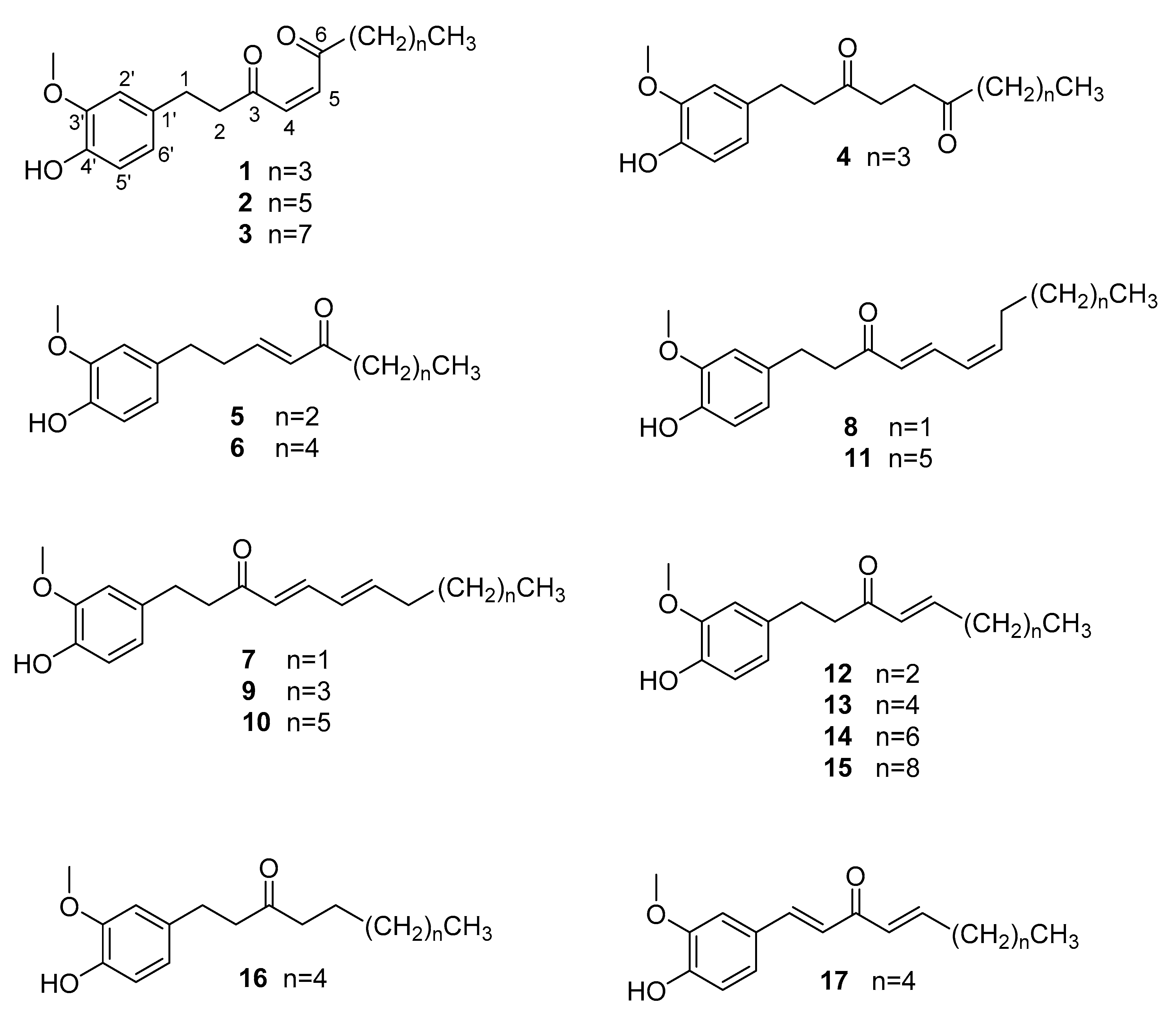
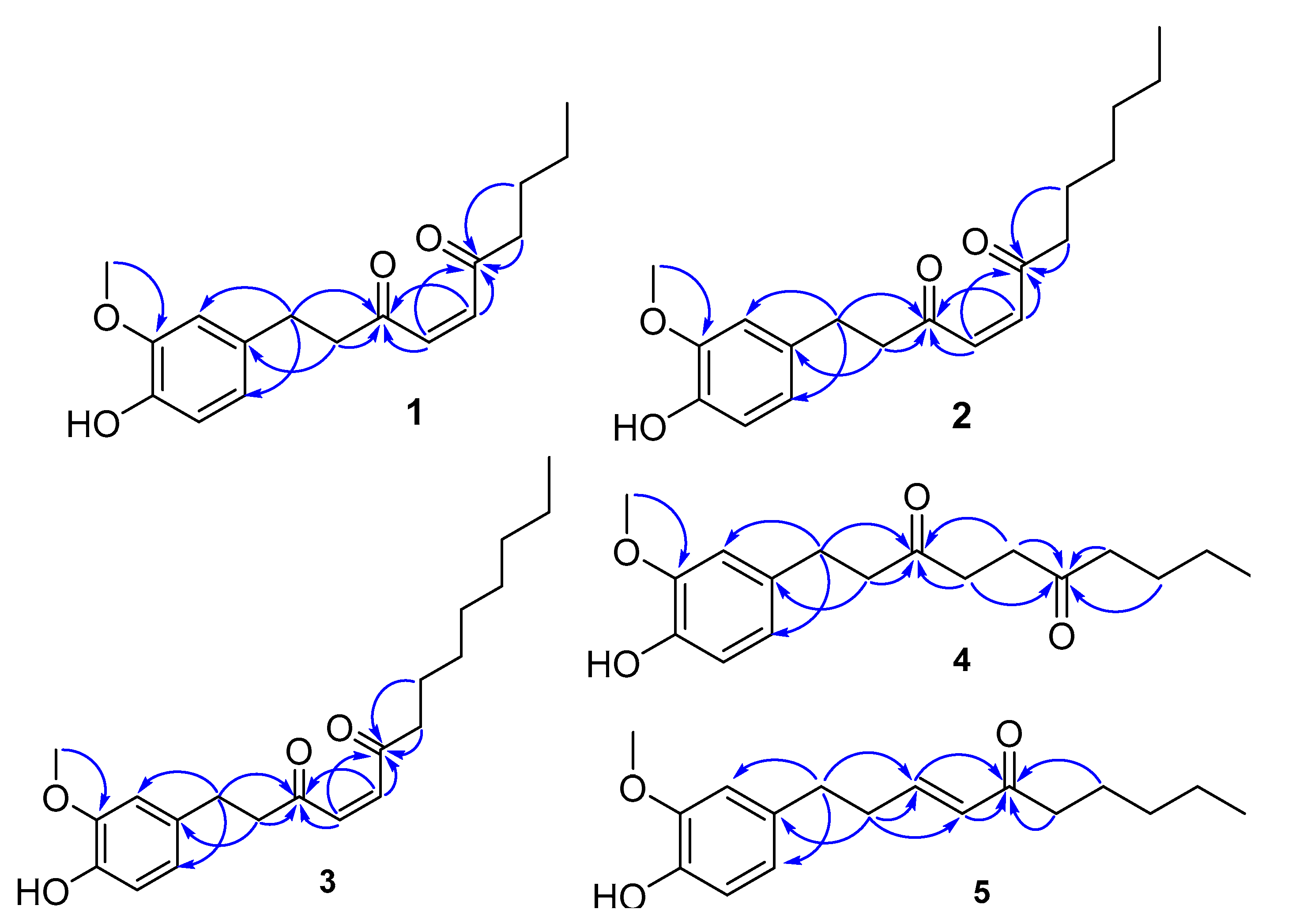
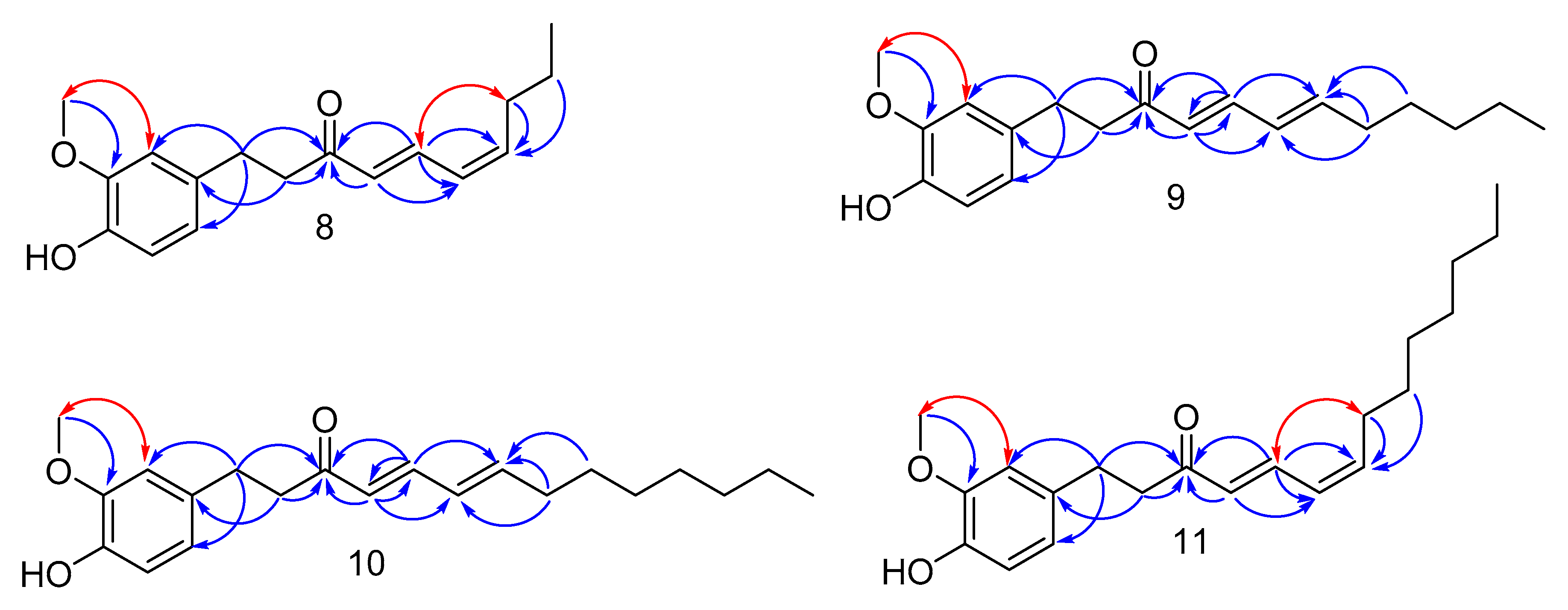
Structure Elucidation of the Compounds

The EtOAc extract of Z. officinale rhizoma was submitted to a combination of column chromatography to produce compounds 117 (Figure 1).
Compound 2 was isolated as yellow oil and is demonstrated by a molecular formula of C19H26O4, based on the HRESIMS ion peak at m/z 341.1727 [M + Na]+ (calcd. for 341.1729).The 1H-NMR (Table 1) spectrum of 2 shows the typical pattern of a coupling group of 1,3,4-trisubstituted benzene rings at δH 6.81 (1H, d, J = 8.0 Hz, H-5′), 6.67 (1H, d, J = 1.5 Hz, H-2′), 6.66 (1H, dd, J = 8.0, 1.5 Hz, H-6′), two olefinic protons [δH 6.83 (2H, s, H-4, H-5)], a methoxy group [δH 3.85 (3H, s, 3′-OCH3)], seven methylene groups [δH 2.86 (2H, m, H-1), 2.94 (2H, m, H-2), 2.59 (2H, t, J = 7.4 Hz, H-7), 1.60 (2H, m, H-8), 1.28 (6H, m, H-9, H-10, H-11)] and a methyl group [δH 0.85 (3H, t, J = 6.4 Hz, H-12)]. In the 13C-NMR spectrum of 2, 19 carbon signals were observed, which included two carbonyls [δC 200.7 (C-3) and 199.8 (C-6)], six aromatic carbons [δC 146.4 (C-3′), 144.0 (C-4′), 132.4 (C-1′), 120.8 (C-6), 114.4 (C-5), 111.0 (C-2)], two olefinic carbons [δC 136.4 (C-4) and 136.1 (C-5)], and eight aliphatic carbons [δC 43.5 (C-2), 41.7 (C-7), 31.5 (C-10), 29.3 (C-1), 28.8 (C-9), 23.7 (C-8), 22.4 (C-11), 14.0 (C-12)]. The planar structure of 2 was further demonstrated by analyses of 2D NMR spectra. The HSQC showed that there is a correlation from δH 6.83 (2H, s, H-4, H-5) to two olefinic carbons (δC 136.4, C-4 and 136.1, C-5); while the HMBC presented a correlation from δH 6.83 (2H, s, H-4, H-5) to two carbonyl carbons [δC 200.7 (C-3) and 199.8 (C-6)], as well as a correlation from the methylene protons (H-1, H-2, H-7 and H-8) to two carbonyl carbons, but lacking the correlations from H-4 and H-5 to C-2 and C-7 or from H-2 and H-7 to C-4 and C-5 (Figure 2). It was found that there are two olefinic carbons between two carbonyl carbons. In addition, the methoxy group at δH 3.85 (3′-OCH3) was located at C-3′ by the HMBC correlations from 3′-OCH3 to C-3′, the substituent at C-4′was identified as a hydroxy group because of its 13C-NMR chemical shift, and the side chain was located at C-1′ by HMBC correlations from H-1 to C-1′, C-2′ and C-3′ and from H-2 to C-1′.
Finally, our focus was on determining the cis/trans configuration of double bonds. Due to the molecular symmetry, the magnetically equivalent vinylic protons of 2 behave as a singlet peak. Thus, it was impossible to directly determine the E/Z-configuration of the double bonds by the size of the vicinal coupling constants. We tested a variety of methodologies to solve this problem, including trying to cultivate single crystals, changing different deuterated reagents, and looking for more evidence on 2D NMR spectra, but none of them succeeded. After reviewing relevant literature [14], we found that olefinic protons split obviously when the compounds containing 1,4-enedione are in NMR spectra with deuterated benzene as a solvent. Subsequently, we changed the solvent to deuterated benzene, and we found that the singlet peak of olefinic protons became a quadruplet of AB system. The AB system is an advanced coupling and the coupling constant cannot be calculated directly. We calculated the actual chemical shift of the quadruplet, and the coupling constant was calculated to be 10.4 Hz by the chemical shift. Therefore, the two olefinic protons were cis-oriented, and the structure of 2 was determined to be (Z)-6-oxo-[8]-shogaol.
Compound 3 was obtained as yellow oil and shows a [M + Na]+ ion at m/z 369.2042 in the HRESIMS, consistent with a molecular formula of C21H30O4 (calcd. for 369.2042). Compound 1 was obtained as yellow oil, with the molecular formula C17H22O4, as determined by the HREIMS at m/z 313.1414 for the [M + Na]+ (calcd. for 313.1416). The 1H- and 13C-NMR data (Table 1) of 3 and 1 are similar to those of 2, suggesting that they have the same carbon skeleton. The significant differences in these three compounds are the number of carbons in the aliphatic region of the 13C-NMR and the 1H-NMR spectra. Compound 3 has two more methylene groups in its side-chain than 2, while 1 has two fewer methylene groups in side-chain than 2. Similarly, the double bond moieties of 3 and 1 both behaved as a typical AB system in their 1H-NMR spectrum when deuterated benzene was used as the solvent, and the coupling constants of these two compounds were calculated to be 10.4 Hz. On the basis of this evidence and literature comparison, the structures of 3 and 1 were determined to be (Z)-6-oxo-[10]-shogaol and (Z)-6-oxo-[6]-shogaol, respectively.
Compound 4 was obtained as brown oil. The molecular formula of 4 is assigned as C17H24O4 on the basis of HRESIMS (m/z 315.1572 [M + Na]+, calcd. for 315.1573). The NMR data (Table 1) of 4 closely resemble those of 1; the analysis of the 1H-NMR and HSQC data of 1 revealed that the significant differences in 4 are the absence of two olefinic protons at C-4 and C-5 and the presence of two additionally multiplet methylene groups at δH 2.65 and δH 2.62. This indicates that the 1,4-enedione-like moiety in 1 was replaced by a 1,4-dicarbonyl-like group in 4. This deduction was supported by the HMBC correlations from the methylene protons, H-4 and H-5 (δH 2.65 and 2.62), to two carbonyl carbons (δC 209.7, C-3 and 208.7, C-6), from H-1 (δH 2.73) and H-2 (δH 2.80), to C-3 and from H-7 (δH 2.42), and H-8 (δH 1.54) to C-6 (Figure 2). Accordingly, the structure of 4 was further confirmed by the combined analyses of HSQC and HMBC data, and elucidated as 6-oxo-[6]-paradol.
Compound 5 was obtained as yellow oil. Its molecular formula, C15H20O3, was established by HRESIMS at m/z 271.1312 for the [M + Na]+ (calcd. for 271.1310). The 1H-NMR spectrum (Table 2) of 5 exhibited the 1,3,4-tetrasubstituted aromatic moiety [δH 6.82(1H, d, J = 8.5 Hz, H-5′), 6.65 (1H, dd, J = 8.5, 1.9 Hz, H-6′), 6.64 (1H, s, H-2′)], two trans-conformational olefinic protons [δH 6.81(1H, m, H-3), 6.10 (1H, dd, J = 16.0, 1.5 Hz, H-4)], a methoxy group [δH 3.85 (3H, s, 3′-OCH3)], four methylene groups [δH 2.70 (2H, t, J = 7.2 Hz, H-1), 2.48 (2H, t, J = 7.2 Hz, H-2), 2.47 (2H, t, J = 7.0 Hz, H-6) and 1.61 (2H, m, H-7)], and a methyl group [δH 0.83 (3H, t, J = 7.4 Hz, H-8) ]. The 13C-NMR and HSQC spectra of 5 revealed the presence of a carbonyl carbon [δC 200.7 (C-5) ], two olefinic carbons [δC 145.9 (C-3) and δC 130.7 (C-4) ], six aromatic carbons [δC 146.4 (C-3′), 143.9 (C-4′), 132.7 (C-1′), 120.9 (C-6′), 114.3 (C-5′) and 110.9 (C-2′)], a methoxy carbon [δC 55.85 (3′-OCH3)], four methylene carbons [δC 42.0 (C-6), 34.47 (C-2), 34.16 (C-1), and 17.5 (C-7)], a methyl carbon [δC 13.8 (C-8)]. The NMR data of 5 are similar to those of [5]-shogaol [15], a typical α,β-unsaturated ketone-type structure. The significant difference in 5 is that the position of the olefinic carbons have changed. This deduction was supported by the HMBC correlations from the methylene protons, H-1 to C-2 and C-3, from H-2 to C-1, C-3 and C-4 and from H-6 and H-7 to C-5. In addition, H–H COSY correlations between H-2 and H-3 also proved this deduction (Figure 3). Accordingly, the structure of 5 was further confirmed by the combined analyses of HSQC and HMBC data. Thus, the structure of 5 was determined as (E)-[4]-isoshogaol.
Compound 9, obtained as yellow oil has the molecular formula, C19H26O3, based on the HRESIMS showing the [M + Na]+ ion at m/z 325.1780 (calcd. for 325.1782). The 1H-NMR data (Table 2) of 9 suggest the presence of a typical pattern of a coupling group of 1,3,4-trisubstituted benzene rings at δH 6.80 (1H, d, J = 8.0 Hz, H-5′), 6.68 (1H, d, J = 1.5 Hz, H-2′) and, 6.66 (1H, dd, J = 8.0, 1.5 Hz, H-6′), four olefinic protons [δH 7.10(1H, m, H-5), 6.14 (1H, m, H-6) at 6.13 (1H, m, H-7) and 6.05 (1H, d, J = 15.5 Hz, H-4)], a methoxy group [δH 3.85 (3H, s, 3′-OCH3)], six methylene groups [δH 2.85 (2H, m, H-2) at 2.82 (2H, m, H-1), 2.15 (2H, m, H-8), 1.40 (2H, m, H-9) and 1.27 (4H, m, H-10 and H-11)], and a methyl group [δH 0.86 (3H, t, J = 7.0 Hz, H-12)]. The 13C-NMR spectrum of 9 displays 19 signals, assigned by HSQC data to six aromatics [δC 146.4 (C-3′), 143.8 (C-4′), 133.2 (C-1′), 120.8 (C-6′), 114.3 (C-5′) and 111.1 (C-2′)], six methylene [δC 42.4 (C-2), 33.1 (C-8), 31.3 (C-10), 29.9 (C-1), 28.3 (C-9), 22.4 (C-11)], four olefinic carbons [δC 146.0 (C-7), 143.3 (C-5), 128.8 (C-6), 127.7 (C-4)], one ketone carbonyl [δC 199.9 (C-3)], and two methyls [δC 55.83 (3′-OCH3), 14.0 (C-12)], with the former being a methoxy. The linkage of the conjugate double bond moiety to carbonyl carbon (C-3) was determined by the HMBC correlations of H-4 and H-5 to C-3. In addition, the significant chemical shift differences in 13C-NMR between C-4 and C-5, C-6 and C-7 also proved this deduction (Figure 3).
A large coupling constant between H-4 and H-5 (J = 15.5 Hz) indicated that the two olefinic protons are trans-oriented. We made a bold guess to determine the orientation of the other two olefins in the conjugated double bonds. It is well known that a conjugated double bond forms a conjugated π system by resonance hybrid, where the double bond between carbon and carbon tends to become longer and the single bond tends to become shorter; thus, the bond between each carbon has the characteristics of double bonds, and each bond cannot be rotated. Therefore, a NOESY correlation should be effective in this conjugated double bond moiety. So, if H-6 and H-7 are cis-oriented, the NOESY correlation between H-5 and H-8 can be observed, but if they are trans-oriented, this correlation cannot occur. Hence, the absolute configuration of 9 was determined by detailed analysis of its NOESY spectrum, and we did not observe the NOESY interaction between H-5 and H-8 in the NOESY spectrum; in this case, we speculated that H-6 and H-7 must be trans-oriented, and previously reported literature [16,17,18] with similar structures confirms our conjecture. Thus, on the basis of this evidence and literature comparison, the structure of 9 was determined to be (4E,6E)-[6]-paradoldiene.
The HRESIMS of Compound 10 was obtained as yellow oil and showed a [M + Na]+ ion at m/z 353.2091, consistent with a molecular formula of C21H30O3 (calcd. for 353.2093). The 1H- and 13C-NMR data (Table 2) of 10 closely resemble those of 9, with a slight difference in the number of carbons in the aliphatic region of the NMR spectra. From this, it can be seen that the side chain of compound 10 should have two more methylene groups than that of compound 9. Thus, the structure of 10 was determined to be (4E,6E)-[8]-paradoldiene.
Compound 8 was obtained as yellow oil, and has the molecular formula, C17H22O3, by HRESIMS [found m/z 297.1465 (calcd. for 297.1467) [M + Na]+].The 1H- and 13C-NMR spectroscopic data (Table 2) of 8 are similar to those of 9. The main difference is the chemical shift of the carbons and protons of the conjugated double bond. A large coupling constant between H-4 and H-5 (J = 15.3 Hz) indicated that the two protons were trans-oriented, and a smaller coupling constant between H-6 and H-7 (J = 11 Hz) indicated that the two protons were cis-oriented. In addition, the NOESY correlation of H-5 and H-8 demonstrates that H-6 and H-7 are cis-oriented (Figure 3). Thus, the structure of 8 was determined to be (4E,6Z)-[4]-paradoldiene.
Compound 11 was obtained as yellow oil and has the same molecular formula (C21H30O3) as 10 by HRESIMS analysis. The 1H- and 13C-NMR data (Table 2) of 11 closely resemble those of 8, with a slight difference in the number of carbons in the aliphatic region of the NMR spectra. From this, it can be seen that the side chain of compound 11 should have four more methylene groups than that of compound 8. In addition, the NOESY correlation of H-2′ and 3′-OCH3 also demonstrates that the methoxy is located at C-3′ (Figure 3). Thus, the structure of 11 was determined to be (4E,6Z)-[8]-paradoldiene.
Known metabolites were identified as (4E)-[4]-shogaol (12) [15], (4E)-[6]-shogaol (13) [15], (4E)-[8]-shogaol (14) [15], (4E)-[10]-shogaol (15) [15,19], [8]-paradol (16) [15], (1E,4E)-[6]-dehydroshogaol (17) [15] (Figure 1) by comparing their spectroscopic data (1H-NMR, 13C-NMR, 1H-1H COSY, HSQC, HMBC, and NOESY in Supplementary Materials) with literature values. In addition, by comparing the spectral data of the new compounds that have been identified, we have identified the following two compounds: (3E)-[6]-isoshogaol (6) and (4E,6Z)-[4]-paradoldiene (7).

3. Experimental Section

3.1. General Experimental Procedures

UV spectra were measured on a Thermo EVO 300 spectrophotometer (Thermo Fisher Scientific, Madison, WI, USA). IR spectra were recorded on a Thermo Nicolet IS 10 spectrometer (Thermo Fisher Scientific, Madison, WI, USA). NMR spectra were recorded on a Bruker Avance III 500 spectrometer (Bruker Biospin, Fallanden, Switzerland) with TMS as an internal standard. HRESIMS data were recorded on a Bruker maxis HD Mass Q-TOF LC/MS spectrometer (Bruker Daltonics, Billerica, MA, USA). Preparative HPLC was performed on a Sepuruisi LC-52 instrument (Beijing Sepurusi Scientific Co., Ltd., Beijing, China) with an UV200 detector (Beijing Sepurusi Scientific Co., Ltd., Beijing, China), using a YMC-Pack ODS-A column (250 mm × 20 mm, 5 μm). Column chromatography was undertaken on HP-20 macroporous resin (Mitsubishi Chemical Co., Ltd., Tokyo, Japan), SephadexLH-20 (Amersham Pharmacia, Uppsala, Sweden), ODS (50 μm, YMC Co. Ltd., Kyoto, Japan), and silica gel (200–300 mesh, 100–200 mesh, Qingdao Marine Chemical Inc., Qingdao, China). TLC was carried out with glass that was pre-coated with silica gel GF 254 (Qingdao Marine Chemical Inc.) The human heptocelluar (HepG-2) cell line, human mammary cancer (MCF-7) cell line, and human esophageal cancer (KYSE-150) cell line were purchased from the Institute of Materia Medica, Chinese Academy of Medical Sciences and Peking Union Medical College, Beijing, China.

3.2. Plant Material

The rhizoma of Z. officinale was collected from Bozhou herbal medicine market in Anhui province, China in November 2014, and the locality of growth was Luoping County, Yunnan Province. It was identified by Prof. Sui-Qing Chen at Henan University of Chinese Medicine. A voucher specimen (No. 20141192A) was deposited at the Department of Natural Medicinal Chemistry, School of Pharmacy, Henan University of Chinese Medicine, Zhengzhou, China.

3.3. Extraction and Isolation

The dried rhizoma of Z. officinale (50 kg) was crushed into coarse powder and macerated for 1 h with 200 L EtOAc, refluxed at 75 °C (3 × 200 × 1.5 h). After removing the solvent under reduced pressure, the EtOAc extract (4 kg) was dissolved in 60% EtOH and divided into three parts (Fr.1–Fr.3) using Diaion HP-20 macroporous resin column eluted with gradient aqueous EtOH (60%, 80%, and 100%). Fr.1 (1.5 kg) was applied to a silica gel (100–200 mesh) column and eluted successively with CH2Cl2: MeOH (100:0, 30:1, 20:1, 10:1, 5:1, 2:1, 0:100) to yield seven subfractions (Fr.1.1–Fr.1.7). Fraction Fr.1.1 (500 g) was subjected to another silica gel (200–300 mesh) column eluting with a step-gradient of petroleum ether/acetone (from 100:0 to 0:100) to provide nine subfractions: Fr.1.1.1–Fr.1.1.9. Fr.1.1.4 (80 g) was separated by a MCI gel column (MeOH/H2O, 10–70%) to get five fractions (Fr.1.1.4.1–Fr.1.1.4.5), of which, fraction Fr.1.1.4.3 (12 g) was purified by Sephadex LH-20 (MeOH), followed by semi-preparative HPLC (MeCN/H2O, 65%, flow rate: 3 mL/min) to yield 17 (21.2 mg, Rt = 16.8 min), 2 (11.2 mg, Rt = 17.6 min), 13 (33.6 mg, Rt = 20.1 min), 16 (18.1 mg, Rt = 23.5 min) and 14 (78.5 mg, Rt = 43.8 min). Fr.1.1.4.2 (10 g) was chromatographed over a silica gel (200–300 mesh) column, eluted with a step-gradient of petroleum ether/EtOAc (from 100:0 to 0:100) and was further purified with semi-preparative HPLC (MeCN/H2O, 55%, flow rate: 3 mL/min) to yield 1 (91.2 mg, Rt = 8.9 min), 4 (43.3 mg, Rt = 9.6 min), 12 (16.7 mg, Rt = 10.7 min) and 3 (27.3 mg, Rt = 35.5 min). Fr.1.1.4.5 (9 g) was subjected to a silica gel (400–500 mesh) column, eluted with a step-gradient of petroleum ether/EtOAc (from 100: 0 to 0: 100) to get six fractions (Fr.1.1.4.5.1–Fr.1.1.4.5.6). The Fr.1.1.4.5.3 (1.2 g) was further purified by semi-preparative HPLC (MeCN/H2O, 65%, flow rate: 3 mL/min) to yield 5 (22.3 mg, Rt = 19.8 min) and 6 (17.6 mg, Rt = 14.3 min). Fr.1.1.4.5.1 (1.7 g) was further purified by semi-preparative HPLC (MeCN/H2O, 80%, flow rate: 3 mL/min) to yield 7 (28.8 mg, Rt = 8.7 min), 8 (34.7 mg, Rt = 9.1 min), 9 (59.9 mg, Rt = 15.2 min), 10 (71.6 mg, Rt = 27.2 min), 11 (55.7 mg, Rt = 27.7 min), and 15 (59.1 mg, Rt = 35.6 min).

3.4. Spectroscopic and Physical Data

Compound 1: yellow oil; UV (MeOH) λmax (logε) 281 (2.83), 202(3.91) nm; IR (neat) νmax 3391, 2957, 1682, 1516, 1272, 1033 cm−1. 1H (500 MHz, CDCl3) and 13C (125 MHz, CDCl3) data are presented in Table 1. HREIMS m/z: 313.1414 [M + Na] + (calcd. for C17H22O4, 313.1416).
Compound 2: brown oil; UV (MeOH) λmax (logε) 281 (3.09), 203 (4.15) (0.2) nm; IR (neat) νmax 3407, 2931, 1682, 1516, 1272, 1033 cm−1. 1H (500 MHz, CDCl3) and 13C (125 MHz, CDCl3) data are presented in Table 1. HREIMS m/z: 341.1727 [M + Na] + (calcd. for C19H26O4, 341.1729).
Compound 3: yellow oil; UV (MeOH) λmax (logε) 281 (2.85), 203 (3.95) nm; IR (neat) νmax 3372, 2922, 1681, 1521, 1278, 1030 cm−1. 1H (500 MHz, CDCl3) and 13C (125 MHz, CDCl3) data are presented in Table 1. HREIMS m/z: 369.2042 [M + Na]+ (calcd. for C21H30O4, 369.2042).
Compound 4: yellow oil; UV (MeOH) λmax (logε) 281 (2.85), 206 (3.96) nm; IR (neat) νmax 3409, 2956, 1707, 1515, 1268, 1033 cm−1. 1H (500 MHz, CDCl3) and 13C (125 MHz, CDCl3) data are presented in Table 1. HREIMS m/z: 315.1572 [M + Na]+ (calcd. for C17H24O4, 315.1573).
Compound 5: yellow oil; UV (MeOH) λmax (logε) 281 (2.79), 203 (3.98) nm; IR (neat) νmax 3356, 2963, 2940, 1664, 1516, 1273, 1035 cm−1. 1H (500 MHz, CDCl3) and 13C (125 MHz, CDCl3) data are presented in Table 2. HREIMS m/z: 271.1312 [M + Na]+ (calcd. for C15H20O3, 271.1310).
Compound 8: yellow oil; UV (MeOH) λmax (logε) 279 (3.58), 202 (4.02) nm; IR (neat) νmax 3381, 2957, 1631, 1593, 1515, 1271, 1034 cm−1. 1H (500 MHz, CDCl3) and 13C (125 MHz, CDCl3) data are presented in Table 2. HREIMS m/z: 297.1465 [M + Na]+ (calcd. for C17H22O3, 297.1467).
Compound 9: yellow oil; UV (MeOH) λmax (logε) 279(3.44), 203 (3.93) nm; IR (neat) νmax 3427, 2929, 1633, 1595, 1516, 1271, 1033 cm−1. 1H (500 MHz, CDCl3) and 13C (125 MHz, CDCl3) data are presented in Table 2. HREIMS m/z: 325.1780 [M + Na]+ (calcd. for C19H26O3, 325.1782).
Compound 10: yellow oil; UV (MeOH) λmax (logε) 278 (3.60), 202 (4.07) nm; IR (neat) νmax 3368, 2927, 1634, 1595, 1516, 1271, 1034 cm−1. 1H (500 MHz, CDCL3) and 13C (125 MHz, CDCl3) data are presented in Table 2. HREIMS m/z: 353.2091 [M + Na]+ (calcd. for C21H30O3, 353.2093).
Compound 11: yellow oil; UV (MeOH) λmax (logε) 279 (3.47), 202 (4.15) nm; IR (neat) νmax 3358, 2927, 1597, 1515, 1270, 1035 cm−1. 1H (500 MHz, CDCl3) and 13C (125 MHz, CDCl3) data are presented in Table 2. HREIMS m/z: 353.2094 [M + Na]+ (calcd. for C21H30O3, 353.2093).

3.5. Cytotoxic Assay

Tumor cells were maintained in RPMI-1640 medium containing 10% heat-inactivated fetal bovine serum, penicillin (100 U/mL) and streptomycin (100 μg/mL), under humidified air with 5% CO2 at 37 °C. Exponentially growing cells were seeded into 96-well tissue culture-treated plates and precultured for 24 h; the cells were treated with serum-free medium containing various concentrations of the compounds. After 48 h of incubation, 20 μL of MTT (5 mg/mL in PBS) was added to each well. The cells were incubated at 37 °C for 4 h. After removal of the medium, the cells were treated with 100 μL dimethyl sulfoxide (DMSO) for 10 min, and then the optical density was measured at 570 nm using a microplate reader (iMARK TM microplate reader, Bio-Rad, Hercules, CA, USA). The cytotoxic activities of isolated compounds were tested against MCF-7, HepG-2 and KYSE-150 cell lines. The positive control was 5-fluorouracil.
As the major effective constituent of Z. officinale, gingerols have attracted considerable interest in the fields of chemistry and biology. A rising number of papers have reported the biological activities of gingerols, especially on their antitumor activity [2,3,4,5,6,7,8]. Among these compounds, 6-oxo-shogaol derivatives, paradoldiene derivatives and 6-oxo-paradol derivatives are three rare kinds of structures and there are few reports of their biological activities.
We tested all the newly isolated compounds against HepG-2, MCF-7 and KYSE-150 tumor cells (Table 3). The 6-oxo-shogaol derivatives showed cytotoxic activities against both HepG-2 and MCF-7 cells. Among them, (Z)-6-oxo-[6]-shogaol (1) was the most prominent one, in terms of its cytotoxic activities, whereas this kind of compound has little cytotoxic effect on the KYSE-150 cell line. In addition, we also found that the cytotoxic activities of these compounds against HepG-2 and MCF-7 cells gradually decreased with an increase in the carbon chain. In contrast, among the paradoldiene derivatives, only (4E,6Z)-[4]-paradoldiene (8) showed better activity against HepG-2 and MCF-7 cells, while the other compounds had no obvious effects on these three kinds of cells. It indicated that with an increase in the carbon chain, the cytotoxic activities of HepG-2 and MCF-7 cells decreased dramatically. We only isolated one 6-oxo-paradol derivative, 6-oxo-[6]-paradol (4), which was very similar to the structure of (Z)-6-oxo-[6]-shogaol (1), with only one double bond between the carbonyl groups on the carbon chain, but it had no significant activity against these three tumor cells. We also isolated one isoshogaol derivative, [4]-isoshogaol (5), which was found to produce better activity on all three tested cells.

3.6. Statistical Analysis

The results of each group are expressed as mean ± SD values. Data were analyzed through the one-way ANOVA between control and sample treated groups, in Microsoft Excel. A statistically significant difference among groups was considered to be represented by p < 0.05.

4. Conclusions

In-depth research on ginger has been conducted by scholars across the world. In addition, gingerols, the main component of ginger, have been studied and applied in many fields. Though gingerols contain many varieties, it is not an easy task to find new gingerols. In this experiment, nine new compounds were isolated, and among them, 6-oxo-shogaol, 6-oxo-paradol and paradoldiene are rare structures in gingerol derivatives. Moreover, isolation of 6-oxo-shogaol derivatives from the rhizoma of Z. officinale is reported here for the first time, which also enriches our knowledge about the chemical diversity of this plant.
A wide range of gingerol derivatives, and many kinds of activities have been reported [1,2,3,4,5,6,7,8,9,10]. However, the 6-oxo-shogaol derivatives, paradoldiene derivatives and 6-oxo-paradol derivatives were rarely investigated, and the tested activity of these three derivatives against HepG-2, MCF-7 and KYSE-150 cells is reported here for the first time. To evaluate the efficacy of these nine compounds, the MTT cytotoxicity assay was performed, and significant inhibitory effects were found on the proliferation of these three kinds of cell lines. By comparing the activity of these three kinds of derivatives, we hypothesized that the α,β-unsaturated ketone structure is a key construction unit to cytotoxic activities of gingerol compounds, and the cytotoxic activity decreases rapidly with an increase in the carbon chain.
Our research enriched the number of types of gingerol derivatives, and these results will broaden the application field of gingerols.

Supplementary Materials

The following are available online.

Acknowledgments

This work was supported by following grants: Technological Innovation Team of Henan University of Chinese Medicine, Science and Technology Innovation Talent Program Funding Project (2015XCXTD02).

Author Contributions

Yanzhi Wang, Zezhi Li and Yongxian Cheng designed research; Zezhi Li, Meiling Gao, Wanhua Cui, Mengnan Zeng and Juan Li performed research and analyzed the data; Yongxian Cheng gave suggestions on paper writing, Zezhi Li wrote the paper. All authors read and approved the final manuscript.

Conflicts of Interest

The authors declare no conflicts of interest.

References

  1. Chang, T.T.; Chen, K.C.; Chang, K.W.; Chen, H.Y.; Tsai, F.J.; Sun, M.F.; Chen, C.Y. In silico pharmacology suggests ginger extracts may reduce stroke risks. Mol. Biosyst. 2011, 7, 2702–2710. [Google Scholar] [CrossRef] [PubMed]
  2. Li, Q.Q.; Cai, P.W.; Fu, M.W.; Ying, P. Combined administration of the mixture of honokiol and magnolol and ginger oil evokes antidepressant-like synergism in rats. Arch. Pharm. Res. 2009, 32, 1281–1292. [Google Scholar]
  3. Therkleson, T. Ginger compress therapy for adults with osteoarthritis. J. Adv. Nurs. 2010, 66, 2225–2233. [Google Scholar] [CrossRef] [PubMed]
  4. Masuda, Y.; Hisamoto, M.; Nakatani, N. Antioxidant properties of gingerol related compounds from ginger. Biofactors 2004, 21, 293–296. [Google Scholar] [CrossRef] [PubMed]
  5. Wang, L.X.; Yang, S.W.; Li, B.; Zhang, K.; Li, Y.; Hu, Y.N. Analysis of antioxidant activity of gingerol from ginger by supercritical CO2 fluid extraction. China Food Addit. 2014, 8, 96–101. [Google Scholar]
  6. Chrubasik, S.; Pittler, M.H.; Roufogalis, B.D. Zingiber is rhizoma: A comprehensive review on the ginger effect and efficacy profiles. Phytomedicine 2005, 12, 684. [Google Scholar] [CrossRef] [PubMed]
  7. Weng, C.J.; Wu, C.F.; Huang, H.W.; Ho, C.T.; Yen, G.C. Anti-invasion effects of 6-shogaol and 6-gingerol, two active components in ginger, on human hepatocarcinoma cells. Mol. Nutr. Food Res. 2010, 54, 1618. [Google Scholar] [CrossRef] [PubMed]
  8. Hong, L.J.; Zheng, Z.X.; Hyun, J.K.; Steven, P.M.; Barbara, N.T.; David, R.G. Metabolic profiling and phylogenetic analysis of medicinal Zingiber species: Tools for authentication of ginger (Zingiber officinale Rosc.). Phytochemistry 2006, 67, 1673–1685. [Google Scholar]
  9. Shukla, Y.; Singh, M. Cancer preventive properties of ginger: A brief review. Food Chem. Toxicol. 2007, 45, 683. [Google Scholar] [CrossRef] [PubMed]
  10. Zhou, J.; Yang, W.P.; Li, Y.L.; Yang, M.; Lei, Z.X. Effects of dried ginger decoction on plasma angiotensin II, serum tumor necrosis factor alpha, malondialdehyde and nitric oxide in rats with acute myocardial ischemia. Lishizhen Med. Mater. Med. Res. 2014, 25, 288–290. [Google Scholar]
  11. Nafiseh, S.M.; Reza, G.; Gholamreza, A.; Mitra, H.; Leila, D.; Mohammad, R.M. Anti-oxidative and anti-inflammatory effects of ginger in health and physical activity: Review of current evidence. Int. J. Prev. Med. 2013, 4, 36. [Google Scholar]
  12. Jiang, S.Z.; Liao, K. Effect of alcohol extract from dried ginger on experimental gastric ulcer. Chin. J. Ethnomed. Ethnopharm. 2010, 19, 79–80. [Google Scholar]
  13. Yu, S.J.; Qin, H.L.; Yong, Z.; Yi, Q.L.; Jian, B.L. Transcriptome analysis reveals the genetic basis underlying the biosynthesis of volatile oil, gingerols, and diarylheptanoids in ginger (Zingiber officinale Rosc.). Bot. Stud. 2017, 58, 41. [Google Scholar] [CrossRef]
  14. Roberto, B.; Giovanna, B. A New Stereoselective Synthesis of (E)-α,β-Unsaturated-γ-dicarbonyl Compounds by the Henry Reaction. J. Org. Chem. 1994, 59, 5466–5467. [Google Scholar]
  15. Hung, C.S.; Ching, Y.C.; Ping, C.K.; You, C.W.; Yu, Y.C. Synthesis of analogues of gingerol and shogaol, the active pungent principles from the rhizomes of Zingiber officinale and evaluation of their anti-platelet aggregation effects. Int. J. Mol. Sci. 2014, 15, 3926–3951. [Google Scholar]
  16. Christopher, J.H.; Steven, V.L.; Edward, A. 1,5-Asymmetric induction of chirality using π-allyltricarbonyliron lactone complexes: Highly diastereoselective synthesis of π-functionalised carbonyl compounds. Org. Biomol. Chem. 2003, 1, 3208–3216. [Google Scholar]
  17. Alberto, A.; Rosanna, C.; Gianluca, N.; Orso, V.P.; Sergio, Q. A new strain of streptomyces: An anthracycline containing a C-Glucoside moiety and a chiral decanol. Phytochemistry 1988, 27, 3611–3617. [Google Scholar]
  18. Guillermo, T.; Carmen, S.; Pilar, R.; Marta, P.; Librada, M.C.; Carmen, C. Streptenols F-I isolated from the marine-derived Streptomyces misionensis BAT-10-03-023. J. Nat. Prod. 2017, 80, 1034–1038. [Google Scholar]
  19. Chen, C.C.; Rosen, R.T.; Ho, C.T. Chromatographic analyses of isomeric shogaol compounds derived from isolated gingerol compounds of ginger (Zingiber Officinale, roscoe). J. Chromatogr. A 1986, 360, 175–184. [Google Scholar] [CrossRef]
Sample Availability: Samples of the compounds 4, 815 are available from the authors.
Figure 1. Structures of compounds 117.
Molecules 23 00315 g001
Figure 2. Key HMBC correlations of compounds 15.
Molecules 23 00315 g002
Figure 3. Key HMBC (blue) and NOESY (red) correlations of compounds 811.
Molecules 23 00315 g003
Table 1. 1H- (500 MHz) and 13C-NMR (125 MHz) data for compounds 14 in CDCl3 (δ in ppm, J in Hz).
1 2 3 4
PositionδCδHδCδHδCδHδCδH
1′132.4 132.4 132.4 132.9
2′111.06.67, d (1.5)111.06.67, d (1.5)111.06.67, s111.06.66, d (1.9)
3′146.4 146.4 146.4 146.4
4′144.0 144.0 144.0 143.9
5′114.46.81, d (8.0)114.46.81, d (8.0)114.46.80, d (8.0)114.26.79, d (8.0)
6′120.86.65, dd (8.0,1.5)120.86.66, dd (8.0,1.5)120.86.65, d (8.0)120.76.62, dd (8.0,1.9)
129.32.86, m29.32.86, m29.32.88, m29.42.73, m
243.42.92, m43.52.94, m43.52.92, m44.62.80, m
3200.7 200.7 200.7 208.8
4136.46.82, s136.46.83, s136.46.83, s36.12.65, m
5136.06.82, s136.16.83, s136.06.83, s35.92.62, m
6199.8 199.8 199.8 209.7
741.32.59, t (7.4)41.72.59, t (7.4)41.62.58, t (7.4)42.52.42, t (7.4)
825.71.58, m23.71.60, m23.71.59, m25.91.54, m
922.11.31, m28.81.28, m29.01.24, m22.1.27, m
1013.70.88, t (7.3)31.51.28, m29.31.24, m13.80.87, t (7.4)
11 22.41.28, m29.31.24, m
12 14.00.85, t (6.4)31.71.24, m
13 22.31.24, m
14 14.00.85, t (6.4)
3′-OCH355.83.85, s55.93.85, s55.83.85, s55.83.85, s
Table 2. 1H- (500 MHz) and 13C-NMR (125 MHz) data for compounds 5 and 811 in CDCl3 (δ in ppm, J in Hz).
5 8 9 10 11
PositionδCδHδCδHδCδHδCδHδCδH
1′132.7 133.1 133.2 133.2 133.2
2′110.96.64, s111.16.69, d(1.5)111.16.68, d(1.5)111.16.69, d(1.6)111.16.68, d (1.5)
3′146.4 146.4 146.4 146.4 146.4
4′143.9 143.8 143.8 143.8 143.9
5′114.36.82, d (8.5)114.36.81, d (8.0)114.36.80, d (8.0)114.36.80, d (8.0)114.36.81, d (8.0)
6′120.96.65, dd (8.5, 1.9)120.76.66, dd (8.0, 1.5)120.86.66, dd (8.0, 1.5)120.76.66, dd (8.0, 1.6)120.86.66, dd (8.0, 1.5)
134.22.70, t (7.2)29.92.83, m29.92.82, m29.92.82, m29.92.83, m
234.52.48, t (7.2)42.82.85, m42.42.85, m42.32.85, m42.902.85, m
3145.96.81, m200.0 199.9 199.9 199.9
4130.76.10, dd (16.0,1.5)129.36.14, d (15.3)127.76.05, d (15.5)127.76.05, d (15.5)129.36.14, d (15.5)
5200.7 137.47.48, m143.37.10, m143.47.10, m137.47.45, m
642.02.47, t (7.0)127.06.11, q (11.2)128.86.14, m128.76.14, m126.86.09, q (11.0)
717.51.61, m142.65.89, m146.06.13, m146.06.13, m143.05.88, m
813.80.83, t (7.4)30.32.25, m33.12.15, m33.12.15, m29.32.26, q (7.5)
9 22.51.42, m28.31.40, m28.61.40, m28.31.38, m
10 13.60.90, t (7.4)31.31.27, m29.11.25, m29.11.25, m
11 22.41.27, m29.01.25, m29.01.25, m
12 14.00.86, t (7.0)31.61.25, m31.71.25, m
13 22.61.25, m22.61.25, m
14 14.00.86, t (6.7)14.10.85, t (6.1)
3′-OCH355.853.8555.813.85, s55.833.8, s55.83.85, s55.853.85, s
Table 3. Cytotoxicities of compounds 15 and 1316 against MCF-7, HepG-2 and KYSE-150 cell lines (IC50, μM).
CompoundIC50 (μM)
HepG-2Mcf-7KYSE-150
18.92 ± 0.346.27 ± 0.21>50
245.14 ± 1.6947.22 ± 2.31>50
3>50>50>50
4>50>50>50
514.87 ± 0.57>50>50
1321.56 ± 1.4722.85 ± 1.0120.41 ± 0.53
14>50>50>50
15>50>50>50
16>50>50>50
5-Fluorouracil8.18 ± 0.537.35 ± 0.3713.26 ± 0.47

