
Silver Nanoparticles Synthesized by Using the Endophytic Bacterium Pantoea ananatis are Promising Antimicrobial Agents against Multidrug Resistant Bacteria

 , ,
, ,
Abstract
1. Introduction
2. Results and Discussion
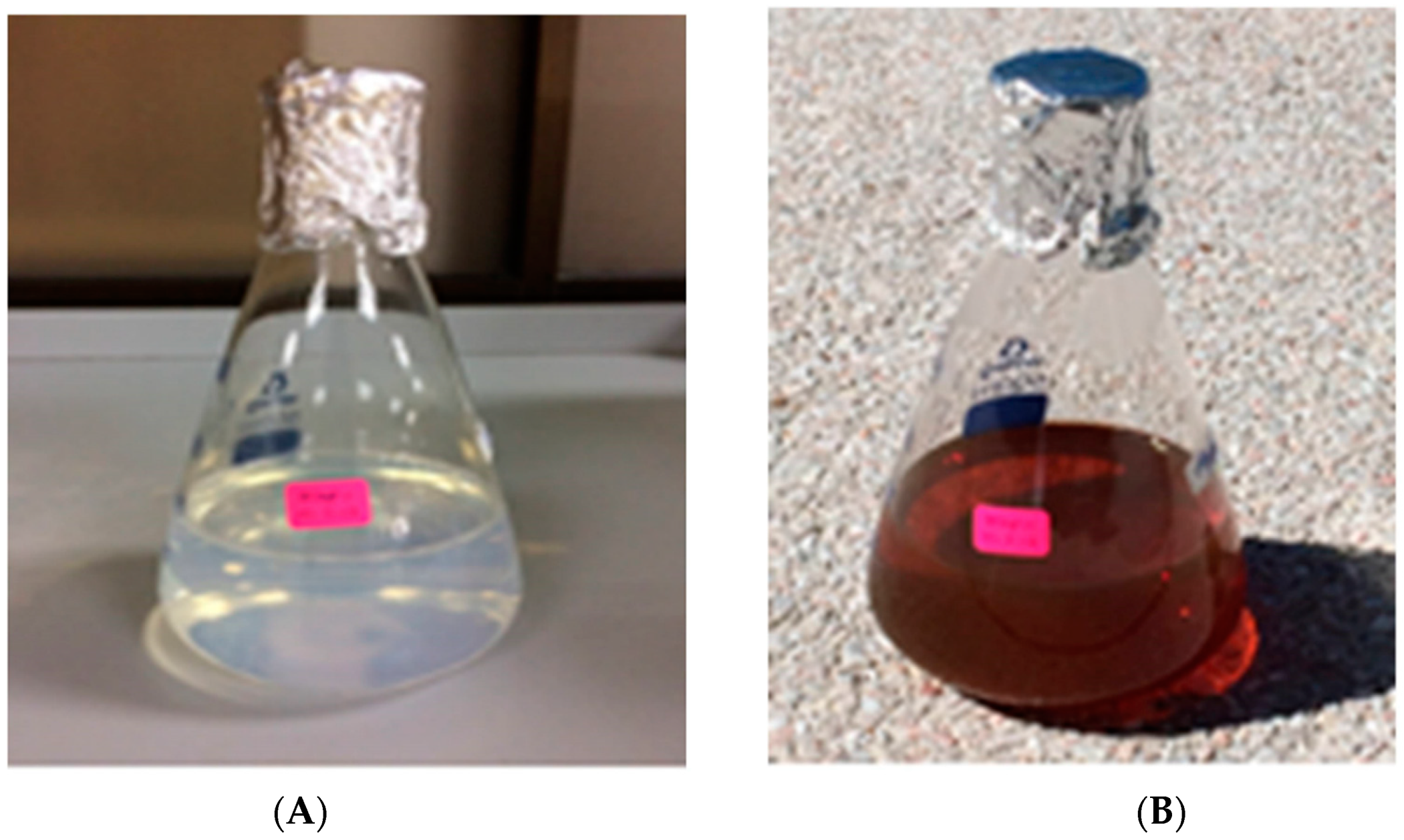
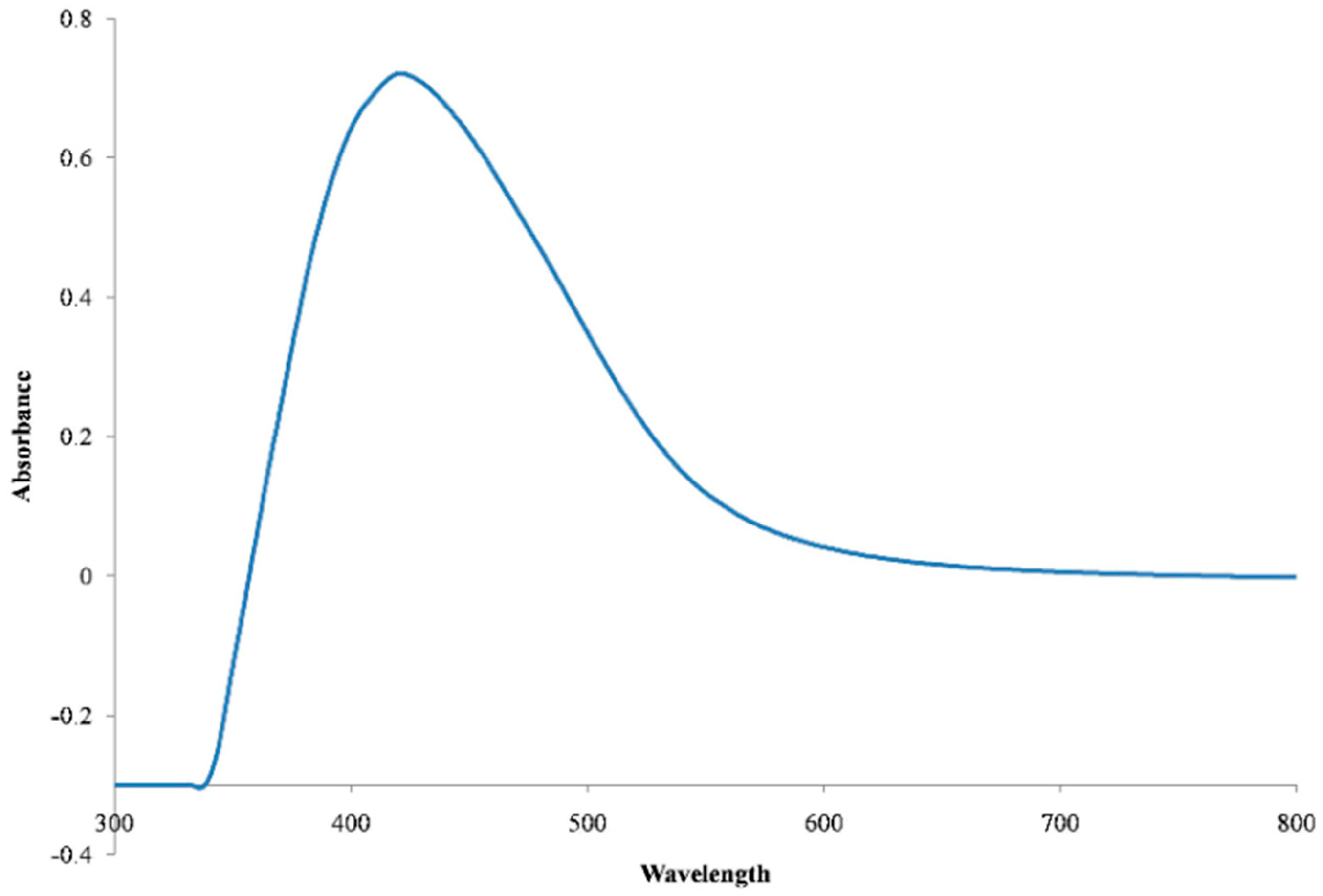
2.1. UV-Vis Spectrometry
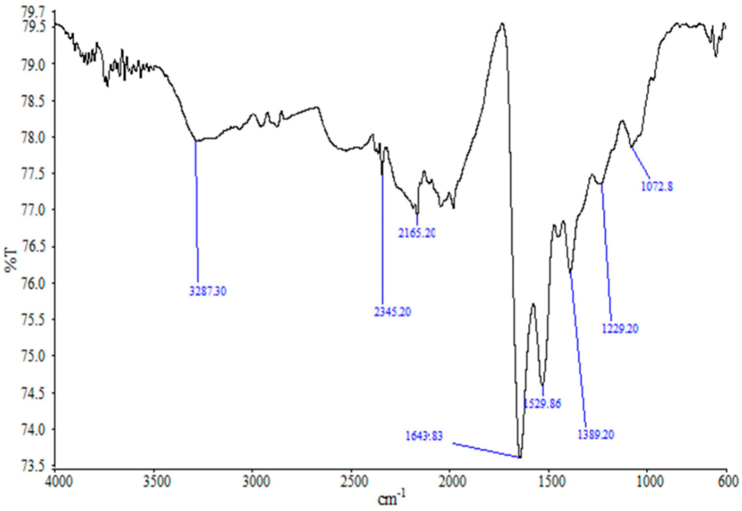
2.2. FTIR Study
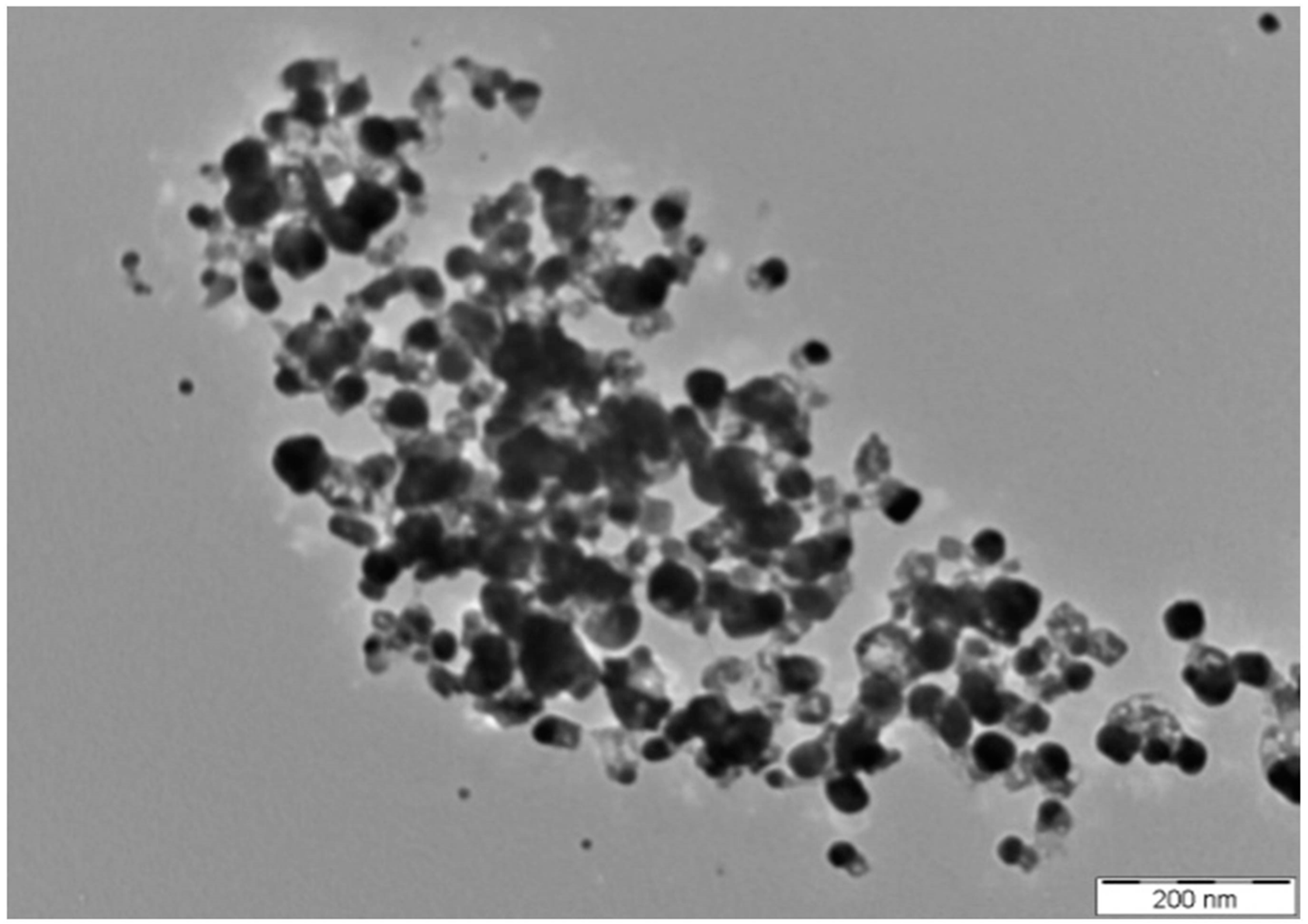
2.3. TEM Study
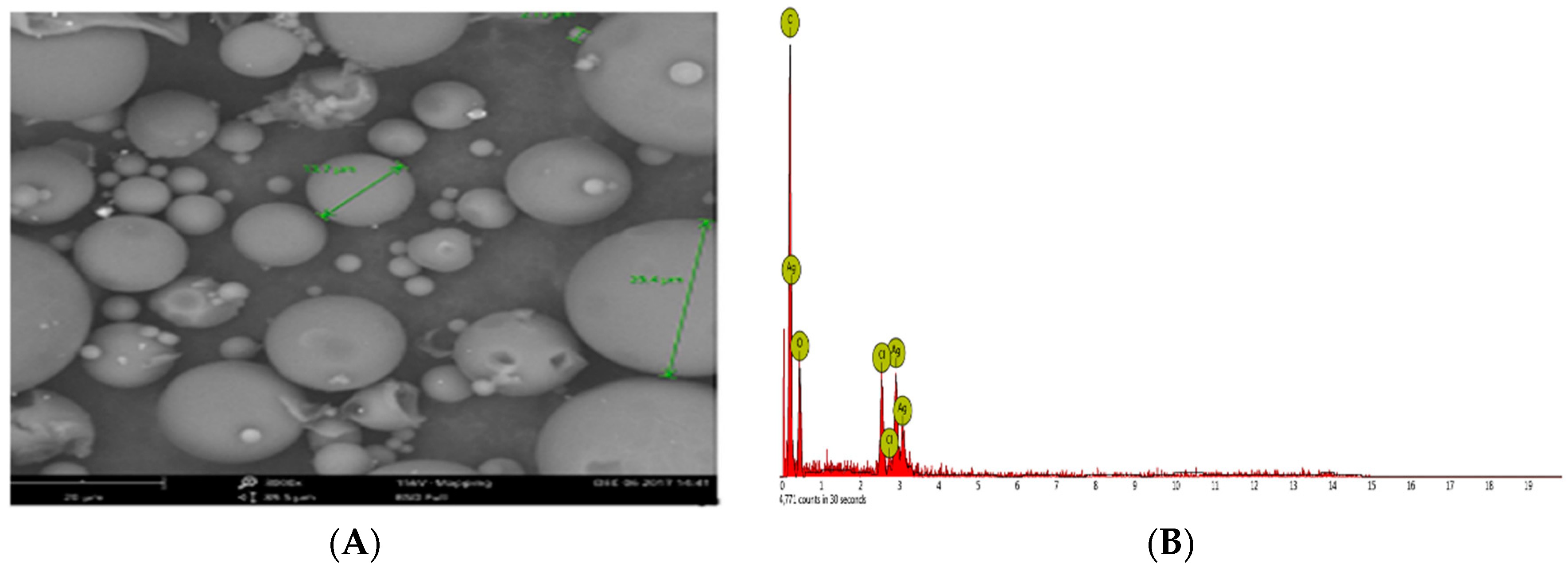
2.4. SEM-EDX Studies
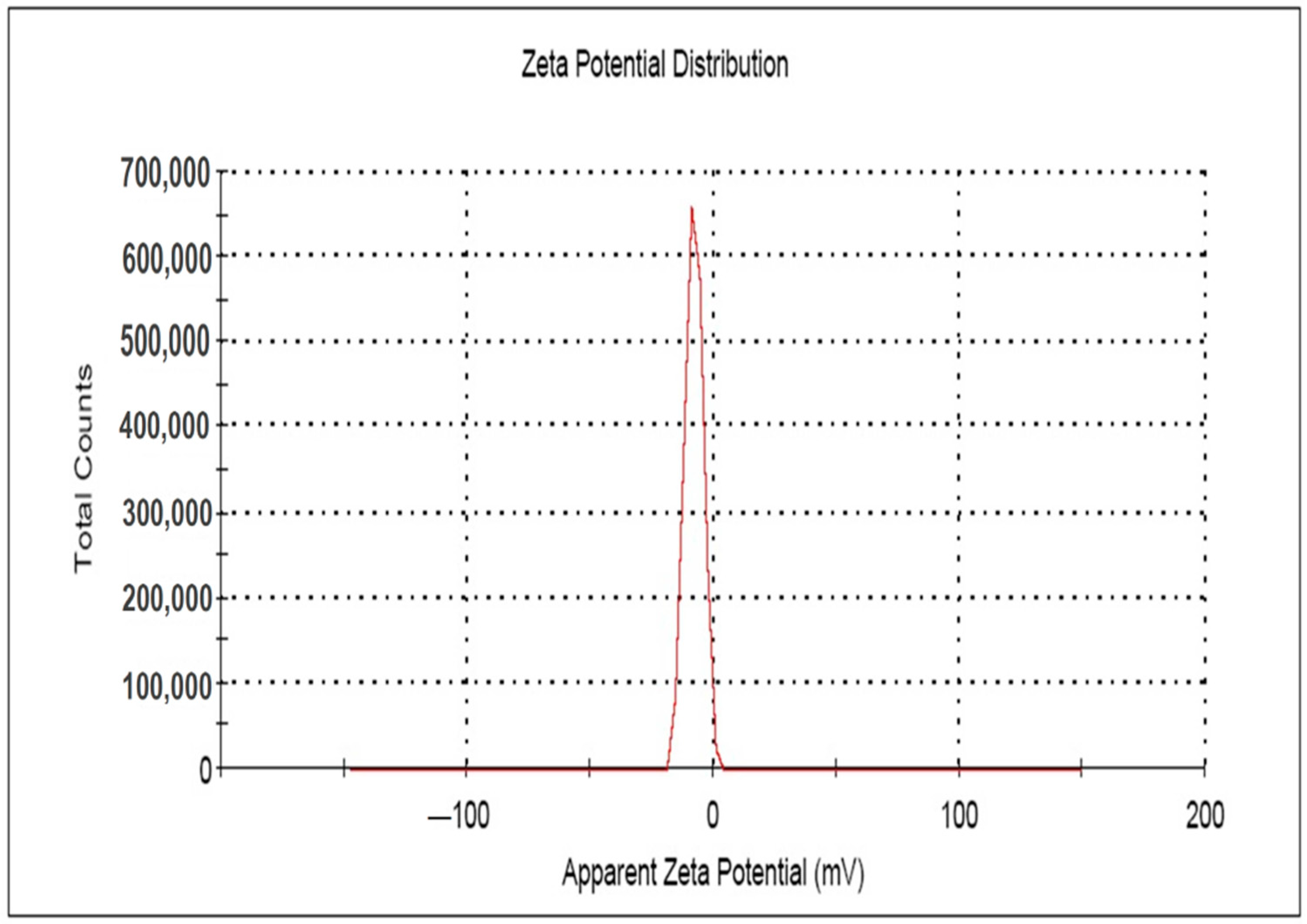
2.5. Zeta Potential Study
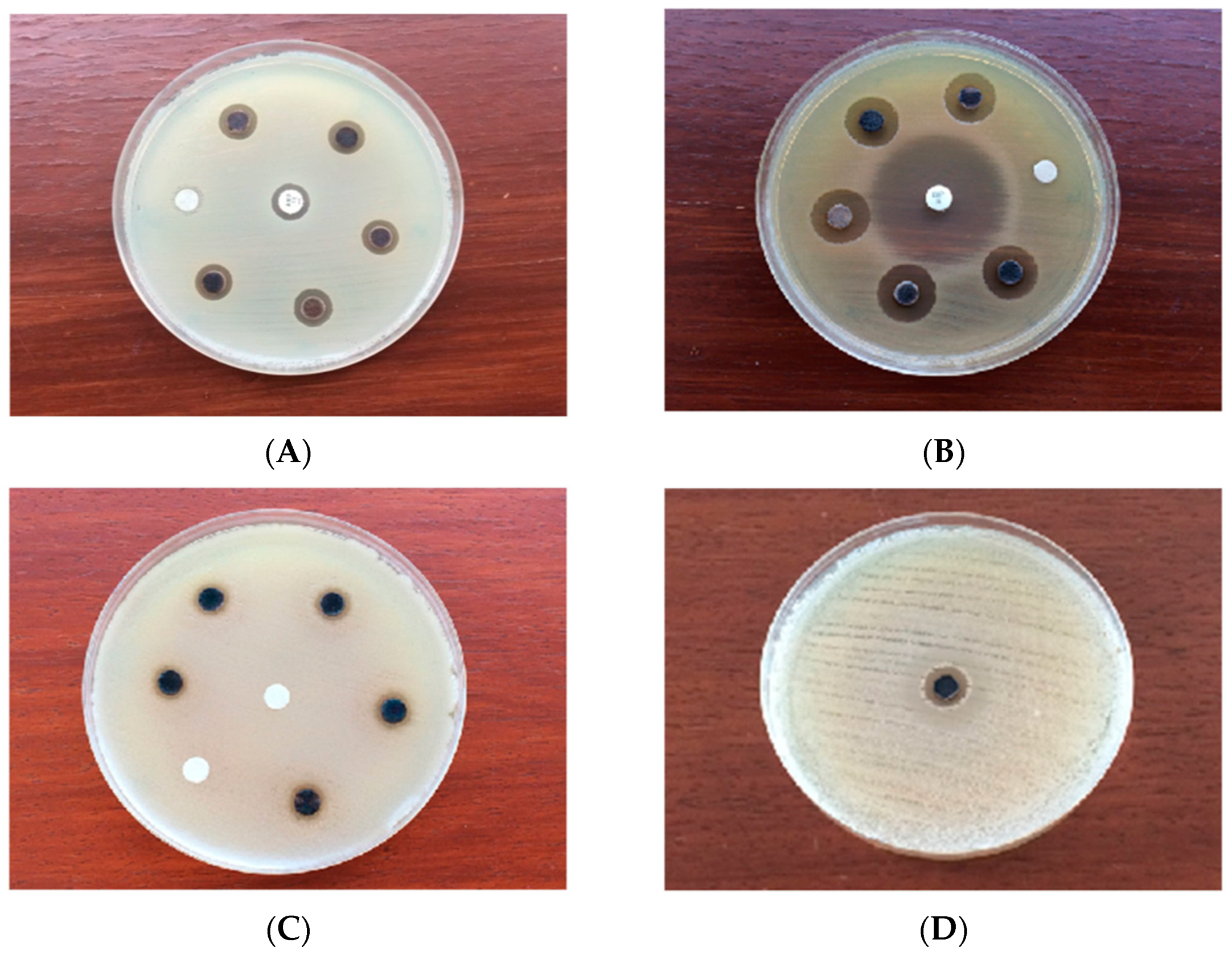
2.6. Antimicrobial Assay
3. Materials and Methods
3.1. Chemicals, Endophytic Bacteria and Microbial Strains
3.2. Culture of the Endophytic Bacteria and Preparation of Cell Free Extract
3.3. Synthesis of AgNPs
3.4. Characterization of AgNPs
3.5. Antimicrobial Assay
3.6. Statistical Analysis
4. Conclusions
Author Contributions
Acknowledgments
Conflicts of Interest
References
- Proia, L.; Anzil, A.; Subirats, J.; Borrego, C.; Farrè, M.; Llorca, M.; Balcázar, J.L.; Servais, P. Antibiotic resistance along an urban river impacted by treated wastewaters. Sci. Total Environ. 2018, 628, 453–466. [Google Scholar] [CrossRef] [PubMed]
- Qiao, M.; Ying, G.G.; Singer, A.C.; Zhu, Y.G. Review of antibiotic resistance in China and its environment. Environ. Int. 2018, 110, 160–172. [Google Scholar] [CrossRef]
- WHO Fact Sheets: Antibiotic Resistance; World Health Organization: Geneva, Switzerland; Available online: http://www.who.int/news-room/fact-sheets/detail/antibiotic-resistance (accessed on 19 July 2018).
- Cantón, R.; González-Alba, J.M.; Galán, J.C. CTX-M Enzymes: Origin and Diffusion. Front. Microbiol. 2012, 3, 110. [Google Scholar] [CrossRef] [PubMed]
- Hiramatsu, K.; Kayayama, Y.; Matsuo, M.; Aiba, Y.; Saito, M.; Hishinuma, T.; Iwamoto, A. Vancomycin-intermediate resistance in Staphylococcus aureus. J. Glob. Antimicrob. Resist. 2014, 2, 213–224. [Google Scholar] [CrossRef] [PubMed]
- Howden, B.P.; Peleg, A.Y.; Stinear, T.P. The evolution of vancomycin intermediate Staphylococcus aureus (VISA) and heterogenous-VISA. Infect. Genet. Evol. 2014, 21, 575–582. [Google Scholar] [CrossRef] [PubMed]
- Kinross, P.; Petersen, A.; Skov, R.; Van Hauwermeiren, E.; Pantosti, A.; Laurent, F.; Voss, A.; Kluytmans, J.; Struelens, M.J.; Heuer, O.; et al. The European human LA-MRSA study group Livestock-associated meticillin-resistant Staphylococcus aureus (MRSA) among human MRSA isolates, European Union/European Economic Area countries, 2013. Eurosurveillance 2017, 22. [Google Scholar] [CrossRef]
- Palomino, J.; Martin, A. Drug Resistance Mechanisms in Mycobacterium tuberculosis. Antibiotics 2014, 3, 317–340. [Google Scholar] [CrossRef]
- Streicher, E.M.; Müller, B.; Chihota, V.; Mlambo, C.; Tait, M.; Pillay, M.; Trollip, A.; Hoek, K.G.P.; Sirgel, F.A.; Gey van Pittius, N.C.; et al. Emergence and treatment of multidrug resistant (MDR) and extensively drug-resistant (XDR) tuberculosis in South Africa. Infect. Genet. Evol. 2012, 12, 686–694. [Google Scholar] [CrossRef]
- Davies, J. Antibiotic Resistance and the Future of Antibiotics. In Microbial Evolution and Co-Adaptation: A Tribute to the Life and Scientific Legacies of Joshua Lederberg; National Academies Press: Washington, DC, USA, 2008; pp. 160–172. [Google Scholar]
- Rios, A.C.; Moutinho, C.G.; Pinto, F.C.; Del Fiol, F.S.; Jozala, A.; Chaud, M.V.; Vila, M.M.D.C.; Teixeira, J.A.; Balcão, V.M. Alternatives to overcoming bacterial resistances: State-of-the-art. Microbiol. Res. 2016, 191, 51–80. [Google Scholar] [CrossRef]
- Jørgensen, P.S.; Wernli, D.; Folke, C.; Carroll, S.P. Changing antibiotic resistance: Sustainability transformation to a pro-microbial planet. Curr. Opin. Environ. Sustain. 2017, 25, 66–76. [Google Scholar] [CrossRef]
- Bos, J.; Austin, R.H. A bacterial antibiotic resistance accelerator and applications. In Methods in Cell Biology; Elsevier: New York, NY, USA, 2018; Vol. 147, pp. 41–57. ISBN 978-0-12-814282-0. [Google Scholar]
- Cailes, B.; Vergnano, S.; Kortsalioudaki, C.; Heath, P.; Sharland, M. The current and future roles of neonatal infection surveillance programmes in combating antimicrobial resistance. Early Hum. Dev. 2015, 91, 613–618. [Google Scholar] [CrossRef] [PubMed]
- Rai, M.; Ingle, A.P.; Pandit, R.; Paralikar, P.; Gupta, I.; Chaud, M.V.; dos Santos, C.A. Broadening the spectrum of small-molecule antibacterials by metallic nanoparticles to overcome microbial resistance. Int. J. Pharm. 2017, 532, 139–148. [Google Scholar] [CrossRef] [PubMed]
- de Smalen, A.W.; Ghorab, H.; Abd El Ghany, M.; Hill-Cawthorne, G.A. Refugees and antimicrobial resistance: A systematic review. Travel Med. Infect. Dis. 2017, 15, 23–28. [Google Scholar] [CrossRef] [PubMed]
- Chaudhary, A.S. A review of global initiatives to fight antibiotic resistance and recent antibiotics’ discovery. Acta Pharm. Sin. B 2016, 6, 552–556. [Google Scholar] [CrossRef] [PubMed]
- Borah, S.; Tripathy, S.; Bhuyan, B.; Kaishap, P.P.; Sharma, H.K.; Choudhury, G.; Saikia, L. Metal nanoparticles as potent antimicrobial nanomachetes with an emphasis on nanogold and nanosilver. In Design of Nanostructures for Versatile Therapeutic Applications; Elsevier: New York, NY, USA, 2018; pp. 487–521. ISBN 978-0-12-813667-6. [Google Scholar]
- Mordorski, B.; Friedman, A. Metal Nanoparticles for Microbial Infection. In Functionalized Nanomaterials for the Management of Microbial Infection; Elsevier: New York, NY, USA, 2017; pp. 77–109. ISBN 978-0-323-41625-2. [Google Scholar]
- WHO World Health Statistics 2018: Monitoring Health for the SDGs; World Health Organization: Geneva, Switzerland; Available online: http://www.who.int/gho/publications/world_health_statistics/2018/en/ (accessed on 30 August 2018).
- Dong, Z.Y.; Narsing Rao, M.P.; Xiao, M.; Wang, H.F.; Hozzein, W.N.; Chen, W.; Li, W.J. Antibacterial Activity of Silver Nanoparticles against Staphylococcus warneri Synthesized Using Endophytic Bacteria by Photo-irradiation. Front. Microbiol. 2017, 8. [Google Scholar] [CrossRef] [PubMed]
- Mathew, S.; Prakash, A.; Radhakrishnan, E.K. Sunlight mediated rapid synthesis of small size range silver nanoparticles using Zingiber officinale rhizome extract and its antibacterial activity analysis. Inorg. Nano-Met. Chem. 2018, 48, 139–145. [Google Scholar] [CrossRef]
- Saravanakumar, A.; Peng, M.M.; Ganesh, M.; Jayaprakash, J.; Mohankumar, M.; Jang, H.T. Low-cost and eco-friendly green synthesis of silver nanoparticles using Prunus japonica (Rosaceae) leaf extract and their antibacterial, antioxidant properties. Artif. Cells Nanomedicine Biotechnol. 2017, 45, 1165–1171. [Google Scholar] [CrossRef] [PubMed]
- Shaker, M.A.; Shaaban, M.I. Synthesis of silver nanoparticles with antimicrobial and anti-adherence activities against multidrug-resistant isolates from Acinetobacter baumannii. J. Taibah Univ. Med. Sci. 2017, 12, 291–297. [Google Scholar] [CrossRef]
- Khatoon, N.; Mazumder, J.A.; Sardar, M. Biotechnological Applications of Green Synthesized Silver Nanoparticles. J. Nanosci. Curr. Res. 2017, 77–109. [Google Scholar] [CrossRef]
- Zhang, X.F.; Liu, Z.G.; Shen, W.; Gurunathan, S. Silver Nanoparticles: Synthesis, Characterization, Properties, Applications, and Therapeutic Approaches. Int. J. Mol. Sci. 2016, 17, 1534. [Google Scholar] [CrossRef]
- Pulit-Prociak, J.; Banach, M. Silver nanoparticles—Amaterial of the future…? Open Chem. 2016, 14, 76–91. [Google Scholar] [CrossRef]
- Souza, L.R.R.; da Silva, V.S.; Franchi, L.P.; de Souza, T.A.J. Toxic and Beneficial Potential of Silver Nanoparticles: The Two Sides of the Same Coin. In Cellular and Molecular Toxicology of Nanoparticles; Saquib, Q., Faisal, M., Al-Khedhairy, A.A., Alatar, A.A., Eds.; Springer: Cham, Switzerland, 2018; Vol. 1048, pp. 251–262. ISBN 978-3-319-72040-1. [Google Scholar]
- de Matteis, V.; Cascione, M.; Toma, C.; Leporatti, S. Silver Nanoparticles: Synthetic Routes, In Vitro Toxicity and Theranostic Applications for Cancer Disease. Nanomaterials 2018, 8, 319. [Google Scholar] [CrossRef] [PubMed]
- Siddiqi, K.S.; Husen, A.; Rao, R.A.K. A review on biosynthesis of silver nanoparticles and their biocidal properties. J. Nanobiotechnology 2018, 16. [Google Scholar] [CrossRef] [PubMed]
- Firdhouse, M.J.; Lalitha, P. Biosynthesis of Silver Nanoparticles and Its Applications. J. Nanotechnol. 2015, 2015, 1–18. [Google Scholar] [CrossRef]
- Rónavári, A.; Kovács, D.; Igaz, N.; Vágvölgyi, C.; Boros, I.; Kónya, Z.; Pfeiffer, I.; Kiricsi, M. Biological activity of green-synthesized silver nanoparticles depends on the applied natural extracts: A comprehensive study. Int. J. Nanomed. 2017, 12, 871–883. [Google Scholar] [CrossRef] [PubMed]
- Syafiuddin, A.; Salmiati; Salim, M.R.; Beng Hong Kueh, A.; Hadibarata, T.; Nur, H. A Review of Silver Nanoparticles: Research Trends, Global Consumption, Synthesis, Properties, and Future Challenges: A Review of Silver Nanoparticles. J. Chin. Chem. Soc. 2017, 64, 732–756. [Google Scholar] [CrossRef]
- Rauwel, P.; Küünal, S.; Ferdov, S.; Rauwel, E. A Review on the Green Synthesis of Silver Nanoparticles and Their Morphologies Studied via TEM. Adv. Mater. Sci. Eng. 2015, 2015, 1–9. [Google Scholar] [CrossRef]
- Kumar, V.; Bano, D.; Mohan, S.; Singh, D.K.; Hasan, S.H. Sunlight-induced green synthesis of silver nanoparticles using aqueous leaf extract of Polyalthia longifolia and its antioxidant activity. Mater. Lett. 2016, 181, 371–377. [Google Scholar] [CrossRef]
- Manikprabhu, D.; Cheng, J.; Chen, W.; Sunkara, A.K.; Mane, S.B.; Kumar, R.; das, M.; Hozzein, W.N.; Duan, Y.-Q.; Li, W.-J. Sunlight mediated synthesis of silver nanoparticles by a novel actinobacterium (Sinomonas mesophila MPKL 26) and its antimicrobial activity against multi drug resistant Staphylococcus aureus. J. Photochem. Photobiol. B 2016, 158, 202–205. [Google Scholar] [CrossRef]
- Neethu, S.; Midhun, S.J.; Sunil, M.A.; Soumya, S.; Radhakrishnan, E.K.; Jyothis, M. Efficient visible light induced synthesis of silver nanoparticles by Penicillium polonicum ARA 10 isolated from Chetomorpha antennina and its antibacterial efficacy against Salmonella enterica serovar Typhimurium. J. Photochem. Photobiol. B 2018, 180, 175–185. [Google Scholar] [CrossRef]
- Gouda, S.; Das, G.; Sen, S.K.; Shin, H.-S.; Patra, J.K. Endophytes: A Treasure House of Bioactive Compounds of Medicinal Importance. Front. Microbiol. 2016, 7, 1538. [Google Scholar] [CrossRef] [PubMed]
- Schneider, C.; Hutter, I.; Döring, M. Commercial use of endophytes in micropropagation. Acta Hortic. 2017, 483–490. [Google Scholar] [CrossRef]
- Sudha, V.; Govindaraj, R.; Baskar, K.; Al-Dhabi, N.A.; Duraipandiyan, V. Biological properties of Endophytic Fungi. Braz. Arch. Biol. Technol. 2016, 59. [Google Scholar] [CrossRef]
- Christina, A.; Christapher, V.; Bhore, S. Endophytic bacteria as a source of novel antibiotics: An overview. Pharmacogn. Rev. 2013, 7, 11. [Google Scholar] [CrossRef] [PubMed]
- Fariq, A.; Khan, T.; Yasmin, A. Microbial synthesis of nanoparticles and their potential applications in biomedicine. J. Appl. Biomed. 2017, 15, 241–248. [Google Scholar] [CrossRef]
- Adur, A.J.; Nandini, N.; Mayachar, K.S.; Ramya, R.; Srinatha, N. Bio-synthesis and antimicrobial activity of silver nanoparticles using anaerobically digested parthenium slurry. J. Photochem. Photobiol. B 2018, 183, 30–34. [Google Scholar] [CrossRef]
- Chahar, V.; Sharma, B.; Shukla, G.; Srivastava, A.; Bhatnagar, A. Study of antimicrobial activity of silver nanoparticles synthesized using green and chemical approach. Colloids Surf. Physicochem. Eng. Asp. 2018, 554, 149–155. [Google Scholar] [CrossRef]
- Seetharaman, P.K.; Chandrasekaran, R.; Gnanasekar, S.; chandrakasan, G.; gupta, M.; Babu, D.; Sivaperumal, S. Antimicrobial and larvicidal activity of eco-friendly silver nanoparticles synthesized from endophytic fungi Phomopsis liquidambaris. Biocatal. Agric. Biotechnol. 2018. [Google Scholar] [CrossRef]
- Birla, S.S.; Gaikwad, S.C.; Gade, A.K.; Rai, M.K. Rapid Synthesis of Silver Nanoparticles from Fusarium oxysporum by Optimizing Physicocultural Conditions. Sci. World J. 2013, 2013, 1–12. [Google Scholar] [CrossRef]
- Lee, S.J.; Heo, M.; Lee, D.; Han, S.; Moon, J.-H.; Lim, H.-N.; Kwon, I.K. Preparation and characterization of antibacterial orthodontic resin containing silver nanoparticles. Appl. Surf. Sci. 2018, 432, 317–323. [Google Scholar] [CrossRef]
- Praphakar, R.A.; Jeyaraj, M.; Ahmed, M.; Kumar, S.S.; Rajan, M. Silver nanoparticle functionalized CS-g-(CA-MA-PZA) carrier for sustainable anti-tuberculosis drug delivery. Int. J. Biol. Macromol. 2018, 118, 1627–1638. [Google Scholar] [CrossRef]
- Taghavi, S.; Barac, T.; Greenberg, B.; Borremans, B.; Vangronsveld, J.; van der Lelie, D. Horizontal Gene Transfer to Endogenous Endophytic Bacteria from Poplar Improves Phytoremediation of Toluene. Appl. Environ. Microbiol. 2005, 71, 8500–8505. [Google Scholar] [CrossRef] [PubMed]
- van Elsas, J.D.; Turner, S.; Bailey, M.J. Horizontal gene transfer in the phytosphere. New Phytol. 2003, 157, 525–537. [Google Scholar] [CrossRef]
- Dupont, P.-Y.; Cox, M.P. Genomic Data Quality Impacts Automated Detection of Lateral Gene Transfer in Fungi. G3 Genes Genomes Genet. 2017, 7, 1301–1314. [Google Scholar] [CrossRef] [PubMed]
- Frank, A.C. The Genomes of Endophytic Bacteria. In Endophytes of Forest Trees; Pirttilä, A.M., Frank, A.C., Eds.; Springer: Dordrecht, The Netherlands, 2011; Vol. 80, pp. 107–136. ISBN 978-94-007-1598-1. [Google Scholar]
- Jaramillo, V.D.A.; Vargas, W.A.; Sukno, S.A.; Thon, M.R. Horizontal Transfer of a Subtilisin Gene from Plants into an Ancestor of the Plant Pathogenic Fungal Genus Colletotrichum. PLoS ONE 2013, 8, e59078. [Google Scholar] [CrossRef]
- Nongkhlaw, F.M.W.; Joshi, S.R. Horizontal Gene Transfer of the Non-ribosomal Peptide Synthetase Gene Among Endophytic and Epiphytic Bacteria Associated with Ethnomedicinal Plants. Curr. Microbiol. 2016, 72, 1–11. [Google Scholar] [CrossRef]
- Shinozuka, H.; Hettiarachchige, I.K.; Shinozuka, M.; Cogan, N.O.I.; Spangenberg, G.C.; Cocks, B.G.; Forster, J.W.; Sawbridge, T.I. Horizontal transfer of a β-1,6-glucanase gene from an ancestral species of fungal endophyte to a cool-season grass host. Sci. Rep. 2017, 7. [Google Scholar] [CrossRef] [PubMed]
- Keese, P. Risks from GMOs due to Horizontal Gene Transfer. Environ. Biosafety Res. 2008, 7, 123–149. [Google Scholar] [CrossRef]
- Ryan, R.P.; Germaine, K.; Franks, A.; Ryan, D.J.; Dowling, D.N. Bacterial endophytes: Recent developments and applications. FEMS Microbiol. Lett. 2008, 278, 1–9. [Google Scholar] [CrossRef] [PubMed]
- Venieraki, A.; Dimou, M.; Katinakis, P. Endophytic fungi residing in medicinal plants have the ability to produce the same or similar pharmacologically active secondary metabolites as their hosts. Hell. Plant Prot. J. 2017, 10, 51–66. [Google Scholar] [CrossRef]
- Kumari, M.; Pandey, S.; Giri, V.P.; Bhattacharya, A.; Shukla, R.; Mishra, A.; Nautiyal, C.S. Tailoring shape and size of biogenic silver nanoparticles to enhance antimicrobial efficacy against MDR bacteria. Microb. Pathog. 2017, 105, 346–355. [Google Scholar] [CrossRef] [PubMed]
- Jung, J.; Kasi, G.; Seo, J. Development of functional antimicrobial papers using chitosan/starch-silver nanoparticles. Int. J. Biol. Macromol. 2018, 112, 530–536. [Google Scholar] [CrossRef] [PubMed]
- Shao, Y.; Wu, C.; Wu, T.; Yuan, C.; Chen, S.; Ding, T.; Ye, X.; Hu, Y. Green synthesis of sodium alginate-silver nanoparticles and their antibacterial activity. Int. J. Biol. Macromol. 2018, 111, 1281–1292. [Google Scholar] [CrossRef] [PubMed]
- Smékalová, M.; Panáček, A.; Jančula, D.; Maršálek, B.; Kolařík, J.; Prucek, R.; Kvítek, L.; Zbořil, R. Culture medium mediated aggregation and re-crystallization of silver nanoparticles reduce their toxicity. Appl. Mater. Today 2018, 12, 198–206. [Google Scholar] [CrossRef]
- Ahmed, M.J.; Murtaza, G.; Mehmood, A.; Bhatti, T.M. Green synthesis of silver nanoparticles using leaves extract of Skimmia laureola: Characterization and antibacterial activity. Mater. Lett. 2015, 153, 10–13. [Google Scholar] [CrossRef]
- Al-Asfar, A.; Zaheer, Z.; Aazam, E.S. Eco-friendly green synthesis of Ag@Fe bimetallic nanoparticles: Antioxidant, antimicrobial and photocatalytic degradation of bromothymol blue. J. Photochem. Photobiol. B 2018, 185, 143–152. [Google Scholar] [CrossRef] [PubMed]
- Gade, A.; Gaikwad, S.; Duran, N.; Rai, M. Green synthesis of silver nanoparticles by Phoma glomerata. Micron 2014, 59, 52–59. [Google Scholar] [CrossRef]
- Elgorban, A.M.; Al-Rahmah, A.N.; Sayed, S.R.; Hirad, A.; Mostafa, A.A.F.; Bahkali, A.H. Antimicrobial activity and green synthesis of silver nanoparticles using Trichoderma viride. Biotechnol. Biotechnol. Equip. 2016, 30, 299–304. [Google Scholar] [CrossRef]
- Bose, S.; Chakraborty, S.; Sanyal, D. Water-Ethylene Glycol Mediated Synthesis of Silver Nanoparticles for Conductive Ink. Mater. Today Proc. 2018, 5, 9941–9947. [Google Scholar] [CrossRef]
- Ansari, A.; Pervez, S.; Javed, U.; Abro, M.I.; Nawaz, M.A.; Qader, S.A.U.; Aman, A. Characterization and interplay of bacteriocin and exopolysaccharide-mediated silver nanoparticles as an antibacterial agent. Int. J. Biol. Macromol. 2018, 115, 643–650. [Google Scholar] [CrossRef]
- Saeb, A.T.M.; Alshammari, A.S.; Al-Brahim, H.; Al-Rubeaan, K.A. Production of Silver Nanoparticles with Strong and Stable Antimicrobial Activity against Highly Pathogenic and Multidrug Resistant Bacteria. Sci. World J. 2014, 2014, 1–9. [Google Scholar] [CrossRef] [PubMed]
- Ahmed, K.B.A.; Senthilnathan, R.; Megarajan, S.; Anbazhagan, V. Sunlight mediated synthesis of silver nanoparticles using redox phytoprotein and their application in catalysis and colorimetric mercury sensing. J. Photochem. Photobiol. B 2015, 151, 39–45. [Google Scholar] [CrossRef] [PubMed]
- Spacciapoli, P.; Buxton, D.; Rothstein, D.; Friden, P. Antimicrobial activity of silver nitrate against periodontal pathogens. J. Periodontal Res. 2001, 36, 108–113. [Google Scholar] [CrossRef] [PubMed]
- Fard, J.K.; Jafari, S.; Eghbal, M.A. A Review of Molecular Mechanisms Involved in Toxicity of Nanoparticles. Adv. Pharm. Bull. 2015, 5, 447–454. [Google Scholar] [CrossRef] [PubMed]
- Kim, J.S.; Sung, J.H.; Ji, J.H.; Song, K.S.; Lee, J.H.; Kang, C.S.; Yu, I.J. In vivo Genotoxicity of Silver Nanoparticles after 90-day Silver Nanoparticle Inhalation Exposure. Saf. Health Work 2011, 2, 34–38. [Google Scholar] [CrossRef] [PubMed]
- Helmlinger, J.; Sengstock, C.; Groß-Heitfeld, C.; Mayer, C.; Schildhauer, T.A.; Köller, M.; Epple, M. Silver nanoparticles with different size and shape: Equal cytotoxicity, but different antibacterial effects. RSC Adv. 2016, 6, 18490–18501. [Google Scholar] [CrossRef]
- Khafaga, A.F.; Abu-Ahmed, H.M.; El-Khamary, A.N.; Elmehasseb, I.M.; Shaheen, H.M. Enhancement of Equid Distal Limb Wounds Healing by Topical Application of Silver Nanoparticles. J. Equine Vet. Sci. 2018, 61, 76–87. [Google Scholar] [CrossRef]
- Kumar, S.S.D.; Houreld, N.N.; Kroukamp, E.M.; Abrahamse, H. Cellular imaging and bactericidal mechanism of green-synthesized silver nanoparticles against human pathogenic bacteria. J. Photochem. Photobiol. B 2018, 178, 259–269. [Google Scholar] [CrossRef]
- Karlsson, H.; Gliga, A.R.; Kohonen, P.; Wallberg, A.; Fadeel, B. Genotoxic and epigenetic effects of silver nanoparticles. Toxicol. Lett. 2012, 211, S40. [Google Scholar] [CrossRef]
- Kumari, M.; Mukherjee, A.; Chandrasekaran, N. Genotoxicity of silver nanoparticles in Allium cepa. Sci. Total Environ. 2009, 407, 5243–5246. [Google Scholar] [CrossRef]
- Loh, C.Y.; Tan, Y.Y.; Rohani, R.; Weber, J.F.F.; Bhore, S.J. Diversity of endophytic bacteria in Malaysian plants as revealed by 16S rRNA encoding gene sequence based method of bacterial identification. J. Young Pharm. 2013, 5, 95–97. [Google Scholar] [CrossRef] [PubMed]
- Deljou, A.; Goudarzi, S. Green Extracellular Synthesis of the Silver Nanoparticles Using Thermophilic Bacillus Sp. AZ1 and its Antimicrobial Activity Against Several Human Pathogenetic Bacteria. Iran. J. Biotechnol. 2016, 14, 25–32. [Google Scholar] [CrossRef] [PubMed]
- Bauer, A.W.; Kirby, W.M.M.; Sherris, J.C.; Turck, M. Antibiotic Susceptibility Testing by a Standardized Single Disk Method. Am. J. Clin. Pathol. 1966, 45, 493–496. [Google Scholar] [CrossRef] [PubMed]
- CLSI. Performance Standards for Antimicrobial Susceptibility Testing, 28th ed.; CLSI supplement M100; Clinical and Laboratory Standards Institute: Wayne, PA, USA, 2018. [Google Scholar]
- CLSI. Method for Antifungal Disk Diffusion Susceptibility Testing of Yeasts, 2nd ed.; CLSI supplement M44; Clinical and Laboratory Standards Institute: Wayne, PA, USA, 2009. [Google Scholar]
- Finch, R.G.; Greenwood, D.; Rorrby, S.R.; Whitley, R.J. Antibiotic and Chemotherapy: Anti-infective Agents and Their Use in Therapy, 9th ed.; Saunders: St. Louis, MO, USA, 2010; ISBN 978-0-7020-4064-1. [Google Scholar]
Sample Availability: Samples of the endophytic bacterial strain are available from the authors. |


| Element Number | Element Symbol | Element Name | Atomic Conc. | Weight Conc. |
|---|---|---|---|---|
| 6 | C | Carbon | 70.08 | 49.02 |
| 47 | Ag | Silver | 3.74 | 23.50 |
| 8 | O | Oxygen | 23.46 | 21.86 |
| 17 | Cl | Chlorine | 2.72 | 5.61 |
| Triplicate Experiments with the Synthesized AgNPs | Zeta Potential (ζ) (mV) | Mean (mV) | Area (%) | Conductivity (mS/cm) |
|---|---|---|---|---|
| 1 | −6.75 | 3.49 | 100 | 0.0127 |
| 2 | −7.92 | 3.54 | 100 | 0.0129 |
| 3 | −7.77 | 2.97 | 100 | 0.0355 |
| Pathogenic Microbes 1 | ||
| Microorganisms | Synthesized AgNPs | Control |
| B. cereus (ATCC 10876) | 9.16 ± 0.05 c (2.25) | Ampicilin (10 μg): resistant |
| S. aureus subsp. aureus (ATCC 11632) | 11.30 ± 0.07 b (2.75) | Ampicilin (10 μg): 10.14±0.05 |
| E. coli (ATCC 10536) | 15.12 ± 0.08 a (3.25) | Ciprofloxacin (5 μg): 30.48±0.08 |
| P. aeruginosa (ATCC 10145) | 8.02 ± 0.08 d (1.75) | Ciprofloxacin (5 μg): 30.10±0.07 |
| C. albicans (ATCC 10231) | 7.16 ± 0.09 e (1.75) | Itraconazole (10 μg): resistant |
| MDR bacteria 2 | ||
| S. pneumoniae (ATCC700677) | 10.20 ± 0.07 B (2.75) | - |
| E. faecium (ATCC 700221) | 12.16 ± 0.05 A (2.25) | - |
| S. aureus subsp. aureus (ATCC33592) | 10.16 ± 0.05 B (3.75) | - |
| E. coli (NCTC 13351) | 12.24 ± 0.05 A (3.50) | - |
© 2018 by the authors. Licensee MDPI, Basel, Switzerland. This article is an open access article distributed under the terms and conditions of the Creative Commons Attribution (CC BY) license (http://creativecommons.org/licenses/by/4.0/).
Share and Cite
Monowar, T.; Rahman, M.S.; Bhore, S.J.; Raju, G.; Sathasivam, K.V. Silver Nanoparticles Synthesized by Using the Endophytic Bacterium Pantoea ananatis are Promising Antimicrobial Agents against Multidrug Resistant Bacteria. Molecules 2018, 23, 3220. https://doi.org/10.3390/molecules23123220
Monowar T, Rahman MS, Bhore SJ, Raju G, Sathasivam KV. Silver Nanoparticles Synthesized by Using the Endophytic Bacterium Pantoea ananatis are Promising Antimicrobial Agents against Multidrug Resistant Bacteria. Molecules. 2018; 23(12):3220. https://doi.org/10.3390/molecules23123220
Chicago/Turabian StyleMonowar, Tahmina, Md. Sayedur Rahman, Subhash J. Bhore, Gunasunderi Raju, and Kathiresan V. Sathasivam. 2018. "Silver Nanoparticles Synthesized by Using the Endophytic Bacterium Pantoea ananatis are Promising Antimicrobial Agents against Multidrug Resistant Bacteria" Molecules 23, no. 12: 3220. https://doi.org/10.3390/molecules23123220
APA StyleMonowar, T., Rahman, M. S., Bhore, S. J., Raju, G., & Sathasivam, K. V. (2018). Silver Nanoparticles Synthesized by Using the Endophytic Bacterium Pantoea ananatis are Promising Antimicrobial Agents against Multidrug Resistant Bacteria. Molecules, 23(12), 3220. https://doi.org/10.3390/molecules23123220