Article

Selection of Specific Peptides for Coccidioides spp. Obtained from Antigenic Fractions through SDS-PAGE and Western Blot Methods by the Recognition of Sera from Patients with Coccidioidomycosis

by
Esperanza Duarte Escalante
1,
María Guadalupe Frías De León
2,
Luz Gisela Martínez García
1,
Jorge Herrera
3,
Gustavo Acosta Altamirano
2,
Carlos Cabello
4,
Gabriel Palma
4 and
María del Rocío Reyes Montes
1,*
1
Departamento de Microbiología y Parasitología, Facultad de Medicina, Universidad Nacional Autónoma de México (UNAM), Ciudad Universitaria No. 3000, México Cd. Mx. 04510, Mexico
2
Hospital Regional de Alta Especialidad de Ixtapaluca, Carretera Federal México—Puebla Km. 34.5, Pueblo de Zoquiapan, Ixtapaluca 56530, Mexico
3
Unidad de Servicios de Apoyo a la Investigación y la Industria (USAII), Facultad de Química, Universidad Nacional Autónoma de México (UNAM), Ciudad Universitaria No. 3000, México Cd. Mx. 04510, Mexico
4
Instituto Nacional de Enfermedades Respiratorias (INER), Calzada de Tlalpan 4502, Belisario Domínguez Sección XVI, Tlalpan, México Cd. Mx 14080, Mexico
*
Author to whom correspondence should be addressed.
Molecules 2018, 23(12), 3145; https://doi.org/10.3390/molecules23123145
Submission received: 24 October 2018 / Revised: 26 November 2018 / Accepted: 29 November 2018 / Published: 30 November 2018

Abstract

:
Antigenic fractions of 100, 50, 37, and 28 kDa obtained through the SDS-PAGE method that were more frequently recognized by anti-Coccidioides antibodies in the sera of coccidioidomycosis patients were selected using western blotting. Subsequently, these bands were sequenced, and the obtained proteins were analysed by BLAST to choose peptides specific for Coccidioides spp. from among the shared aligned sequences of related fungi. A peptide specific for C. immitis was selected from the “GPI anchored serine-threonine rich protein OS C. immitis”, while from the “uncharacterized protein of C. immitis”, we selected a peptide for C. immitis and C. posadasii. These proteins arose from the 100 kDa antigenic fraction. From the protein “fatty acid amide hydrolase 1 of C. posadasii” that was identified from the 50 kDa antigenic fraction, a peptide was selected that recognized C. immitis and C. posadasii. In addition, the analysis of all the peptides (353) of each of the assembled proteins showed that only 35 had 100% identity with proteins of C. immitis and C. posadasii, one had 100% identity with only C. immitis, and one had 100% identity with only C. posadasii. These peptides can be used as diagnostic reagents, vaccines, and antifungals.

1. Introduction

Coccidioidomycosis or San Joaquin valley fever is a disease caused by fungi of the genus Coccidioides, in which two species with almost identical phenotypes, C. immitis and C. posadasii, are recognized [1]. This disease is one of the most important endemic mycoses in North America. However, there are also significant endemic areas in other countries, such as Guatemala, Honduras, Venezuela, Colombia, Brazil, and Argentina. In these countries, coccidioidomycosis is not a disease with mandatory reporting; therefore, epidemiological data regarding this mycosis are still scarce, and investigations are restricted to retrospective studies and to the publication of clinical cases [2,3,4,5,6,7,8,9,10,11,12,13,14,15,16]. Therefore, the actual incidence of this disease is unknown.
The clinical manifestations of coccidioidomycosis can be confused with those of other nosological entities, which hinders its diagnosis. Traditionally, the diagnosis of coccidioidomycosis is made considering clinical and radiological results and laboratory analyses. The conventional diagnosis for coccidioidomycosis has been based on fungal identification via direct examination using freshly stained samples or different staining techniques and by the culture of tissue or fluid samples. However, the retrieval rate of this pathogen through culture varies from 0.4% in blood to 8.3% in respiratory tract samples [17]. The use of immunodiagnostic methods is still recommended, such as immunodiffusion (ID), complement fixation (CF), and tube precipitation (TP). These methods traditionally use coccidioidin, a crude extract formed from a large number of fungal components that favours cross-reactivity between the antibodies of patients with different fungal (Blastomyces dermatitides, Histoplasma capsulatum) and bacterial infections [18]. Lindsley et al. [19] showed that coccidioidin detects IgG antibodies, but when coccidioidin is treated with heat, it detects IgM antibodies. Therefore, the immunodiffusion-complement fixation (IDCF) sensitivity is 77%, while the immunodiffusion-tube precipitation (IDTP) sensitivity is between 75% and 91% [20]. These findings led to the production of pure antigens with well-defined physicochemical properties for the diagnosis of coccidioidomycosis, such as the coccidioidal CF and IDCF antigen, corresponding to a chitinase. The gene encoding this antigen was cloned, and a functional recombinant antigen called 47 kDa CF-chitinase (rCF) was obtained [21]. This antigen has shown a sensitivity of 96.9% and a specificity of 100% in an enzyme-linked immunosorbent assay (ELISA) [22]. However, it shares an epitope with H. capsulatum and B. dermatitidis [23]. The proline-rich antigen (Ag2/PRA), or antigen 2, is another specific antigen for Coccidioides spp., obtained by the deglycosylation of a spherule extract. This antigen has been favourably evaluated (in mice) as a vaccine candidate [24]. The gene encoding this antigen has been cloned and the recombinant antigen (rPRA) obtained, which recognizes IgG antibodies from patients with progressive pulmonary or extrapulmonary coccidioidomycosis. It has a high sensitivity (88%) and specificity (97%) for the diagnosis of coccidioidomycosis [25]. Filho et al. [26] evaluated the reactivity of a local antigen that was extracted from a strain of C. posadasii. This antigen was extracted with ammonium sulfate and was characterized by 1-D electrophoresis, showing good specificity and reactivity by radial immunodiffusion and western blot. Two well-defined bands were observed, with one corresponding to β-glucosidase and the other to glutamine synthetase; therefore, the authors proposed them to be a rapid diagnostic tool with a low cost and without the risk of the direct manipulation of the microorganism. Likewise, De Aguiar Cordeiro et al. [27] described the preparation of two antigens (F0-90 and F60-90) for the detection of human anti-Coccidioides antibodies by ID and EIA (enzyme immunoassay). The antigens were tested against serum samples from patients with coccidioidomycosis, histoplasmosis, and paracoccidioidomycosis, as well as from healthy individuals. The highest reactivity in the ID tests was observed with the F0-90 antigen, while the ELISA showed the best results with the F60-90 antigen. The F0-90 antigen showed cross-reactivity with Paracoccidioides, whereas F60-90 had cross-reactivity with Paracoccidioides and Histoplasma. Therefore, the authors suggested the commercial use of the F0-90 and F60-90 antigens for the presumptive diagnosis of coccidioidomycosis by ID or EIA, respectively.
Due to the disadvantages of cross-immunity, several researchers have oriented their work towards obtaining biomarkers with specificity and immunodominance properties, as in the case of Navalkar et al. [28], who applied the immunosignature technique. These authors used two peptide types to classify coccidioidomycosis: (1) a peptide set created by tiling four of the most immunodominant proteins of C. immitis to create 83 “epitope peptides” so that these peptides interact specifically with anti-Coccidioides antibodies; and (2) a microarray chip containing 10,000 printed, randomly sequenced peptides, which serve as artificial Coccidioides antigens to randomly interact with patient antibodies. The authors showed that, compared with the epitope peptides, the randomly sequenced peptides had a greater precision when classifying the different stages of coccidioidomycosis. Currently, there is a greater interest in molecular mimicry, particularly in using low molecular weight peptides, which have generated considerable expectations as substitutes for natural antigens [29]. Therefore, this work aims to select specific peptides for Coccidioides spp. that are obtained from antigenic fractions through SDS-PAGE and western blot by the recognition of sera from patients with coccidioidomycosis.

2. Results

2.1. Species Identification for Isolates of Coccidioides spp.

Six isolates were identified as C. posadasii (HU-1, 073129, 083376, GGM, ZVJ, QR) and five as C. immitis (M40-05, PRA, 30.1, 43.3, 62).

2.2. Coccidioidins Characterization

The coccidioidins obtained from the isolates of C. immitis and C. posadasii included in this study revealed antigenic identity by ID, and all showed double identity bands (Figure 1).

2.3. Electrophoresis (SDS-PAGE)

For the SDS-PAGE assays, the protein concentration was adjusted to 3.3 mg/mL for all samples. Despite using different polyacrylamide concentrations in the SDS-PAGE gels (7.5, 10, and 12%), it was not possible to identify species-specific bands. However, when using 12% gels, bands with a wide range of molecular weights (10 to 250 kDa) were observed for exoantigens of C. immitis and C. posadasii (Figure 2). Table 1 shows the binary data matrix for the presence/absence of bands identified in the electrophoretic patterns for the evaluated coccidioidins (Figure 2).

2.4. Reactivity of Antigenic Fractions by WB

To optimise the antigen and antibody concentrations in the WB assays, the antigenic fractions were tested via dot blot using 11 serum samples that recognized C. immitis and C. posadasii antigens. The optimal antigen concentration was 0.66 mg/mL; the dilution of the primary antibody was 1/250, and the dilution of the secondary antibody was 1/5000. To demonstrate the recognition of antigenic fractions with 27 anti-Coccidioides sera samples obtained from patients, they were only tested with one coccidioidin of C. immitis (M40-05) and one of C. posadasii (HU-1). From the presence/absence data matrix (Supplementary Material Table S1), four bands were chosen for sequencing, according to the highest recognition frequency with each of the tested sera: 100, 50, 37, and 28 kDa (Table 2).

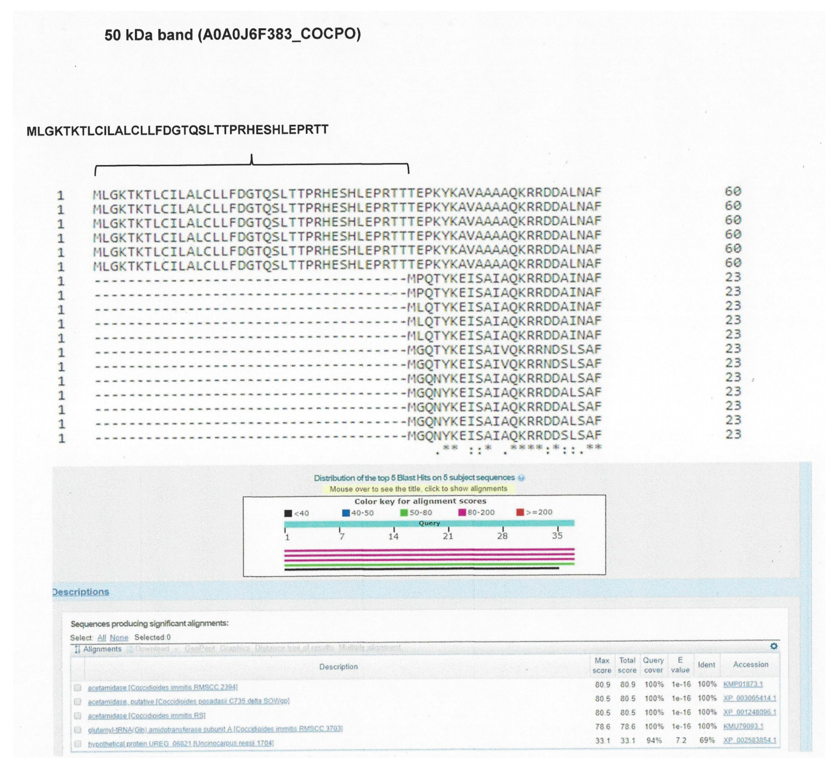
2.5. Analysis of Assembled Sequences

We identified eight proteins (assembled sequences) corresponding to the 100 kDa band, 14 proteins (assembled sequences) corresponding to the 50 kDa band, 14 proteins (assembled sequences) corresponding to the 37 kDa band, and 13 proteins (assembled sequences) corresponding to the 28 kDa band. For the sequences of proteins corresponding to the 100 kDa band, the analysis performed with the BLAST program showed two sequences: one as “GPI anchored serine-threonine rich protein OS C. immitis”, with access number A0A0J6YAG6_COCIT; and the other as “uncharacterized protein OS C. immitis”, with access number A0A0J8R1I4_COCIT. From the 50 kDa band, a sequence was identified as “fatty acid amide hydrolase 1 OS C. posadasii”, with access number A0A0J6F383_COCPO. From the 37 and 28 kDa bands, no sequence was identified with the conditions proposed for selection (Table 3).
From the alignment result of the protein “GPI anchored serine-threonine rich membrane of C. immitis” with the related fungi, the peptide MTVSCCDKRGKALVGAVSELSRDITPPSPDFT was selected, which was specific for C. immitis. From the “uncharacterized protein of C. immitis”, with access number A0A0J8R1I4_COCIT, peptide GNASSIESRSTDVEPTKTPTGTDGGASLTNAPCNGPNCPSQSP was selected, which was present in both C. immitis and C. posadasii. Likewise, from the alignment of the protein “fatty acid amide hydrolase 1 of C. posadasii”, with access number A0A0J6F383_COCPO, the peptide MLGKTKTLCILALCLLFDGTQSLTTPRHESHLEPRTT was selected, which was present in C. immitis and C. posadasii (Figure 3, Figure 4 and Figure 5). To confirm the specificity of the selected peptides, they were analysed using the BLAST protein program.

2.6. Peptide Analysis

The analysis of all the peptides from each assembled protein showed that out of the 91 identified peptides corresponding to the 100 kDa band, nine had 100% identity with C. immitis and C. posadasii (Supplementary Material, Table S2). For the 50 kDa band, 50 peptides were identified; four showed 100% identity with C. immitis and C. posadasii and one showed 100% identity with only C. posadasii (Supplementary Material, Table S3). For the 37 kDa band, 152 peptides were identified; eight showed 100% identity with C. immitis and C. posadasii, and one showed 100% identity with only C. immitis (Supplementary Material, Table S4). For the 28 kDa band, 60 peptides were identified; 14 showed 100% identity with C. immitis and C. posadasii, and one showed 100% identity with only C. posadasii (Supplementary Material, Table S5).

3. Discussion

The diagnosis of coccidioidomycosis can be difficult in patients with acute and fulminant disease, especially if they are immunosuppressed. Commonly, antibody detection is performed based on traditional techniques, including CF and ID assays. Although ELISA and a lateral flow immunoassay (LFI) have been developed in an effort to improve the diagnostic accuracy and to improve the performance of laboratory tests, CF and ID continue to be widely used due to their simplicity and reliability [30]. Despite these advantages, ID tests can present great variability in terms of sensitivity [31], and cross-reactions with Histoplasma antigens can occur [32], even against purified antigens [23]. In addition, ID tests can have negative results at the beginning of the infection and in immunosuppressed patients.
Therefore, in the search for biomarkers for the diagnosis of coccidioidomycosis, our work identifies antigens of Coccidioides spp. by the recognition of sera from patients with coccidioidomycosis through the SDS-PAGE and western blot methods. In this study, coccidioidins were used because they are antigens containing proteins and polysaccharides. The coccidioidins are crude culture filtrates obtained from prolonged incubation and autolysis of the mycelial phase of the Coccidioidess spp. fungus in liquid media. On the other hand, the spherulin are extracts of the spherules of the fungus produced in vitro in a chemically defined liquid medium. The advantage of this antigen is that it is obtained from the parasitic phase of the fungus, which is in contact with the tissue of the host, while the mycelial phase of the fungus exists in the soil, so it is assumed that the antigens made from spherules would contain more relevant antigens in their participation of the host immune response; in addition, the spherulin is more sensitive than mycelial-based coccidioidin [33]. However, the utility of both preparations is recognized by clinicians, so in this study, we use the coccdidioidin, which is the antigen most commonly used in the diagnosis of this disease [26,34,35]. On the other hand, it has been shown that this antigen is considerably more specific than spherulin [36].
Of all the coccidioidins of both C. immitis and C. posadasii, no specific band was found for either species. However, all coccidioidins showed bands of 20, 25, 28, 50, 75, and 100 kDa; therefore, only one coccidioidin representative of each species was chosen (HU-1 for C. posadasii and M40-05 for C. immitis) to carry out the western blot assays with 27 serum samples from patients with proven coccidioidomycosis. Most serum samples recognized the 28, 37, 50, and 100 kDa bands, which were sequenced and analysed. A specific peptide was identified for C. immitis from the GPI anchored serine-threonine rich protein OS Coccidioides immitis, corresponding to the 100 kDa band, and another peptide corresponding to the “uncharacterized protein OS Coccidioides immitis”, which recognizes C. immitis and C. posadasii. Additionally, a peptide from the protein “fatty acid amide hydrolase 1 OS Coccidioides posadasii” was chosen, corresponding to the 50 kDa band, which is present in both C. immitis and C. posadasii. Importantly, the selected peptides were chosen based on the elimination of shared regions with closely related fungi, such as H. capsulatum, Aspergillus spp., Blastomyces spp., and Paracoccidioides spp. These are the fungi with which the selected peptides share the antigenic fractions responsible for cross-immunity [20].
In turn, the sequenced peptides of each band were also analysed by the BLAST protein program. Of the 353 peptides, only 35 showed 100% identity with sequences of Coccidioides spp. and an E-value <0.001, while the remainder shared sequences with related fungi (Histoplasma, Blastomyces, Paracoccidioides, Aspergillus, and Criptococcus). Of the selected peptides, one showed identity with only C. immitis, and another showed identity with only C. posadasii, so these peptides can be used for epidemiological studies because they discriminate between the two species. The low percentage found for species-specific peptides coincides with that reported by Mitchell et al. [37], who reported that both species of Coccidioides are closely related at the proteomic level. On the other hand, the rest of the peptides that were specific for the genus Coccidioides can represent an important tool for the diagnosis of coccidioidomycosis, with the advantage that they originate from antigenic fractions that are recognized by antibodies present in patients with coccidioidomycosis. In addition, peptides are now considered to be important; previously, an extensive area for the recognition of the antigen was assumed in the antibody-antigen interaction, where relatively large interaction sites are involved (>6 amino acids). For this reason, the biotechnology industry has only produced products with large recognition sites—either the complete ligand or receptor, or a substantial part of these. Today, it has been shown to be beneficial to use only one molecule representative of the binding site involved in the recognition event instead of an entire protein [29]. Such molecules, for example, peptides or mimotope peptides, with a size between six and 20 amino acids, have multiple advantages compared to the native protein in terms of production, storage, and safety. Moreover, very large protein molecules turn out to be pleiotropic; that is, they have more than one function. Often, these functions are found in separate sites of the molecule [38,39], and therefore, they can be separated using molecules that mimic the appropriate site. Thus, unwanted immune responses are less likely to occur with small peptides than with large proteins. Consequently, peptides can be used as diagnostic reagents, vaccines, and antifungals [29,40,41,42,43].
As a perspective of this study, the peptides selected as specific for Coccidioides spp., could be analyzed through immune-informatics and computational tools to find epitopes that are capable of inducing a humoral immune and cellular response and evaluating its usefulness in obtaining a vaccine or for diagnosis. In the case of reaching a diagnosis, this will be important, since the use of chemically defined antigens, such as peptides, can replace the classical immunological tests, which use undefined antigens that show variability in the sensitivity and specificity of the results [44,45].

4. Materials and Methods

4.1. Isolates

Eleven clinical isolates of Coccidioides spp. were used, consisting of nine isolates from Mexico and two isolates from Argentina. The clinical isolates were identified at the species level according to Duarte-Escalante et al. [46], using the method of Bialek et al. [47]. The isolates were preserved at 4 °C in vials with sterile water and in tubes with mycobiotic agar (Bioxón, México, MX), with and without mineral oil. All the procedures of this study were carried out in a class-3 biosecurity area. The isolates are found in the fungal collection of the Molecular Mycology Laboratory, Microbiology and Parasitology Department, School of Medicine, National Autonomous University of Mexico (Universidad Nacional Autónoma de México—UNAM). Their source and geographic origin are shown in Table 4.

4.2. Biological Samples

Twenty-seven serum samples from patients with proven coccidioidomycosis were used. The serum samples were provided by Dr. Carlos Cabello from the National Institute of Respiratory Diseases (Instituto Nacional de Enfermedades Respiratorias—INER) (Table 5).

4.3. Obtaining Coccidioidins

To demonstrate the antigenic identity of the study isolates, coccidioidins were obtained according to Smith et al. [48]. The mycelial phase of each isolate was inoculated in Smith’s medium (0.3 g of ferric citrate, 1.31 g of potassium dibasic phosphate, 0.4 g of anhydrous sodium citrate, 1.5 g of magnesium sulphate, 10 g of glucose, 25.6 mL of glycerine, 7 g of asparagine, 7 g of ammonium chloride, brought to 1000 mL) and incubated at 28 °C for three months. Next, each culture was inactivated with thimerosal (1:5000) for 20 days, and subsequently, the mycelial mass was separated by filtration. Each culture’s filtrate was concentrated 100 times using the Amicon system (Sigma-Aldrich, St. Louis, MO, USA) and a 10 kDa exclusion membrane (Millipore Corporation, Burlington, MA, USA). The concentrated filtrate was dialyzed against deionized water for 24 h, with several changes, to finally obtain concentrated exoantigens, known as crude antigens (coccidioidins). The antigenic identification was carried out by the ID immune method, as described by Ouchterlony and Nilsson [49]. Positive and negative sera were used, as well as a reference antigen. The immunological identification of coccidioidins was carried out based on the appearance of identity bands. For subsequent trials, the protein concentration of each antigenic extract was determined by the method of Bradford et al. [50], using bovine serum albumin (BSA) as a control.

4.4. Sodium Dodecyl Sulfate-Polyacrylamide Gel Electrophoresis (SDS-PAGE)

To determine the components and molecular weights of the different coccidioidins, one-dimensional electrophoresis was carried out under denaturing conditions using the batch method [51], as described below. The coccidioidins were adjusted to a concentration of 80 µg/mL with 0.5 M Tris-HCl buffer pH 6.8 and 0.1% SDS, and the samples were heated at 100 °C for 5 min. Next, the coccidioidins were run on gels with different acrylamide concentrations (7.5%, 10%, and 12%) in buffer containing 0.025 M Tris-HCl, 0.2 M glycine, and 1% SDS. A molecular size marker of 250–15 kDa was used (Bio-Rad Laboratories Inc., Hercules, CA, USA). Electrophoresis was carried out with a Bio-Rad vertical electrophoresis system, with a constant current of 20 mA for 2 h. The gels were stained with Coomassie blue R250 (Sigma-Aldrich, St. Louis, MO, USA). A binary data matrix for the presence/absence of bands was developed based on the analysis with the Bio-Rad imaging system of the electrophoretic patterns obtained with the analysed exoantigens to identify, with greater precision, the existence of species-specific bands.

4.5. Recognition of Antigenic Fractions of Coccidioidins by Western Blot (WB)

The WB assays were carried out according to the method described by Towbin et al. [52]. The SDS-PAGE gels were placed on 0.45 µm pore nitrocellulose membranes (Bio-Rad, Hercules, CA, USA). The transfer of the antigens to the membranes was carried out in a Mini Trans-Blot Cell device (Bio-Rad, Hercules, CA, USA) at 4 °C, with a constant voltage of 100 V for 2 h using transfer buffer (0.025 M Tris-base pH 8.3, 0.192 M glycine, 0.37 g of SDS and 20% methanol in one litre of deionized water). After the transfer, the nitrocellulose membrane was washed three times for 5 min with TBS (50 mM Tris, 150 mM NaCl, 0.1% Tween-20). Free reactive sites were blocked overnight with 5.0% BSA in TBS/Tween at 4 °C with occasional shaking. The membranes were washed with deionized water and with TBS-Tween three times, 10 min per wash. The test serum was added (previously diluted (1/250) with TBS-Tween) and incubated at room temperature for 2 h. The membrane was washed three times with TBS-Tween for 5 min. Next, peroxidase-conjugated anti-human gamma globulin was added (Sigma-Aldrich, St. Louis, MO, USA) at a dilution of 1/5000 and incubated for 1 h at room temperature, followed by washing with TBS for 5 min. Subsequently, we added 6 mL of Luminata Crescendo substrate (Merck, Kenilworth, NJ, USA) and incubated the sample for 2–5 min. Finally, to develop the sample, the membrane was exposed to a Kodak X-OMAT AR FILM radiography plate (10 × 10 cm). A presence/absence matrix was built to identify bands recognized by sera from patients with proven coccidioidomycosis and to select those that presented the highest recognition frequency with the tested sera.

4.6. Protein Identification of the Antigenic Fractions of C. immitis and C. posadasii by Mass Spectrometry

Differential bands were selected for identification by mass spectrometry. The sample treatment prior to identification was carried out according to the protocol developed in the Research and Industry Support Services Unit (Unidad de Servicios de Apoyo a la Investigación y la Industria- USAII) of the School of Chemistry at the National Autonomous University of Mexico. Briefly, the gel bands were cut into small fragments and destained with 50% methanol (v/v) and 5% acetic acid (v/v) for 12 h. They were then washed with distilled water and incubated for 15 min (twice) in 100 mM ammonium bicarbonate. They were reduced in 50 mM dithiothreitol (DTT) for 45 min and alkylated with 30 mM iodoacetamide for 2 h. Subsequently, they were washed three times with 100 mM ammonium bicarbonate. The gel samples were then completely dehydrated with 100% acetonitrile and brought to full dryness. The in-gel digestion was performed with 30 μL of porcine trypsin (Sigma-Aldrich, St. Louis, MO, USA) modified from a solution containing 20 ng/μL and incubated for 18 h at 37 °C.
The peptides were extracted by sonication in 50% acetonitrile (v/v) and 5% formic acid (v/v) and brought to full dryness. Subsequently, they were resuspended in 20 μL of 1% formic acid (v/v), desalted on Ziptip C18 columns, and eluted in 12 μL of mobile phase (97% water, 3% acetonitrile, and 0.1% formic acid).
Peptide analysis by mass spectrometry was carried out using an integrated high-definition SYNAPT G2 nano-LC-ESI-MS/MS system (Waters Corporation, Milford, MA, USA) equipped with a NanoLockSpray ion source and coupled in line to a nanoACQUITY Ultra Performance Liquid Chromatograph (UPLC, Waters Corporation, Milford, MA, USA). The binary solvent system was 2% acetonitrile in Milli-Q water with 0.1% formic acid (mobile phase A) and 98% acetonitrile in Milli-Q water with 0.1% formic acid (mobile phase B). The sample was injected into a C18 UPLC trap column (5 μm, 180 μmx, and 20 mm; Waters Corporation, Milford, MA, USA) and washed with 100% mobile phase A at a flow rate of 15 μL/min. The peptides were separated on a BEH, C18 UPLC column (1.7 μm, 75 μm and 100 mm; Waters Corporation, Milford, MA, USA) using a linear gradient up to 40% of mobile phase B with a flow rate of 0.3 μL/min.
We searched the UNIPROT database (https://www.uniprot.org) using trypsin as a specific protease. Peptides with a Protein Lynx Global Server (PLGS) score of >95% confidence were accepted as correct. The scoring threshold under the previous conditions was 52 for p < 0.05.

4.7. Analysis of Assembled Sequences

Assembled sequences corresponding to the bands at 100, 50, 37, and 28 kDa were analysed by the BLAST protein program (Basic Local Alignment Search Tool) [53] (www.blast.ncbi.nlm.nih.gov/blast.cgi) to choose sequences that showed an identity and similarity of 100% with Coccidioides spp. and an identity <50% with related fungi. Subsequently, the selected sequences were aligned with the sequences that presented <50% identity to select a fragment that was not shared with the related fungi. The alignment was carried out with the UNIPROT program (https://www.uniprot.org/align).

4.8. Peptide Analysis

All of the peptides from each assembled protein identified in the sequencing were analysed through the BLAST protein program. The identity, similarity, and e-value parameters were considered for analysis. Peptides that had 100% identity and similarity with C. immitis and C. posadasii and a p-value < 0.001 were selected.

5. Conclusions

The peptides identified in this work can be an important tool for the diagnosis of coccidioidomycosis since they arise from antigenic fractions that are recognized by antibodies present in the serum of patients with coccidioidomycosis. In addition, the identification of specific-species peptides can be useful for the epidemiological study of this disease.

Supplementary Materials

Supplementary data associated with this article can be found in the online version.

Author Contributions

M.d.R.R.M. and E.D.E. were involved in the study design, analyzed and interpreted the results, and drafted the manuscript. L.G.M.G. and M.G.F.D.L. performed the experiments. J.H. participated in the protein identification of the antigenic fractions of Coccidioides by mass spectrometry. E.D.E. and G.A.A. conducted data analysis, participated in drafting the manuscript, and provided a critical review of the manuscript. C.C. and G.P. participated in the typing of human sera and provided a critical review of the manuscript. M.d.R.R.M. conceptualized and coordinated the project. All authors read and approved the final manuscript.

Funding

This research was funded by PAPIIT-DGAPA grant number IN217415.

Conflicts of Interest

The authors declare no conflict of interest.

References

  1. Fisher, M.G.; Koening, G.I.; White, T.J.; Taylor, J.W. Molecular and phenotypic description of Coccidioides posadasii sp. Nov., previously recognized as the non-California population of Coccidioides immitis. Mycologia 2002, 94, 73–84. [Google Scholar] [CrossRef] [PubMed]
  2. Canteros, C.E.; Toranzo, A.; Ibarra-Camou, B.; David, V.; Carrizo, S.G.; Santillán-Iturres, A.; Serrano, J.; Fernández, N.; Capece, P.; Gorostiaga, J.; et al. Coccidioidomycosis in Argentina, 1892–2009. Rev. Argent. Microbiol. 2010, 42, 261–268. [Google Scholar] [PubMed]
  3. de Aguiar Cordeiro, R.; Bezerra Fechine, M.A.; Nogueira Brilhante, R.S.; Gadelha Rocha, M.F.; Freire da Costa, A.K.; Tiemi Dias Nagao, M.A.; Pires de Camargo, Z.; Costa Sidrim, J.J. Serologic detection of coccidioidomycosis antibodies in northeast Brazil. Mycopathologia 2009, 167, 187–190. [Google Scholar] [CrossRef] [PubMed]
  4. Hideo-Togashi, R.; Batista-Aguiar, F.M.; Barros-Ferreira, D.; Matos de Moura, C.; Montezuma-Sales, M.T.; Ximenes-Rios, N. Pulmonary and extrapulmonary coccidioidomycosis: Three cases in an endemic area in the state of Ceará, Brazil. J. Bras. Pneumol. 2009, 35, 275–279. [Google Scholar]
  5. Baptista-Rosas, R.C.; Hinojosa, A.; Riquelme, M. Ecological niche modeling of Coccidioides spp. in western North American deserts. Ann. N. Y. Acad. Sci. 2007, 1111, 35–46. [Google Scholar] [CrossRef] [PubMed]
  6. Baptista-Rosas, R.C.; Riquelme, M. Epidemiología de la coccidioidomicosis en México. Rev. Iberoam. Micol. 2007, 24, 100–105. [Google Scholar] [CrossRef]
  7. Laniado-Laborín, R. Expanding understanding of epidemiology of coccidioidomycosis in the western hemisphere. Ann. N. Y. Acad. Sci. 2007, 1111, 19–34. [Google Scholar] [CrossRef] [PubMed]
  8. Laniado-Laborín, R. Coccidioidomycosis and other endemic mycoses in México. Rev. Iberoam. Micol. 2007, 24, 249–258. [Google Scholar] [CrossRef]
  9. Méndez-Tovar, L.J.; Ramos-Hernández, J.M.; Manzano-Gayosso, P.; García-González, A.; Hernández-Hernández, F.; Bazán-Mora, E.; López-Martínez, R. Micosis sistémicas: Experiencia de once años en el Hospital de Especialidades del Centro Médico Nacional Siglo XXI del IMSS, México. Rev. Mex. Mic. 2007, 25, 15–19. [Google Scholar]
  10. Herrera, L.E.; Gómez, V.; Morales-Blanhir, J.E. Coccidioidomicosis: Serie de casos. Neumol. Cir. Torax 2006, 65, 206–213. [Google Scholar]
  11. Torres-Nájera, M.; de la Garza-Galván, S.; Cerda-Flores, R.M.; Nocedal-Rustrián, F.C.; Calderón-Garcidueñas, A.L. Coccidioidomicosis osteoarticular: Estudio clinicopatológico de una serie de 36 pacientes mexicanos. Rev. Invest. Clin. 2006, 58, 211–216. [Google Scholar] [PubMed]
  12. Mondragón-González, R.; Méndez-Tovar, L.J.; Bernal-Vázquez, E.; Hernández-Hernández, F.; López-Martínez, R.; Manzano-Gayosso, P.; Ríos-Rosas, C.; Contreras-Pérez, C.; Anides-Fonseca, A.E. Detección de infección por Coccidioides immitis en Coahuila, México. Rev. Argent. Microbiol. 2005, 37, 135–138. [Google Scholar]
  13. Reviákina, V.; Panizo, M.; Dolande, M.; Maldonado, B. Micosis profundas sistémicas: Casuística del Departamento de Micología del Instituto Nacional de Higiene “Rafael Rangel” durante 5 años (1997–2001). Rev. Soc. Ven. Microbiol. 2002, 22, 164–168. [Google Scholar]
  14. Domínguez-Cota, G.; Zamora-Ângulo, L.E.; Osuna-Beltrán, E.; Vega-Rosas, C.E. Coccidioidomicosis cutánea primaria. Estudio epidemiológico y clínico. Serie de 16 casos. CIMEL 2001, 6, 49–53. [Google Scholar]
  15. Borelli, D.; Pérez, M.; Molina, T. Coccidioidomicosis: Un caso más en el bosque muy seco tropical. Dermatol. Venez. 1991, 29, 119–123. [Google Scholar]
  16. Laniado-Laborín, R.; Cárdenas-Moreno, R.P.; Álvarez-Cerro, M. Tijuana: Zona endémica de infección por Coccidioides immitis. Salud Pub. Méx. 1991, 33, 235–239. [Google Scholar]
  17. Saubolle, M.A. Laboratory aspects in the diagnosis of coccidioidomycosis. Ann. N. Y. Acad. Sci. 2007, 1111, 301–314. [Google Scholar] [CrossRef] [PubMed]
  18. Kaufman, L.; Clark, M.J. Value of the concomitant use of complement fixation and immunodiffusion tests in the diagnosis of coccidioidomycosis. Appl. Microbiol. 1974, 28, 641–643. [Google Scholar] [PubMed]
  19. Lindsley, M.D.; Warnock, D.W.; Morrison, C.J. Serological and molecular diagnosis of fungal infections. In Manual of Molecular and Clinical Laboratory Immunology, 7th ed.; Detrick, B.H., Hamilton, R.G., Folds, J.D., Eds.; ASM Press: Washington, DC, USA, 2006; pp. 569–605. [Google Scholar]
  20. Pappagianis, D. Serologic studies in coccidioidomycosis. Semin. Respir. Infect. 2001, 16, 242–250. [Google Scholar] [CrossRef] [PubMed]
  21. Zimmermann, C.R.; Johnson, S.M.; Martens, G.W.; White, A.G.; Pappagianis, D. Cloning and characterization of the complement fixation antigen-chitinase of Coccidioides immitis. Infect. Immun. 1996, 64, 4967–4975. [Google Scholar]
  22. Johnson, S.M.; Zimmermann, R.; Pappagianis, D. Use of a recombinant Coccidioides immitis complement fixation antigen-chitinase in conventional serological assays. J. Clin. Microbiol. 1996, 34, 3160–3164. [Google Scholar] [PubMed]
  23. Yang, M.C.; Magee, M.; Kaufman, L.; Zhu, Y.; Cox, R.A. Recombinant Coccidioides immitis complement-fixing antigen: Detection of an epitope shared by Coccidiodes immitis, Histoplasma capsulatum, and Blastomyces dermatitidis. Clin. Diag. Lab. Immunol. 1997, 4, 19–22. [Google Scholar]
  24. Peng, T.; Orsborn, K.I.; Orbach, M.J.; Galgiani, J.N. Proline-rich vaccine candidate antigen of Coccidioides immitis: Conservation among isolates and differential expression with spherule maturation. J. Infect. Dis. 1999, 179, 518–521. [Google Scholar] [CrossRef] [PubMed]
  25. Orsborn, K.I.; Galgiani, J.N. Detecting serum antibodies to a purified recombinant proline-rich antigen of Coccidioides immitis in patients with coccidioidomycosis. Clin. Infect. Dis. 1998, 27, 1475–1478. [Google Scholar] [CrossRef] [PubMed]
  26. Filho, R.E.; Bandeira, S.P.; Brillhante, R.S.; Rocha, M.F.; Vasconcelos, I.M.; Pereira, M.L.; Castelo-Branco, D.D.S.C.; Rocha, F.A.; Camargo, Z.P.; Ramos, M.V.; et al. Biochemical characterization of an in-house Coccidioides antigen: Perspectives for the immunodiagnosis of coccidioidomycosis. Molecules 2012, 17, 7854–7863. [Google Scholar] [CrossRef] [PubMed] [Green Version]
  27. de Aguiar Cordeiro, R.; Patoilo, K.R.; Praciano, S.B.; Medrano, D.J.; de Farias Marques, F.J.; Martins, L.M.; Eulalio, K.D.; de Deus Filho, A.; Cavalvanti Mdo, A.; Fechine, M.A.; et al. Antigens of Coccidioides posadasii as an important tool for the immunodiagnosis of coccidioidomycosis. Mycopathologia 2013, 175, 25–32. [Google Scholar] [CrossRef] [PubMed]
  28. Navalkar, K.A.; Johnston, S.A.; Stafford, P. Peptide based diagnostics: Are random-sequence peptides more useful than tiling proteome sequences? J. Immunol. Methods 2015, 417, 10–21. [Google Scholar] [CrossRef] [PubMed]
  29. Meloen, R.H.; Puijk, W.C.; Slootstra, J.W. Mimotopes: Realization of an unlikely concept. J. Mol. Recognit. 2000, 13, 352–359. [Google Scholar] [CrossRef]
  30. Ampel, N.M. The diagnosis of coccidioidomycosis. F1000 Med. Rep. 2010, 2, 2. [Google Scholar] [CrossRef] [PubMed]
  31. Johnson, J.E.; Jeffery, B.; Huppert, M. Evaluation of five commercially available immunodiffusion kits for detection of Coccidioides immitis and Histoplasma capsulatum antibodies. J. Clin. Microbiol. 1984, 20, 530–532. [Google Scholar] [PubMed]
  32. Pappagianis, D.; Zimmer, B.L. Serology of coccidioidomycosis. Clin. Microbiol. Rev. 1990, 3, 247–268. [Google Scholar] [CrossRef] [PubMed]
  33. Stevens, D.A.; Levine, H.B.; TenEyck, D.R. Dermal sensitivity to different doses of spherulin and coccidioidin. Chest 1974, 65, 530–533. [Google Scholar] [CrossRef] [PubMed]
  34. Brilhante, R.S.; Cordeiro, R.A.; Rocha, M.F.; Fechine, M.A.; Furtado, F.M.; Nagao-Dias, A.T.; Camargo, Z.P.; Sidrim, J.J. Coccidioidal pericarditis: A rapid presumptive diagnosis by an in-house antigen confirmed by mycological and molecular methods. J. Med. Microbiol. 2008, 57, 1288–1292. [Google Scholar] [CrossRef] [PubMed]
  35. Welsh, O.; Vera-Cabrera, L.; Rendon, A.; González, G.; Bonifaz, A. Coccidioidomycosis. Clin. Dermatol. 2012, 30, 573–591. [Google Scholar] [CrossRef] [PubMed]
  36. Huppert, M.; Krasnow, I.; Vukovich, K.R.; Sun, S.H.; Rice, E.H.; Kutner, L.J. Comparison of coccidioidin and spherulin in complement fixation tests for coccidioidomycosis. J. Clin. Microbiol. 1977, 6, 33–41. [Google Scholar] [PubMed]
  37. Mitchell, N.M.; Sherrard, A.L.; Dasari, S.; Magee, D.M.; Grys, T.E.; Lake, D.F. Proteogenomic re-annotation of Coccidioides posadasii strain silveira. Proteomics 2018, 18, 1700173. [Google Scholar] [CrossRef] [PubMed]
  38. Antoni, G.; Presentini, R.; Perin, F.; Tagliabue, A.; Ghiara, P.; Censini, S.; Volpini, G.; Villa, L.; Boraschi, D. A short synthetic peptide fragment of human interleukin 1 with immunostimulatory but not inflammatory activity. J. Immunol. 1986, 137, 3201–3204. [Google Scholar] [PubMed]
  39. Schotanus, K.; Meloen, R.H.; Puijk, W.C.; Berkenbosch, F.; Binnekade, R.; Tilders, F.J.H. Effects of monoclonal antibodies to specific epitopes of rat interleukin-1 beta (IL-β) on IL-1β-induced ACTH, corticosterone and IL-6 responses in rats. J. Neuroendocrinol. 1995, 7, 255–262. [Google Scholar] [CrossRef] [PubMed]
  40. Meloen, R.H.; Casal, J.I.; Dalsgaard, K.; Langeveld, J.P.M. Synthetic peptide vaccines: Success at last. Vaccine 1995, 13, 885–886. [Google Scholar] [CrossRef]
  41. Meloen, R.H.; Langedijk, J.P.M.; Langeveld, J.P.M. Synthetic peptides for diagnostic use. Vet. Q. 1997, 19, 122–126. [Google Scholar] [CrossRef] [PubMed] [Green Version]
  42. Langedijk, J.P.M.; Brandenburg, A.H.; Middel, W.G.J.; Osterhaus, A.; Meloen, R.H.; van Oirschot, J.T. A subtype-specifc peptide-based enzyme immunoassay for detection of antibodies to the G protein of human respiratory syncytial virus is more sensitive than routine serological tests. J. Clin. Microbiol. 1997, 55, 1656–1660. [Google Scholar]
  43. Grunsven, W.M.; Spaan, W.J.; Middeldorp, J.M. Localisation and diagnostic application of immunodominant domains of BFRF3 encoded EBV capsid protein. J. Infect. Dis. 1994, 170, 13–19. [Google Scholar] [CrossRef] [PubMed]
  44. Brusic, V.; Petrovsky, N. Immunoinformatics and its relevance to understanding human immune disease. Expert Rev. Clin. Immunol. 2005, 1, 145–157. [Google Scholar] [CrossRef] [PubMed]
  45. Tomar, N.; De, R.K. Immunoinformatics: An integrated scenario. Immunology 2010, 131, 153–168. [Google Scholar] [CrossRef] [PubMed]
  46. Duarte-Escalante, E.; Zúñiga, G.; Frías-De-León, M.G.; Canteros, C.; Castañón-Olivares, L.R.; Reyes-Montes, M.R. AFLP analysis reveals high genetic diversity but low population structure in Coccidioides posadasii isolates from Mexico and Argentina. BMC Infect. Dis. 2013, 13, 411. [Google Scholar] [CrossRef] [PubMed]
  47. Bialek, R.; Kern, J.; Herrmann, T.; Tijerina, R.; Ceceñas, L.; Reischl, U.; González, G.M. PCR assays for identification of Coccidioides posadasii based on the nucleotide sequence of the antigen 2/proline-rich antigen. J. Clin. Microbiol. 2004, 42, 778–783. [Google Scholar] [CrossRef] [PubMed]
  48. Smith, C.E.; Saito, M.T.; Beard, R.R.; Kepp, R.M.; Clark, R.W.; Eddie, B.U. Serological tests in the diagnosis and prognosis of coccidioidomycosis. Am. J. Hyg. 1950, 52, 1–20. [Google Scholar] [PubMed]
  49. Ouchterlony, O.; Nilsson, L.A. Immunodiffusion and immunoelectrophoresis. In Handbook of Experimental Immunology, 1st ed.; Weir, D.M., Ed.; Blackwell Scientific Publications: Oxford, UK, 1978; Volume 1, pp. 19.1–19.44. ISBN 0632014997. [Google Scholar]
  50. Bradford, M.M. A rapid and sensitive method for the quantitation of microgram quantities of protein utilizing the principle of protein-dye binding. Anal. Biochem. 1976, 72, 248–254. [Google Scholar] [CrossRef]
  51. Laemmli, U.K. Cleavage of structural proteins during the assembly of the bacteriophage T4. Nature 1970, 227, 679–685. [Google Scholar] [CrossRef]
  52. Towbin, H.; Staehelin, T.; Gordon, J. Electrophoretic transfer of proteins from polyacrylamide gels to nitrocellulose sheets: Procedure and some applications. Proc. Natl. Acad. Sci. USA 1979, 76, 4350–4354. [Google Scholar] [CrossRef] [PubMed]
  53. Altschul, S.F.; Madden, T.L.; Schäffer, A.A.; Zhang, J.; Zhang, Z.; Miller, W.; Lipman, D.J. Gapped BLAST and PSI-BLAST: A new generation of protein database search programs. Nucleic Acids Res. 1997, 25, 3389–3402. [Google Scholar] [CrossRef] [PubMed]
Sample Availability: Samples of the compounds are not available from the authors.
Figure 1. Immunological identification of coccidioidins. The identification of coccidioidins, based on the appearance of identity bands compared to the reference antigen through ID, was carried out according to the Methods section. Ag (+): Reference antigen; Ab (+): anti-Coccidioides-positive serum; SS (-): Saline solution.
Molecules 23 03145 g001
Figure 2. Electrophoresis (SDS-PAGE) of coccidioidins of C. immitis and C. posadasii. The molecular weight of each band of the protein pattern was analysed with Quantity One® 1-D Analysis software (Bio-Rad). A concentration of 12% polyacrylamide was used. M: Molecular size marker (250 kDa).
Molecules 23 03145 g002
Figure 3. Alignment map with the BLAST protein program. The selected peptide of the assembled protein corresponding to the 100 kDa band is shown, identified through aligned sequences of Coccidioides spp. and related fungi. The selected peptide was compared through the BLAST program to confirm its specificity.
Molecules 23 03145 g003
Figure 4. Alignment map with the BLAST protein program. The selected peptide of the assembled protein corresponding to the 100 kDa band is shown, identified through aligned sequences of Coccidioides spp. and related fungi. The selected peptide was compared through the BLAST program to confirm its specificity.
Molecules 23 03145 g004
Figure 5. Alignment map with the BLAST protein program. The selected peptide of the assembled protein corresponding to the 50 kDa band is shown, identified through aligned sequences of Coccidioides spp. and related fungi. The selected peptide was compared through the BLAST program to confirm its specificity.
Molecules 23 03145 g005
Table 1. Matrix of presence/absence of bands in the electrophoretic patterns of coccidioidins of C. immitis and C. posadasii.
Coccidioidins
kDaQRHU-1GGMZVJ073329083376M40-05PRA30.143.362
10011111010110
7511111111111
6511111011110
5511111111111
5011111111111
371111101100
3501111111111
3011111111111
2811111111111
2301111100001
2101000100001
2011111111111
1911100101000
1800111111110
1611111110111
Table 2. Recognition frequency of anti-Coccidioides sera with Coccidioides spp. antigens.
kDaFrequency (%) Anti-C. immitisFrequency (%) Anti-C. posadasii
25021.42 (6/28)10.71 (3/28)
15010.71 (3/28)10.71 (3/28)
10046.42 (13/28)46.42 (13/28)
7510.71 (3/28)10.71 (3/28)
5042.85 (12/28)42.85 (12/28)
3728.57 (8/28)28.57 (8/28)
2820.0 (5/28)25.0 (7/28)
Table 3. Identification of proteins that recognize Coccidioides spp.
Access NumberDescriptionAlignmentQuery
Cover
E-ValueIdentity
100 kDa band
A0A0J6YAG6_COCITGPI anchored serine threonine-rich protein
OS C. immitis
C. immitis93%7 × 10−9675%
C. posadasii93%1 × 10−9474%
H. capsulatum68%8 × 10−3852%
A. fumigatus63%6 × 10−4557%
P. brasiliensis58%2 × 10−3049%
A0A0J8R1I4_COCITUncharacterized protein OS C. immitisC. immitis100%090%
C. posadasii100%089%
P. lutzi49%1 × 10−6760%
50 kDa band H. capsulatum47%1 × 10−6458%
A0A0J6F383_COCPOFatty acid amide hydrolase 1 OS C. posadasiiC. posadasii100%097%
C. immitis100%096%
P. brasiliensis91%054%
P. lutzi91%054%
H. capsulatum88%055%
Table 4. Source and geographical origin of Coccidioides spp. isolates.
IsolateSpecieSourceGeographical Origin
HU1C. posadasiiNDMonterrey, MX
073129C. posadasiiBiopsySalta, AR
083376C. posadasiiBALCatamarca, AR
30.1C. immitisSputumTijuana, MX
43.3C. immitisSputumGuerrero, MX
62C. immitisSputumND
GGMC. posadasiiSputumBaja California, MX
ZVJC. posadasiiPulmonary noduleMichoacán, MX
QRC. posadasiiKnee punctureND
PRAC. immitisBALTijuana, MX
M40-05C. immitisWound secretionGuadalajara, MX
BAL: bronchoalveolar lavage.
Table 5. Serum samples from patients with acute pulmonary or disseminated coccidioidomycosis.
SeraGenderAgeIDGPTCELISACulture
1F31Positive1:32NDPositive
2F39Negative1:32NDNegative
3M75Positive1:161:256Positive
5F53Positive1:16NDNegative
9F37Negative1:32NDNegative
10F38Negative1:2NDNegative
14M41Positive1:8NDNegative
17F38Negative1:16NDNegative
18F46Negative1:64NDNegative
23M79Negative1:32NDNegative
29M42Positive1:321:128Positive
31M19Positive1:128NDPositive
32M32Positive1:21:256Positive
35M44Positive1:2NDPositive
45F47Negative1:8NDNegative
52M42Negative1:256NDNegative
53F19Negative1:64NDNegative
55M45Positive1:32NDNegative
59M11Negative1:16NDNegative
61F26NegativeNegativeNDNegative
64F63PositiveNegative1:128Negative
69M35Negative1:8NDPositive
74F35Negative1:8NDNegative
75F57Positive1:64NDNegative
81M45Positive1:321:256Positive
82M67Negative1:64NDNegative
85F77Positive1:64NDNegative

