Article

The Constituents of the Stems of Cissus assamica and Their Bioactivities

1
Department of Biotechnology, Southern Taiwan University of Science and Technology, Tainan 71005, Taiwan
2
Graduate Institute of Natural Products, School of Traditional Chinese Medicine, College of Medicine, Chang Gung University, Taoyuan 333, Taiwan
3
Research Center for Chinese Herbal Medicine, Research Center for Food and Cosmetic Safety, Graduate Institute of Health Industry Technology, College of Human Ecology, Chang Gung University of Science and Technology, Taoyuan 333, Taiwan
4
Department of Anesthesiology, Chang Gung Memorial Hospital, Taoyuan 333, Taiwan
5
Department of Pharmacy, Tajen University, Pingtung 90741, Taiwan
6
School of Pharmacy, College of Medicine, National Cheng Kung University, Tainan 701, Taiwan
7
Department of Cosmetic Science, Providence University, Taichung 433, Taiwan
*
Author to whom correspondence should be addressed.
Molecules 2018, 23(11), 2799; https://doi.org/10.3390/molecules23112799
Submission received: 24 September 2018 / Revised: 23 October 2018 / Accepted: 26 October 2018 / Published: 28 October 2018
(This article belongs to the Section Natural Products Chemistry)

Abstract

:
Fifty-five compounds were isolated from the fresh stems of Cissus assamica, including 14 benzenoids, 11 triterpenes, nine steroids, five tocopherols, five chlorophylls, four flavonoids, two benzoquinones, two tannins, and three other compounds. Their structures were constructed by 1D and 2D nuclear magnetic resonance (NMR) and mass spectral data, and were also identified by a comparison of their spectral data with those reported in the literature. Among these isolates, 1,2-bis-(5-γ-tocopheryl) ethane (51) was reported for the first time from natural sources. Some purified compounds were examined for their anti-inflammatory and anticancer bioactivities. The results indicated that betulinic acid (16) exhibited strong inhibition of superoxide anion generation with IC50 value of 0.2 ± 0.1 μM, while betulinic acid (16) and pheophytin-a (47) inhibited elastase release with IC50 value of 2.7 ± 0.3 and 5.3 ± 1.0 μM, respectively. In addition, betulinic acid (16) and epi-glut-5(6)-en-ol (18) exhibited potential cytotoxicity to non-small-cell lung carcinoma (NCI-H226) and colon cancer (HCT-116) cell lines with IC50 values in the range of 1.6 to 9.1 μM.

1. Introduction

Cissus assamica L. belong to the Vitaceae family and is distributed in mainland China, Vietnam, India, Thailand, Indonesia, the southern part of Taiwan, and Lanyu Island [1]. Traditional Chinese medical literature records that the stem of C. assamica can activate the circulation to remove blood stasis and treat bruises, fractures, and rheumatoid arthritis [2]. Moreover, several active constituents, such as quinolizidine alkaloids, triterpenes, sterols, flavonoids, stilbenes, and saponins, were isolated and reported from the Cissus genus [3,4,5,6,7,8,9,10,11]. Previous biological investigations indicated that the extract of C. sicyoides showed moderate cytostatic activity against HEp-2 cells [12] and a significant anti-inflammatory effect [13]. In addition, studies of biological activities also showed hypoglycemic, anti-dyslipidemic, and anti-allergic effects of this genus [14,15,16,17,18,19,20,21,22,23,24,25,26]. However, research related to the species C. assamica L. is scarce. Only a few reported the effects of antagonizing the vasoconstriction induced by endothelin-1 [22,23,24,25,26]. Therefore, this study aimed at the purification and identification of anticancer and anti-inflammatory principles from the stem of C. assamica.

2. Results and Discussion

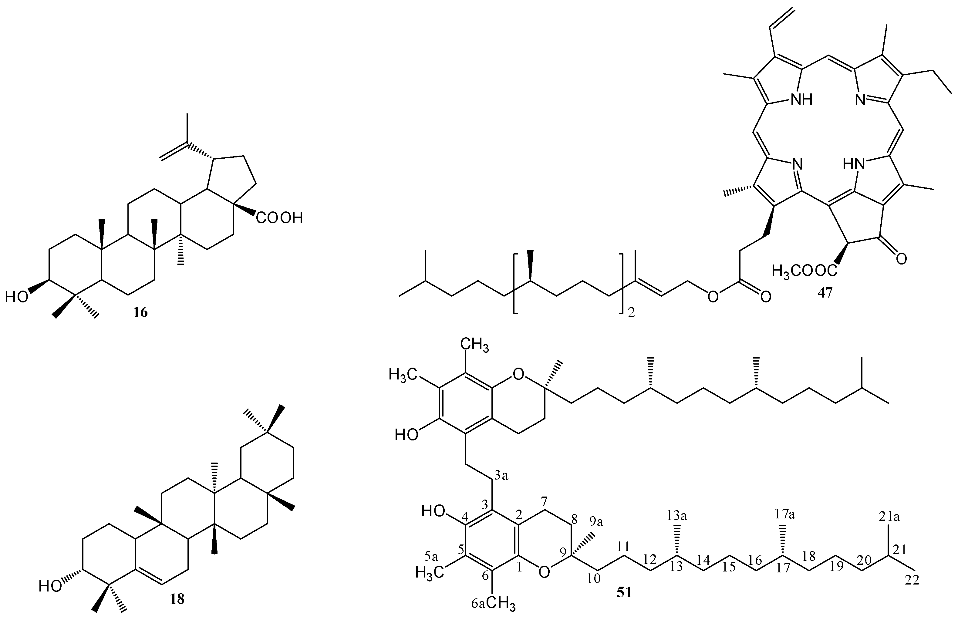
2.1. Isolation and Characterization of Compounds

The fresh stems of C. assamica L. were extracted with methanol and refluxed for 8 h. The filtrate was concentrated under reduced pressure to yield a dark brown syrup. The crude extract was suspended in water and then partitioned with chloroform and n-butanol successively to afford chloroform, n-butanol, and water layers, respectively. Purification of the three layers by column chromatography yielded a mixture of β-sitosterol (1) and stigmasterol (2) [27], β-sitosteryl glucoside (3) [28], a mixture of 3β-hydroxyl stigmast-5-en-7-one (4) and 3β-hydroxystigmast-5, 22-dien-7-one (5) [29], a mixture of β-sitostenone (6) [30] and stigmasta-4,22-dien-3-one (7) [31], 6β-hydroxy-β-sitostenone (8) [32], ergosterol peroxide (9) [33], 3,5,7,4′-tetramethoxyflavone (10) [34], 3′,4′,3,6,7-pentamethoxyflavone (11) [35], 3′,4′,5,6,7-pentamethoxyflavone (12) [35], 4′,5,6,7-tetramethoxy-flavone (13) [36], a mixture of oleanolic acid (14) [30] and ursolic acid (15) [28], betulinic acid (16) [37], friedelin (17) [38], epi-glut-5(6)-en-ol (18) [39], taraxerol (19) [9], epi-friedelinol (20) [40], glutinone (21) [41], lup-28-al-20(29)-en-3-ol (22) [42], a mixture of α-amyrin (23) and β-amyrin (24) [43], bergenin (25) [44], p-hydroxybenzaldehyde (26) [45], vanillin (27) [30], methyl gallate (28) [46], gallic acid (29) [47], 4-methoxybenzoic acid (30) [48], vanillic acid (31) [30], a mixture of 4-hydroxy-trans-cinnamic acid methyl ester (32) and 4-hydroxy-cis-cinnamic acid methyl ester (33) [49], a mixture of octadecyl-trans-ferulate (34) and octadecyl-cis-ferulate (35) [50], 1-(4-methoxy-phenyl)undecan-1-one (36) [51], 3-hydroxy-4-methoxybenzoic acid (37) [52], hexadecyl ferulate (38) [45], 2-hydroxybenzoquinone (39) [53], 2,6-dimethyoxybenzoquinone (40) [49], 3,3′,4-tri-O-methyl-ellagic acid (41) [54], 3,3′,4,4′-tetra-O-methylellagic acid (42) [55], methyl pheophorbide-a (43) [49], a mixture of methyl-21-hydroxy-(21S)-pheophorbide-a (44) and methyl-21-hydroxy-(21R)-pheophorbide-a (45) [49], methyl-21-hydroxyl-(21S)-pheophorbide-b (46) [49], pheophytin-a (47) [49], α-tocopherol (48) [56], tocopherol trimer IVa (49) [57], tocopherol trimer IVb (50) [57], 1,2-bis-(5-γ-tocopheryl)ethane (51) [58], α-tocospirol B (52) [59], 5,6-dimethoxy-3-methyl-2-cyclohexa-2,5-dien-1,4-dione (53) [60], 3-methyl-8-hydroxy-3,4-dihydroisocoumarin (54) [61], and methyl linoleate (55) [62], respectively. Among them, 1,2-bis-(5-γ-tocopheryl)ethane (51) (Figure 1) is reported from natural sources for the first time.

2.2. Structural Elucidation of Compound 51

1,2-Bis-(5-γ-tocopheryl)ethane (51) was isolated as a light yellow syrup. Its UV spectrum had an absorption maximum at 294 nm. The IR spectrum suggested the presence of hydroxyl (3444 cm−1) and an aromatic conjugated double bond (1458 and 1377 cm−1). The 13C-NMR and DEPT spectra exhibited a benzene ring partial structure that has two oxygenated substituents at δ 117.0 (s), 122.1 (s), 123.4 (s), 124.1 (s), 145.5 (s), and 146.3 (s). The 1H-NMR spectrum of 51 exhibited signals for two methyl and one methylene groups attached to a benzene ring at δ 2.13, 2.18, and 2.73. Comparing all the 1H- and 13C-NMR spectral signals carefully, the structure of 51 was similar to that of α-tocopherol (48) [56]. It indicated that they are very closely related analogues, differing only in the presence of a methylene group (δH 2.73, δC 26.7) in 51, instead of the methyl group (δH 2.17, δC 11.2) found in 48 (Table 1). To establish the structure of 51, 2D NMR including correlation spectroscopy (COSY), nuclear Overhauser enhancement spectroscopy (NOESY), heteronuclear multiple quantum correlation (HMQC), and heteronuclear multiple bond correlation (HMBC) experiments were conducted. In the HMBC experiment, the correlations observed for H-5a (δH 2.18)/C-4, C-5, C-6 and H-6a (δH 2.13)/C-1, C-5, C-6 indicate that two methyl groups are located in the ortho position on the benzene ring. Moreover, the correlation of the methylene proton at δH 2.73 with C-3 in HMBC spectrum suggests that the location C-3a in the dimerization of alpha-tocopherol forms the dimer 51. Conclusively, the structure of 51 was assigned as 1,2-bis-(5-γ-tocopheryl)ethane, which had been reported by synthesis [58], but is reported from natural sources for the first time. The NMR spectra are presented Figures S1–S6.

2.3. Anti-Inflammatory Activity

Neutrophils are the most abundant white blood cells and participate in the development of the inflammatory reactions in human body; they are important factors in the immune defense against various diseases. Some cytotoxins—for example, the superoxide anion radical, bioactive lipids, granule proteases, and elastase—can be secreted when the different stimuli activate neutrophils. Moreover, they are also major contributors to tissue destruction in chronic inflammatory diseases. It has been proposed that inhibiting neutrophil activation is a method of enhancing inflammatory disorders [63,64,65,66]. Most of the purified compounds in this study were inspected for the inhibition of elastase release and superoxide anion generation by human neutrophils in response to N-formyl-l-methionyl-phenylalanine/cytochalasin B (fMLP/CB). Only compound 16 (Figure 1) displayed significant inhibition of superoxide anion generation, with an IC50 value of 0.2 ± 0.1 µM (Table 2). In addition, compounds 16 and 47 also exhibited an inhibitory effect on elastase release with an IC50 value of 2.7 ± 0.3 and 5.3 ± 1.0 µM, respectively (Table 2). The inhibitory effects of all the tested compounds on superoxide anion generation and elastase release by human neutrophils in response to fMLP/CB are presented in Table S1. The cytotoxicity of compounds 16, 47, and LY294002 (a PI3K inhibitor, as a positive control) was examined in human neutrophils using an LDH release assay (Figure S7). All these compounds did not induce LDH release, suggesting that the inhibitory effects did not result from cytotoxicity in human neutrophils.

2.4. Cytotoxicity

In order to evaluate the growth inhibitory activity of the purified compounds against cancer cells, this study selected three different cell lines from malignant tumors including human nasopharyngeal carcinoma (NPC-TW01), non-small-cell lung carcinoma (NCI-H226), and colon cancer cell lines (HCT116). The results showed that betulinic acid (16) and epi-glut-5(6)-en-ol (18) (Figure 1) exhibited significant cytotoxicity with IC50 values ranged from 1.6 to 9.1 μM (Table 3). Moreover, betulinic acid (16) exhibited powerful inhibitory activity against NCI-H226 and HCT116 with IC50 values of 2.0 and 1.6 μM, respectively. Our study suggested the stem extracts of C. assamica and the purified compounds are potential candidates for the development of anti-cancer drugs. The preliminary growth inhibitory activity of all the tested compounds is presented in Table S2.

3. Materials and Methods

3.1. General Information

UV spectra were obtained with a Hitachi UV-3210 and UV-3010 spectrophotometer (Hitachi, Tokyo, Japan), and IR spectra were measured with a Shimadzu FTIR Prestige-21 spectrometer (Shimadzu, Kyoto, Japan). Optical rotations were measured with a HORIBA SEPA-300 digital polarimeter in a 0.5 dm cell (Horiba, Kyoto, Japan). The ESIMS and HRESIMS were taken on a Bruker Daltonics APEX II 30e spectrometer (Bruker, Billerica, MA, USA). 1H- and 13C-NMR spectra were measured using Bruker Avance-300, AMX-400, and AV-500 spectrometers (Bruker, Billerica, MA, USA) with TMS as the internal reference, and chemical shifts are expressed in δ (ppm). Silica gel (70–230 and 230–400 mesh; Merck, Darmstadt, Germany) and Spherical C18 100 Å reversed phase silica gel (RP-18; particle size 20–40 μm; Silicycle, Quebec City, QC, Canada) were used for column chromatography (CC), and silica gel 60 F254 and RP-18 F254S thin-layer chromatography (TLC) plates (Merck, Darmstadt, Germany) were used for preparative TLC, respectively.

3.2. Materials

The fresh stems of C. assamica L. were collected from Taitung Hsien, Taiwan, in October 2009 and verified by Prof. Chang-Sheng Kuoh (Department of Biology, National Cheng Kung University, Tainan, Taiwan). A voucher specimen (TSWu 20091016) has been deposited in the Herbarium of School of Pharmacy, National Cheng Kung University, Tainan, Taiwan.

3.3. Extraction and Isolation

The fresh stems of C. assamica L. (15 kg) were extracted with methanol (15 × 20 L) and refluxed for 8 h. The filtrate was evaporated under reduced pressure to yield a dark brown syrup (418 g). The residue was suspended in water and then partitioned with chloroform (5 × 2 L) and n-butanol (5 × 2 L) successively to afford chloroform (63 g), n-butanol (145 g) and water (210 g) soluble fractions respectively.
The chloroform soluble extracts were fractionated via silica gel column chromatography eluting with n-hexane/acetone (9:1) to afford seven fractions, on the basis of TLC monitoring. Fraction 1 was subjected to silica gel column chromatography eluted with n-hexane/acetone (79:1) to yield a mixture of β-sitosterol (1) and stigmasterol (2) (3.1 g), 3,5,7,4′-tetramethoxyflavone (10, 2.9 mg), 3′,4′,3,6,7-pentamethoxyflavone (11, 12.2 mg), 3′,4′,5,6,7-pentamethoxyflavone (12, 6.1 mg), 4′,5,6,7-tetra-methoxyflavone (13, 3.1 mg), friedelin (17, 1.8 g), taraxerol (19, 3.9 mg), glutinone (21, 3.2 mg) and methyl linoleate (55, 20.7 mg).
Purification of fraction 2 by column chromatography with silica gel was eluted by a gradient of benzene/ethyl acetate (79:1) to afford epi-glut-5(6)-en-ol (18, 23.2 mg), α-tocopherol (48, 200 mg), tocopherol trimer IVa (49, 24.3 mg), tocopherol trimer IVb (50, 28.1 mg), 1,2-bis-(5-γ-tocopheryl)- ethane (51, 15.4 mg), α-tocospirol B (52, 6.1 mg) and 5,6-dimethoxy-3-methyl-2-cyclohexa-2,5-dien-1,4-dione (53, 52.2 mg).
Separation of fraction 3 by column chromatography with silica gel eluted by n-hexane/ethyl acetate (9:1) yielded a mixture of 3β-hydroxystigmast-5-en-7-one (4) and 3β-hydroxystigmast- 5,22-dien-7-one (5, 4.5 mg), a mixture of β-sitostenone (6) and stigmasta-4,22-dien-3-one (7, 13.7 mg), 6β-hydroxy-β-sitostenone (8, 16.9 mg), ergosterol peroxide (9, 35 mg), epi-friedelinol (20, 200 mg), lup-28-al-20(29)-en-3-ol (22, 3.2 mg), a mixture of α-amyrin (23) and β-amyrin (24, 60 mg), a mixture of 4-hydroxy-trans-cinnamic acid methyl ester (32) and 4-hydroxy-cis-cinnamic acid methyl ester (33, 190 mg), a mixture of octadecyl-trans-ferulate (34) and octadecyl-cis-ferulate (35, 45.3 mg), hexadecyl ferulate (38, 20.7 mg) and 3-methyl-8-hydroxy-3,4-dihydroisocoumarin (55, 3.5 mg).
Fraction 4 was chromatographed over silica gel eluted with a benzene/acetone gradient (49:1) to give a mixture of oleanolic acid (14) and ursolic acid (15, 49.6 mg), betulinic acid (16, 63.6 mg), 4-methoxybenzoic acid (30, 4.7 mg), 1-(4-methoxyphenyl)undecan-1-one (36, 2.3 mg) and pheophytin-a (47, 11.2 mg).
Purification of fraction 5 by column chromatography with silica gel was eluted by a gradient of chloroform/acetone (49:1) to afford 2-hydroxybenzoquinone (39, 3.5 mg), methyl pheophorbide-a (43, 3.1 mg), a mixture of methyl-21-hydroxy-(21S)-pheophorbide-a (44) and methyl-21-hydroxy-(21R)-pheophorbide-a (45, 3.5 mg).
Separation of fraction 6 by column chromatography with a silica gel eluted by chloroform/acetone (49:1) yielded p-hydroxybenzaldehyde (26, 4.0 mg), 3,3′,4-tri-O-methylellagic acid (41, 37.7 mg), 3,3′,4,4′-tetra-O-methylellagic acid (42, 1.5 mg) and methyl-21-hydroxy-(21S)-pheophorbide-b (46, 3.6 mg).
Fraction 7 was subjected to silica gel column chromatography eluted with chloroform/methanol (49:1) to yield β-sitosteryl glucoside (3, 700 mg), vanillin (27, 7.8 mg), vanillic acid (31, 1.7 mg), 3-hydroxy-4-methoxybenzoic acid (37, 3.2 mg), 2, 6-dimethyoxybenzoquinone (40) (3.5 mg).
The n-butanol layer was subjected directly to Diaion HP-20 column chromatography, eluted with water containing increasing proportions of methanol, to give six fractions. Fraction 1 was chromatographed over Sephadex LH-20 eluted with gradient of water/methanol to give gallic acid (29, 600 mg). Fraction 2 was chromatographed on Sephadex LH-20 eluted with gradient of water/methanol to afford bergenin (25, 6.2 g). Fraction 4 was chromatographed on Sephadex LH-20 with water/methanol to give methyl gallate (28, 32.1 mg).
1,2-Bis-(5-γ-tocopheryl)ethane (51): light yellow syrup; UV λmax (MeOH) nm (log ε) 294; IR (KBr) νmax cm−1 3444, 2920, 2850, 1458, 1377, 1257, 1087; 1H- and 13C-NMR data, see Table 1.

3.4. Anti-Inflammatory Bioactivity Examination

3.4.1. Preparation of Human Neutrophils

Human neutrophils study was approved by Chang Gung Memorial Hospital Institutional Review Board, Taoyuan, Taiwan. It was conducted according to the Declaration of Helsinki. Blood was obtained from healthy donors (20–32 years old) who provided written informed consent before blood was drawn. Briefly, neutrophils were isolated by dextran sedimentation, Ficoll‒Hypaque gradient centrifugation, and hypotonic lysis of the erythrocytes [67].

3.4.2. Measurement of Superoxide Anion Generation and Elastase Release

The superoxide anion generation and elastase release were measured using the reduction of ferricytochrome c and elastase substrate, methoxysuccinyl-Ala-Ala-Pro-Val-p-nitroanilide, respectively, as described previously [68,69,70]. Human neutrophils were suspended in HBSS containing ferricytochrome c (0.6 mg/mL) or elastase substrate (100 μM) at 37 °C and treated with DMSO or tested compounds for 5 min. The cells were then activated using fMLF (0.1 μM)/cytochalasin B (CB, 1 μg/mL for superoxide generation and 0.5 μg/mL for elastase release) and the change of absorbance was continually measured at 550 nm and 405 nm by a spectrophotometer (U-3010, Hitachi) to determine the superoxide anion generation and elastase release, respectively.

3.4.3. Detection of Cytotoxicity

Human neutrophils were treated with DMSO or tested compounds and incubated at 37 °C for 15 min. The supernatant was assayed to detect the released LDH using CytoTox 96 non-radioactive cytotoxicity assay (Promega, Madison, WI, USA). The results are presented in Figure S7.

3.5. Determination of Anticancer Bioactivity

3.5.1. Cell Lines

Human cancer cell lines, non-small cell lung carcinoma (NCI-H226) and colon cancer cell line (HCT116) were obtained from the American Type Culture Collection (Rockville, MD, USA). A nasopharyngeal carcinoma (NPC-TW01) cell line was purchased from Food Industry Research and Development Institute (Hsinchu, Taiwan). Tumor cells were maintained in proper medium supplemented with 10% fetal bovine serum (FBS) at 37 °C in a humidified atmosphere of 5% CO2.

3.5.2. Growth Inhibition Assay

The evaluation of cell growth and survival was carried out according to Hansen et al. [71] with some modifications.

4. Conclusions

In summary, 55 compounds were characterized from the fresh stems of C. assamica, including 14 benzenoids, 11 triterpenes, nine steroids, five tocopherols, five chlorophylls, four flavonoids, two benzoquinones, two tannins, and three other compounds. Among these isolates, 1,2-bis-(5-γ-tocopheryl)ethane was reported for the first time from natural sources. Furthermore, the inhibitory activity on superoxide anion generation and elastase release and the cytotoxicity on three cancer cells were analyzed. The present study suggests that the stems of C. assamica and several compounds of its isolation could be further developed as candidates for the treatment or prevention of cancer and various inflammatory diseases. Thus, the detailed mechanism of action of these compounds appears worthy of follow-up investigation.

Supplementary Materials

The following are available online. Tables S1 and S2: Anti-inflammatory and cytotoxic effects of all the tested compounds from C. assamica; Figures S1–S6: NMR spectra of compound 51; Figure S7: Cytotoxicity of compounds 16, 47 and LY294002.

Author Contributions

Conceptualization, T.-S.W.; Data curation, Y.-Y.C.; Investigation, C.-Y.W., H.-Y.H., P.-C.K.; Methodology, T.-L.H., S.-H.J., and P.-J.C.; Writing—original draft, Y.-Y.C., H.-Y.H.; Writing—review & editing, P.-C.K. and T.-S.W. All authors read and approved the final manuscript.

Funding

This study was sponsored by the Ministry of Science and Technology, Taiwan via a grant to T.-S.W. and Y.-Y.C. (NSC97-2320-B-218-001-MY3). The authors are also thankful to Chang Gung Memorial Hospital [CMRPD1B0281~3, CMRPF1D0442~3, CMRPF 1F0011~3, CMRPF1F0061~3 and BMRP450 granted to T.-L.H.] for the partial financial support for the present research.

Acknowledgments

Authors were thankful to the Instruments Center of NCKU and the Joint Center for High Valued Instruments at NSYSU of the Ministry of Science and Technology, Taiwan for the assistance of recording NMR and MS spectra.

Conflicts of Interest

The authors declare no conflict of interest.

References

  1. Yang, T.Y. Flora of Taiwan, 2nd ed.; Editorial Committee of the Flora of Taiwan: Taipei, Taiwan, 1998; pp. 701–703.
  2. Zhang, Y.Q.; Xie, Y.H.; Huang, L.P. Studies on the chemical constituents and biological activities from Cissus L. Lishizhen Medicine and Materia Medica Research 2006, 17, 107–114. [Google Scholar]
  3. Otshudi, A.L.; Foriers, A.; Vercruysse, A.; Van, Z.A.; Lauwers, S. In vitro antimicrobial activity of six medicinal plants traditionally used for the treatment of dysentery and diarrhoea in Democratic Republic of Congo. Phytomedicine 2000, 7, 167–172. [Google Scholar] [CrossRef]
  4. Beltrame, F.L.; Sartoretto, J.L.; Bazotte, R.B.; Cuman, R.N.; Cortez, D.A.G. Phytochemical study and evaluation of the antidiabetic potential of Cissus sicyoides L. Quim. Nova 2001, 24, 783–785. [Google Scholar] [CrossRef]
  5. Beltrame, F.L.; Ferreira, A.G.; Cortez, D.A. Coumarin glycoside from Cissus sicyoides. Nat. Prod. Lett. 2002, 16, 213–216. [Google Scholar] [CrossRef] [PubMed]
  6. Adesanya, S.A.; Nia, R.; Martin, M.T.; Boukamcha, N.; Montagnac, A.; Pais, M. Stilbene derivatives from Cissus quadrangularis. J. Nat. Prod. 1999, 62, 1694–1695. [Google Scholar] [CrossRef]
  7. Bhutani, K.K.; Kapoor, R.; Atal, C.K. Two unsymmetric tetracyclic triterpenoids from Cissus quadrangulari. Phytochemistry 1984, 23, 407–410. [Google Scholar] [CrossRef]
  8. Gupta, M.M.; Verma, R.K. Unsymmetric tetracyclic triterpenoid from Cissus quadrangularis. Phytochemistry 1990, 29, 336–337. [Google Scholar] [CrossRef]
  9. Gupta, M.M.; Verma, R.K. Lipid constituents of Cissus quadrangularis. Phytochemistry 1991, 30, 875–878. [Google Scholar] [CrossRef]
  10. Khan, M.A.; Nabi, S.G.; Prakash, S.; Zaman, A. Pallidol, a resveratrol dimer from Cissus pallida. Phytochemistry 1986, 25, 1945–1948. [Google Scholar] [CrossRef]
  11. Al-Said, M.S.; Khalifa, A.S.; Al-Azizi, M.M. Flavonoids from Cissus digitat. Int. J. Pharm. 1991, 29, 281–283. [Google Scholar]
  12. Saenz, M.T.; Garcia, M.D.; Quilez, A.; Ahumada, M.C. Cytotoxic activity of Agave intermixta L. (Agavaceae) and Cissus sicyoides L. (Vitaceae). Phytother. Res. 2000, 14, 552–554. [Google Scholar] [CrossRef]
  13. Garcia, M.D.; Quilez, A.M.; Saenz, M.T.; Martinez, D.M.E.; De, P.R. Anti-inflammatory activity of agave intermixta trel and Cissus sicyoides L., species used in the caribbean traditional medicine. J. Ethnopharmacol. 2000, 71, 395–400. [Google Scholar] [CrossRef]
  14. Atawodi, S.E.; Arneh, D.A.; Ibrahim, S.; Andrew, J.N.; Nzelibe, H.C.; Onyike, E.O.; Anigo, K.M.; Abu, E.A.; James, D.B.; Njoku, G.C.; Sallau, A.B. Indigenous knowledge system for treatment of trypanosomiasis in Kaduna state of Nigeria. J. Ethnopharmacol. 2002, 79, 279–282. [Google Scholar] [CrossRef]
  15. Jainu, M.; Devi, C.S.S. Effect of Cissus quadrangularis on gastric mucosal defensive factors in experimentally induced gastric ulcer–A comparative study with sucralfate. J. Med. Food 2004, 7, 372–376. [Google Scholar] [PubMed]
  16. Quilez, A.M.; Saenz, M.T.; Garcia, M.D.; de la Puerta, R. Phytochemical analysis and anti-allergic study of agave intermixta trel and Cissus sicyoides L. J. Pharm. Pharmacol. 2004, 56, 1185–1189. [Google Scholar] [CrossRef] [PubMed]
  17. Viana, G.S.; Medeiros, A.; Lacerda, A.M.; Leal, L.K.; Vale, T.G.; Matos, F.J. Hypoglycemic and anti-lipemic effects of the aqueous extract from Cissus sicyoides. BMC Pharmacol. 2004, 4, 9–15. [Google Scholar] [CrossRef] [PubMed] [Green Version]
  18. Chidambara, M.K.N.; Vanitha, A.; Mahadeva, S.M.; Ravishankar, G.A. Antioxidant and antimicrobial activity of Cissus quadrangularis L. J. Med. Food 2003, 6, 99–105. [Google Scholar] [CrossRef] [PubMed]
  19. Pepato, M.T.; Baviera, A.M.; Vendramini, R.C.; Perez, M.P.M.S.; Kettelhut, I.C.; Brunetti, I.L. Cissus sicyoides in the long-term treatment of streptozotocin-diabetic rats. Biotechnol. Appl. Biochem. 2003, 37, 15–20. [Google Scholar] [CrossRef] [PubMed]
  20. Alzoreky, N.S.; Nakahara, K. Antibacterial activity of extracts from some edible plants commonly consumed in Asia. Int. J. Food. Microbiol. 2003, 80, 223–230. [Google Scholar] [CrossRef]
  21. Garcia, X.; Cartas, H.L.; Lorenzana, J.M.; Gijon, E. Vasoconstrictor effect of Cissus sicyoides on guinea Pig aortic rings. Gen. Pharmacol. 1997, 29, 457–462. [Google Scholar] [CrossRef]
  22. Mori, T.; Nishikawa, Y.; Takata, Y.; Kashuichi, N.; Ishihara, N. Effect of insulina leaf extract on development of diabetes comparison between normal, streptozotocin-induced diabetic rats and hereditary diabetic mice. Nippon Eiyo Shokuryo Gakkaishi 2001, 54, 197–203. [Google Scholar] [CrossRef]
  23. Yang, L.; Wang, F.; Liu, M.; Lu, M.; Jia, H.; Fang, X.; Li, Z. Isolation of resveratrol and its antagonistic effects on endothelin-1. Huaxi Yaoxue Zazhi 2000, 15, 81–84. [Google Scholar]
  24. Wang, F.; Yang, L.; Cheng, Y.; Lu, M.; Liu, M.; Ji, X.; Jia, H. Antagonism effects of extracts of Chinese herb Cissus assamica and monomer CA-1201 on endothelin-1 responses. Zhongguo Yaoxue Zazhi 1998, 33, 337–340. [Google Scholar]
  25. Yang, L.C.; Wang, F.; Liu, M. A study of an endothelin antagonist from a Chinese anti-snake venom medicinal herb. J. Cardiovasc. Pharmacol. 1998, 31, S249–S250. [Google Scholar] [CrossRef] [PubMed]
  26. Wang, F.; Yang, L.; Liu, M.; Lu, M.; Cheng, Y.; Jia, H. A primary study on antagonizing effects of anti-snake venom Chinese herb on endothelin-1 and sarafotoxin 6b. Zhongguo Zhong Yao Za Zhi 1997, 22, 620–622. [Google Scholar] [PubMed]
  27. Shen, D.Y.; Juang, S.H.; Kuo, P.C.; Huang, G.J.; Chan, Y.Y.; Damu, A.G.; Wu, T.S. Chemical constituents from Andrographis echioides and their anti-inflammatory activity. Int. J. Mol. Sci. 2013, 14, 496–514. [Google Scholar] [CrossRef] [PubMed]
  28. Wu, T.S.; Chan, Y.Y. Constituents of leaves of Uncaria hirsuta Haviland. J. Chin. Chem. Soc. 1994, 41, 209–212. [Google Scholar] [CrossRef]
  29. Kuo, Y.H.; Chu, P.H. Studies on the constituents from the bark of Bauhinia purpurea. J. Chin. Chem. Soc. 2002, 49, 269–274. [Google Scholar] [CrossRef]
  30. Wu, S.J.; Chan, Y.Y. Five New Iridoids from Roots of Salvia digitaloides. Molecules 2014, 19, 15521–15534. [Google Scholar] [CrossRef] [PubMed]
  31. Kuo, P.C.; Yang, M.L.; Hwang, T.L.; Lai, Y.Y.; Li, Y.C.; Thang, T.D.; Wu, T.S. Anti-inflammatory Diterpenoids from Croton tonkinensis. J. Nat. Prod. 2013, 76, 230–236. [Google Scholar] [CrossRef] [PubMed]
  32. Chang, Y.C.; Chang, F.R.; Wu, Y.C. The Constituents of Lindera Glauca. J. Chin. Chem. Soc. 2000, 47, 373–380. [Google Scholar] [CrossRef]
  33. Thang, T.D.; Kuo, P.C.; Hwang, T.L.; Yang, M.L.; Ngoc, T.B.; Han, T.N.; Lin, C.W.; Wu, T.S. Triterpenoids and steroids from Ganoderma mastoporum and their inhibitory effects on superoxide anion generation and elastase release. Molecules 2013, 18, 14285–14292. [Google Scholar] [CrossRef] [PubMed]
  34. Dong, H.; Gou, Y.L.; Cao, S.G.; Chen, S.X.; Sim, K.Y. Eicosenones and methylated flavonols from Amomum koenigii. Phytochemistry 1999, 50, 899–902. [Google Scholar] [CrossRef]
  35. Machida, K.; Osawa, K. On the flavonoid constituents from the peels of Citrus hassaku Hort.ex Tanaka. Chem. Pharm. Bull. 1989, 37, 1092–1094. [Google Scholar] [CrossRef]
  36. Weber, B.; Hartmann, B.; Stockigt, D.; Schreiber, K.; Roloff, M.; Bertram, H.J.; Schmidt, C.O. Liquid chromatography/mass spectrometry and liquid chromatography nuclear magnetic resonance as complementary analytical techniques for unambiguous identification of polymethoxylated flavones in residues from molecular distillation of orange peel oils (Citrus sinensis). J. Agric. Food Chem. 2006, 54, 274–278. [Google Scholar] [PubMed]
  37. Chatterjee, P.; Kouzi, S.A.; Pezzuto, J.P.; Hamann, M.T. Biotransformation of the antimelanoma agent betulinic acid by Bacillus megaterium ATCC 13368. Appl. Environ. Microbiol. 2000, 66, 3850–3855. [Google Scholar] [CrossRef] [PubMed]
  38. Wu, T.S.; Chan, Y.Y.; Liou, M.J.; Lin, F.W.; Shi, L.S.; Chen, K.T. Platelet aggregation inhibitor from Murraya euchrestifolia. Phytother. Res. 1998, 12, S80–S82. [Google Scholar] [CrossRef]
  39. Tanabe, Y.; Sinoda, R.; Horikoshi, Y.; Takahashi, K. Studies on constituents of medicinal plants. XVI. The constituents of Spiracea species. Yakugaku Zasshi 1976, 96, 248–250. [Google Scholar] [CrossRef] [PubMed]
  40. Li, K.H.; Chang, C.R.; Chang, Y.S. Chemical components from Triumfetta bartramia. J. Chin. Chem. Soc. 1995, 42, 93–95. [Google Scholar]
  41. Zhang, J.; Yin, Z.Q.; Cao, P.; Li, Y.B.; Duan, J.A. A new flavonol derivative from Fagopyrum dibotrys. Chem. Nat. Compd. 2008, 44, 701–703. [Google Scholar] [CrossRef]
  42. Hata, K.; Hori, K.; Takahashi, S. Differntion and apoptosis-inducing activities by pentacyclic triterpenes on a mouse melanoma cell line. J. Nat. Prod. 2009, 65, 645–648. [Google Scholar] [CrossRef]
  43. Bolleddula, J.; Mulabagal, V.; Yanjun, Z.; David, L.D.; Muraleedharan, G.N. Impact of alkyl esters of caffeic and ferulic acids on tumor cell proliferation cyclooxygenase enzyme and lipid peroxidation. J. Agric. Food Chem. 2006, 54, 5375–5381. [Google Scholar]
  44. Xie, Y.H.; Zhang, Y.Q.; Deng, P.; Yu, W.S. Determination of bergenin in Cissus assamica by HPLC. Shizhen Guoyi Guoyao 2009, 20, 1086–1087. [Google Scholar]
  45. Chiang, C.Y.; Leu, Y.L.; Chan, Y.Y.; Wu, T.S. Sodium aristolochates from the flowers and fruits of Aristolochia zollingeriana. J. Chin. Chem. Soc. 1998, 45, 93–97. [Google Scholar] [CrossRef]
  46. Choi, S.E.; Yoon, J.H.; Choi, H.K.; Lee, M.W. Phenolic compounds from the root of Phragmites communis. Chem. Nat. Compd. 2009, 45, 893–895. [Google Scholar] [CrossRef]
  47. Jun, X.L.; Duo, L.D.; Yan, P.S. Diversity of chemical constituents from Saxifraga montana H. J. Chin. Chem. Soc. 2008, 55, 863–870. [Google Scholar]
  48. Cui, L.Q.; Liu, K.; Zhang, C. Effective oxidation of benzylic and alkane C–H bonds catalyzed by sodium O-iodobenzenesulfonate with oxone as a terminal oxidant under phase-transfer conditions. Org. Biomol. Chem. 2011, 9, 2258–2265. [Google Scholar] [CrossRef] [PubMed]
  49. Wu, T.S.; Tsanga, Z.J.; Wu, P.L.; Lin, F.W.; Li, C.Y.; Temg, C.M.; Lee, K.H. New constituents and antiplatelet aggregation and anti-HIV principles of Artemisia capillaris. Bioorg. Med. Chem. 2001, 9, 77–83. [Google Scholar] [CrossRef]
  50. Wei, H.; Wen, D.X.; Liu, X.S.; Tang, R.J. Constituents in petroleum ether and ethyl acetate extract fractions of Dracaena cochinensis (Lour.). Zhong Guo Zhong Yao Za Zhi 1998, 23, 616–618. [Google Scholar]
  51. Loupy, A.; Chatti, S.; Delamare, S.; Lee, D.-Y.; Chung, J.-H.; Jun, C.-H. Solvent-free chelation-assisted hydroacylation of olefins rhodium(Ι) catalyst under microwave irradiation. J. Chem. Soc. 2002, 10, 1280–1285. [Google Scholar]
  52. Ding, H.Y.; Lin, H.C.; Teng, C.M.; Wu, Y.C. Phytochemical and pharmacological studies on Chinese Paeonia species. J. Chin. Chem. Soc. 2000, 47, 381–388. [Google Scholar] [CrossRef]
  53. Magdziak, D.; Rodriguez, A.A.; Van, D.W.; Ryan, W.; Pettus, T.R.R. Regioselective oxidation of phenols to O-quinones with O-iodoxybenzoic Acid (IBX). Org. Lett. 2002, 4, 285–288. [Google Scholar] [CrossRef] [PubMed]
  54. Gao, X.; Wu, J.; Zou, W.; Dai, Y. Two ellagic acids isolated from roots of Sanguisorba officinalis L. promote hematopoietic progenitor cell proliferation and megakaryocyte differentiation. Molecules 2014, 19, 5448–5458. [Google Scholar] [CrossRef] [PubMed]
  55. Reitze, J.D.; Przewloka, S.R.; Shearer, B.J. The further chemistry of ellagic acid: I. synthesis of tetramethylellagic acid and associated polymer precursors. Holzforschung 2005, 55, 171–175. [Google Scholar] [CrossRef]
  56. Almeida, E.R.D.; Rafael, K.R.D.O.; Couto, G.B.L.; Ishigmi, A.B. Anxiolytic and anticonvulsant effects on mice of flavonoids, linalool, and α-tocopherol presents in the extract of leaves of Cissus sicyoides L. J. Biomed. Biotechnol. 2009, 1, 1–6. [Google Scholar] [CrossRef] [PubMed]
  57. Lie, C.R.; Jiau, C.H.; Chiu, M.C. Cerebrosides and tocopherol trimers from the seeds of Euryale ferox. J. Nat. Prod. 2007, 70, 1214–1217. [Google Scholar]
  58. Rosenau, T.; Kloser, E.; Gille, L.; Mazzini, F.; Netscher, T. Vitamin E chemistry. studies into initial oxidation intermediates of α-tocopherol: disproving the involvement of 5a-C-centered chromanol methide radicals. J. Org. Chem. 2007, 72, 3268–3281. [Google Scholar] [CrossRef] [PubMed]
  59. Chiang, Y.M.; Kuo, Y.H. Two novel α-tocopheroids from the aerial roots of Ficus microcarpa. Tetrahedron Lett. 2003, 44, 5125–5128. [Google Scholar] [CrossRef]
  60. Lipshutz, B.H.; Butler, T.; Lower, A. Controlling regiochemistry in negishi carboaluminations fine tuning the ligand on zirconium. J. Am. Chem. Soc. 2006, 128, 15396–15398. [Google Scholar] [CrossRef] [PubMed]
  61. Zeng, Z.X.; Ma, W.H.; Li, Y.L.; Han, T.; Zheng, C.J.; Qin, L.P. Two new diterpenes from Solidago canadensis. Helv. Chim. Acta. 2012, 95, 1121–1125. [Google Scholar] [CrossRef]
  62. Zeng, Z.X.; Li, Y.L.; Dong, L.L.; Fan, G.X.; Fei, D.Q. Chemical constituents of the roots of Ligularia lapathifolia. Chem. Nat. Compd. 2015, 51, 375–377. [Google Scholar] [CrossRef]
  63. Witko-Sarsat, V.; Rieu, P.; Descamps-Latscha, B.; Lesavre, P.; Halbwachs-Mecarelli, L. Neutrophils: Molecules, functions and pathophysiological aspects. Lab. Investig. 2000, 80, 617–653. [Google Scholar] [CrossRef] [PubMed]
  64. Borregaard, N. The human neutrophil. Function and dysfunction. Eur. J. Haematol. 1998, 41, 401–413. [Google Scholar] [CrossRef]
  65. Roos, D.; van Bruggen, R.; Meischl, C. Oxidative killing of microbes by neutrophils. Microbes Infect. 2003, 5, 1307–1315. [Google Scholar] [CrossRef] [PubMed]
  66. Faurschou, M.; Borregaard, N. Neutrophil granules and secretory vesicles in inflammation. Microbes Infect. 2003, 5, 1317–1327. [Google Scholar] [CrossRef] [PubMed]
  67. Chen, P.J.; Wang, Y.L.; Kuo, L.M.; Lin, C.F.; Chen, C.Y.; Tsai, Y.F.; Shen, J.J.; Hwang, T.L. Honokiol suppresses TNF-α-induced neutrophil adhesion on cerebral endothelial cells by disrupting polyubiquitination and degradation of IκBα. Sci. Rep. 2016, 6, 26554. [Google Scholar] [CrossRef] [PubMed] [Green Version]
  68. Yang, S.C.; Chung, P.J.; Ho, C.M.; Kuo, C.Y.; Hung, M.F.; Huang, Y.T.; Chang, W.Y.; Chang, Y.W.; Chan, K.H.; Hwang, T.L. Propofol inhibits superoxide production, elastase release, and chemotaxis in formyl peptide-activated human neutrophils by blocking formyl peptide receptor 1. J. Immunol. 2013, 190, 6511–6519. [Google Scholar] [CrossRef] [PubMed]
  69. Babior, B.M.; Kipnes, R.S.; Curnutte, J.T. Biological defense mechanisms. The production by leukocytes of superoxide, a potential bactericidal agent. J. Clin. Investig. 1973, 52, 741–744. [Google Scholar] [CrossRef] [PubMed]
  70. Hwang, T.L.; Li, G.L.; Lan, Y.H.; Chia, Y.C.; Shieh, P.W.; Wu, Y.H.; Wu, Y.C. Potent inhibition of superoxide anion production in activated human neutrophils by isopedicin, a bioactive component of the Chinese medicinal herb Fissistigma oldhamii. Free Radic. Biol. Med. 2009, 46, 520–528. [Google Scholar] [CrossRef] [PubMed]
  71. Hansen, M.B.; Nielsen, S.E.; Berg, K. Re-examination and further development of a precise and rapid dye method for measuring cell growth/cell kill. J. Immunol. Methods 1989, 119, 203–210. [Google Scholar] [CrossRef]
Sample Availability: Samples of all the isolated compounds are available from the authors.
Figure 1. Structures of compounds 16, 18, 47 and 51.
Molecules 23 02799 g001
Table 1. 1H- and 13C-NMR spectra data of 48 and 51 (CDCl3, 400 MHz).
Position4851
δH (mult., J in Hz)δCδH (mult., J in Hz)δC
1 145.5 146.3
2 117.2 117.0
3 118.5 123.4
4 144.5 145.5
5 121.1 122.1
6 122.6 124.1
72.64 (t, 4.5)21.02.74 (m)21.7
81.79 (m)31.51.83 (m)32.3
9 74.5 75.3
101.56 (m)39.81.56 (m)40.7
111.54 (m)22.61.54 (m)21.3
121.26~1.25 (m)37.41.28~1.25 (m)38.2
131.31 (d, 7.4)32.71.32 (d, 7.2)33.4
141.26~1.25 (m)37.21.28~1.25 (m)37.9
151.26~1.25 (m)24.81.28~1.25 (m)25,5
161.26~1.25 (m)37.51.28~1.25 (m)38.2
171.31 (d, 7.4)32.81.32 (d, 7.2)33.5
181.16~1.13 (m)37.41.16~1.13 (m)38.2
191.16~1.13 (m)25.11.16~1.13 (m)25,2
201.16~1.13 (m)39.41.16~1.13 (m)40.1
211.52 (m)27.91.52 (m)28.7
220.91 (d, 6.4)23.70.86 (d, 7.2)23.4
3a2.17 (s)11.22.73 (s)26.7
5a2.22 (s)12.82.18 (s)12.8
6a2.19 (s)11.82.13 (s)12.6
9a1.28 (s)24.41.24 (s)24.5
13a0.90 (d, 7.4)20.30.85 (d, 7.2)20.3
17a0.89 (d, 7.4)20.70.83 (d, 7.2)20.5
21a0.93 (d, 6.5)22.70.87 (d, 7.2)23.3
OH4.25 (s) 5.41 (s)
Table 2. Inhibitory effects of isolated compounds on superoxide anion generation and elastase release by human neutrophils in response to fMLP/CB.
CompoundSuperoxide Anion GenerationElastase Release
IC50 (μM) aIC50 (μM)
160.2 ± 0.1 ***2.7 ± 0.3 ***
47>105.3 ± 1.0 ***
LY294002b0.4 ± 0.1 ***1.5 ± 0.3 ***
Results are presented as mean ± S.D. (n = 3~4). *** p < 0.001 compared with the control (DMSO). a Concentration necessary for 50% inhibition (IC50). b A phosphatidylinositol-3-kinase inhibitor was used as a positive control.
Table 3. Cytotoxicity of compounds 16, 18, 20, 21, 41 and 52.
CompoundsCell Lines
NCI-H226HCT-116
IC50 (μM)IC50 (μM)
162.01.6
189.16.0
2015.816.7
2138.024.0
4131.630.3
52>5039.4

