Review

Thermodynamic Model for B-Z Transition of DNA Induced by Z-DNA Binding Proteins

Department of Chemistry and RINS, Gyeongsang National University, Gyeongnam 52828, Korea
*
Author to whom correspondence should be addressed.
Molecules 2018, 23(11), 2748; https://doi.org/10.3390/molecules23112748
Submission received: 28 September 2018 / Revised: 21 October 2018 / Accepted: 23 October 2018 / Published: 24 October 2018
(This article belongs to the Special Issue Protein-DNA Interactions: From Biophysics to Genomics)

Abstract

:
Z-DNA is stabilized by various Z-DNA binding proteins (ZBPs) that play important roles in RNA editing, innate immune response, and viral infection. In this review, the structural and dynamics of various ZBPs complexed with Z-DNA are summarized to better understand the mechanisms by which ZBPs selectively recognize d(CG)-repeat DNA sequences in genomic DNA and efficiently convert them to left-handed Z-DNA to achieve their biological function. The intermolecular interaction of ZBPs with Z-DNA strands is mediated through a single continuous recognition surface which consists of an α3 helix and a β-hairpin. In the ZBP-Z-DNA complexes, three identical, conserved residues (N173, Y177, and W195 in the Zα domain of human ADAR1) play central roles in the interaction with Z-DNA. ZBPs convert a 6-base DNA pair to a Z-form helix via the B-Z transition mechanism in which the ZBP first binds to B-DNA and then shifts the equilibrium from B-DNA to Z-DNA, a conformation that is then selectively stabilized by the additional binding of a second ZBP molecule. During B-Z transition, ZBPs selectively recognize the alternating d(CG)n sequence and convert it to a Z-form helix in long genomic DNA through multiple sequence discrimination steps. In addition, the intermediate complex formed by ZBPs and B-DNA, which is modulated by varying conditions, determines the degree of B-Z transition.

1. Introduction

Left-handed Z-DNA is a higher energy conformation than right-handed B-DNA. Z-DNA was first found in a polymer of alternating d(CG)n DNA duplexes observed in high salt conditions [1]; its crystal structure was reported in 1979 [2]. The Z-DNA helix is built from d(CG)-repeats, with the dC in the anti-conformation and the dG in the unusual syn-conformation, which causes the backbone to follow a zigzag path [3,4]. Z-DNA can also be stabilized by negative supercoiling generated behind a moving RNA polymerase during transcription [5].
A distinct biological function of Z-DNA is suggested by the discovery of various Z-DNA binding proteins (ZBPs). Double stranded (ds) RNA deaminase 1 (ADAR1) deaminates adenine in pre-mRNA to yield inosine, which codes as guanine [6,7,8]. ADAR1 has two left-handed Z-DNA binding domains (ZBDs), Zα and Zβ, at its NH2-terminus [7,9]. High binding affinity of this Zα domain to Z-DNA was shown by a band-shift assay and confirmed by CD and Raman spectroscopic measurement [10,11,12]. The DNA-dependent activator of IFN-regulatory factors (DAI; also known as ZBP1 or DLM-1) also contains two tandem ZBDs (Zα and Zβ) at the NH2-terminus, such as ADAR1 [13,14]. It has been shown that ZBDs regulate the localization of DAI and its association with stress granules [15,16]. All poxviruses have a gene called E3L that consists of two domains: An N-terminal ZBD and a C-terminal RNA binding domain [17,18]. This ZBD shows sequence homology to the Zα domains found in human ADAR1 and in the DAI of mammals (Figure 1a). The Z-DNA binding affinity of E3L protein is essential for pathogenesis in the poxviruses [17,18,19]. The RNA-dependent protein kinase (PKR) plays an important role in the innate immune response against viral infections by recognizing dsRNA in the cytosol [20,21,22]. In fish species, a functional analogue of PKR, PKZ contains two ZBDs instead of dsRNA binding domains [23,24,25,26]. Similar to PKR, the phosphorylation function of PKZ is activated by Z-DNA binding [24].
These biological data, along with the results of more recent structural studies of Z-DNA induced by various ZBPs, have provided insights into Z-DNA recognition and ZBP-induced B-Z transition carried out by the innate immune response, viral infection, and RNA editing that are influenced by the nature of the Z-DNA. Crystallographic and NMR studies have provided detailed three-dimensional (3D) structures and dynamic information of various ZBPs complexed with Z-DNA. The structural and dynamic data summarized in this review have yielded a rich understanding of the mechanisms by which ZBPs selectively recognize the d(CG)-repeat DNA sequences in genomic DNA and efficiently convert them to left handed Z-DNA to achieve their biological function.

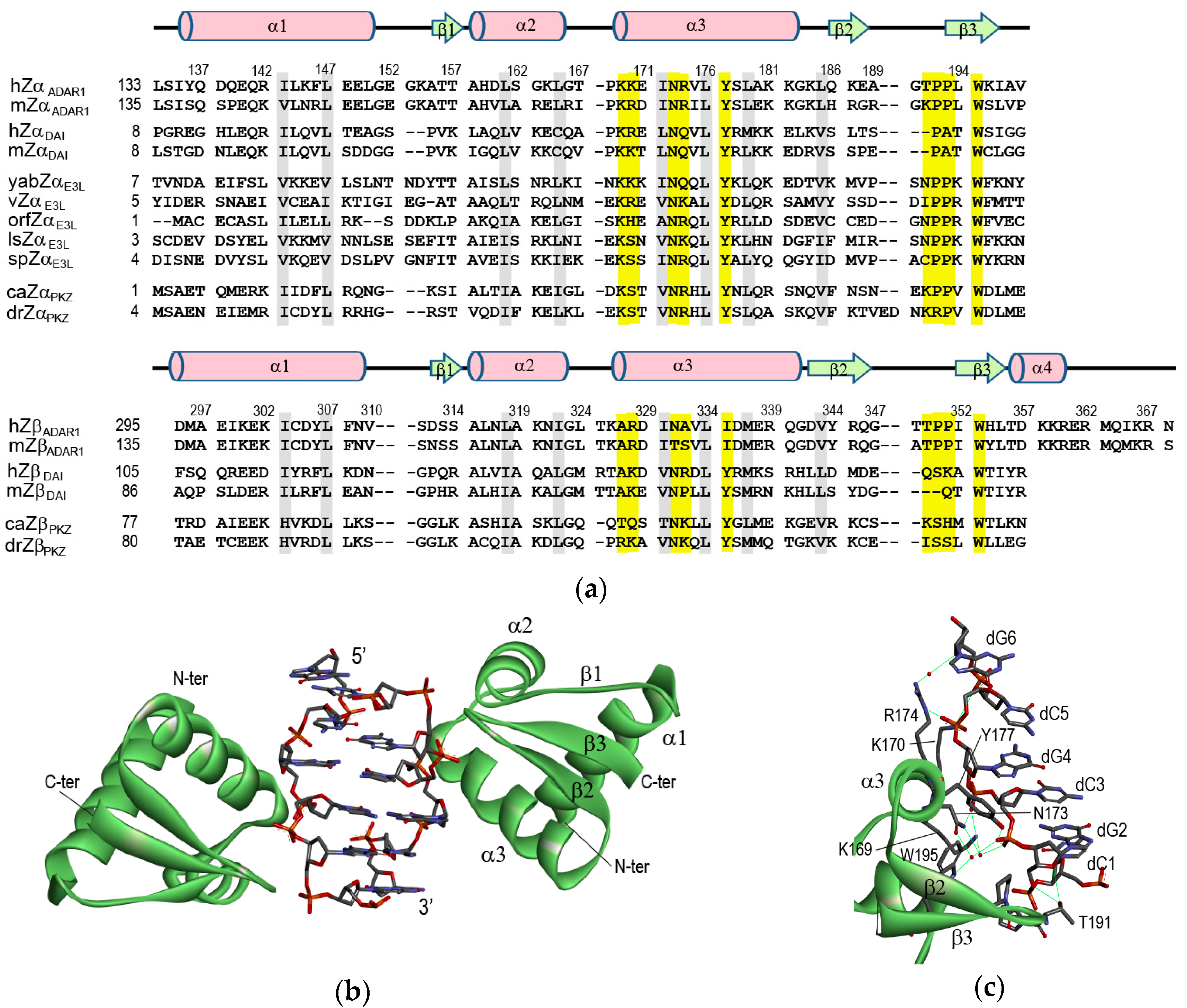
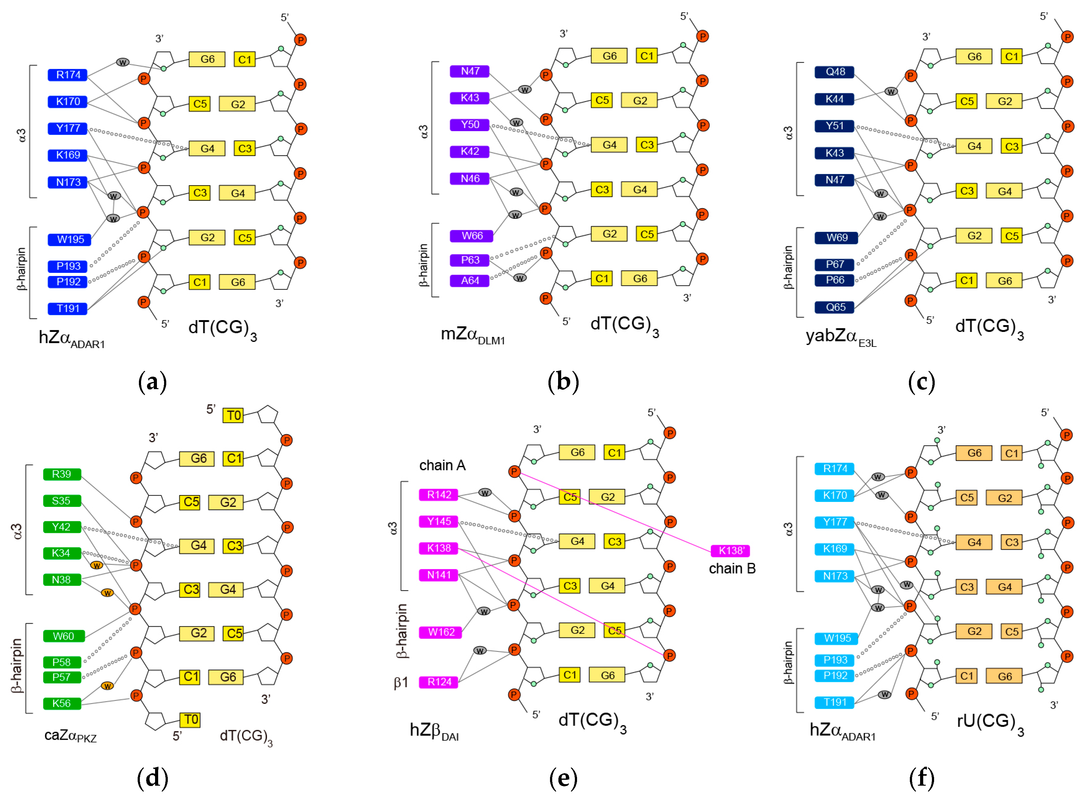
2. Crystal Structures of ZBPs Complexed with DNA Duplexes

2.1. hZαADAR1-Z-DNA Complex

In 1999, Alexander Rich and his colleagues first reported the crystal structure of the Zα domain of human ADAR1 (hZαADAR1) complexed with a six-base-pair (bp), double-stranded (ds) DNA fragment d(TCGCGCG)2 [7]. The monomeric hZαADAR1 domain binds to one strand of the palindromic dsDNA, in which the conformation of the DNA substrate is very similar to the canonical Z-DNA structure. (Figure 1b) [7]. A second monomer binds to the opposite strand of DNA yielding two-fold symmetry with respect to the DNA helical axis (Figure 1b) [7]. The hZαADAR1 domain has a compact α/β architecture containing a three-helix bundle (α1 to α3) and twisted antiparallel β sheets (β1 to β3) (Figure 1b). The arrangement of a β-hairpin with hydrogen-bonds (H-bonds) between the β2 (L185–A188) and β3 strands (P193–I197) is a common feature of helix-turn-helix (HTH) proteins with a/β topology [7]. Aliphatic residues from the three helices, together with the W195 in strand β3, form a hydrophobic core [7]. The NMR studies reported that the free hZαADAR1 protein in the solution adopts a similar fold in its complex structure [9,27].
The contact of hZαADAR1 with the Z-DNA strand is mediated through a single continuous recognition surface, which consists of residues from an α3 helix and a β-hairpin (β2-loop-β3, also called a β-wing) (Figure 1c) [7]. The electrostatic interactions in the complex are made between K169, K170, N173, R174, and Y177 in an α3 helix as well as between T191 and W195 in a β-hairpin and five consecutive phosphate-backbones of Z-DNA (Figure 1c) [7]. K169 and N173 form the direct and water-mediated H-bonds to the dC3pdG4 and dG2pdC3 phosphates, respectively (Figure 2a) [7]. The N173A mutant displays the most dramatic decrease in Z-DNA binding affinity, suggesting that it plays an important role in the Zα function [27,28]. Similarly, the K169A mutant also has a significantly lower Z-DNA binding affinity than a wild-type protein [27,28]. K170 forms direct H-bonds to the dG4pdC5 and dC5pdG6 phosphates (Figure 2a) [7]. The K170A mutant binds to Z-DNA with a lower affinity than wild-type protein, but better than the K169A and N173A mutants [27,28]. Interestingly, R174 and T191 bind to the furanose oxygens of dG6 and dG2, respectively (Figure 2a) [7]. However, the R174A and T191A mutations have little effect on the B-Z transition activities of hZαADAR1 [28].
In addition to polar interactions, the aromatic ring of Y177 and the side-chains of P192 and P193 make the important van der Waals interactions with Z-DNA (Figure 2a) [7]. Interestingly, Y177 displays CH-π interaction with the C8 position of dG4 (Figure 2a) [7]. The Y177A mutant, which is unable to form both H-bonding and hydrophobic interactions, exhibits a significantly low Z-DNA binding affinity [27]. The Y177I, and Y177F mutants, which are capable only of hydrophobic interactions, could bind better to Z-DNA than Y177A, but still worse than wild-type protein [17,27,28]. Furthermore, the aromatic ring of W195, which forms a water-mediated H-bond to the dG2pdC3 phosphate, is almost perpendicular to Y177 and positioned in the center of the hydrophobic core (Figure 1c) [7]. The W195F mutant has 2-fold lower B-Z transition activity than wild type protein [28].
The crystal structural study reported that hZαADAR1 is also able to bind to 6-bp Z-DNA duplexes with non-CG-repeat sequences, such as d(CACGTG)2, d(CGTACG)2, and d(CGGCCG)2 [34]. In these structures, N173, Y177, P192, P193, and W195 contribute to the recognition of Z-DNA-like CG-repeat DNA [34]. However, R174 and T191 did not show intermolecular interaction with Z-DNA in most structures [34]. Similarly, K169 and K170 are only in contact with Z-DNA within some structures of these complexes [34]. Thus, these four residues might play an important role in the sequence discrimination step for the B-Z transition of DNA.
The second Z-DNA binding domain of human ADAR1 (hZβADAR1) adopts a winged-HTH fold like hZαADAR1, with the addition of a C-terminal α4 helix (see sequence in Figure 1a) [35]. Superposition of the hZβADAR1 with the hZαADAR1 structure reveals that A327, A332, and I335 (corresponding to K169, R174, and Y177 in hZαADAR1, respectively) did not perform H-bonding to the backbone of Z-DNA. Mutagenesis studies of these residues have shown that all three residues are important for Z-DNA-binding by Zα proteins [17,27,28]. This study suggested that hZβADAR1 is unable to interact with nucleic acids in a manner similar to that seen in the hZαADAR1-Z-DNA complex [35]. Instead, this region participates in self-association protein–protein interactions [35].

2.2. mZαDLM1-Z-DNA Complex

The amino acid sequence of the Zα domain of murine DLM-1 (mZαDLM1) is ~35% identical to that of the hZαADAR1 (Figure 1a). The overall structures of the complexes with Z-DNA are very similar to each other [29]. The core of the Zα-DNA interface in both proteins consists of three identical residues: N173, Y177, and W195 in hZαADAR1 and N46, Y50, and W66 in mZαDLM1, respectively (Figure 2a,b) [7,29]. K43 and Q47 form direct or water-mediated H-bonds to three phosphates of the dC3pdG4pdC5pdG6 sequence like the corresponding K170 and R174 of hZαADAR1 (Figure 2b) [29]. The structural differences between the two domains are found in the α1-β1 loop and the β-hairpin [29]. The β-hairpin of mZαDLM1 is two residues shorter than that of hZαADAR1 (Figure 1a), indicating that the β-hairpin is apparently tolerant of greater sequence variability than the α3 helix without loss of function [29].

2.3. yabZαE3L-Z-DNA Complex

The Zα domain of the Yaba-like disease virus E3L (yabZαE3L) stabilizes the Z-DNA conformation in a manner similar to that of hZαADAR1 and mZαDLM1 [30], although it shares only 26% sequence identity with hZαADAR1 (Figure 1a). The crystal structural study revealed that two yabZαE3L domains are found in the asymmetric unit, each bound to one strand of double-stranded DNA in the Z- conformation [30]. The intermolecular interaction of one asymmetric unit with Z-DNA is summarized in Figure 2c [30]. Three residues, N47, Y51, and W69 (corresponding to N173, Y177, and W195 in hZαADAR1), play central roles in the interaction with Z-DNA, as with other members of the Zα family (Figure 2c). K43, K44, and Q48 also participate in DNA recognition via direct or water-mediated H-bonds to the phosphate backbone of Z-DNA (Figure 2c).

2.4. caZαPKZ-Z-DNA Complex

The Zα domain of PKZ from Carassius auratus (caZαPKZ), which shows limited identity with other ZBPs (28% for hZαADAR1, 20% for hZαDAI, and 22% for yabZαE3L, respectively), is able to convert d(CG)-repeat DNA from B-DNA to Z-DNA [36,37]. The interaction between caZαPKZ and Z-DNA is mediated by five residues in the α3 helix and four residues in the β-hairpin, similar to other Zα proteins (Figure 2d) [31]. Unlike the positively charged Lys or Arg in other ZBPs, the S35 in the α3 helix forms electrostatic interaction with the dC3pdG4 phosphate (Figure 2d) [31]. Interestingly, K56 of caZαPKZ interacts not only with dC1pdG2 but also with dT0pdC1 (Figure 2d) [31], whereas a polar residue, like Ser or Thr at the corresponding position in other mammalian ZBPs, could not form these interactions (Figure 2). Generally, the B-Z transition activity by ZBPs was decreased when the ionic strength was increased. Surprisingly, the reduction of the B-Z transition rate is more severe in caZαPKZ than in hZαADAR1, suggesting that the effect of charge-charge interactions on B-to-Z transition activity plays a more critical role in the case of caZαPKZ [31].

2.5. hZβDAI-Z-DNA Complex

The second ZBD of human DAI (hZβDAI) was also shown to bind Z-DNA based on its binding specificity for Z-DNA and its ability to convert B-DNA to Z-DNA [16]. Although hZβDAI also has α/β topology with three helices packed against three β-stands, like other ZBPs, hZβDAI has a 310 helix at the N terminus of α3, instead of the long continuous α3 helix [32]. In the hZβDAI-Z-DNA complex, protein-DNA interactions are mediated by the most conserved core residues, N141, Y145, and W162 (corresponding to N173, Y177, and W195) [32]. However, except for those core residues, other interactions with Z-DNA seem to be different for hZβDAI (Figure 2) [32]. For example, K138, located in the region between K169 and K170 of hZαADAR1, forms an H-bond to the dC3pdG4 phosphate of one Z-DNA strand, whereas the two Lys residues of hZαADAR1 contact the 4 phosphate groups of Z-DNA (Figure 2) [32]. Interestingly, K138 spans the length of the Z-DNA molecule and interacts with the dC5pdG6 phosphate on the opposite DNA strand (Figure 2e) [32]. An NMR study found that free hZβDAI has notable alterations in the α3 helix, the β-hairpin, and Y145 which are critical in Z-DNA recognition [38]. These results indicate that, unlike some other Zα domains, structural flexibility of hZβDAI is required for Z-DNA binding [38].

2.6. hZαADAR1-Z-RNA Complex

ADAR1 edits dsRNA in vitro at significantly higher levels when dsRNA contains the purine-pyrimidine repeat sequence in dsRNA [39]. The hZαADAR1 protein can bind to Z-RNA like Z-DNA [40]. It was first reported that the crystal structure of hZαADAR1 complexed with 6-bp dsRNA, r(UCGCGCG)2 [33]. Interestingly, hZαADAR1 exhibited significantly different binding modes when bound to Z-RNA versus Z-DNA (Figure 2). First, in the Z-RNA binding conformation, Y177 showed H-bonding interaction with the rG2prC3 phosphate and the O2’ of rG2, whereas it H-bonded with only the dG2pdC3 phosphate in the Z-DNA binding structure (Figure 2) [7,33]. Second, in the Z-DNA binding structure, R174 showed a direct, water-mediated H-bonding interactions with the dC5pdG6 phosphate and the O4’ of dG6, respectively (Figure 2a) [7]. However, when binding to Z-RNA, a water-mediated H-bond with the rC5prG6 phosphate as well as an H-bond with the E171 side-chain were formed (Figure 2f) [33]. Third, in the Z-RNA binding conformation, H159 exhibited a distinct orientation due to a water-mediated H-bonding interaction with the K169 side-chain compared to the Z-DNA binding conformation [33]. Fourth, in the Z-RNA binding conformation, T191 showed H-bonding interaction with only the rC3prG4 phosphate, whereas it formed H-bonds with both the phosphate and the O4’ of dC3 in the Z-DNA binding structure (Figure 2) [7,33].

3. Molecular Mechanism of B-Z Transition of 6-bp DNA Induced by ZBPs

3.1. B-Z Transition of a 6-bp CG-Repeat DNA by hZαADAR1

NMR studies on the hZαADAR1-Z-DNA interaction first proposed the B-Z transition mechanism of a 6-bp DNA, d(CGCGCG)2, by hZαADAR1, in which the hZαADAR1 plays two independent roles: (i) one molecule first binds to B-DNA and shifts the equilibrium from B-DNA to Z-DNA (BP to ZP, where B, Z, and P indicate B-DNA, Z-DNA, and protein); and (ii) the second molecule selectively binds to and stabilizes the Z-DNA conformation (Figure 3a) [41]. This study confirmed the existence of a one-to-one complex of Z-DNA and hZαADAR1 (ZP) by gel filtration chromatography, NMR dynamics data, and diffusion coefficient values as functions of the [P]t/[N]t molar ratio, where [P]t and [N]t are the total concentrations of the hZαADAR1 and DNA, respectively [41]. It was found that the Z-DNA produced was half the total amount of the added hZαADAR1 when the [P]t/[N]t ratio was ≤2 (that is Zt = 1/2[P]t), where Zt is the total amount of the Z-DNA conformation (Figure 3b) [41]. To satisfy this relation, the BP and ZP complexes must exist as intermediate states with a correlation of [ZP] = [BP] (that is KBZ,1 = [ZP]/[BP] ≈1). Based on this correlation, the observed exchange rate constant (kex) for the imino proton in the Z-DNA conformation could be expressed as a function of the [P]t/[N]t ratio by the following Equation:
k ex = k ex , ZP 2 + k ex , ZP k ex , ZP 2 ( 1 α ) χ { 1 1 4 ( 1 α ) ( χ 2 χ 2 4 ) }
where kex,ZP and kex,ZP2 are the exchange rate constants of the imino protons for the ZP and ZP2 complexes, respectively, χ is the [P]t/[N]t ratio, and α (= Kd,ZP2/Kd,BP) is the ratio of the dissociation constants of BP and ZP2 complexes [41]. The kex dataset was fitted using Equation (1) to obtain the α value of 1.15 × 10⁻² (Table 1) [41].

3.2. B-Z Transition of a 6-bp Non-CG-Repeat DNA by hZαADAR1

The hZαADAR1 protein can also convert the B-form of non-CG-repeat DNA, d(CACGTG)2 and d(CGTACG)2, to Z-form with lower activities compared to CG-repeat DNA, d(CGCGCG)2 (Figure 3b) [41,42]. Equation (1) could not be used to analyze the hydrogen exchange data of these DNA complexed with hZαADAR1 because the non-CG-repeat DNA did not satisfy the relation, Zt = 1/2[P]t. Instead, the observed kex value for the imino proton in the B-DNA (not Z-DNA) conformation could be expressed as a function of the relative Z-DNA population (fZ = Zt/[N]t) by the following equation:
k ex = k ex , B + k ex , BP k ex , B 2 ( 1 α ) ( 1 f z ) { 1 + ( K BZ , 1 1 ) f z ( 1 + ( K BZ , 1 1 ) f z ) 2 4 K BZ , 1 ( 1 α ) f z ( 1 f z ) }
where kex,B and kex,BP are the exchange rate constants of the imino protons for free B-DNA and the BP complex, respectively [42]. The NMR dynamics studies found that hZαADAR1 binds to non-CG-repeat DNA with weak binding affinity through the α3 helix as well as through the loop-β1-loop (151–158) and the α3-loop-β2 regions (178–191) [46]. Then, the B-form helix of non-CG-repeat DNA duplexes can be converted to a Z-conformation via these multiple intermolecular interactions with hZαADAR1 proteins [46]. These studies explained how hZαADAR1 exhibited the sequence preference of d(CGCGCG)2 >> d(CACGTG)2 > d(CGTACG)2 during the B-Z transition [42,46]. First, the P binds to the B, with a sequence preference of d(CGCGCG)2 >> d(CACGTG)2 > d(CGTACG)2 [42], even though the structural features of these three DNA duplexes complexed with hZαADAR1 are very similar to each other [34]. Second, the BP of d(CGCGCG)2 and d(CACGTG)2 convert to ZP. In d(CGTACG)2, however, this process is less efficient as a way of discriminating d(TA)-containing DNA sequences from alternating pyrimidine-purine sequences [42]. Third, the ZP of d(CGCGCG)2 and d(CACGTG)2 binds to the P and forms the stable ZP2 complex with a sequence preference of d(CGCGCG)2 >> d(CACGTG)2, which acts as the third sequence discrimination step [42]. Taken together, it was suggested that hZαADAR1 selectively recognizes the alternating d(CG)n sequence and then converts it to a Z-form helix in long genomic DNA through its multiple sequence discrimination steps [42,46].

3.3. B-Z Transition of a 6-bp DNA by yabZαE3L

The yabZαE3L could efficiently change the B-form helix of the d(CGCGCG)2 to left-handed Z-DNA like hZαADAR1 (Figure 3c) [43]. In this study, because the B-Z transition activity of yabZαE3L did not satisfy the relation, Zt = 1/2[P]t, the observed kex value for the imino proton in the Z-DNA conformation could be expressed by the following equation instead of Equation (1):
k ex = k ex , ZP 2 + k ex , ZP k ex , ZP 2 2 K Bz , 1 ( 1 α ) f z { 1 + ( K BZ , 1 1 ) f z ( 1 + ( K 1 , BZ 1 ) f z ) 2 4 K BZ , 1 ( 1 α ) f z ( 1 f z ) }
The kex dataset was fitted using Equation (3) to obtain the α value of 0.154 and the KBZ,1 value of 1.02 (Table 1) [43]. This KBZ,1 value means that yabZαE3L and hZαADAR1 have the same B-Z transition efficiency [43], which is consistent with their structural similarity in complexes with Z-DNA [7,30].

3.4. B-Z Transition of a 6-bp DNA by hZβDAI

NMR studies have revealed that hZβDAI had significantly lower B-Z transition activity than hZαADAR1 and yabZαE3L (Figure 3c) [44]. In addition, the imino proton and 31P-NMR spectra of d(CGCGCG)2 complexed with hZβDAI are completely different from those of the d(CGCGCG)2-hZαADAR1 complex [44]. These indicate that the base pair geometry and backbone conformation of the hZβDAI-induced Z-DNA helix are significantly different from those of the Z-DNA-hZαADAR1 complex, similar to their crystal structures [7,32]. The hydrogen exchange study of the d(CGCGCG)2-hZβDAI complex found that the exchange rates of imino protons in B-DNA as well as Z-DNA conformations are not affected by complex formation [A4]. In addition, diffusion optimized spectroscopy experiments confirmed that the Z-form of d(CGCGCG)2 complexed with hZβDAI exhibited one major complex state (perhaps ZP2), even at various [P]t/[N]t [44]. Based on these results, they proposed the distinct B-Z transition mechanism where two molecules of hZβDAI initially bind directly to the B-form DNA and form the BP2 complex; subsequently, there is a conformational change from BP2 to ZP2. (Figure 3a) [44].

3.5. B-Z Transition of a 6-bp DNA by caZαPKZ

The caZαPKZ domain can convert the dsDNA, d(CGCGCG)2 to Z-DNA with lower activity rates than hZαADAR1 and yabZαE3L (Figure 3d) [45]. Instead, caZαPKZ exhibits full B-Z transition activity when binding to d(TCGCGCG)2 (Figure 3d) [45]. This indicates that the H-bonding interaction of K56 with the dT0pdC1 phosphate plays an important role in the B-Z transition of DNA by caZαPKZ [45]. In this study, instead of the kex value, the 1H and 15N chemical shift changes (Δδobs) of amide protons of caZαPKZ and relative Z-DNA population (fZ) were determined as the functions of [N]t and [P]t expressed by the following functions, respectively:
Δ δ obs = [ BP ] [ P ] t Δ δ B + [ ZP ] + 2 [ ZP 2 ] [ P ] t Δ δ Z
f Z = [ ZP ] + [ ZP 2 ] [ P ] t
where ΔδB and ΔδZ are the 1H and 15N chemical shift differences of the B-DNA- and Z-DNA-bound forms relative to free form, respectively, [BP], [ZP], and [ZP2] are the concentration of the BP, ZP, and ZP2 complex states, respectively, which are described as:
[ BP ] = [ N ] t K d , ZP 2 [ P ] K d , BP K d , ZP 2 + ( 1 + K BZ , 1 ) K d , ZP 2 [ P ] + K BZ , 1 [ P ] 2
[ ZP ] = [ N ] t K BZ , 1 K d , ZP 2 [ P ] K d , BP K d , ZP 2 + ( 1 + K BZ , 1 ) K d , ZP 2 [ P ] + K BZ , 1 [ P ] 2
[ ZP 2 ] = [ N ] t K BZ , 1 [ P ] 2 K d , BP K d , ZP 2 + ( 1 + K BZ , 1 ) K d , ZP 2 [ P ] + K BZ , 1 [ P ] 2
and [P] is the concentration of free caZαPKZ, solvable via the following cubic equation [45]:
[ P ] 3 + a [ P ] 2 + b [ P ] + c = 0
a = 2 [ N ] t [ P ] t + ( 1 + 1 K BZ , 1 ) K d , ZP 2
b = ( 1 + 1 K BZ , 1 ) K d , ZP 2 ( [ N ] t [ P ] t ) + K d , BP K d , ZP 2 K BZ , 1
c = K d , BP K d , ZP 2 K BZ , 1 [ P ] t
The closed-form solution of Equation (9) has been expressed by [45]:
[ P ] = a 3 + 2 3 a 2 3 b cos θ 3
where
θ = arccos ( 2 a 3 + 9 a b 27 c 2 ( a 2 3 b ) 2 )
In order to obtain accurate Kd values, all 1H and 15N titration curves and the fZ data were globally fitted with Equation (4) and Equation (5), respectively. This approach successfully provides two binding constants, Kd,BP and Kd,ZP2, not the relative ratio (α) of these two constants as in previous studies.
At 10 mM NaCl, the global fitting gave a Kd,BP and a Kd,ZP2 of 28 and 350 nM, respectively, and a KBZ,1 of 0.87 (Table 2) [45]. As [NaCl] was increased, the Kd,BP and Kd,ZP2 values became increased, but the KBZ,1 value became smaller (Table 2) [45,47]. The NMR dynamics studies found that increasing the ionic strength interferes more with the association of ZP with caZαPKZ via intermolecular electrostatic interactions rather than the dissociation of ZP2 [45]. In addition, the global fitting method using Equation (4) also provides the 1H and 15N chemical shift differences between the free and the bound forms for both B-DNA (ΔδB) and Z-DNA binding (ΔδZ). At higher concentrations of NaCl, the B-DNA-bound state exhibited completely different results than at 10 mM NaCl, whereas the Z-DNA binding conformation was not affected by the change of ionic strength [45,47]. These results meant that the B-DNA binding state of caZαPKZ exhibited distinct structural features under high and low salt conditions which might be related to reduced B-Z transition activity at higher [NaCl]. Taken together, these studies suggest that the intermediate complex formed by caZαPKZ and B-DNA can be used as a molecular ruler to measure the degree to which DNA transitions to the Z isoform [45,47].

4. Conclusions

Z-DNA is induced by various Z-DNA binding proteins (ZBPs) that play important roles in RNA editing, innate immune response, and viral infection. We summarized the structural and dynamics data of various ZBPs complexed with Z-DNA to understand the mechanisms by which ZBPs selectively recognize d(CG)-repeat DNA sequences in genomic DNA and efficiently convert it to left handed Z-DNA to achieve their biological function. The contact of ZBPs with Z-DNA strands mediated through a single continuous recognition surface consists of an α3 helix and a β-hairpin. In the ZBP-Z-DNA complexes, three conserved identical residues of ZBPs (N173, Y177, and W195 in hZαADAR1) play a central role in interactions with Z-DNA. ZBPs convert a 6-bp DNA to a Z-form helix via a B-Z transition mechanism in which the ZBP first binds to B-DNA and then shifts the equilibrium from B-DNA to Z-DNA, a conformation that is then selectively stabilized by the additional binding of a second ZBP molecule. During B-Z transition, ZBPs selectively recognize the alternating d(CG)n sequence and then convert it to a Z-form helix in long genomic DNA through its multiple sequence discrimination steps. In addition, the intermediate complex formed by ZBPs and B-DNA, which is modulated by varying conditions, determines the degree of B-Z transition.

Author Contributions

A.-R.L. and J.-H.L. designed the review; A.-R.L., N.-H.K., S.-R.C., Y.-J.S., and J.-H.L. wrote the paper.

Funding

This work was supported by the National Research Foundation of Korea [2017R1A2B2001832] and the Samsung Science and Technology Foundation [SSTF-BA1701-10].

Conflicts of Interest

The authors declare no conflicts of interest.

References

  1. Pohl, F.M.; Jovin, T.M. Salt-induced co-operative conformational change of a synthetic DNA: Equilibrium and kinetic studies with poly(dG-dC). J. Mol. Biol. 1972, 67, 375–396. [Google Scholar] [CrossRef]
  2. Wang, A.H.; Quigley, G.J.; Kolpak, F.J.; Crawford, J.L.; van Boom, J.H.; van der Marel, G.; Rich, A. Molecular structure of a left-handed double helical DNA fragment at atomic resolution. Nature 1979, 282, 680–686. [Google Scholar] [CrossRef] [PubMed]
  3. Herbert, A.; Rich, A. The biology of left-handed Z-DNA. J. Biol. Chem. 1996, 271, 11595–11598. [Google Scholar] [CrossRef] [PubMed]
  4. Herbert, A.; Rich, A. Left-handed Z-DNA: Structure and function. Genetica 1999, 106, 37–47. [Google Scholar] [CrossRef] [PubMed]
  5. Liu, L.F.; Wang, J.C. Supercoiling of the DNA template during transcription. Proc. Natl. Acad. Sci. USA 1987, 84, 7024–7027. [Google Scholar] [CrossRef] [PubMed]
  6. Herbert, A.; Lowenhaupt, K.; Spitzner, J.; Rich, A. Chicken double-stranded RNA adenosine deaminase has apparent specificity for Z-DNA. Proc. Natl. Acad. Sci. USA 1995, 92, 7550–7554. [Google Scholar] [CrossRef] [PubMed]
  7. Schwartz, T.; Rould, M.A.; Lowenhaupt, K.; Herbert, A.; Rich, A. Crystal structure of the Zα domain of the human editing enzyme ADAR1 bound to left-handed Z-DNA. Science 1999, 284, 1841–5948. [Google Scholar] [CrossRef] [PubMed]
  8. Rich, A.; Zhang, S. Z-DNA: The long road to biological function. Nat. Rev. Genet 2003, 4, 566–572. [Google Scholar] [CrossRef] [PubMed]
  9. Schade, M.; Turner, C.J.; Kühne, R.; Schmieder, P.; Lowenhaupt, K.; Herbert, A.; Rich, A.; Oschkinat, H. The solution structure of the Zα domain of the human RNA editing enzyme ADAR1 reveals a prepositioned binding surface for Z-DNA. Proc. Natl. Acad. Sci. USA 1999, 96, 12465–12470. [Google Scholar] [CrossRef] [PubMed]
  10. Herbert, A.G.; Rich, A. A method to identify and characterize Z-DNA binding proteins using a linear oligodeoxynucleotide. Nucleic Acids Res. 1993, 21, 2669–2672. [Google Scholar] [CrossRef] [PubMed] [Green Version]
  11. Herbert, A.; Alfken, J.; Kim, Y.G.; Mian, I.S.; Nishikura, K.; Rich, A. A Z-DNA binding domain present in the human editing enzyme, double-stranded RNA adenosine deaminase. Proc. Natl. Acad. Sci. USA 1997, 94, 8421–8426. [Google Scholar] [CrossRef] [PubMed] [Green Version]
  12. Herbert, A.; Schade, M.; Lowenhaupt, K.; Alfken, J.; Schwartz, T.; Shlyakhtenko, L.S.; Lyubchenko, Y.L.; Rich, A. The Zα domain from human ADAR1 binds to the Z-DNA conformer of many different sequences. Nucleic Acids Res. 1998, 26, 3486–3493. [Google Scholar] [CrossRef] [PubMed]
  13. Yanai, H.; Savitsky, D.; Tamura, T.; Taniguchi, T. Regulation of the cytosolic DNA-sensing system in innate immunity: A current view. Curr. Opin. Immunol. 2009, 21, 17–22. [Google Scholar] [CrossRef] [PubMed]
  14. Wang, Z.; Choi, M.K.; Ban, T.; Yanai, H.; Negishi, H.; Lu, Y.; Tamura, T.; Takaoka, A.; Nishikura, K.; Taniguchi, T. Regulation of innate immune responses by DAI (DLM-1/ZBP1) and other DNA-sensing molecules. Proc. Natl. Acad. Sci. USA 2008, 105, 5477–5482. [Google Scholar] [CrossRef] [PubMed] [Green Version]
  15. Pham, H.T.; Park, M.Y.; Kim, K.K.; Kim, Y.-G.; Ahn, J.H. Intracellular localization of human ZBP1: Differential regulation by the Z-DNA binding domain, Zα, in splice variants. Biochem. Biophys. Res. Commun. 2006, 348, 145–152. [Google Scholar] [CrossRef] [PubMed]
  16. Deigendesch, N.; Koch-Nolte, F.; Rothenburg, S. ZBP1 subcellular localization and association with stress granules is controlled by its Z-DNA binding domains. Nucleic Acids Res. 2006, 34, 5007–5020. [Google Scholar] [CrossRef] [PubMed] [Green Version]
  17. Kim, Y.-G.; Muralinath, M.; Brandt, T.; Pearcy, M.; Hauns, K.; Lowenhaupt, K.; Jacobs, B.L.; Rich, A. A role for Z-DNA binding in vaccinia virus pathogenesis. Proc. Natl. Acad. Sci. USA 2003, 100, 6974–6979. [Google Scholar] [CrossRef] [PubMed] [Green Version]
  18. Kim, Y.-G.; Lowenhaupt, K.; Oh, D.-B.; Kim, K.K.; Rich, A. Evidence that vaccinia virulence factor E3L binds to Z-DNA in vivo: Implications for development of a therapy for poxvirus infection. Proc. Natl. Acad. Sci. USA 2004, 101, 1514–1518. [Google Scholar] [CrossRef] [PubMed] [Green Version]
  19. Kwon, J.-A.; Rich, A. Biological function of the vaccinia virus Z-DNA binding protein E3L: Gene transactivation and antiapoptotic activity in HeLa cells. Proc. Natl. Acad. Sci. USA 2005, 102, 12759–12764. [Google Scholar] [CrossRef] [PubMed]
  20. Williams, B.R. PKR: A sentinel kinase for cellular stress. Oncogene 1999, 18, 6112–6120. [Google Scholar] [CrossRef] [PubMed]
  21. Wu, S.; Kaufman, R.J. A model for the double-stranded RNA (dsRNA)-dependent dimerization and activation of the dsRNA-activated protein kinase PKR. J. Biol. Chem. 1997, 272, 1291–1296. [Google Scholar] [CrossRef] [PubMed]
  22. Tan, S.L.; Gale, M.J.; Katze, M.G. Double-stranded RNA-independent dimerization of interferon-induced protein kinase PKR and inhibition of dimerization by the cellular P58IPK inhibitor. Mol. Cell. Biol. 1998, 18, 2431–2443. [Google Scholar] [CrossRef] [PubMed]
  23. Su, J.; Zhu, Z.; Wang, Y. Molecular cloning, characterization and expression analysis of the PKZ gene in rare minnow Gobiocypris rarus. Fish Shellfish Immunol. 2008, 25, 106–113. [Google Scholar] [CrossRef] [PubMed]
  24. Bergan, V.; Jagus, R.; Lauksund, S.; Kileng, O.; Robertsen, B. The Atlantic salmon Z-DNA binding protein kinase phosphorylates translation initiation factor 2α and constitutes a unique orthologue to the mammalian dsRNA-activated protein kinase R. FEBS J. 2008, 275, 184–197. [Google Scholar] [CrossRef] [PubMed]
  25. Rothenburg, S.; Deigendesch, N.; Dittmar, K.; Koch-Nolte, F.; Haag, F.; Lowenhaupt, K.; Rich, A. A PKR-like eukaryotic initiation factor 2α kinase from zebrafish contains Z-DNA binding domains instead of dsRNA binding domains. Proc. Natl. Acad. Sci. USA 2005, 102, 1602–1607. [Google Scholar] [CrossRef] [PubMed]
  26. Hu, C.Y.; Zhang, Y.B.; Huang, G.P.; Zhang, Q.Y.; Gui, J.F. Molecular cloning and characterization of a fish PKR-like gene from cultured CAB cells induced by UV-inactivated virus. Fish Shellfish Immunol. 2004, 17, 353–366. [Google Scholar] [CrossRef] [PubMed]
  27. Schade, M.; Turner, C.J.; Lowenhaupt, K.; Rich, A.; Herbert, A. Structure–function analysis of the Z-DNA-binding domain Zα of dsRNA adenosine deaminase type I reveals similarity to the (α + β) family of helix–turn–helix proteins. EMBO J. 1999, 18, 470–479. [Google Scholar] [CrossRef] [PubMed]
  28. Jeong, M.; Lee, A.-R.; Kim, H.-E.; Choi, Y.-G.; Choi, B.-S.; Lee, J.-H. NMR study of the Z-DNA binding mode and B-Z transition activity of the Zα domain of human ADAR1 when perturbed by mutation on the α3 helix and β-hairpin. Arch. Biochem. Biophys. 2014, 558, 95–103. [Google Scholar] [CrossRef] [PubMed]
  29. Schwartz, T.; Behlke, J.; Lowenhaupt, K.; Heinemann, U.; Rich, A. Structure of the DLM-1–Z-DNA complex reveals a conserved family of Z-DNA-binding proteins. Nat. Struct. Biol. 2001, 8, 761–765. [Google Scholar] [CrossRef] [PubMed]
  30. Ha, S.C.; Lokanath, N.K.; Quyen, D.V.; Wu, C.A.; Lowenhaupt, K.; Rich, A.; Kim, Y.-G.; Kim, K.K. A poxvirus protein forms a complex with left-handed Z-DNA: Crystal structure of a Yatapoxvirus Zα bound to DNA. Proc. Natl. Acad. Sci. USA 2004, 101, 14367–14372. [Google Scholar] [CrossRef] [PubMed]
  31. Kim, D.; Hur, J.; Park, K.; Bae, S.; Shin, D.; Ha, S.C.; Hwang, H.-Y.; Hohng, S.; Lee, J.-H.; Lee, S.; et al. Distinct Z-DNA binding mode of a PKR-like protein kinase containing a Z-DNA binding domain (PKZ). Nucleic Acids Res. 2014, 42, 5937–5948. [Google Scholar] [CrossRef] [PubMed]
  32. Ha, S.C.; Kim, D.; Hwang, H.-Y.; Rich, A.; Kim, Y.-G.; Kim, K.K. The crystal structure of the second Z-DNA binding domain of human DAI (ZBP1) in complex with Z-DNA reveals an unusual binding mode to Z-DNA. Proc. Natl. Acad. Sci. USA 2008, 105, 20671–20676. [Google Scholar] [CrossRef] [PubMed] [Green Version]
  33. Placido, D.; Brown II, B.A.; Lowenhaupt, K.; Rich, A.; Athanasiadis, A. A left-handed RNA double helix bound by the Zα domain of the RNA-editing enzyme ADAR1. Structure 2007, 15, 395–404. [Google Scholar] [CrossRef] [PubMed]
  34. Ha, S.C.; Choi, J.; Hwang, H.-Y.; Rich, A.; Kim, Y.-G.; Kim, K.K. The structures of non-CG-repeat Z-DNAs co-crystallized with the Z-DNA-binding domain, hZαADAR1. Nucleic Acids Res. 2009, 37, 629–637. [Google Scholar] [CrossRef] [PubMed]
  35. Athanasiadis, A.; Placido, D.; Maas, S.; Brown II, B.A.; Lowenhaupt, K.; Rich, A. The crystal structure of the Zβ domain of the RNA-editing enzyme ADAR1 reveals distinct conserved surfaces among Z-domains. J. Mol. Biol. 2005, 351, 496–507. [Google Scholar] [CrossRef] [PubMed]
  36. Wu, C.X.; Wang, S.J.; Lin, G.; Hu, C.Y. The Zα domain of PKZ from Carassius auratus can bind to d(GC)(n) in negative supercoils. Fish Shellfish Immunol. 2010, 28, 783–788. [Google Scholar] [CrossRef] [PubMed]
  37. Kim, D.; Hwang, H.Y.; Kim, Y.G.; Kim, K.K. Crystallization and preliminary X-ray crystallographic studies of the Z-DNA-binding domain of a PKR-like kinase (PKZ) in complex with Z-DNA. Acta Crystallogr. F Struct. Biol. Cryst. Commun. 2009, 65, 267–270. [Google Scholar] [CrossRef] [PubMed] [Green Version]
  38. Kim, K.; Khayrutdinov, B.I.; Lee, C.-K.; Cheong, H.-K.; Kang, S.W.; Park, H.; Lee, S.; Kim, Y.-G.; Jee, J.G.; Rich, A.; et al. Solution structure of the Zβ domain of human DNA-dependent activator of IFN-regulatory factors and its binding modes to B- and Z-DNAs. Proc. Natl. Acad. Sci. USA 2011, 108, 6921–6926. [Google Scholar] [CrossRef] [PubMed]
  39. Koeris, M.; Funke, L.; Shrestha, J.; Rich, A.; Maas, S. Modulation of ADAR1 editing activity by Z-RNA in vitro. Nucleic Acids Res. 2005, 33, 5362–5370. [Google Scholar] [CrossRef] [PubMed] [Green Version]
  40. Brown, B.A., II; Lowenhaupt, K.; Wilbert, C.M.; Hanlon, E.B.; Rich, A. The Zα domain of the editing enzyme dsRNA adenosine deaminase binds left-handed Z-RNA as well as Z-DNA. Proc. Natl. Acad. Sci. USA 2000, 97, 13532–13536. [Google Scholar] [CrossRef] [PubMed]
  41. Kang, Y.-M.; Bang, J.; Lee, E.-H.; Ahn, H.-C.; Seo, Y.-J.; Kim, K.K.; Kim, Y.-G.; Choi, B.-S.; Lee, J.-H. NMR spectroscopic elucidation of the B-Z transition of a DNA double helix induced by the Zα domain of human ADAR1. J. Am. Chem. Soc. 2009, 131, 11485–11491. [Google Scholar] [CrossRef] [PubMed]
  42. Seo, Y.-J.; Ahn, H.-C.; Lee, E.-H.; Bang, J.; Kang, Y.-M.; Kim, H.-E.; Lee, Y.-M.; Kim, K.; Choi, B.-S.; Lee, J.-H. Sequence discrimination of the Zα domain of human ADAR1 during B-Z transition of DNA duplexes. FEBS Lett. 2010, 584, 4344–4350. [Google Scholar] [CrossRef] [PubMed] [Green Version]
  43. Lee, E.-H.; Seo, Y.-J.; Ahn, H.-C.; Kang, Y.-M.; Kim, H.-E.; Lee, Y.-M.; Choi, B.-S.; Lee, J.-H. NMR study of hydrogen exchange during the B-Z transition of a DNA duplex induced by the Zα domain of yatapoxvirus E3L. FEBS Lett. 2010, 584, 4453–4457. [Google Scholar] [CrossRef] [PubMed]
  44. Kim, H.-E.; Ahn, H.-C.; Lee, Y.-M.; Lee, E.-H.; Seo, Y.-J.; Kim, Y.-G.; Kim, K.K.; Choi, B.-S.; Lee, J.-H. The Zβ domain of human DAI binds to Z-DNA via a novel B-Z transition pathway. FEBS Lett. 2011, 585, 772–778. [Google Scholar] [CrossRef] [PubMed]
  45. Lee, A.-R.; Park, C.-J.; Cheong, H.-K.; Ryu, K.-S.; Park, J.-W.; Kwon, M.-Y.; Lee, J.; Kim, K.K.; Choi, B.-S.; Lee, J.-H. Solution structure of Z-DNA binding domain of PKR-like protein kinase from Carassius auratus and quantitative analyses of intermediate complex during B-Z transition. Nucleic Acids Res. 2016, 44, 2936–2948. [Google Scholar] [CrossRef] [PubMed]
  46. Lee, A.-R.; Kim, H.-E.; Lee, Y.-M.; Jeong, M.; Choi, K.-H.; Park, J.-W.; Choi, Y.-G.; Ahn, H.-C.; Choi, B.-S.; Lee, J.-H. NMR dynamics study of the Z-DNA binding domain of human ADAR1 bound to various DNA duplexes. Biochem. Biophys. Res. Commun. 2012, 428, 137–141. [Google Scholar] [CrossRef] [PubMed]
  47. Lee, A.-R.; Seo, Y.-J.; Choi, S.-R.; Ryu, K.-S.; Cheong, H.-K.; Lee, S.S.; Katahira, M.; Park, C.-J.; Lee, J.-H. NMR elucidation of reduced B-Z transition activity of PKZ protein kinase at high NaCl concentration. Biochem. Biophys. Res. Commun. 2017, 482, 335–340. [Google Scholar] [CrossRef] [PubMed]
Figure 1. (a) Multiple sequence alignment of ZBPs: hZαADAR1, hZβADAR1, human ADAR1; mZαADAR1, mZβADAR1, murine ADAR1; hZαDAI, hZβDAI, human DAI; mZαDAI, mZβDAI, murine DAI; yabZαE3L, Yaba-like disease virus E3L; vZαE3L, vaccinia virus E3L; orfZαE3L, orf virus E3L; lsZαE3L, lumpy skin disease virus E3L; spZαE3L, swinepox virus E3L; caZαPKZ, caZβPKZ, goldfish PKZ; drZαPKZ, drZβPKZ, zebrafish PKZ. Numbering and secondary structural elements for hZαADAR1 and hZβADAR1 are shown above the sequence. Yellow and gray bars indicate residues important for Z-DNA recognition and protein folding, respectively. (b) Overview of the hZαADAR1 domain bound to left-handed Z-DNA (PDB id: 1QBJ) [7]. (c) View of the DNA recognition surface of hZαADAR1 (PDB id: 1QBJ) [7]. The green lines indicate the H-bonding interactions. In (b,c), the backbone structure of hZαADAR1 domain and Z-DNA duplex, d(TCGCGCG)2, are represented by the green ribbon and element-based stick presentation, respectively.
Molecules 23 02748 g001
Figure 2. Protein residues involved Z-DNA/Z-RNA interactions in (a) hZαADAR1–dT(CG)3 [7], (b) mZαDLM1–dT(CG)3 [29], (c) yabZαE3L–dT(CG)3 [30], (d) caZαPKZ–dT(CG)3 [31], (e) hZβDAI–dT(CG)3 [32], and (f) hZαADAR1–rU(CG)3 complexes [33]. Intermolecular H-bonds and van der Waals contacts are indicated by solid lines and open circles, respectively. The water molecules in key positions within the protein–DNA interface are indicated by ovals.
Molecules 23 02748 g002
Figure 3. (a) Mechanism for the B-Z conformational transition of a 6-bpDNA by two ZBPs [41]. (b) Relative Z-DNA populations (fZ) of d(CGCGCG)2 (grey circle) [41], d(CACGTG)2 (dark green circle) [42], and d(CGTACG)2 (brown circle) [42] induced by hZαADAR1 in NMR buffer (pH = 8.0) containing 100 mM NaCl as a function of [P]t/[N]t ratio. (c) fZ of d(CGCGCG)2 induced by hZαADAR1 (grey circle) [41], yabZαE3L (orange triangle) [43], and hZβDAI (purple triangle) [44] in NMR buffer (pH = 8.0) containing 100 mM NaCl as a function of [P]t/[N]t ratio. (d) fZ of d(CGCGCG)2 at 10 mM NaCl (blue square) and d(TCGCGCG)2 at 10 mM (red square), 100 mM (pink square), and 250 mM NaCl (light pink square) induced by caZαPKZ [45].
Molecules 23 02748 g003
Table 1. Equilibrium constants for the ZBP-induced B-Z transition.
ZBPDNAα 1KBZ,1Kd,BP (μM)Kd,ZP2 (μM)Rerefences
hZαADAR1d(CGCGCG)21.15 × 10⁻²~1<0.1<0.1[41]
hZαADAR1d(CACGTG)21.420.4260 ± 87180 ± 62[42]
hZαADAR1d(CGTACG)213.96.3400 ± 14429 ± 11[42]
yabZαE3Ld(CGCGCG)20.1541.02n.d. 2n.d. 2[43]
1 α = Kd,ZP2/Kd,BP; 2 n.d.: not determined.
Table 2. Equilibrium constants for the caZαPKZ-induced B-Z transition.
ZBPDNApH[NaCl]KBZ,1Kd,BP (μM)Kd,ZP2 (μM)Rerefences
caZαPKZd(TCGCGCG)26.010 mM0.87 ± 0.030.028 ± 0.0170.345 ± 0.079[45]
caZαPKZd(TCGCGCG)26.0100 mM0.19 ± 0.0216.4 ± 0.88.76 ± 0.67[45]
caZαPKZd(TCGCGCG)26.0250 mM~0.0164.1 ± 8.39.57 ± 0.85[47]
caZαPKZd(TCGCGCG)28.010 mM1.18 ± 0.030.157 ± 0.0210.129 ± 0.074[45]
caZαPKZd(TCGCGCG)28.0100 mM0.18 ± 0.025.41 ± 0.662.41 ± 0.37[45]
caZαPKZd(CGCGCG)28.010 mM0.11 ± 0.055.18 ± 2.431.79 ± 0.95[45]

