Article

New Thiazoline-Tetralin Derivatives and Biological Activity Evaluation

by
Gülhan Turan-Zitouni
1,*,
Leyla Yurttaş
1,
Aouatef Tabbi
2,
Gülşen Akalın Çiftçi
3,
Halide Edip Temel
3 and
Zafer Asım Kaplancıklı
1
1
Department of Pharmaceutical Chemistry, Faculty of Pharmacy, Anadolu University, 26470 Eskisehir, Turkey
2
Department of Chemistry, Faculty of Sciences, Mentouri University, 25000 Constantin, Algeria
3
Department of Biochemistry, Faculty of Pharmacy, Anadolu University, 26470 Eskisehir, Turkey
*
Author to whom correspondence should be addressed.
Molecules 2018, 23(1), 135; https://doi.org/10.3390/molecules23010135
Submission received: 27 December 2017 / Revised: 7 January 2018 / Accepted: 9 January 2018 / Published: 10 January 2018
(This article belongs to the Section Medicinal Chemistry)

Abstract

:
In this study, novel N′-(3-cyclohexyl/phenyl-4-(substituted phenyl)thiazole-2(3H)-ylidene)-2-[(5,6,7,8-tetrahydronaphthalen-2-yl)oxy]acetohydrazide (4a4k) derivatives were synthesized and their anticancer potency were evaluated on human breast adenocarcinoma cell line (MCF-7), human lung carcinoma cell line (A549) and mouse embryoblast cell line (NIH/3T3) using the MTT method, DNA synthesis inhibition and flow cytometric analysis. Compound 4e bearing 4-methoxyphenyl moiety exhibited the highest antitumor efficiency against MCF-7 cell line with higher DNA synthesis inhibition and apoptotic cell percentages (ealy+late apoptotic cell). On the other hand, compounds 4f, 4g, and 4h bearing 4-bromo, 4-chloro and 4-florophenyl moieties, respectively caused excellent apoptosis levels against A549 cell line when treated with lower concentration even than cisplatin. Anticholinesterase activity of the compounds were also tested, compound 4h showed 49.92% inhibition of acetylcholinesterase (AChE).

Graphical Abstract

1. Introduction

Thiazole ring is an important pharmacologically active heterocylic ring whose mono-, di-, three-substituted, condensed derivatives and reduced analogs (thiazoline, thiazolidine) have been widely studied in medicinal chemistry [1,2,3,4,5,6]. Among these molecules with common origin, thiazoline derivatives have attracted attention due to existing in many biochemical reactions in organisms [7]. Consequently, various thiazoline derivatives were reported with a broad spectrum of activity attributed to this property [8,9]. When considering molecular aspects and biological diversity, a wide variety of thiazoline derivatives were encountered with anticancer activity. From natural sources, oligothiazoline includes some marine products such as tantazole B, mirabazole and thiangazole, and were demonstrated to have high selective cytotoxicity on murine solid tumor [10,11]. Furthermore, bacterial isolates ulbactin F, G, aerugine and pyochelin, which possess tandemly connected thiazoline and thiazolidine ring systems, were declared with tumor cell migration inhibitory activity (Figure 1) [12]. Therewithal, synthetic thiazoline derivatives exhibiting antiproliferative activity were widely studied and well documented in literature [13,14,15,16,17,18,19,20,21,22]. Apoptosis inducing [23] and cell division inhibiting [24] abilities were reported as anticancer activity mechanism.
On the other hand, tetrahydronaftalene (tetralin) is an efficient ring, which is found in structures of anthracycline antibiotics doxorubicin, daunorubicin, epirubicin, and idarubicin (Figure 2). These clinically used anticancer drugs have been known to act as DNA intercalators [25]. Another naturally occurring plant-derived podophyllotoxin glycosides have been established with high cytotoxicity since 1950s [26]. Their semisynthetic derivatives etoposide and teniposide are topoisomerase II inhibitor anticancer drugs in clinical use. Due to high toxicity, low water solubility, acquired drug resistance and gastrointestinal discomfort, numerous structural modifications were performed, consequently NK611, NPF, GL-311 and TOP53 molecules have been discovered which are being tested in clinical trials, presently [27,28,29]. Besides, many studies have been carried out to achieve appropriate tetralin derivatives in terms of all aspects to be an efficacious anticancer drug [30,31,32,33,34,35,36,37,38,39,40].
Acetylcholinesterase is an enzyme responsible for the breakdown of cholinergic neurotransmitter acetylcholine. The ability of inhibiting acetylcholinesterase activity is closely associated with myasthenia gravis, Alzheimer, post-operative ileus, bladder distention, glaucoma diseases and Parkinson’s disease dementia [41]. Since the 1980s, regulation of acetylcholinesterase during tumor formation process including adhesion, differentiation, and proliferation stages have been investigated and it was found to show an overexpression of some cholinesterase activities on some tumor varieties. Accordingly, some researches showed that the importance of cholinergic signal system in carcinogenic tissues and reported acetylcholinesterase (AChE) inhibition-dependent reduction of cell proliferation in primary cell lines derived from lung tumor then suggested that main antitumoral effects may be responsible for AChE activity inhibition [42,43].
Considering the above data, we have combined thiazoline and tetralin rings using a hydrazine bridge. We have synthesized novel N′-[3-cyclohexyl/phenyl-4-(substituted phenyl)thiazole-2(3H)-ylidene]-2-[(5,6,7,8-tetrahydronaphthalen-2-yl)oxy]acetohydrazides (4a4k) and evaluate their anticancer effects on MCF-7 and A549 cell lines. The anticholinesterase activity of the compounds was also studied on acetylcholinesterase (AChE) and butyrylcholinesterase (BuChE) enzymes.

2. Results and Discussion
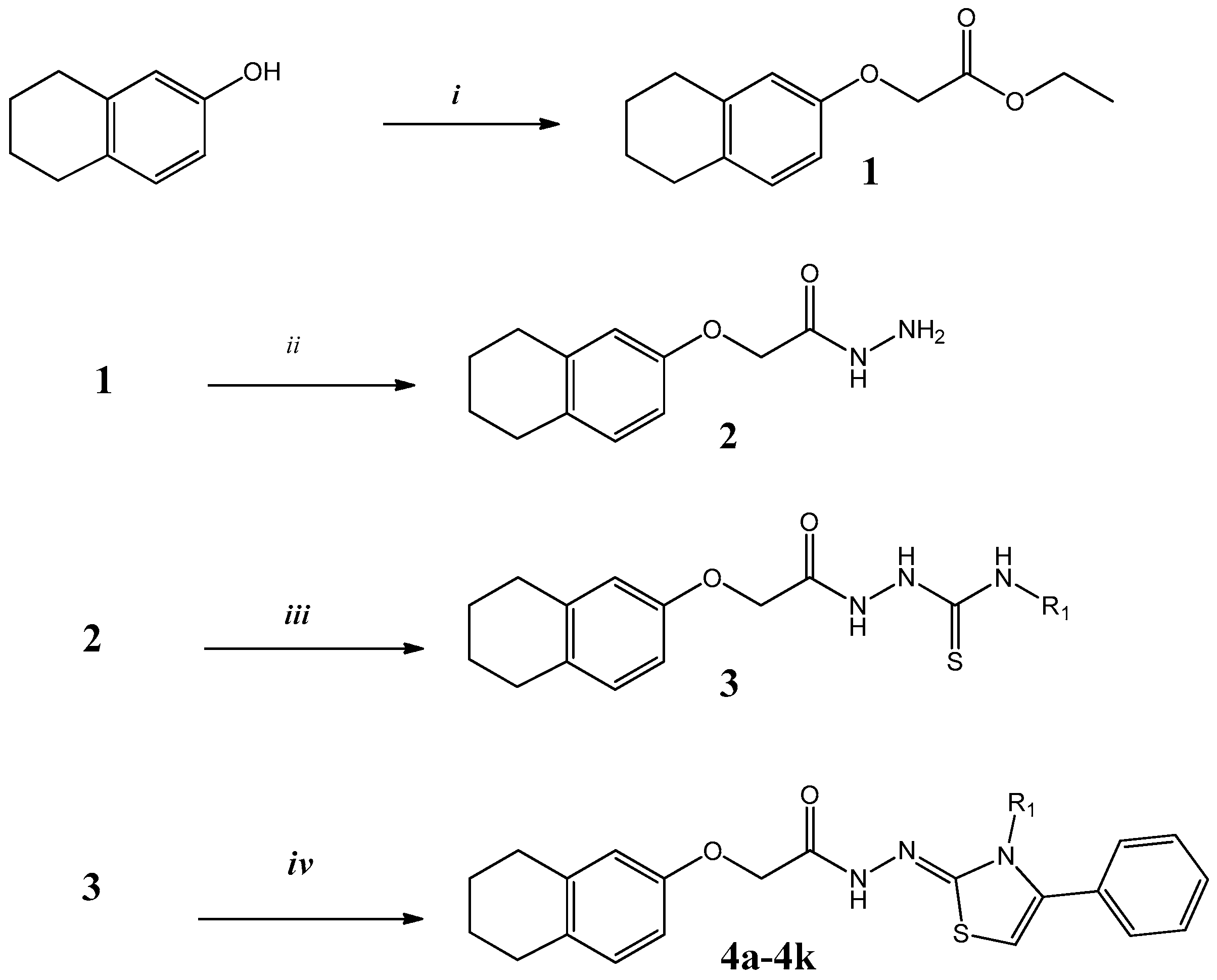
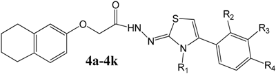
2.1. Chemistry

Eleven new compounds were synthesized in a three step synthetic procedure and this is outlined in Scheme 1. Starting material 5,6,7,8-tetrahydro-2-naphthol was reacted with ethyl 2-chloroethyl acetate to give ethyl 2-[(5,6,7,8-tetrahydronaphthalen-2-yl)oxy]acetate (1), this intermediate was hydrazinated with hydrazine hydrate to give ethyl 2-[(5,6,7,8-tetrahydronaphthalen-2-yl)oxy]acetohydrazide (2) and these intermediates were reported, previously [44]. The resulting hydrazide was boiled with phenyl and cyclohexyl isothiocyanate in ethanol to give N-phenyl/cyclohexyl-2-[2-((5,6,7,8-tetrahydronaphthalen-2-yl)oxy)acetyl]hydrazine carbothioamide (3a, 3b) according to the literature [45]. In the last step these compounds were reacted with phenacyl bromide derivatives to give N′-[3-phenyl/cyclohexyl-4-(substituted phenyl)thiazol-2(3H)-ylidene]-2-[(5,6,7,8-tetrahydronaphthalen-2-yl)oxy]acetohydrazides (4a4k). The substituents were showed in Table 1. The structures of final compounds were confirmed by using spectral and elemental analysis. In the IR spectra of the compounds (see Supplementary materials), characteristic bands belong to N–H and C=O bonds of acetamide moiety were observed at 3109–3199 cm−1 and 1712–1745 cm−1 whereas the protons of acetamide moiety were observed at 4.75–4.61 ppm as singlets belong to methylene and at 11.79–11.98 ppm belong to amino protons in the 1H-NMR spectra of the compounds. –CH2 Protons of tetrahydronaftalene ring were detected at 1.67–1.98 ppm and 2.48–2.69 ppm region. In aromatic field, proton of thiazole ring, C5–H was seen at about 7.0–7.19 ppm as singlet peak, mostly. In the 13C-NMR spectra of the compounds, methylene carbons of acetamide group were resonated at 56.21–66.15 ppm, whereas methylene carbons of tetrahydronaftalene ring were observed at 20.91–31.17 ppm. In the MS, M + 1 peaks were detected for all final compounds. Elemental analysis data was also determined within the accurate range of ±0.4%.

2.2. Biological Results

2.2.1. Cytotocity

In order to determine anticancer efficiency of the synthesized compounds, three-step activity studies were carried out. Accordingly, cytotoxicity and DNA synthesis inhibition experiments and tests to determine apoptosis/necrosis ratios were performed. Tested tumor cells were human breast adenocarcinoma cell line (MCF-7), human lung carcinoma cell line (A549) and healthy cell mouse embryoblast cell line (NIH/3T3) were used to detect cytotoxicity of the compounds according to classical MTT method. The calculated inhibitory concentrations (IC50) and selectivity index (SI) are represented in Table 2.
Compounds 4b (IC50 = 69.2 μM) and 4d (IC50 = 71.8 μM) showed the highest cytotoxicity against MCF-7 cell line. However, compounds 4e and 4k also exhibited considerable cytotoxic activity. None of these compounds were found as active as standard drug cisplatin. Among these four compounds 4b and 4k showed toxicity to normal cells. When we compare, antiproliferative effects of the compounds, their potency against A549 was found to be greater than against MCF-7 cells. It has been determined that compounds 4f, 4g and 4h act at a lower concentration than the inhibition concentration of cisplatin which possessed 4-bromo, 4-chloro and 4-fluoro substituent moieties respectively on A549 cell line. Besides, these compounds exhibited selectivity due to their low cytotoxicity on healthy cells with selectivity index in between 2.7 and 7.8. However, the IC50 values of compounds 4c, 4d, 4i and 4k were not determined at the highest concentration tested against A549. Additionally, compounds 4a, 4b and 4j exhibited moderate antiproliferative activity.

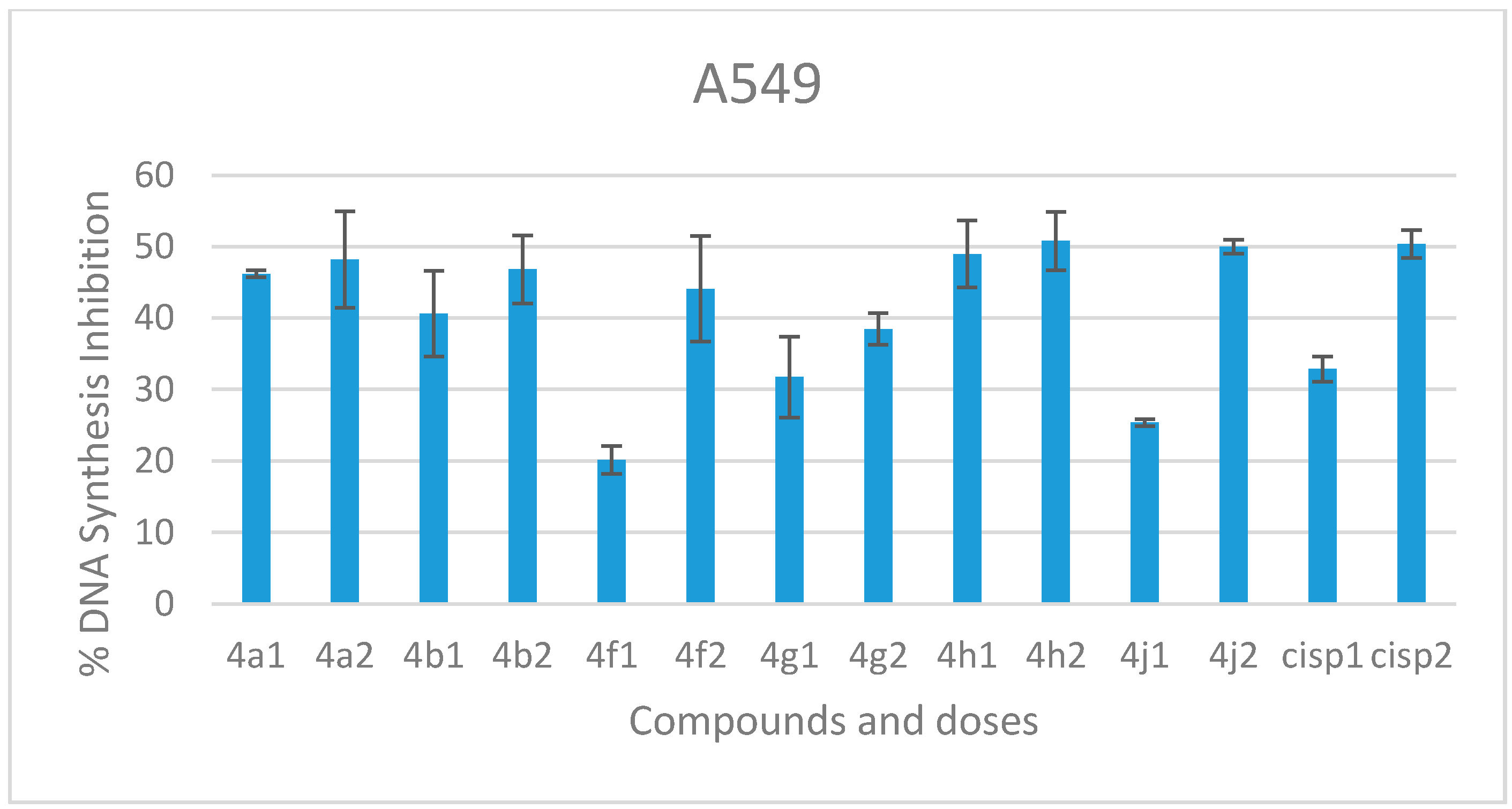
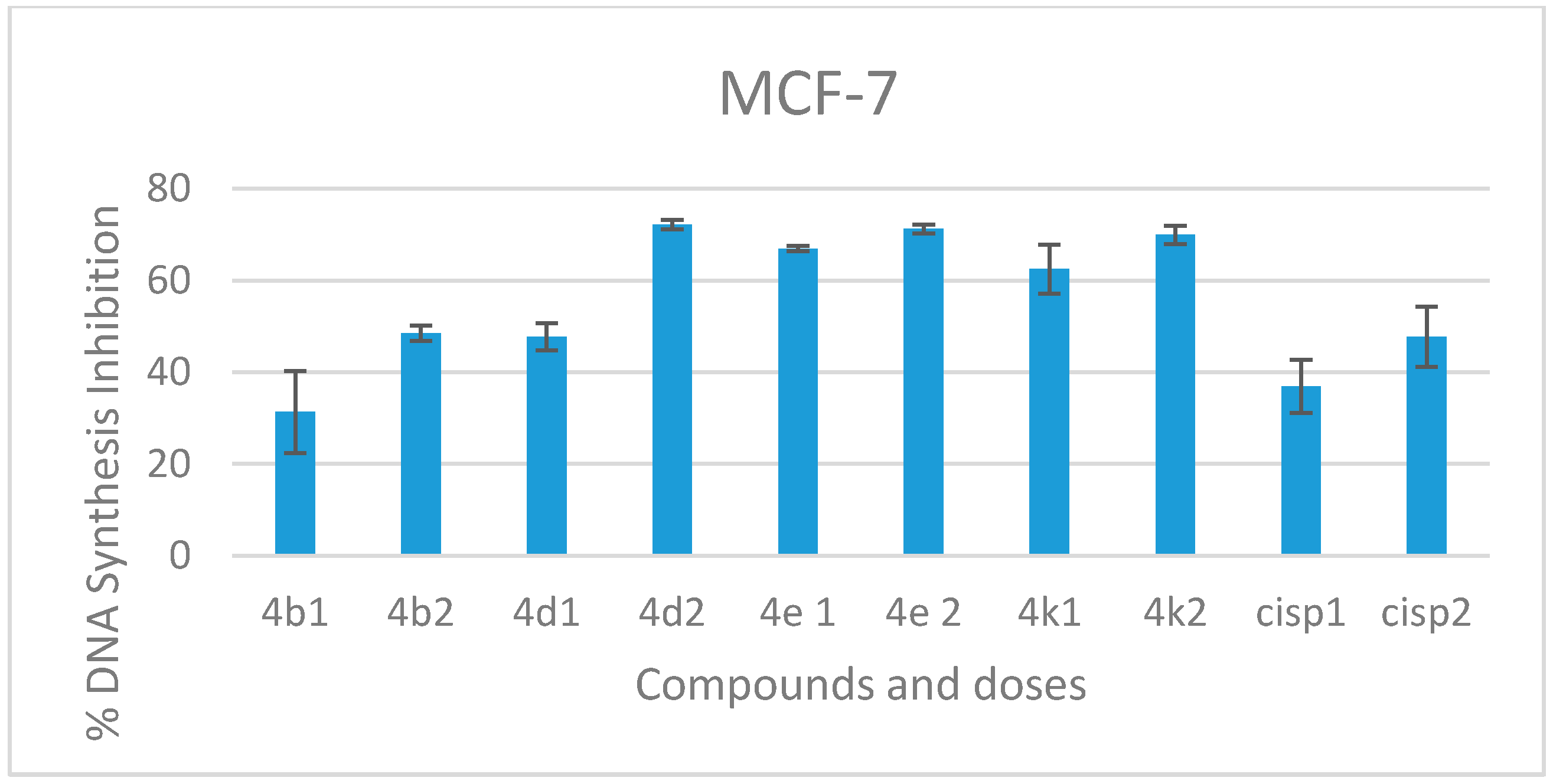
2.2.2. DNA Synthesis Inhibition

DNA synthesis inhibition study, which was considered as the second stage in anticancer activity studies, have been conducted on compounds with high cytotoxicity. Compounds 4b, 4d, 4e, and 4k were determined to possess more cytotoxic activity on the MCF cell line. The graph generated according to the percent inhibitions of the selected compounds at a: IC50/2 and b: IC50 concentrations is shown in Figure 3. Compound 4b inhibited DNA synthesis with ratio of 31.39% at 34.6 μM and 48.50% at 69.2 μM concentration on MCF-7 cell line. It was seen this ratio was very close to the inhibition exhibited by cisplatin. The inhibition rates for compounds 4d, 4e and 4k were calculated higher than the rate of standard, ranging from 47.75% to 72.15%. Among these three, compound 4d exhibited the best inhibition potential of 47.75% at 36.9 μM concentration and 72.15% at 71.8 μM on MCF-7 cells.
DNA synthesis inhibition studies have also been conducted on those compounds that exhibit high cytotoxic activity on A549 cells. Compounds 4a, 4b, 4f, 4g and 4j were selected for this study. The graph based on the obtained results is shown in Figure 4. DNA synthesis inhibition on A549 revealed that these six compounds showed close inhibition values of 20.13–50.33%. The compound 4h bearing 4-fluoro substitution on phenyl ring showed the highest inhibitory effect. The inhibition percentages were specified as 8.97% at 24.3 μM and 50.78% at 48.6 μM whereas these values were established 32.86% at 54.2 μM and 50.33% at 108.3 μM for standard drug cisplatin.

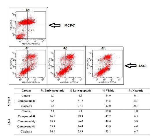
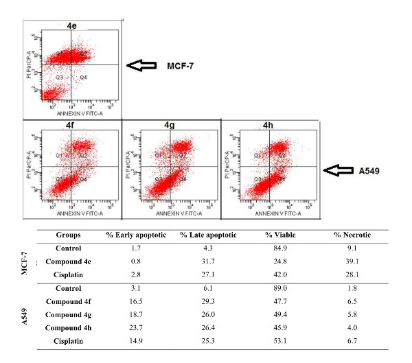
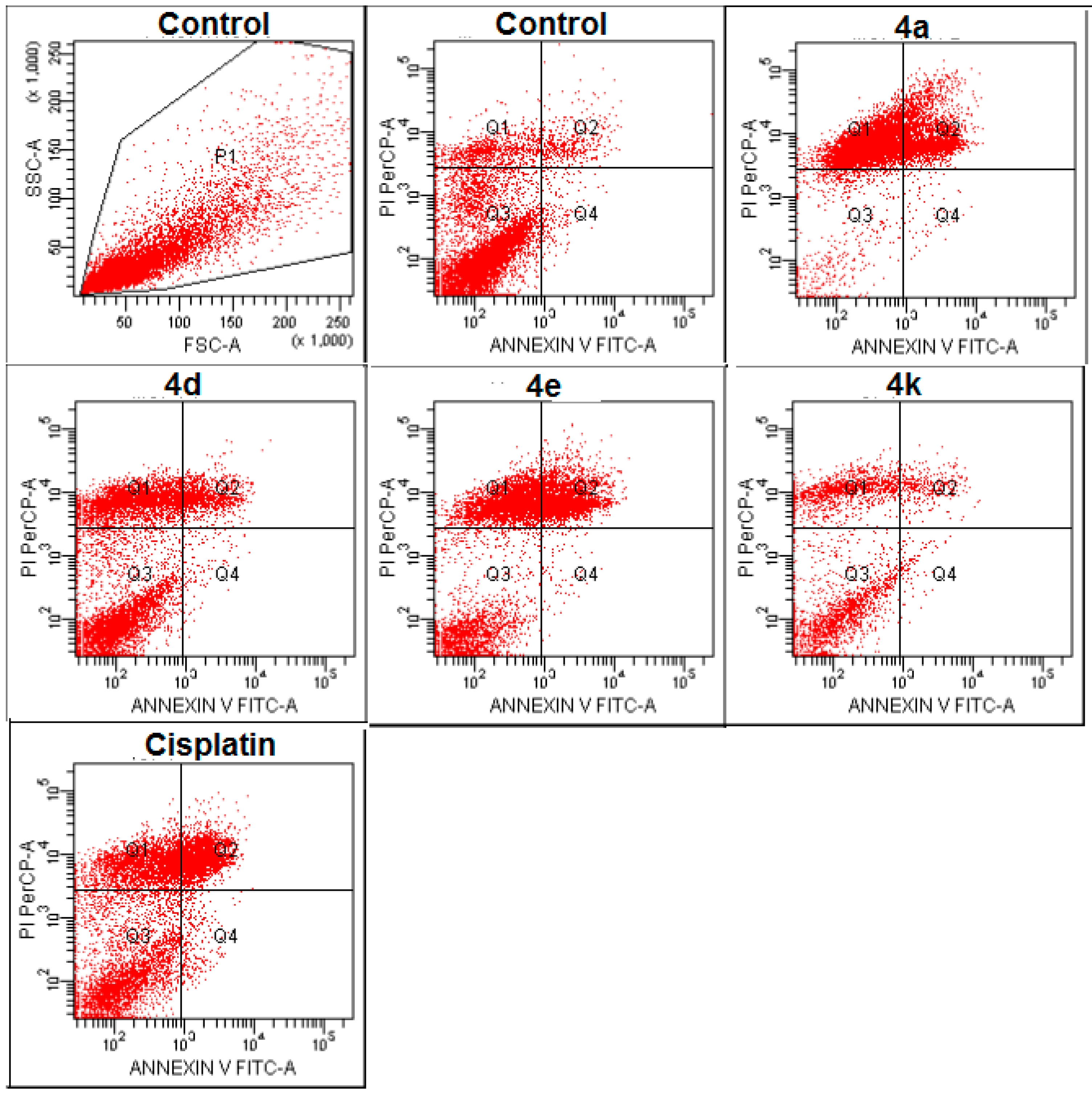
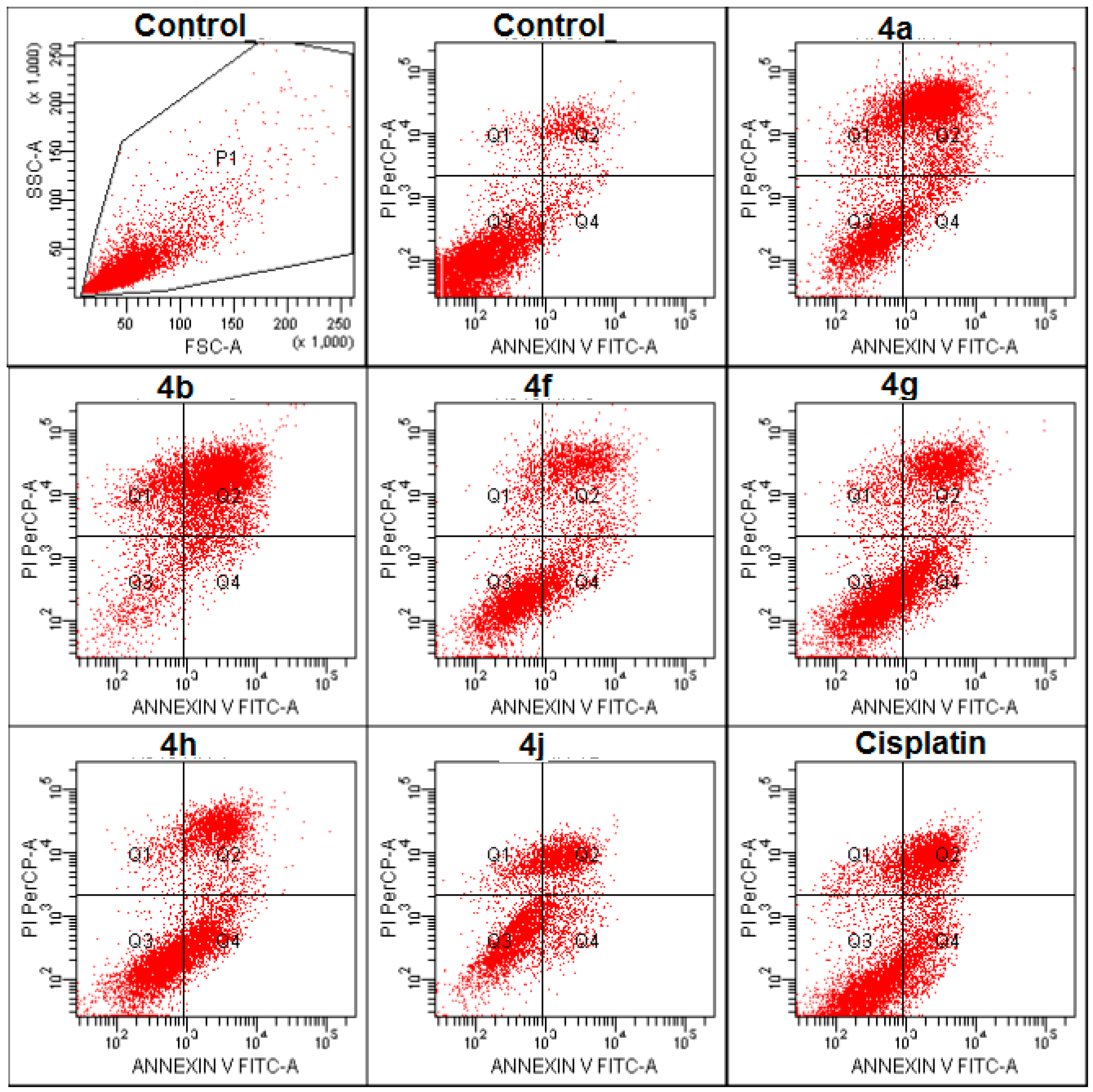
2.2.3. Apoptosis/Necrosis Detection

To determine the death mechanism of the most active compounds, flow cytometric analysis was done according to the instructions of the Annexin V-FITC Apoptosis Detection Kit. The results were calculated in percentage of which the ratios were demonstrated as early apoptotic, late apoptotic, viable and necrotic cells. The numerical values were represented in Table 3 and diagrams in Figure 5 and Figure 6. Compounds 4b, 4d, 4e and 4k against MCF-7 cell line and compounds 4a, 4b, 4f, 4g, 4h and 4j against A549 cells were tested in flow cytometry at IC50 concentrations of the compounds. Against MCF-7 cell line, compounds 4b and 4e exhibited 29.8% and 32.5% apoptosis (ealy + late apoptotic cell percentage), respectively whereas cisplatin caused 29.9% apoptosis of the cells. Other two compounds did not showed prominent activity. All selected compounds for flow cytometric analysis on A549 cell line displayed marvelous activity that apoptotic cell ratios were found even higher than cisplatin caused (40.2%). Among them, compound 4b showed the highest apoptosis level of 73.6% which was a great value for such a study. Besides, compounds 4a and 4h caused apoptosis over 50% and compounds 4f, 4g and 4j caused apoptosis over 41.5%. Among these compounds, the results of compounds 4f, 4g and 4h bearing p-halogenated phenyl moiety are more meaningful, which, is because they have lower IC50 values than cisplatin. Consequently, the dose administered to this test is lesser, but apoptotic cell percentage found is higher. In addition, these three compounds have exhibited non-toxic profile which is a required property for druglikeness of a molecule.

2.2.4. Anticholinesterase Activity

The inhibitory effects of the compounds on the acetylcholinesterase (AChE) and butyrylcholinesterase (BuChE) enzymes were investigated. The efficacy of the compounds is compared to the anticholinesterase effective donepezil drug molecule used in Alzheimer's treatment. The results obtained are shown in Table 4.
Based on the enzyme inhibition studies, AChE and BuChE inhibitions were determined at 80 μg/mL (~160 µM) concentrations of the compounds. Compound 4h showed the highest acetylcholinesterase inhibition with 49.92%. It was followed by compounds 4j with 44.96%, 4d with 43.22%, 4f with 42.87% and 4i with 41.85%. Compounds 4b and 4e exhibited the lowest inhibition among remaining others. Since the compounds did not provide over 50% enzyme inhibition, they were not studied at lower doses to detect inhibition concentrations (IC50). The IC50 value of the standard drug, donepezil, was determined to be (11.8 ± 0.7) × 10−3 μM. The inhibition of butyrylcholinesterase enzyme at concentration of 80 μg/mL was found in between 15.89% and 33.19% for compounds 4a, 4b, 4c and 4d while other compounds did not inhibit this enzyme. Compounds 4f, 4g, 4h and 4j are the most active compounds on A549 cancer cells. The AChE inhibition effect of these substances may enhance antiproliferative activity of them.

3. Materials and Methods

3.1. Chemistry

All chemicals were purchased from Sigma-Aldrich Chemical Co., (Sigma-Aldrich Corp., St. Louis, MO, USA) and Merck (Merck, Darmstadt, Germany). All melting points (m.p.) were determined by Electrothermal 9300 digital melting point apparatus (Electrothermal, Essex, UK) and are uncorrected. All the reactions were monitored by thin-layer chromatography (TLC) using Silica gel 60 F254 TLC plates (Merck, Darmstadt, Germany). Spectroscopic data were recorded with the following instruments: IR, Shimadzu 8400S spectrophotometer (Shimadzu, Tokyo, Japan); 1H-NMR, Agilent 400 MHz NMR spectrometer (Agilent Technologies, Palo Alto, CA, USA); 13C-NMR, Agilent 100 MHz NMR spectrometer (Agilent Technologies, Palo Alto, CA, USA); Shimadzu 8040 LC/MS/MS system (Shimadzu, Tokyo, Japan), and elemental analyses were performed on a Perkin Elmer EAL 240 elemental analyser (Perkin Elmer, Norwalk, CT, USA).
General procedure for the synthesis of N′-[3-cyclohexyl/phenyl-4-(substituted phenyl)thiazole-2(3H)-ylidene]-2-[(5,6,7,8-tetrahydronaphthalen-2-yl)oxy]acetohydrazide (4a4k). The synthesized intermediates N-phenyl/cyclohexyl-2-[2-((5,6,7,8-tetrahydronaphthalen-2-yl)oxy)acetyl]hydrazine carbothioamide (3a, 3b) were refluxed with phenacyl bromide derivatives for 5 h in ethanol. After overnight standing in a cool place, crystals of final thiazole compounds were filtered off.
N′-(3-Cyclohexyl-4-phenylthiazole-2(3H)-ylidene)-2-[(5,6,7,8-tetrahydronaphthalen-2-yl)oxy]acetohydrazide (4a): Yield: % 68. m.p. 277 °C. IR νmax (cm−1): 3199 (N–H), 3070 (aromatic C–H), 2854 (aliphatic C–H), 1745 (amide C=O), 1595–1442 (C=C and C=N), 1215–1004 (C–N and C–O). 1H-NMR (400 MHz, DMSO-d6, ppm): δ 1.13–2.60 (19H, m, CH2), 4.61 (2H, s, OCH2), 6.46–7.51 (9H, m, Ar–H), 11.85 (1H, s, NH). 13C-NMR (100 MHz, DMSO-d6, ppm) δ 22.5, 22.8, 24.3, 24.5, 27.9, 28.9, 31.2, 55.2, 58.5, 66.0, 100.9, 112.1, 114.1, 114.3, 118.9, 129.5, 129.5, 130.1, 137.6, 140.8, 155.1, 160.6, 167.3. HRMS (m/z): [M + H]+ for C27H31N3O2S calculated: 461.62; found: 462.2195. For C27H31N3O2S calculated: 70.25% C, 6.77% H, 9.10% N; found: 70.23% C, 6.76% H, 9.12% N.
N′-[3-Cyclohexyl-4-(4-methoxyphenyl)thiazole-2(3H)-ylidene]-2-[(5,6,7,8-tetrahydronaphthalen-2-yl)oxy]acetohydrazide (4b): Yield: % 71. m.p. 266 °C. IR νmax (cm−1): 3182 (N–H), 3073 (aromatic C–H), 2972 (aliphatic C–H), 1714 (amide C=O), 1602–1502 (C=C and C=N), 1247–1037 (C–N and C–O). 1H-NMR (400 MHz, DMSO-d6, ppm): δ 1.12–2.61 (19H, m, CH2), 4.62 (2H, s, OCH2), 6.45–6.49 (2H, m, Ar–H), 6.85 (1H, d, J = 8.4 Hz, Ar–H), 6.95 (2H, d, J = 8.8 Hz), 7.12 (1H, s, Ar–H), 7.43 (2H, d, J = 8.8 Hz, Ar–H), 11.79 (1H, s, NH). 13C-NMR (100 MHz, DMSO-d6, ppm) δ 22.5, 22.7, 27.8, 28.9, 66.1, 112.0, 114.3, 122.6, 128.1, 129.4, 129.5, 129.9, 130.0, 130.7, 131.4, 132.6, 137.5, 137.7, 155.1, 167.8. HRMS (m/z): [M + H]+ for C28H33N3O3S calculated: 491.65; found: 492.2296. For C28H33N3O3S calculated: 68.40% C, 6.77% H, 8.55% N; found: 68.42% C, 6.76% H, 8.56% N.
N′-[3,4-Diphenylthiazole-2(3H)-ylidene]-2-[(5,6,7,8-tetrahydronaphthalen-2-yl)oxy]acetohydrazide (4c): Yield: % 73. m.p. 253 °C. IR νmax (cm−1): 3120 (N–H), 3053 (aromatic C–H), 2987 (aliphatic C–H), 1714 (amide C=O), 1600–1444 (C=C and C=N), 1217–1055 (C–N and C–O). 1H-NMR (400 MHz, DMSO-d6, ppm): δ 1.98 (4H, br, CH2), 2.56–2.61 (4H, m, CH2), 4.65 (2H, s, OCH2), 6.49–7.58 (13H, m, Ar–H), 7.07 (1H, s, thiazole–H), 11.98 (1H, br, NH). 13C-NMR (100 MHz, DMSO-d6, ppm) δ 22.6, 22.8, 22.9, 24.5, 25.2, 27.9, 29.0, 30.7, 56.2, 60.2, 112.6, 112.6, 114.5, 123.8, 129.6, 129.8, 129.8, 137.7, 140.2, 149.1, 150.1, 151.7, 155.1, 192.6. HRMS (m/z): [M + H]+ for C27H25N3O2S calculated: 455.58; found: 456.1719. For C27H25N3O2S calculated: 71.18% C, 5.53% H, 9.22% N; found: 71.20% C, 5.54% H, 9.23% N.
N′-[3-Phenyl-4-(4-methylphenyl)thiazole-2(3H)-ylidene]-2-[(5,6,7,8-tetrahydronaphthalen-2-yl)oxy]acetohydrazide (4d): Yield: % 72. m.p. 258 °C. IR νmax (cm−1): 3109 (N–H), 3047 (aromatic C–H), 2860 (aliphatic C–H), 1745 (amide C=O), 1599–1496 (C=C and C=N), 1199–1074 (C–N and C–O). 1H-NMR (400 MHz, DMSO-d6, ppm): δ 1.67 (4H, br, CH2), 2.32 (3H, s, CH3), 2.56–2.59 (4H, m, CH2), 4.65 (2H, s, OCH2), 6.48–7.56 (12H, m, Ar–H), 7.00 (1H, s, thiazole–H), 11.98 (1H, br, NH). 13C-NMR (100 MHz, DMSO-d6, ppm) δ 22.4, 22.7, 27.8, 28.9, 66.0, 112.1, 114.1, 122.7, 123.6, 129.3, 129.4, 129.5, 130.1, 133.7, 137.4, 138.1, 147.6, 155.1, 168.0. HRMS (m/z): [M + H]+ for C28H27N3O2S calculated: 469.60; found: 470.1885. For C28H27N3O2S calculated: 71.62% C, 5.80% H, 8.95% N; found: 71.61% C, 5.79% H, 8.94% N.
N′-[3-Phenyl-4-(4-methoxyphenyl)thiazole-2(3H)-ylidene]-2-[(5,6,7,8-tetrahydro naphthalen-2-yl)oxy]acetohydrazide (4e): Yield: % 67. m.p. 240 °C. IR νmax (cm−1): 3109 (N–H), 3047 (aromatic C–H), 2860 (aliphatic C–H), 1745 (amide C=O), 1599–1496 (C=C and C=N), 1199–1074 (C–N and C–O). 1H-NMR (400 MHz, DMSO-d6, ppm): δ 1.68 (4H, br, CH2), 2.48–2.59 (4H, m, CH2), 3.77 (3H, s, OCH3), 4.65 (2H, s, OCH2), 6.47–7.57 (13H, m, Ar–H), 11.98 (1H, br, NH). 13C-NMR (100 MHz, DMSO-d6, ppm) δ 22.6, 22.9, 27.9, 29.0, 66.0, 112.2, 114.4, 123.5, 127.3, 128.4, 128.7, 129.5, 129.7, 129.7, 131.1, 130.3, 137.6, 140.6, 155.2, 167.7. HRMS (m/z): [M + H]+ for C28H27N3O3S calculated: 485.60; found: 486.1834. For C28H27N3O3S calculated: 69.26% C, 5.60% H, 8.65% N; found: 69.24% C, 5.61% H, 8.66% N.
N′-[3-Phenyl-4-(4-bromophenyl)thiazole-2(3H)-ylidene]-2-[(5,6,7,8-tetrahydronaphthalen-2-yl)oxy]acetohydrazide (4f): Yield: % 74. m.p. 272 °C. IR νmax (cm−1): 3122 (N–H), 3039 (aromatic C–H), 2935 (aliphatic C–H), 1712 (amide C=O), 1598–1454 (C=C and C=N), 1313–1006 (C–N and C–O). 1H-NMR (400 MHz, DMSO-d6, ppm): δ 1.67 (4H, br, CH2), 2.50–2.63 (4H, m, CH2), 4.67 (2H, d, J = 4.4 Hz, OCH2), 6.48–6.51 (2H, m, Ar–H), 6.87 (1H, d, J =7.6 Hz, Ar–H), 7.04 (1H, s, thiazole–H), 7.33–7.38 (3H, m, Ar–H), 7.45 (2H, d, J = 8.4 Hz), 7.52–7.56 (2H, m, Ar–H), 7.60 (2H, d, J = 8.4 Hz, Ar–H), 11.89 (1H, br, NH). 13C-NMR (100 MHz, DMSO-d6, ppm) δ 22.6, 22.9, 28.0, 29.0, 66.0, 112.2, 114.4, 123.2, 126.3, 128.8, 129.5, 129.6, 130.1, 130.3, 134.9, 137.6, 139.26, 155.2, 167.9. HRMS (m/z): [M + H]+ for C27H24N3O2SBr calculated: 534.47; found: 536.0798. For C27H24N3O2SBr calculated: 60.68% C, 4.53% H, 7.86% N; found: 60.67% C, 4.52% H, 7.87% N.
N′-[3-Phenyl-4-(4-chlorophenyl)thiazole-2(3H)-ylidene]-2-[(5,6,7,8-tetrahydronaphthalen-2-yl)oxy]acetohydrazide (4g): Yield: % 67. m.p. 268 °C. IR νmax (cm−1): 3122 (N–H), 3032 (aromatic C–H), 2972 (aliphatic C–H), 1712 (amide C=O), 1593–1456 (C=C and C=N), 1313–1055 (C–N and C–O). 1H-NMR (400 MHz, DMSO-d6, ppm): δ 1.68 (4H, br, CH2), 2.50–2.62 (4H, m, CH2), 4.64 and 4.70 (2H, d, J = 15.6 Hz, OCH2), 6.49–6.51 (2H, m, Ar–H), 6.87 (1H, d, J = 8.8 Hz, Ar–H), 7.05 (1H, s, thiazole–H), 7.35–7.56 (9H, m, Ar–H), 11.93 (1H, br, NH). 13C-NMR (100 MHz, DMSO-d6, ppm) δ 20.9, 22.6, 22.9, 29.0, 66.0, 112.2, 114.4, 123.5, 124.4, 128.8, 129.3, 129.5, 129.6, 130.4, 137.6, 139.9, 140.7, 155.2, 167.7. HRMS (m/z): [M + H]+ for C27H24N3O2SCl calculated: 490.02; found: 490.1328. For C27H24N3O2SCl calculated: 66.18% C, 4.94% H, 7.58% N; found: 66.16% C, 4.93% H, 8.57% N.
N′-[3-Phenyl-4-(4-florophenyl)thiazole-2(3H)-ylidene]-2-[(5,6,7,8-tetrahydronaphthalen-2-yl)oxy]acetohydrazide (4h): Yield: % 72. m.p. 257 °C. IR νmax (cm−1): 3116 (N–H), 3043 (aromatic C–H), 2916 (aliphatic C–H), 1714 (amide C=O), 1600–1494 (C=C and C=N), 1313–1035 (C–N and C–O). 1H-NMR (400 MHz, DMSO-d6, ppm): δ 1.67 (4H, br, CH2), 2.57–2.62 (4H, m, CH2), 4.62 and 4.69 (2H, dd, J = 15.6 Hz, OCH2), 6.50–6.53 ( 2H, m, Ar–H), 6.86 (1H, d, J = 8.0 Hz, Ar–H), 7.03 (1H, s, thiazole–H), 7.21–7.56 (9H, m, Ar–H), 11.93 (1H, br, NH). 13C-NMR (100 MHz, DMSO-d6, ppm) δ 22.6, 22.8, 27.9, 29.0, 66.0, 112.1, 114.4, 123.1, 123.6, 126.6, 129.5, 129.6, 130.2, 130.2, 131.6, 137.6, 139.3, 155.2, 167.8. HRMS (m/z): [M + H]+ for C27H24FN3O2S calculated: 473.57; found: 474.1629. For C27H24FN3O2S calculated: 68.48% C, 5.11% H, 8.87% N; found: 68.49% C, 5.12% H, 8.88% N.
N′-[3-Phenyl-4-(3-nitrophenyl)thiazole-2(3H)-ylidene]-2-[(5,6,7,8-tetrahydronaphthalen-2-yl)oxy]acetohydrazide (4i): Yield: % 69. m.p. 252 °C. IR νmax (cm−1): 3118 (N–H), 3045 (aromatic C–H), 2949 (aliphatic C–H), 1710 (amide C=O), 1604–1344 (C=C and C=N), 1217–1062 (C–N and C–O). 1H-NMR (400 MHz, DMSO-d6, ppm): δ 1.64 (4H, br, CH2), 2.47–2.57 (4H, m, CH2), 4.52 and 6.72 (2H, dd, J = 15.6 Hz, OCH2), 6.41–6.43 (2H, m, Ar–H), 6.74 (1H, d, J = 8.0 Hz, Ar–H), 7.16 (1H, s, thiazole–H), 7.30–8.29 (9H, m, Ar–H), 11.95 (1H, br, NH). 13C-NMR (100 MHz, DMSO-d6, ppm) δ 22.6, 22.8, 27.9, 29.0, 55.3, 66.1, 112.2, 114.1, 114.4, 119.4, 123.6, 129.5, 129.6, 130.0, 130.6, 137.6, 140.6, 155.2, 160.5, 167.7. HRMS (m/z): [M + H]+ for C27H24N4O4S calculated: 500.57; found: 501.1567. For C27H24N4O4S calculated: 64.79% C, 4.83% H, 11.19% N; found: 64.78% C, 4.82% H, 11.18% N.
N′-[3-Phenyl-4-(4-nitrophenyl)thiazole-2(3H)-ylidene]-2-[(5,6,7,8-tetrahydronaphthalen-2-yl)oxy]acetohydrazide (4j): Yield: % 67. m.p. 268 °C. IR νmax (cm−1): 3126 (N–H), 3037 (aromatic C–H), 2931 (aliphatic C–H), 1714 (amide C=O), 1602–1338 (C=C and C=N), 1288–1035 (C–N and C–O). 1H-NMR (400 MHz, DMSO-d6, ppm): δ 1.65 (4H, br, CH2), 2.50–2.57 (4H, m, CH2), 4.61 and 4.75 (2H, dd, J = 15.6 Hz, OCH2), 6.48–6.54 ( 2H, m, Ar–H), 6.81 (1H, d, J = 8.0 Hz, Ar–H), 7.19 (1H, s, thiazole–H), 7.30–8.20 (9H, m, Ar–H), 11.94 (1H, br, NH). 13C-NMR (100 MHz, DMSO-d6, ppm) δ 22.6, 22.8, 27.9, 29.0, 66.03, 112.2, 114.3, 115.7, 115.9, 123.3, 123.8, 127.4, 129.6, 129.6, 130.3, 130.8, 130.9, 137.6, 139.5, 155.1, 161.7, 164.2, 167.8. HRMS (m/z): [M + H]+ C27H24N4O4S için calculated: 500.57; found: 501.1572. For C27H24N4O4S calculated: 64.79% C, 4.83% H, 11.19% N; found: 64.80% C, 4.84% H, 11.20% N.
N′-[3-Phenyl-4-(2,3-dichlorophenyl)thiazole-2(3H)-ylidene]-2-[(5,6,7,8-tetrahydronaphthalen-2-yl)oxy]acetohydrazide (4k): Yield: % 75. m.p. 272 °C. IR νmax (cm−1): 3109 (N–H), 3047 (aromatic C–H), 2916 (aliphatic C–H), 1732 (amide C=O), 1610–1305 (C=C and C=N), 1219–1028 (C–N and C–O). 1H-NMR (400 MHz, DMSO-d6, ppm): δ 1.68 (4H, br, CH2), 2.49–2.62 (4H, m, CH2), 4.63 and 4.72 (2H, dd, J = 15.6 Hz, OCH2), 6.48–6.50 (2H, m, Ar–H), 6.84 (1H, d, J = 8.0 Hz, Ar–H), 7.04 (1H, s, thiazole–H), 7.27–7.82 (8H, m, Ar–H), 11.92 (1H, br, NH). 13C-NMR (100 MHz, DMSO-d6, ppm) δ 22.4, 22.7, 27.8, 28.8, 66.2, 111.9, 114.1, 122.9, 124.4, 128.8, 129.4, 129.5, 130.6, 130.2, 134.7, 137.4, 138.0, 147.4, 155.0, 168.0. HRMS (m/z): [M + H]+ for C27H23Cl2N3O2S calculated: 524.46; found: 524.0938. For C27H23Cl2N3O2S calculated: 61.83% C, 4.42% H, 8.01% N; found: 61.82% C, 4.43% H, 8.02%N.

3.2. Biochemistry

3.2.1. MTT Assay (Mitochondrial Activity)

This is a colorimetric assay that measures the reduction of 3-(4,5-dimethylthiazol-2-yl)-2,4,diphenyltetrazolium bromide (MTT) by mitochondria [46]. All final compounds were tested against human breast adenocarcinoma cell line (MCF-7), human lung carcinoma cell line (A549) and mouse embryooblast cell line (NIH/3T3) to determine their cytotoxic profile. Cancer cells were cultured in RPMI medium (Hyclone, Thermo Scientific, Waltham, MA, USA) at 37 °C in a humidified atmosphere of 95% air and 5% CO2. The studied cells were situated at 2 × 10⁴ cells into each well of 96-well microtiter tissue culture plates (Nunc, Denmark) and incubated for 24 h. After that, test compounds were dissolved in DMSO and added to culture wells at varying concentrations (85–1000 µM). After a 24 h incubating period, 20 mL MTT solution (5 mg/mL MTT powder in PBS) was added to each well and the cells were incubated for 4 h at 37 °C. The purple formazan crystals produced were dissolved in DMSO and the absorbance was read by ELISA reader (OD570 nm). The percent values of cell proliferations were calculated relative to controls, whose cell proliferations were accepted as 100% [47].

3.2.2. DNA Synthesis Inhibition

The 5-bromo-2′-deoxy-uridine (BrdU) cell proliferation assay [48] was used to detect the proliferation of A549 and C6 cells. This method was performed in the 96-well flat-bottomed microtiter plates by using cisplatin as positive control drug. A549 and C6 cells were collected from cell cultures by 0.25% trypsin/EDTA solution and counted in a hemocytometer. Suspensions of cell lines were seeded into 96-well flat-bottomed microtiter plates at a density of 1 × 10³ cells/mL. The tumor cell lines were cultured in the presence of various doses of the test compounds or standard drugs. Microtiter plates were incubated at 37 °C in a 5% CO2/95% air humidified atmosphere for 24 h and 48 h. At the end of each day, the cells were labelled with 10 mL BrdU solution for 2 h and then fixed. Anti-BrdU-POD (100 mL) was added and incubated for 90 min. Finally, microtiter plates were washed with phosphate buffered saline (PBS) three times and the cells were incubated with substrate solution until the color was sufficient for photometric detection. Absorbance of the samples was measured with an ELx808-IUBio-Tek apparatus at 492 nm. The growth percentage was evaluated spectrophotometrically versus untreated controls with used cell viability of growth assay. Results for each spectrophotometrical measure were noticed as percent of growth inhibition. All experiments were done in triplicates.

3.2.3. Flow Cytometric Analysis

Evaluation of apoptosis was carried out through the Annexin V-FITC Apoptosis Detection Kit from BD, Pharmingen, according to the manufacturer’s instruction. This assay was performed on C6 and A549 cell lines with selected compounds which crossed the second step of anticancer activity. After induced apoptosis of cancer cells by the addition of IC50 concentrations of selected compounds and positive controls including cisplatin (50 mM) (24 h incubation), cells were collected by centrifugation for 5 min. Then cells were rinsed with cold water twice and resuspended in Annexin V-FITC binding buffer at a concentration of 1 × 10⁶ cells/mL. 5 mL of Annexin V-FITC was used to stain the cells. After 15 min of incubation at 25 °C, the cell suspension was centrifugated for 5 min and cells were resuspended in Annexin V-FITC binding buffer. Propidium iodide (5 mL) was added and the tubes were situated on ice and away from light. The fluorescence was measured using a flow cytometer. The results were analyzed by using FCSExpress software and represented as percentage of normal and apoptotic cells at various stages [49]. The percentage of apoptotic cells was calculated from the number of cells in sub-G1 phase, representing fragmented cell vesicles. The four areas in the diagrams stand for necrotic cells (Q1, positive for PI and negative for annexin/FITC, left square on the top), live cells (Q3, negative for annexin and PI, left square at the bottom), late apoptotic or necrotic cells (Q2, positive for annexin and PI, right square on the top) and apoptotic cells (Q4, negative for PI and positive for annexin, right square at the bottom), respectively. The experiment was repeated three times.

3.2.4. Inhibition of AChE/BuChE Enzymes

In order to determine the acetyl- and butyrylcholinesterase enzyme inhibitions, experiments were carried out by making some changes in the method developed by Ellman [50]. Samples were prepared by dissolving in the appropriate solvent. Adding 20 μL enzyme solution (AChE or BuChE, 1 U/mL) and 10 μL sample solution to 2.4 mL buffer solution and it was incubated for 15 min at 37 °C. After incubation, 50 μL DTNB and 75 mM ATCI solution or 20 μL of 25 mM BTCI solution were added and allowed the mixture to incubate at room temperature for 30 min. For the control group, only 10 μL of solvent was added instead of the sample solution provided all protocols are the same. The absorbance values of the prepared mixture were measured spectrophotometrically using polystyrene forceps at 37 °C at 412 nm. Experiments were run in triplicate. The inhibition fractions of AChE yada BuChE enzymes were calculated using the following equation:
I(%) = 100 − (Abssample − Abscontrol) × 100

3.2.5. Statistical Analysis

All data were recorded as mean ± standard error of mean (S.E.M). Results were analyzed by the one-way ANOVA plus Tukey’s test. p-values lower than 0.05 were considered significant (p < 0.05 *, p < 0.01 **, p < 0.001 ***).

4. Conclusions

In this study, we have synthesized novel eleven compounds combining two ring systems thiazoline and tetralin. N′-[3-cyclohexyl/phenyl-4-(substituted phenyl)thiazole-2(3H)-ylidene]-2-[(5,6,7,8-tetrahydronaphthalen-2-yl)oxy]acetohydrazide (4a4k) derivatives have been tested to determine their anticancer potency. Compounds 4b (IC50 = 69.2 μM) and 4d IC50 = 71.8 μM showed the highest cytotoxicity against MCF-7 cells whereas compounds 4f, 4g and 4h including 4-bromo, 4-chloro and 4-fluoro substitutions act at lower concentration than cisplatin. Compound 4d inhibited DNA synthesis had the best potential of 47.75% at 35.9 μM concentration and 72.15% at 71.8 μM on MCF-7 cells. Compound 4h inhibited DNA synthesis with a percentage of 48.97% at 24.3 μM and 50.78% at 48.6 μM on A549 cell line. With regard to flow cytometric analysis, compounds 4b and 4e exhibited 29.8% and 32.5% apoptosis (ealy + late apoptotic cell percentage), respectively against MCF-7 cell line, whereas cisplatin caused 29.9% apoptosis of the cells. Compounds 4a, 4b, 4f, 4g, 4h and 4j showed significant apoptosis levels whose apoptosis induction ratios were higher than cisplatin against A549 cell line. With regard to anticholinesterase activity of the compounds, compound 4h showed the highest acetylcholinesterase inhibition with 49.92% and compounds 4j with 44.96%, 4d with 43.22%, 4f with 42.87% and 4i with 41.85% followed it.

Supplementary Materials

Supplementary materials are available online.

Acknowledgments

The authors present their thanks to Anadolu University for financial support within the Anadolu University Scientific Research Project, Eskişehir, Turkey (Project No: 1502S058). Also, we thank to Anadolu University Plant, Drug and Scientific Research Center (BIBAM) for providing laboratory to study anticancer activity.

Author Contributions

G.T.-Z. and Z.A.K. designed and organized study. A.T. and L.Y. performed the synthetic work. G.A.Ç. and H.E.T. studied the biological activity assay. G.T.-Z. and L.Y. interpreted the results and wrote the paper. All authors have approved the final version of the manuscript.

Conflicts of Interest

The authors confirm that the content of this article involves no conflict of interests.

References

  1. Zaharia, V.; Ignat, A.; Palibroda, N.; Ngameni, B.; Kuete, V.; Fokunang, C.N.; Moungang, M.L.; Ngadjui, B.T. Synthesis of some p-toluenesulfonyl-hydrazinothiazoles and hydrazino-bis-thiazoles and their anticancer activity. Eur. J. Med. Chem. 2010, 45, 5080–5085. [Google Scholar] [CrossRef] [PubMed]
  2. Ding, H.; Chen, Z.; Zhang, C.; Xin, T.; Wang, Y.; Song, H.; Jiang, Y.; Chen, Y.; Xu, Y.; Tan, C. Synthesis and cytotoxic activity of some novel N-pyridinyl-2-(6-phenylimidazo[2,1-b]thiazol-3-yl)acetamide derivatives. Molecules 2012, 17, 4703–4716. [Google Scholar] [CrossRef] [PubMed]
  3. Liu, Y.; Zhang, L.; Gong, J.; Fang, H.; Liu, A.; Du, G.; Xu, W. Design, synthesis, and biological activity of thiazole derivatives as novel influenza neuraminidase inhibitors. J. Enzym. Inhib. Med. Chem. 2011, 26, 506–513. [Google Scholar] [CrossRef] [PubMed]
  4. Cai, W.X.; Liu, A.L.; Li, Z.M.; Dong, W.L.; Liu, X.H.; Sun, N.B. Synthesis and anticancer activity of novel thiazole-5-carboxamide derivatives. Appl. Sci. 2016, 6, 8. [Google Scholar] [CrossRef]
  5. Kashyap, S.J.; Kumar Garg, V.; Kumar Sharma, P.; Kumar, N.; Dudhe, R.; Kumar Gupta, J. Thiazoles: Having diverse biological activities. Med. Chem. Res. 2012, 21, 2123–2132. [Google Scholar] [CrossRef]
  6. Nastasa, C.; Tiperciuc, B.; Duma, M.; Benedec, D.; Oniga, O. New hydrazones bearing thiazole scaffold: Synthesis, characterization, antimicrobial, and antioxidant investigation. Molecules 2015, 20, 17325–17338. [Google Scholar] [CrossRef] [PubMed]
  7. Bhole, R.P.; Bhusari, K.P. Synthesis and antitumor activity of (4-hydroxyphenyl)[5-substituted alkyl/aryl)-2-thioxo-1,3,4-thiadiazol-3-yl]methanone and [(3,4-disubstituted)-1,3-thiazol-2ylidene]-4-hydroxybenzohydrazide. Med. Chem. Res. 2011, 20, 695–704. [Google Scholar] [CrossRef]
  8. Sondhi, S.M.; Rani, R.; Gupta, P.P.; Agrawal, S.K.; Saxena, A.K. Synthesis, anticancer, and anti-inflammatory activity evaluation of methanesulfonamide and amidine derivatives of 3,4-diaryl-2-imino-4-thiazolines. Mol. Divers. 2009, 13, 357–366. [Google Scholar] [CrossRef] [PubMed]
  9. Makhaeva, G.F.; Boltneva, N.P.; Lushchekina, S.V.; Serebryakova, O.G.; Stupina, T.S.; Terentiev, A.A.; Serkov, I.V.; Proshin, A.N.; Bachurin, S.O.; Richardson, R.J. Synthesis, molecular docking and biological evaluation of N,N-disubstituted 2-aminothiazolines as a new class of butyrylcholinesterase and carboxylesterase inhibitors. Bioorg. Med. Chem. 2016, 24, 1050–1062. [Google Scholar] [CrossRef] [PubMed]
  10. Han, F.S.; Osajima, H.; Cheung, M.; Tokuyama, H.; Fukuyama, T. Novel structural motifs consisting of chiral thiazolines: Synthesis, molecular recognition, and anticancer activity. Chemistry 2007, 13, 3026–3038. [Google Scholar] [CrossRef] [PubMed]
  11. Carmeli, S.; Moore, R.E.; Patterson, G.L. Mirabazoles, minor tantazole-related cytotoxins from the terrestrial blue-green alga scytonema mirabil. Tetrahedron Lett. 1991, 32, 2593–2596. [Google Scholar] [CrossRef]
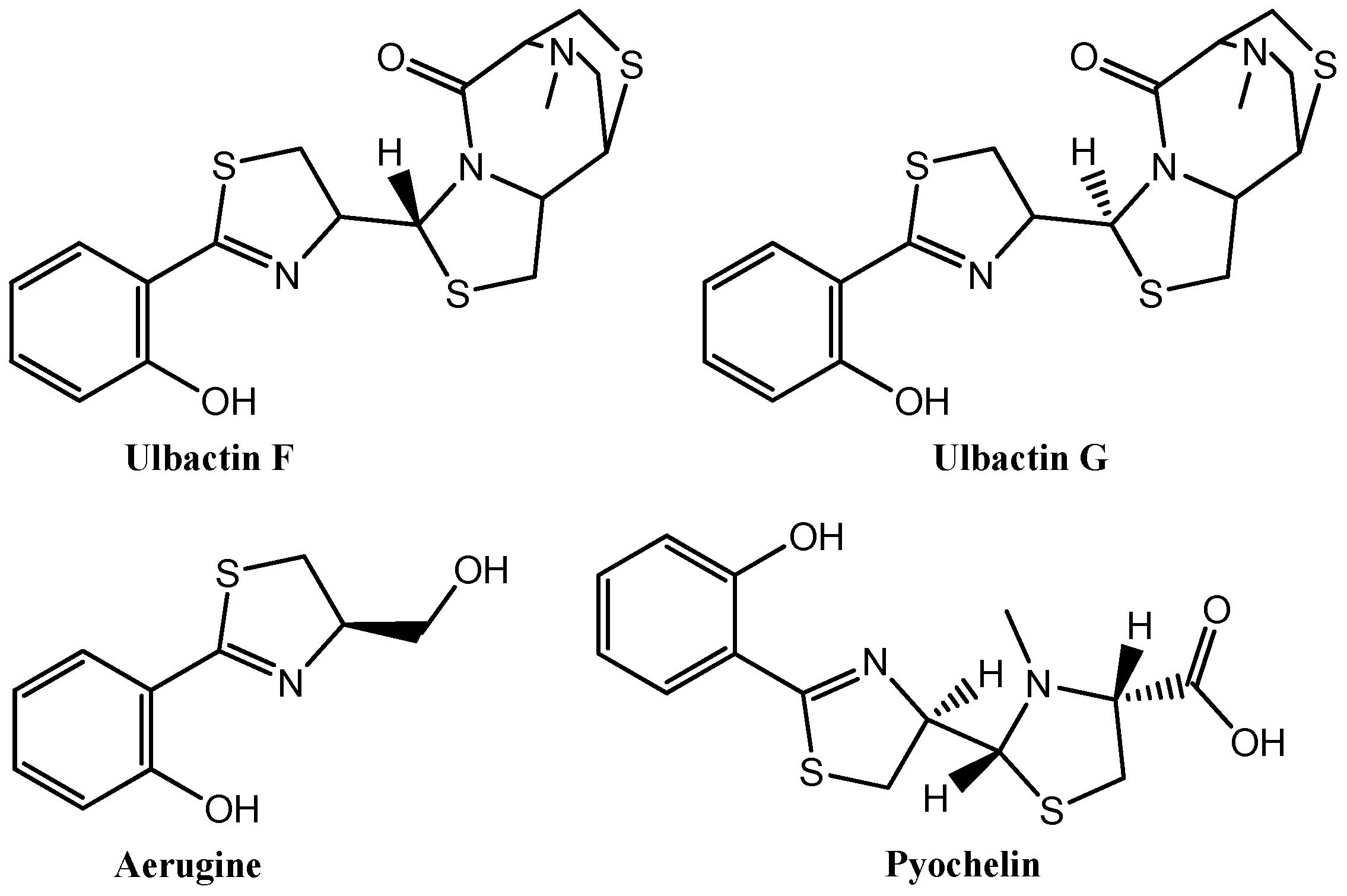
  12. Igarashi, Y.; Asano, D.; Sawamura, M.; In, Y.; Ishida, T.; Imoto, M. Ulbactins F and G, polycyclic thiazoline derivatives with tumor cell migration inhibitory activity from Brevibacillus sp. Org. Lett. 2016, 18, 1658–1661. [Google Scholar] [CrossRef] [PubMed]
  13. Marson, C.M.; Matthews, C.J.; Yiannaki, E.; Atkinson, S.J.; Soden, P.E.; Shukla, L.; Lamadema, N.; Thomas, N.S. Discovery of potent, isoform-selective inhibitors of histone deacetylase containing chiral heterocyclic capping groups and a N-(2-aminophenyl)benzamide binding unit. J. Med. Chem. 2013, 56, 6156–6174. [Google Scholar] [CrossRef] [PubMed]
  14. Patil, P.A.; Amnerkar, N.D.; Pathare, S.S.; Bhusari, K.P. Docking study of p-hydroxybenzohydrazide derivatives as tyrosine kinase inhibitors and anticancer agents. J. Comput. Methods Mol. Des. 2015, 5, 109–114. [Google Scholar]
  15. Wang, W.; Zhao, B.; Xu, C.; Wu, W. Synthesis and antitumor activity of the thiazoline and thiazine multithioether. Int. J. Org. Chem. 2012, 2, 117–120. [Google Scholar] [CrossRef]
  16. Sondhi, S.M.; Johar, M.; Singh, N.; Dastidar, S.G. Synthesis of biscoupled hemin-thiazoline derivatives and their anticancer activity evaluation. Indian J. Chem. 2004, 43B, 162–167. [Google Scholar] [CrossRef]
  17. Mahani, N.M.; Sabermahani, F.; Jahani, P.M.; Jalali, N. A density function theory based quantitative structure activity relationships study of thiazoline derivatives as anticancer agents. Iran. J. Anal. Chem. 2015, 2, 70–76. [Google Scholar]
  18. Taher, A.T.; Khalil, N.A.; Ahmed, E.M. Synthesis of novel isatin-thiazoline and isatin-benzimidazole conjugates as anti-breast cancer agents. Arch. Pharm. Res. 2011, 34, 1615–1621. [Google Scholar] [CrossRef] [PubMed]
  19. Miller, D.D.; Dalton, J.T.; Gududuru, V.; Hurh, E. Thiazoline analogs as cell proliferation inhibitors. Chem. Abstr. 2005, 143, 326352. [Google Scholar]
  20. Ng, R.A.; Sui, Z. Preparation of thiazoline derivatives as selective androgen receptor modulators (SARMs). Chem. Abstr. 2005, 142, 74557. [Google Scholar]
  21. Pine, M.J.; Mirand, E.A.; Ambrus, J.L.; Bock, F.G. Antitumor studies of 2- amino-2-thiazoline and other tumor-modifying agents. J. Med. 1983, 14, 433–449. [Google Scholar] [CrossRef]
  22. Randazzo, A.; Bifulco, G.; Giannini, C.; Bucci, M.; Debitus, C.; Cirino, G.; Gomez-Paloma, L. Halipeptins A and B: Two novel potent anti-inflammatory cyclic depsipeptides from the Vanuatu marine sponge Haliclona species. J. Am. Chem. Soc. 2001, 123, 10870–10876. [Google Scholar] [CrossRef] [PubMed]
  23. Pérez-Perarnau, A.; Preciado, S.; Palmeri, C.M.; Moncunill-Massaguer, C.; Iglesias-Serret, D.; González-Gironès, D.M.; Miguel, M.; Karasawa, S.; Sakamoto, S.; Cosialls, A.M.; et al. A trifluorinated thiazoline scaffold leading to pro-apoptotic agents targeting prohibitins. Angew. Chem. Int. Ed. 2014, 53, 10150–10154. [Google Scholar] [CrossRef] [PubMed]
  24. Shih, M.H.; Ke, F.Y. Syntheses and evaluation of antioxidant activity of sydnonyl substituted thiazolidinone and thiazoline derivatives. Bioorg. Med. Chem. 2004, 12, 4633–4643. [Google Scholar] [CrossRef] [PubMed]
  25. Monneret, C. Recent developments in the field of antitumour anthracyclines. Eur. J. Med. Chem. 2001, 36, 483–493. [Google Scholar] [PubMed]
  26. Cragg, G.M.; Newman, D.J. Plants as a source of anti-cancer agents. J. Ethnopharmacol. 2005, 100, 72–79. [Google Scholar] [CrossRef] [PubMed]
  27. Kusari, S.; Lamshöft, M.; Spiteller, M. Aspergillus fumigatus Fresenius, an endophytic fungus from Juniperus communis L. Horstmann as a novel source of the anticancer pro-drug deoxypodophyllotoxin. J. Appl. Microbiol. 2009, 107, 1019–1030. [Google Scholar] [CrossRef] [PubMed]
  28. Chaitramallu, M.; Devaraju, P. Synthesis of organic nanoparticle of aryl tetralin compounds and study of their biological activities for targeted delivery system. Indian J. Adv. Chem. Sci. 2014, 2, 61–63. [Google Scholar]
  29. Chen, H.; Zuo, S.; Wang, X.; Tang, X.; Zhao, M.; Lu, Y.; Chen, L.; Liu, J.; Liu, Y.; Liu, D.; et al. Synthesis of 4b-triazole-podophyllotoxin derivatives by azideealkyne cycloaddition and biological evaluation as potential antitumor agents. Eur. J. Med. Chem. 2011, 46, 4709–4714. [Google Scholar] [PubMed]
  30. Hamdy, N.A.; Gamel-Eldeen, A.M.; Abdel-Aziz, H.A.; El-Hussieny, A.A.; Fakhr, I.A. Modulation of carcinogen metabolizing enzymes by new fused heterocycles pendant to 5,6,7,8-tetrahydronaphthalene derivatives. Eur. J. Med. Chem. 2010, 45, 463–470. [Google Scholar] [CrossRef] [PubMed]
  31. Dong, Y.; Shi, Q.; Nakagawa-Goto, K.; Wua, P.; Bastow, K.F.; Morris-Natschke, S.L.; Lee, K. Antitumor agents 269. Non-aromatic ring-A neotanshinlactone analog, TNO, asa new class of potent antitumor agents. Bioorg. Med. Chem. Lett. 2009, 19, 6289–6292. [Google Scholar] [CrossRef] [PubMed]
  32. Gamal-Eldeen, A.M.; Hamdy, N.A.; Abdel-Aziz, H.A.; El-Hussieny, A.A.; Fakhr, I.A. Induction of intrinsic apoptosis pathway in colon cancer HCT-116 cells by novel 2-substituted-5,6,7,8-tetrahydronaphthalene derivatives. Eur. J. Med. Chem. 2014, 77, 323–333. [Google Scholar] [CrossRef] [PubMed]
  33. Hamdy, N.A.; Anwar, M.M.; Abu-Zied, K.M.; Awad, H.M. Synthesis, tumor inhibitory and antioxidant activity of new polyfuncctionnaly 2-substituted-5,6,7,8-tetrahydronaphthalene derivatives containing pyridine, thioxopyridine and pyrazolopyridine moieties. Acta Pol. Pharm. 2013, 70, 987–1001. [Google Scholar] [PubMed]
  34. Gurkan-Alp, A.S.; Mumcuoglu, M.; Andac, C.A.; Dayanc, E.; Cetin-Atalay, R.; Buyukbingol, E. Synthesis, anticancer activities and molecular modeling studies of novel indole retinoid derivatives. Eur. J. Med. Chem. 2012, 58, 346–354. [Google Scholar] [CrossRef] [PubMed]
  35. Al-Abdullah, E.S. Synthesis and anticancer activity of some novel tetralin-6-yl-pyrazoline, 2-thioxopyrimidine, 2-oxopyridine, 2-thioxo-pyridine and 2-iminopyridine derivatives. Molecules 2011, 16, 3410–3419. [Google Scholar] [CrossRef] [PubMed]
  36. Ebeid, M.Y.; El Zahar, M.I.; Kamel, M.M.; Omar, M.T.; Anwar, M.M. Novel 5,6,7,8-tetrahydronaphth-2-yl heterocycles of possible biological activity. Egypt. Pharmaceut. J. 2004, 3, 49–65. [Google Scholar]
  37. Gouhar, R.S.; Raafat, E. Synthesis of (3-(naphthalen-1-yl)oxiran-2-yl)(5,6,7,8 tetrahydronaphthalen-2-yl)methanone and reaction with some nucleophiles for anticancer evaluation. Res. Chem. Intermed. 2015, 41, 5529–5543. [Google Scholar] [CrossRef]
  38. Amin, K.M.; El-Zahar, M.I.; Anwar, M.M.; Kamel, M.M.; Mohamed, M.H. Synthesis and anticancer actıvıty of novel tetralin-6-ylpyridine and tetralin-6-ylpyrimidine derivatives. Acta Pol. Pharm. 2009, 66, 279–291. [Google Scholar] [PubMed]
  39. Ali, M.M.; Hassan, S.A. Role of some newly synthesized tetrahydronaphthalenthiazol derivatives as anticancer compounds. Int. J. Cancer Res. 2007, 3, 103–110. [Google Scholar]
  40. Solimana, E.A.; El-Zahar, M.I.; El-Masry, A.H.; Kamel, M.; Gohar, R.S. Synthesis and anticancer evaluation of novel tetrahydronaphthalen-6-ylthiazole heterocycles against human HePG2 and MCF7 cell lines. Der Pharm. Chem. 2010, 2, 507–521. [Google Scholar]
  41. Čolović, M.B.; Krstić, D.Z.; Lazarević-Pašti, T.D.; Bondžić, A.M.; Vasić, V.M. Acetylcholinesterase inhibitors: Pharmacology and toxicology. Curr. Neuropharmacol. 2013, 11, 315–335. [Google Scholar] [CrossRef] [PubMed]
  42. Zovko, A.; Sepcic, K.; Turk, T.; Faimali, M.; Garaventa, F.; Chelossi, E.; Paleari, L.; Falugi, C.; Aluigi, M.G.; Angelini, C.; et al. New aspects of the relationship between acethylcholinesterase activity and cancer I: Poly-Aps experiments. WSEAS Trans. Biol. Biomed. 2009, 6, 58–69. [Google Scholar]
  43. Ciftci, G.A.; Temel, H.E.; Yildirim, S.U.; Kaplancikli, Z.A.; Altintop, M.D.; Genc, L. Apoptotic effects of some carbazole derivatives on lung carcinoma and glioma cell lines. Med. Chem. Res. 2013, 22, 3751–3759. [Google Scholar] [CrossRef]
  44. Turan-Zitouni, G.; Ozdemir, A.; Kaplancikli, Z.A.; Revial, G.; Demirci, F. Synthesis and antimicrobial activity evaluation of new hydrazide derivatives. Turk. J. Pharm. Sci. 2011, 8, 199–206. [Google Scholar]
  45. Turan-Zitouni, G.; Kaplancıklı, Z.A.; Erol, K.; Kılıç, F.S. Synthesis and analgesic activity of some triazoles and triazolothiazines. II Farmaco 1999, 54, 218–223. [Google Scholar] [CrossRef]
  46. Berridge, M.V.; Herst, P.M.; Tan, A.S. Tetrazolium dyes as tools in cell biology: New insights into their cellular reduction. Biotechnol. Annu. Rev. 2005, 11, 127–152. [Google Scholar] [PubMed]
  47. Mossmann, T. Rapid colorimetric assay for cellular growth and survival: Application to proliferation and cytotoxicity assays. J. Immunol. Methods 1983, 65, 55–63. [Google Scholar] [CrossRef]
  48. Maghni, K.; Nicolescu, O.M.; Martin, J.G. Suitability of cell metabolic colorimetric assays for assessment of CD4+ T cell proliferation: Comparison to 5-bromo-2-deoxyuridine (BrdU) ELISA. J. Immunol. Methods 1999, 223, 185. [Google Scholar] [CrossRef]
  49. Lowe, S.W.; Lin, A.W. Apoptosis in cancer. Carcinogenesis 2000, 21, 485–495. [Google Scholar] [CrossRef] [PubMed]
  50. Ellman, G.L.; Courtney, K.D.; Andres, V., Jr.; Feather-Stone, R.M. A new and rapid colorimetric determination of acetylcholinesterase activity. Biochem. Pharmacol. 1961, 7, 88–95. [Google Scholar] [CrossRef]
Sample Availability: Samples of the compounds 4a4k are available from the authors.
Figure 1. Some thiazoline containing anticancer molecules.
Molecules 23 00135 g001
Figure 2. Tetralin including anticancer drugs.
Molecules 23 00135 g002
Scheme 1. The synthesis of the compounds (4a4k). Reactants and reagents: i: ClCH2COOEt, 3–5 h, reflux; ii: NH2NH2·H2O, EtOH, 3 h, reflux; iii: C6H5NCS/C6H11NCS, EtOH, 2 h, reflux; iv: phenacyl bromide derivative, EtOH, 5 h, reflux.
Molecules 23 00135 sch001
Figure 3. DNA synthesis inhibitory activity of compounds 4b, 4d, 4e, 4k and cisplatin on MCF-7 cells. Mean percent absorbance of untreated control cells were assumed 0%. In addition, two different concentrations (1: IC50/2; 2: IC50 µM) of test compounds and cisplatin were given. Data points represent means for three independent wells ± SD. p < 0.05.
Molecules 23 00135 g003
Figure 4. DNA synthesis inhibitory activity of compounds 4a, 4b, 4f, 4g, 4h, 4j and cisplatin on A549 cells. Mean percent absorbance of untreated control cells were assumed 0%. In addition, two different concentrations (1: IC50/2; 2: IC50 µM) of test compounds and cisplatin were given. Data points represent means for three independent wells ± SD. p < 0.05.
Molecules 23 00135 g004
Figure 5. Flow cytometric diagrams of MCF-7 cells treated with compounds 4a, 4d, 4e, 4k, and cisplatin.
Molecules 23 00135 g005
Figure 6. Flow cytometric diagrams of A549 cells treated with compounds 4a, 4b, 4f, 4g, 4h, 4j and cisplatin.
Molecules 23 00135 g006
Table 1. The structures of the compounds (4a4k).
Molecules 23 00135 i001
CompoundsR1R2R3R4
4aCyclohexylHHH
4bCyclohexylHHOCH3
4cPhenylHHH
4dPhenylHHCH3
4ePhenylHHOCH3
4fPhenylHHBr
4gPhenylHHCl
4hPhenylHHF
4iPhenylHNO2H
4jPhenylHHNO2
4kPhenylClClH
Table 2. IC50 values (µM) of the compounds and SI values against MCF-7, A549 and NIH/3T3 cell lines.
CompoundsMCF-7A549NIH/3T3SIMCF-7SIA549
4a>1000157.6 ± 14.8206.1 ± 15.30.21.3
4b69.2 ± 5.8137.5 ± 7.255.7 ± 1.20.80.4
4c296.7 ± 46.6>1000>10003.4
4d71.8 ± 12.5>1000>100013.9
4e91.4 ± 9.8707.9 ± 83.3>100010.91.4
4f280.9 ± 56.254.3 ± 6.8146.7 ± 5.40.52.7
4g483.0 ± 31.228.6 ± 4.1224.5 ± 20.50.57.8
4h243.1 ± 14.948.6 ± 6.0246.7 ± 12.21.05.1
4i>1000>1000>1000
4j>100049.0 ± 1.41>100020.4
4k152.7 ± 27.5>1000165.4 ± 11.01.1
Cisplatin100.0 ± 3.3108.3 ± 11.8-
-: not tested; …:not calculated.
Table 3. Percents of typical quadrant analysis of Annexin V FITC/Propidium Iodide flow cytometry of MCF-7 and A549 cells treated with compounds and Cisplatin.
Groups% Early Apoptotic% Late Apoptotic% Viable% Necrotic
MCF-7 cell lineControl1.74.384.99.1
Compound 4b0.729.16.363.8
Compound 4d1.112.150.736.1
Compound 4e0.831.724.839.1
Compound 4k2.48.954.334.4
Cisplatin2.827.142.028.1
A549 cell lineControl3.16.189.01.8
Compound 4a7.448.331.512.8
Compound 4b6.667.010.915.6
Compound 4f16.529.347.76.5
Compound 4g18.726.049.45.8
Compound 4h23.726.445.94.0
Compound 4j11.230.347.011.4
Cisplatin14.925.353.16.7
Table 4. % AChE and BuChE inhibitions of the compounds at 80 µg/mL concentration.
CompoundsAChE
% Inhibition (80 µg/mL)
BuChE
% Inhibition (80 µg/mL)
4a38.26 ± 2.1828 ± 1.88
4b11.08 ± 1.4815.89 ± 1.72
4c19.30 ± 1.8716.48 ± 0.08
4d43.22 ± 1.5633.19 ± 2.38
4e10.81 ± 0.94---
4f42.87 ± 2.84---
4g24.37 ± 1.20---
4h49.92 ± 1.44---
4i41.85 ± 0.23---
4j44.96 ± 1.60---
4k15.19 ± 1.99---
Donepezil IC50 (µM)(11.8 ± 0.7) × 10−34.85 ± 0.55
---: no inhibition.

