Single-Labeled Oligonucleotides Showing Fluorescence Changes upon Hybridization with Target Nucleic Acids


Abstract
: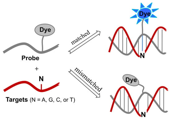
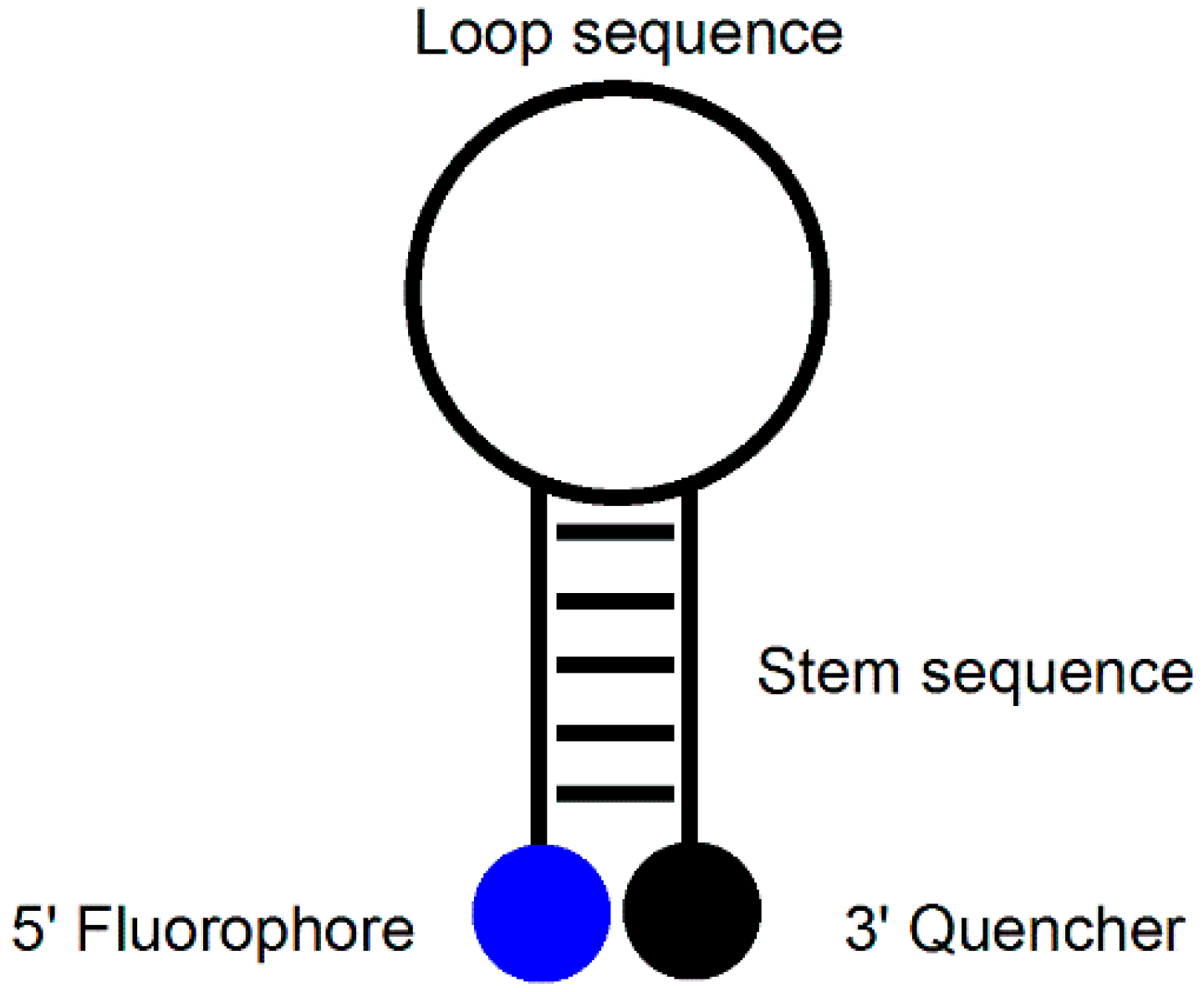
1. Introduction
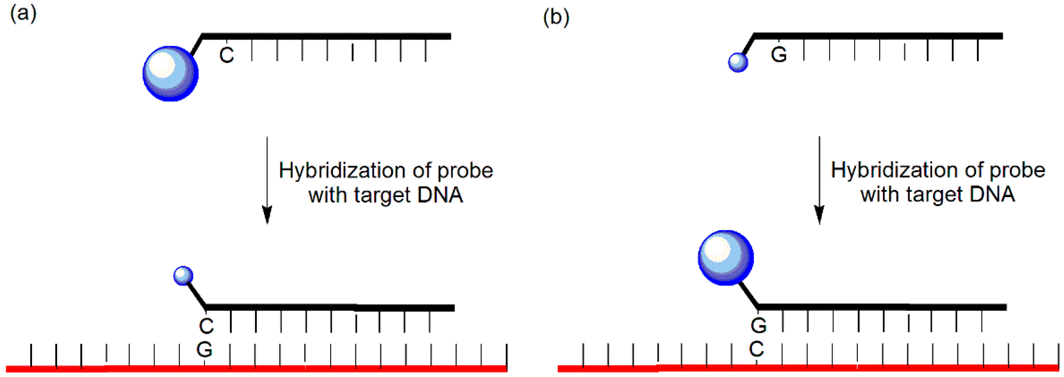
2. Guanine-Quenching Probes
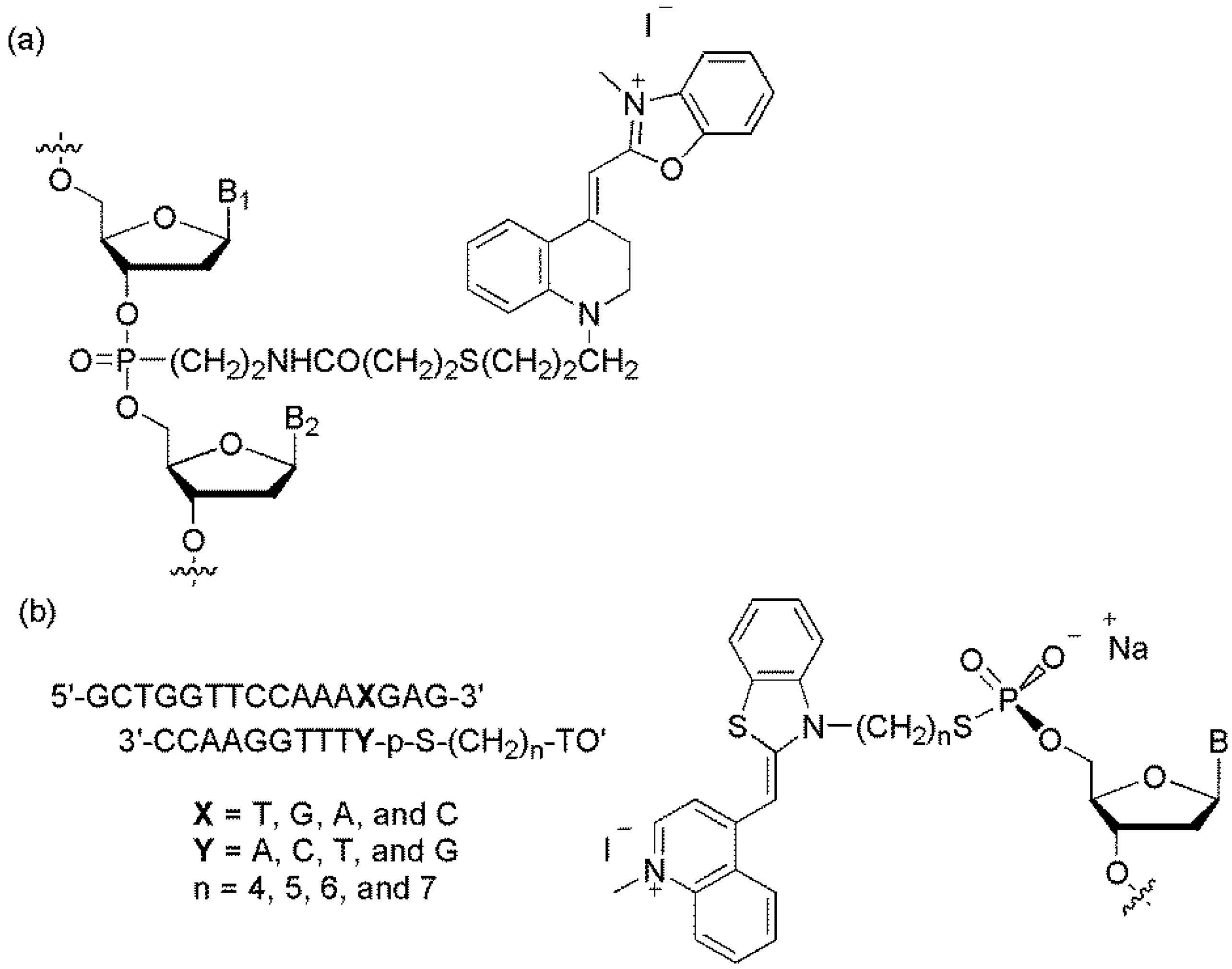
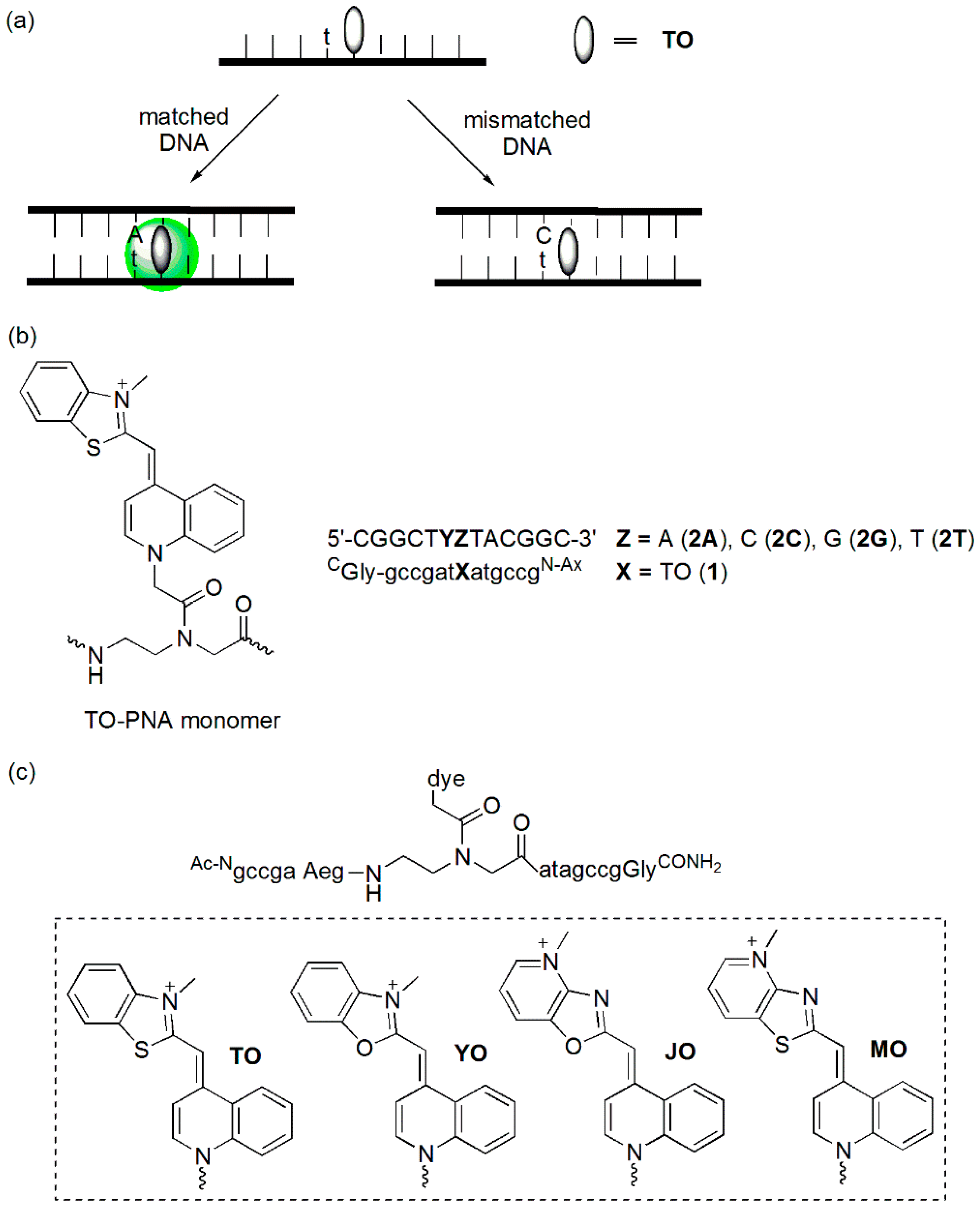
3. Cyanine-Containing Probes
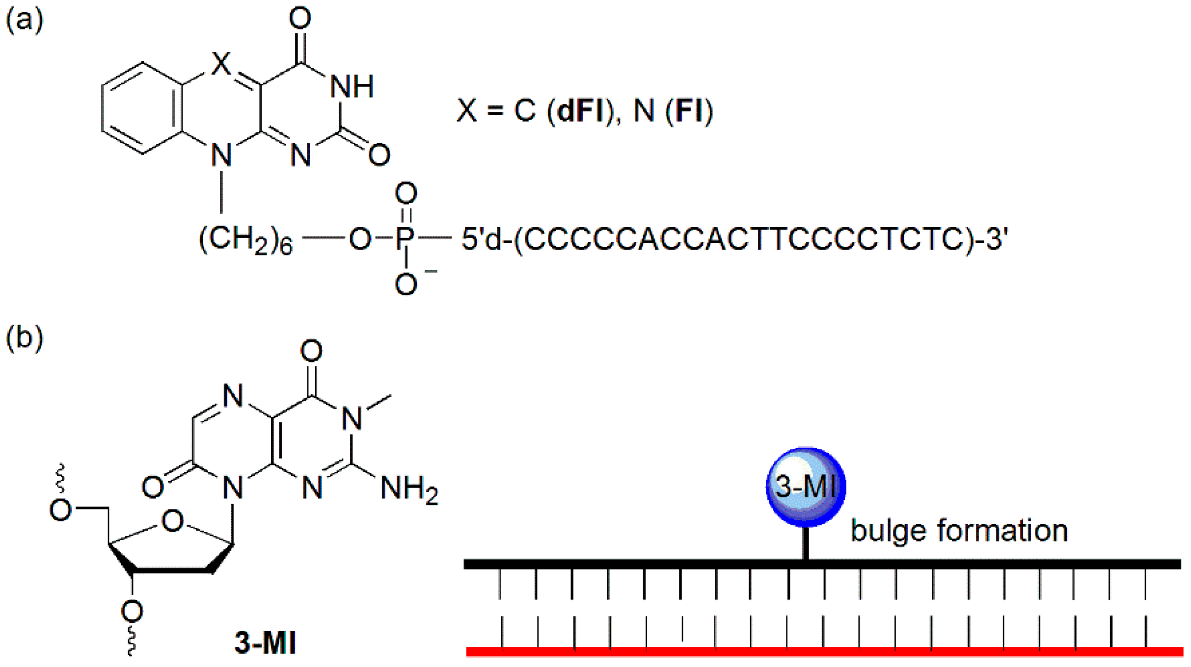
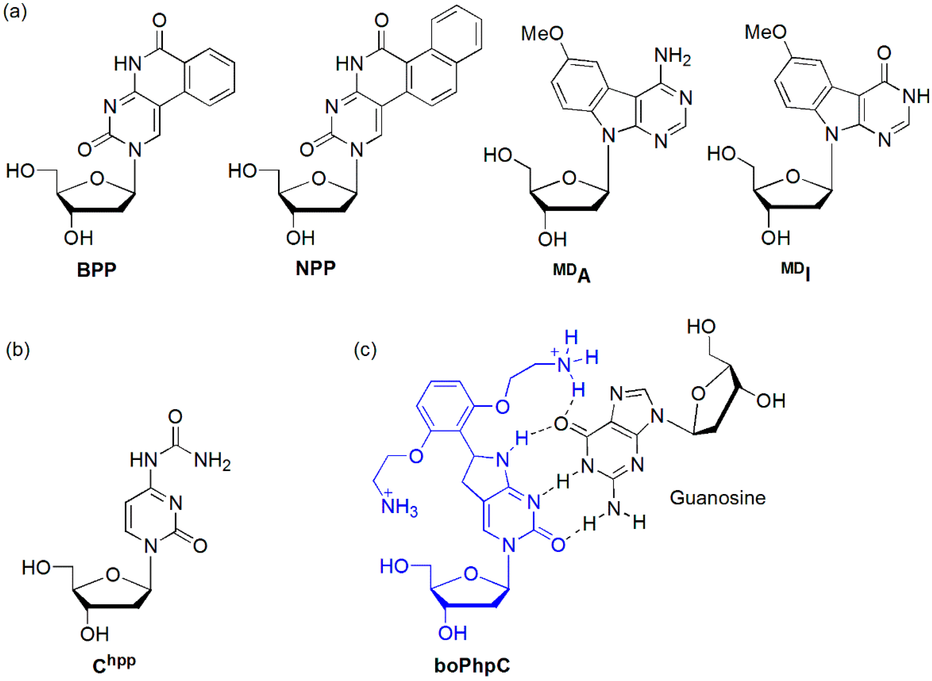
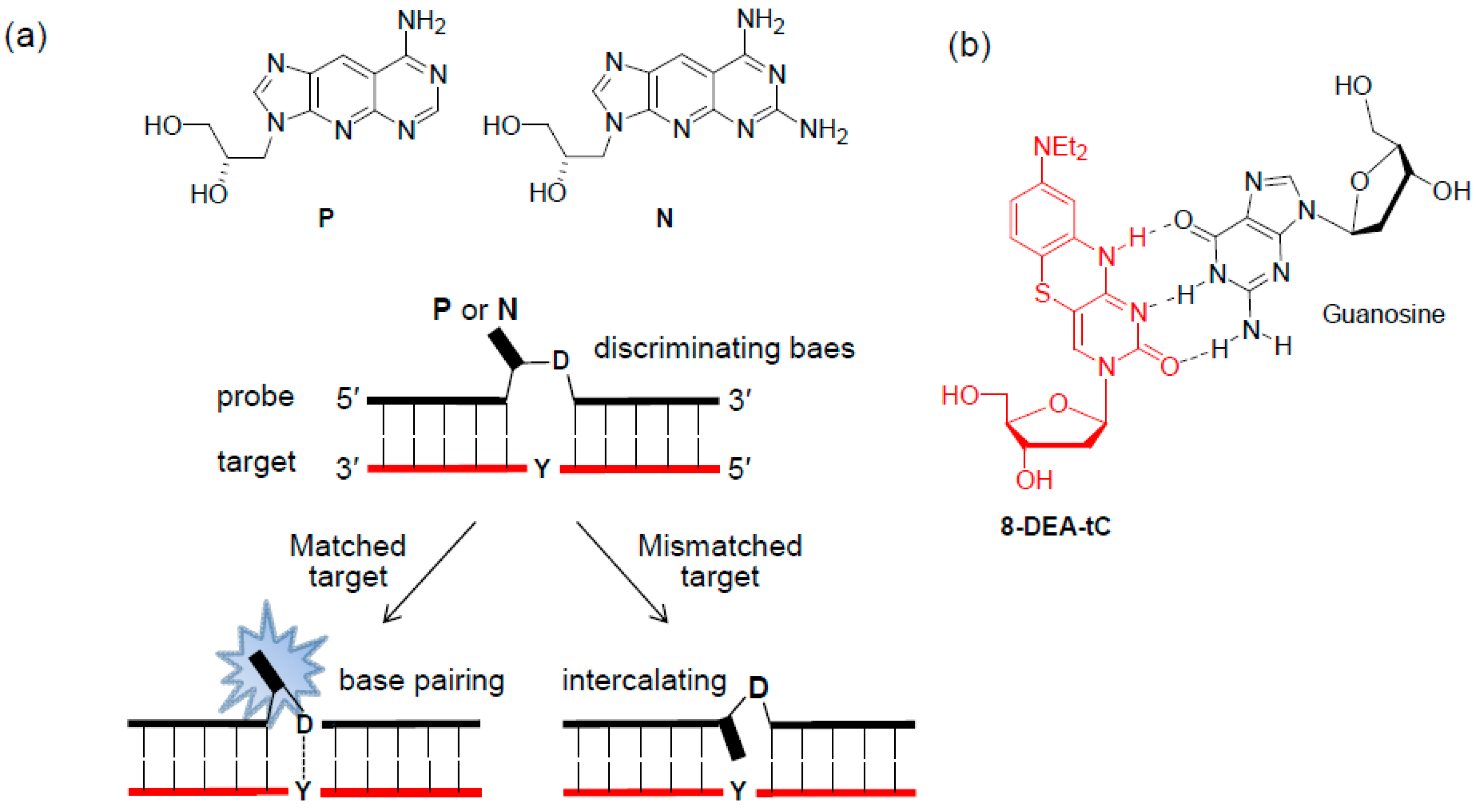
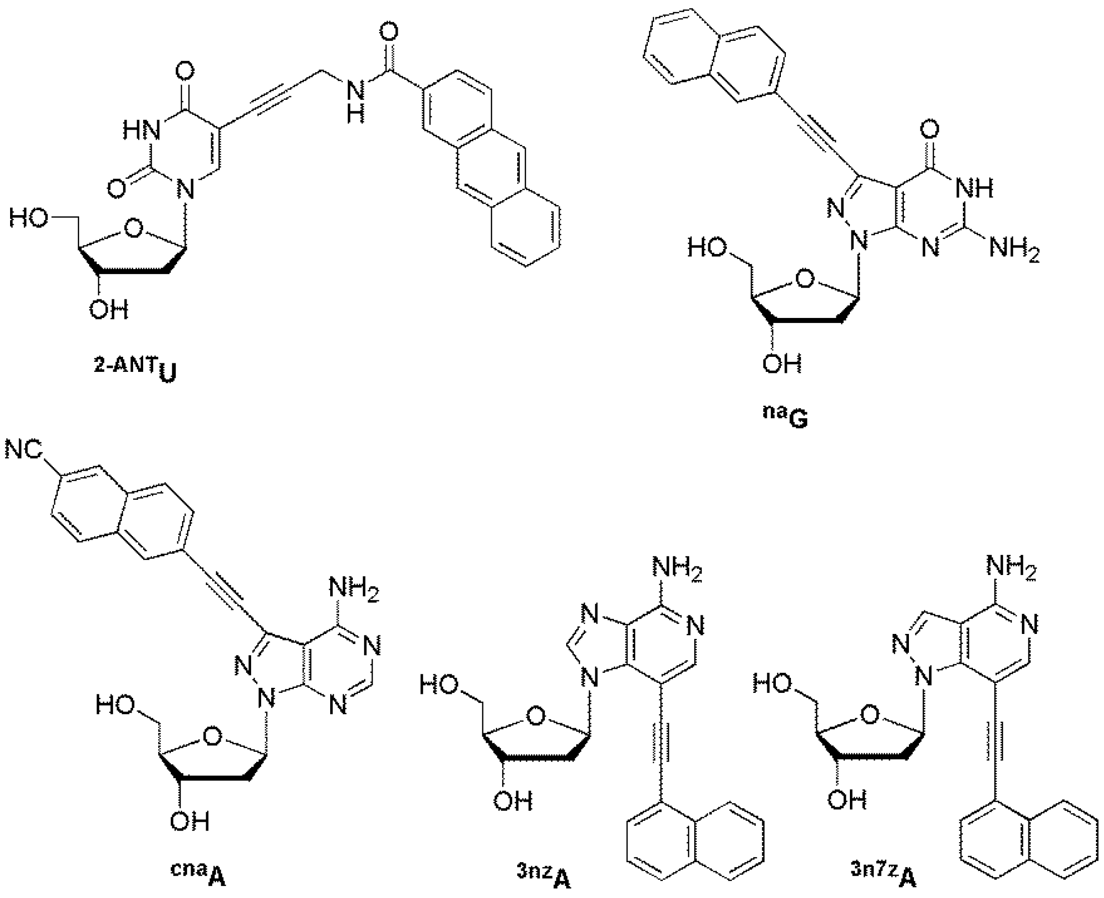
4. Probes Containing a Fluorescent Nucleobase Analog
5. Probes Containing a Fluorophore-Labeled Base
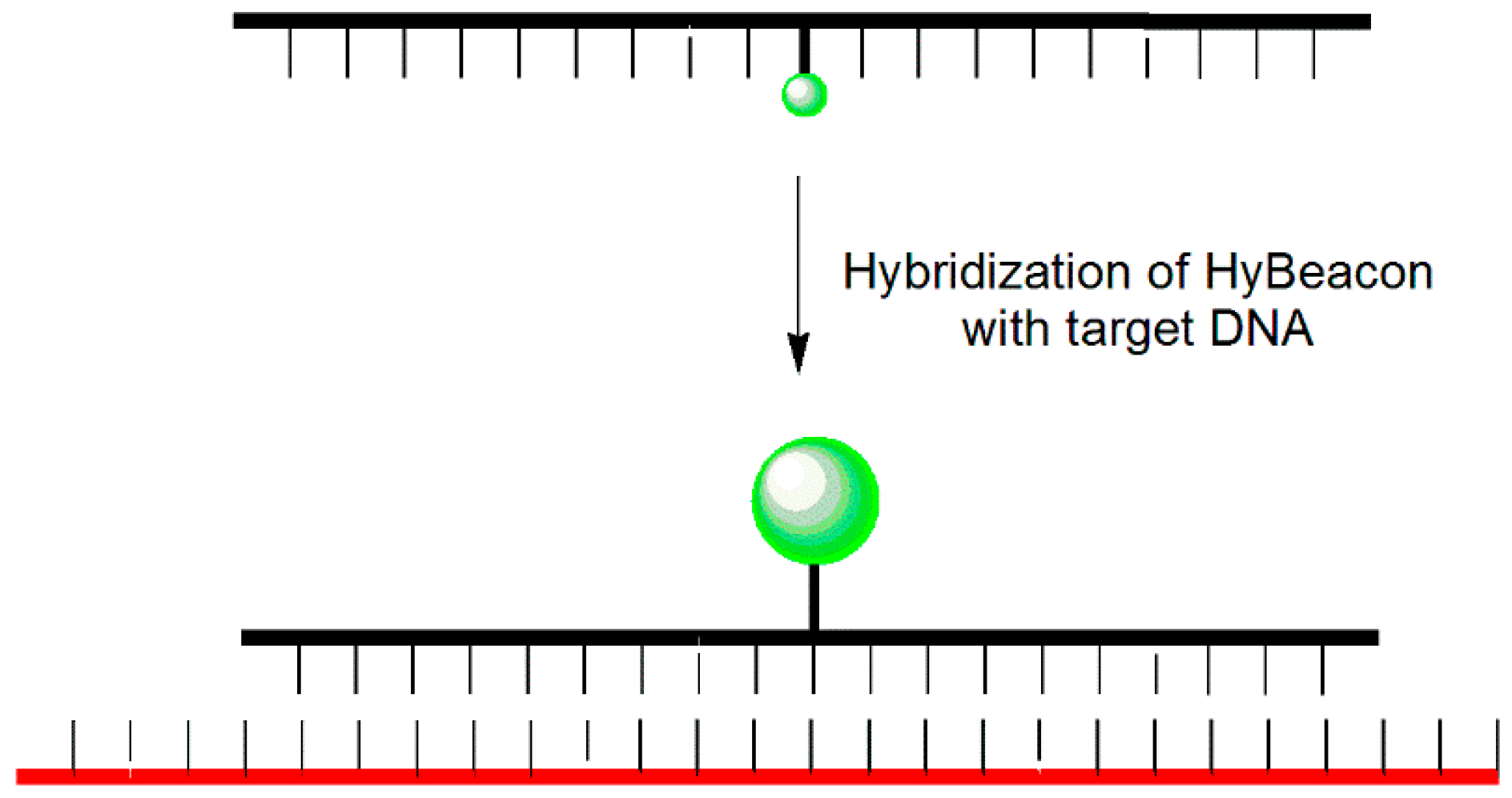
5.1. HyBeacon Probes
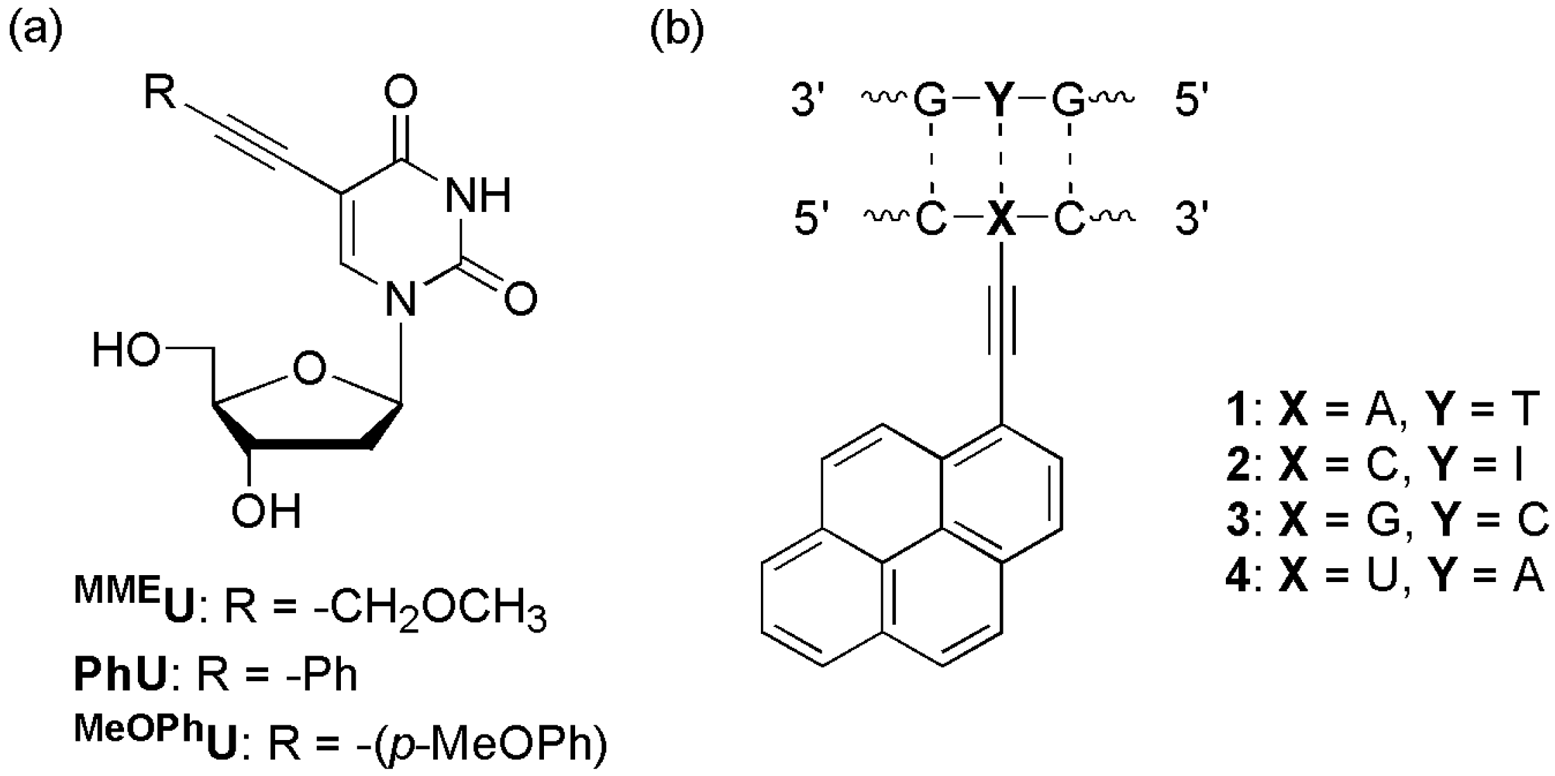
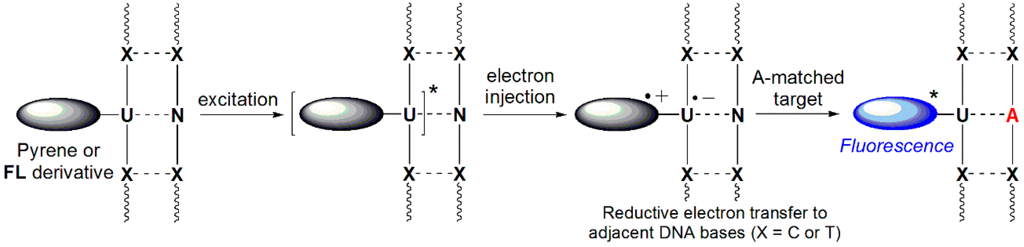
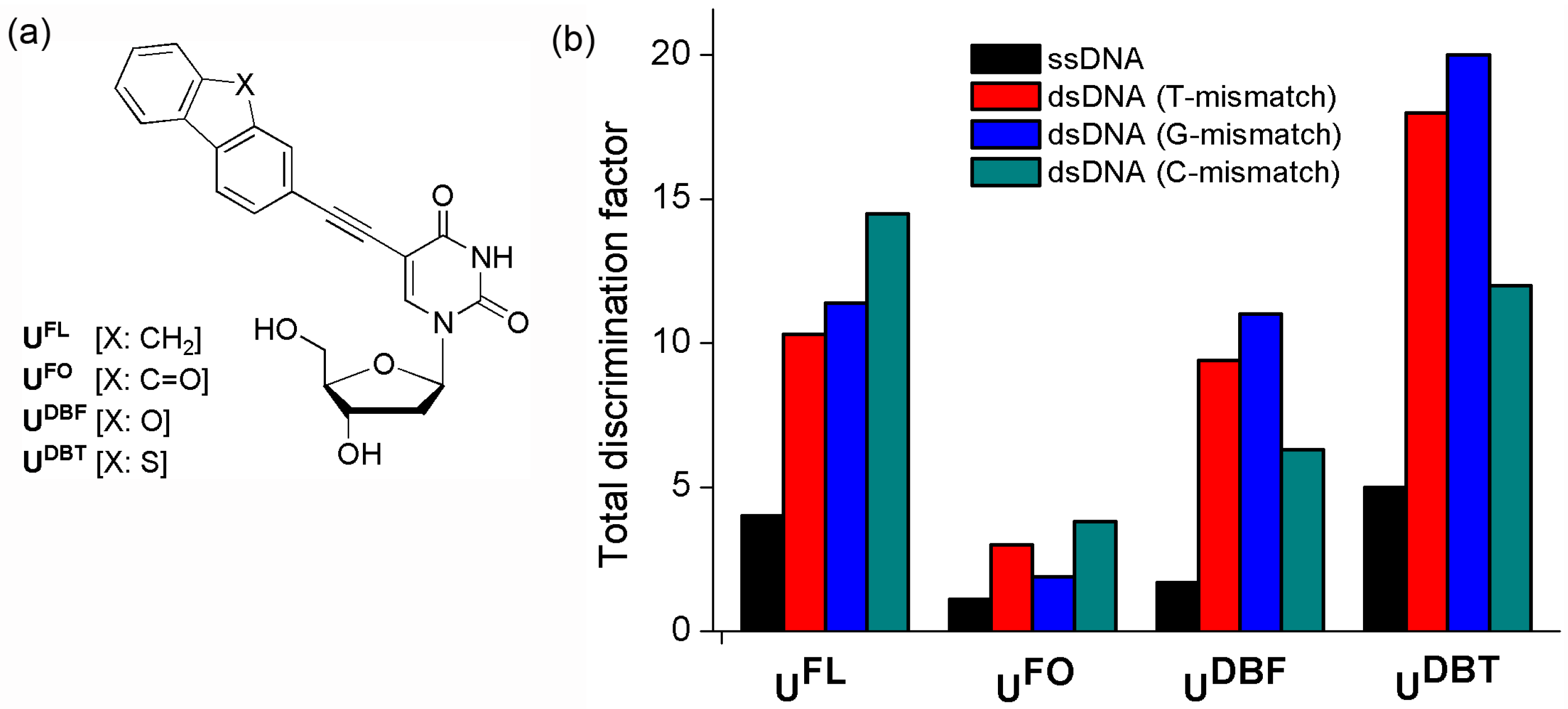
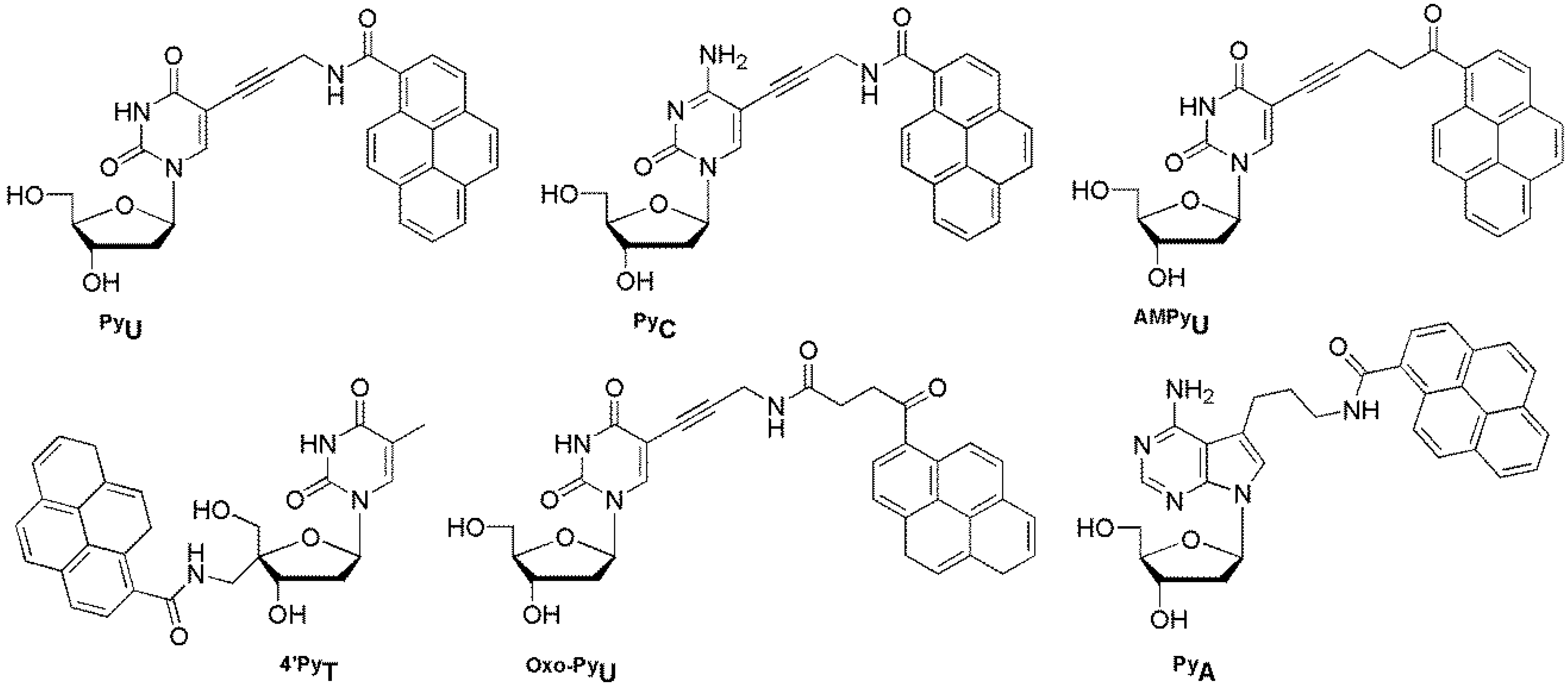
5.2. Probes Containing a Nucleobase-Labeled Fluorophore with an Acetylene Group
5.3. BDF Probes
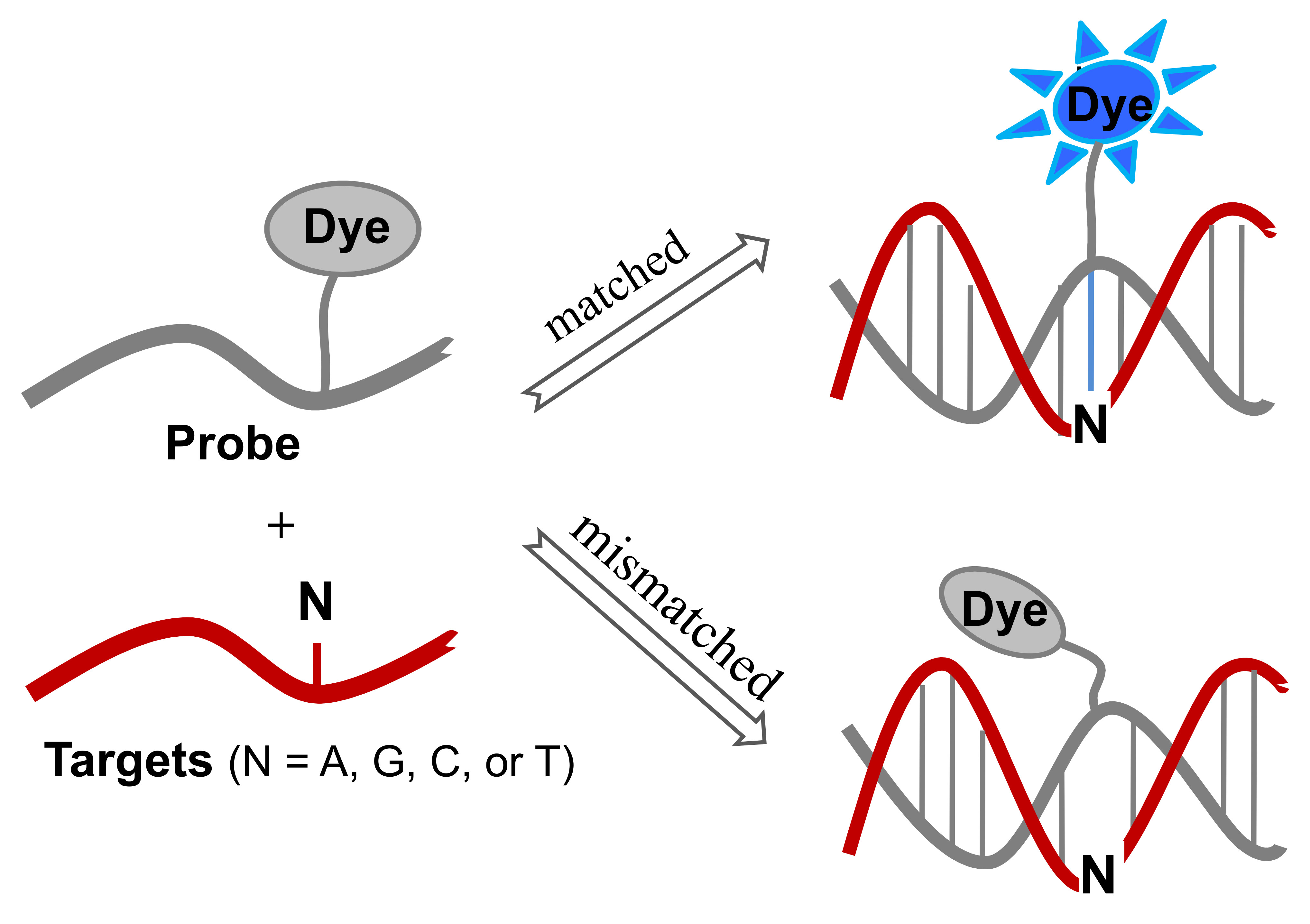
6. Microenvironment-Sensitive Probes
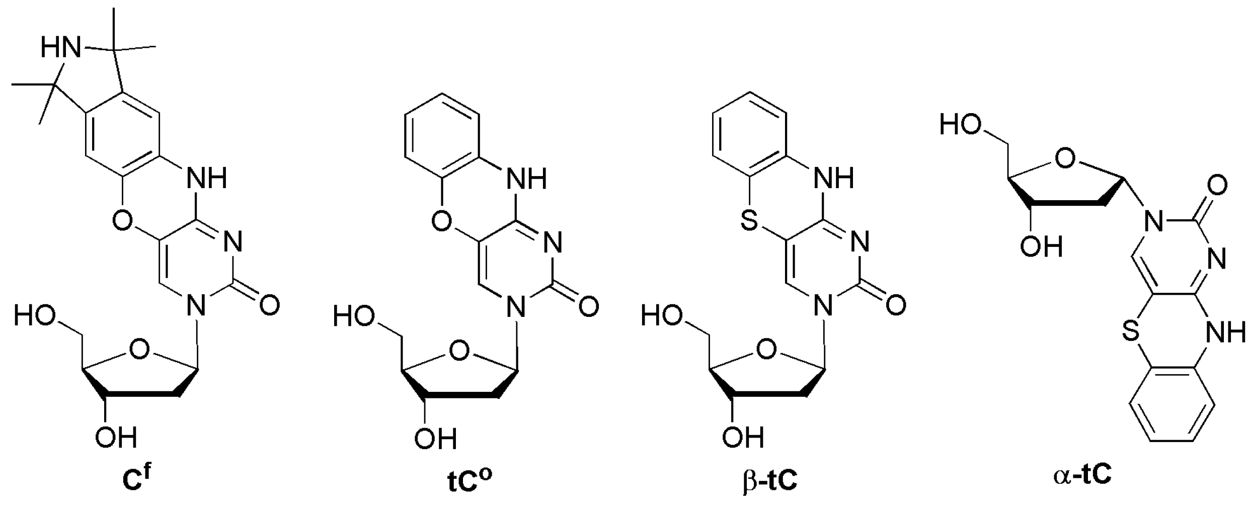
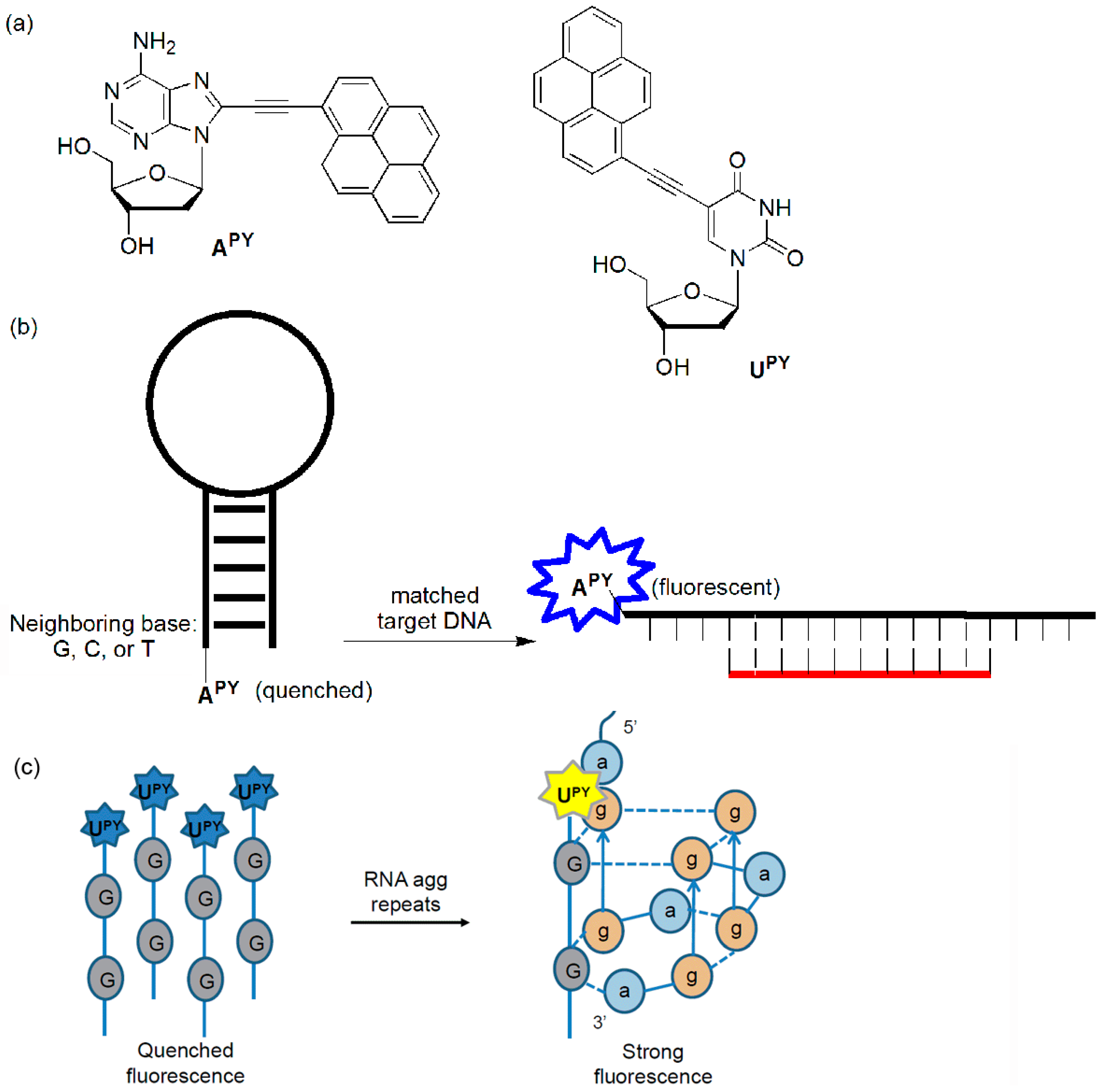
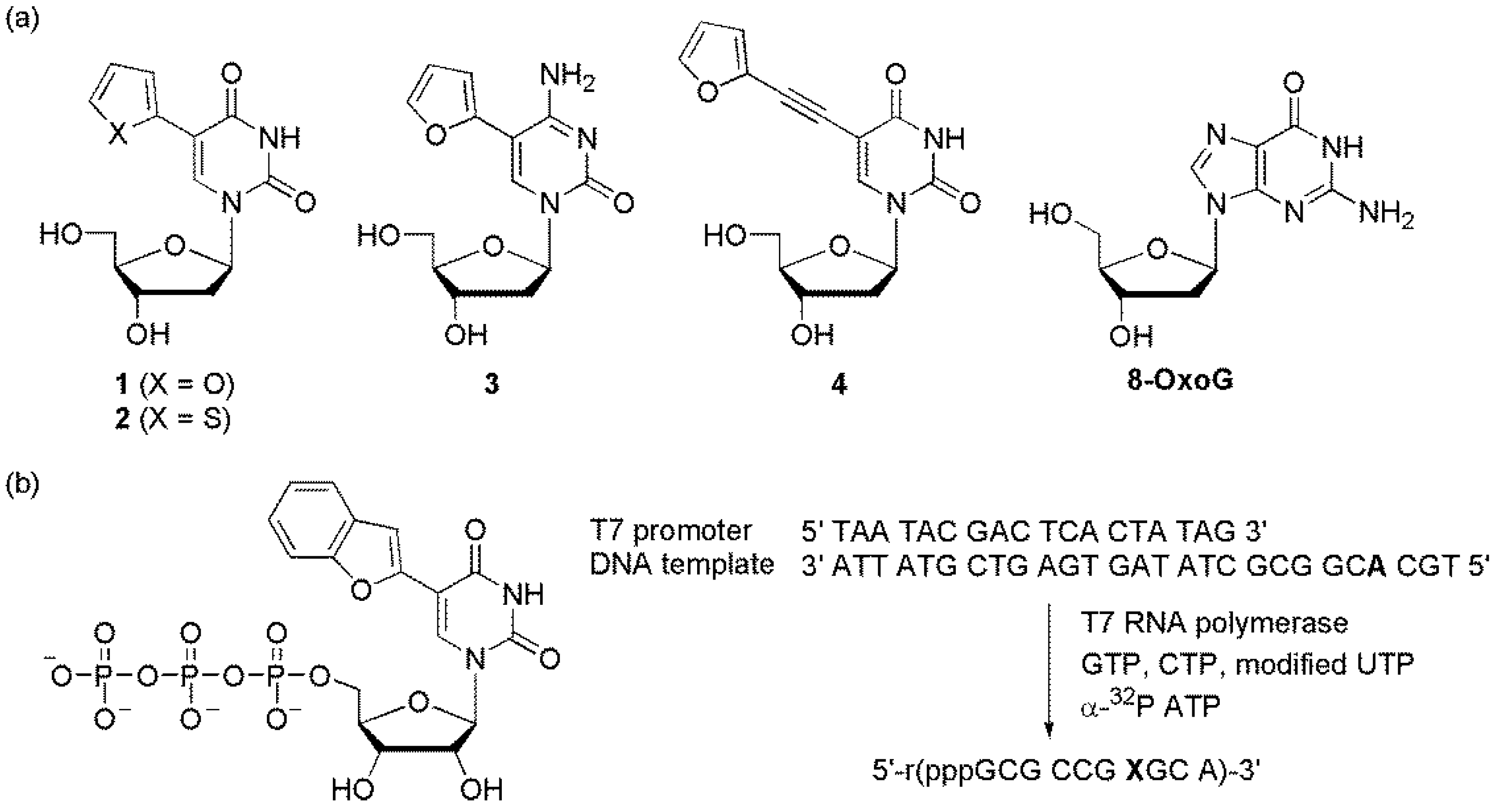
6.1. Probes Containing a Heterocycle-Conjugated Pyrimidine
6.2. ESF Probes
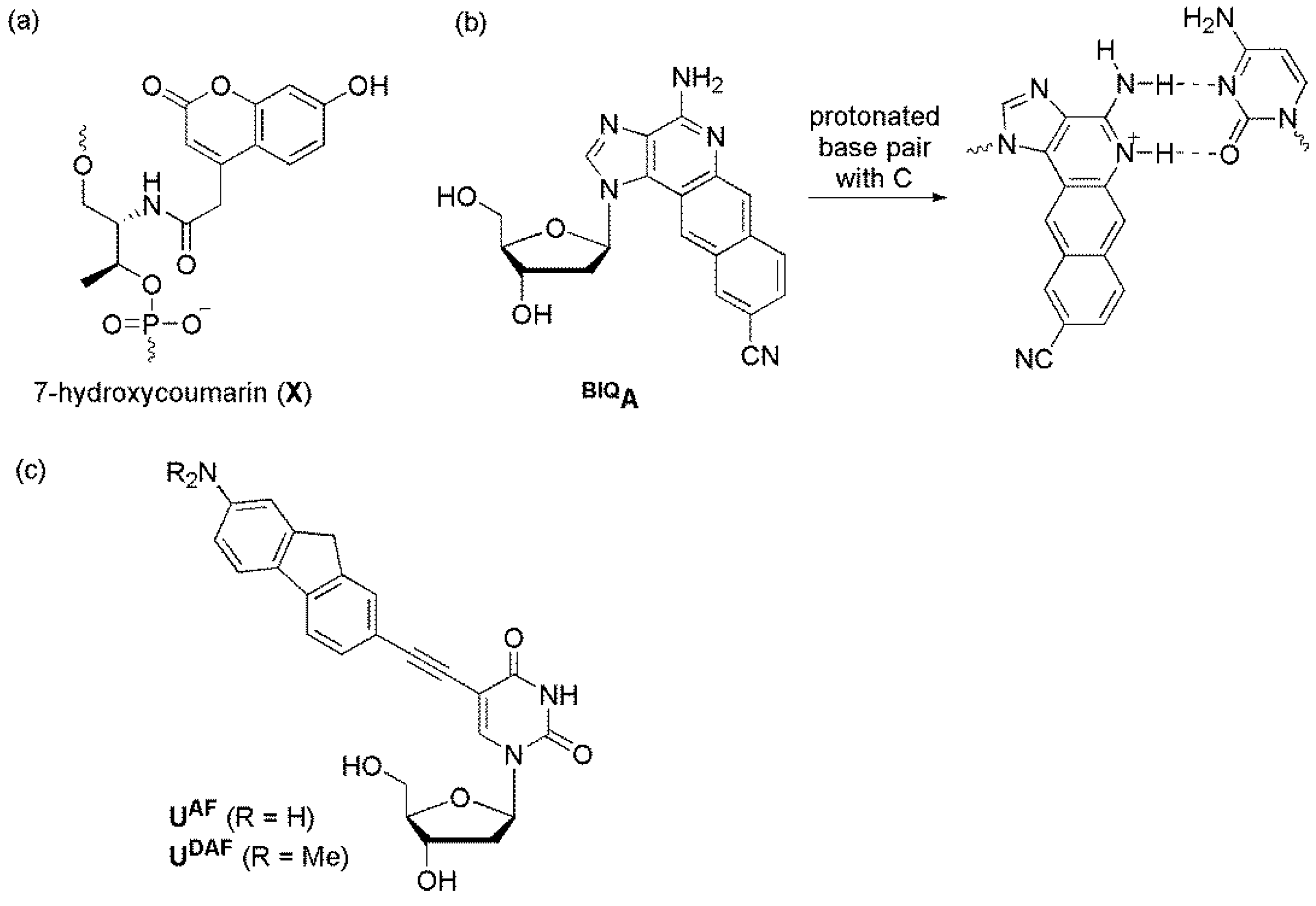
6.3. pH-Sensitive Probes
7. Conclusions
Supplementary Materials
Supplementary File 1Acknowledgments
Conflicts of Interest
References
- Vignal, A.; Milan, D.; SanCristobal, M.; Eggen, A. A review on SNP and other types of molecular markers and their use in animal genetics. Genet. Sel. Evol. 2002, 34, 275–305. [Google Scholar] [CrossRef] [PubMed]
- Sobrino, B.; Brion, M.; Carracedo, A. SNPs in forensic genetics: A review on SNP typing methodologies. Forensic Sci. Int. 2005, 154, 181–194. [Google Scholar] [CrossRef] [PubMed]
- Venkatesan, N.; Seo, Y.J.; Kim, B.H. Quencher-free molecular beacons: A new strategy in fluorescence based nucleic acid analysis. Chem. Soc. Rev. 2008, 37, 648–663. [Google Scholar] [CrossRef] [PubMed]
- Krasheninina, O.A.; Novopashina, D.S.; Apartsin, E.K.; Venyaminova, A.G. Recent advances in nucleic acid targeting probes and supramolecular constructs based on pyrene-modified oligonucleotides. Molecules 2017, 22, 2108. [Google Scholar] [CrossRef] [PubMed]
- Fang, X.; Mi, Y.; Li, J.J.; Beck, T.; Schuster, S.; Tan, W. Molecular beacons: Fluorogenic probes for living cell study. Cell Biochem. Biophys. 2002, 37, 71–81. [Google Scholar] [CrossRef]
- Tan, L.; Li, Y.; Drake, T.J.; Moroz, L.; Wang, K.; Li, J.; Munteanu, A.; Chaoyong, J.Y.; Martinez, K.; Tan, W. Molecular beacons for bioanalytical applications. Analyst 2005, 130, 1002–1005. [Google Scholar] [CrossRef] [PubMed]
- Huang, K.; Marti, A.A. Recent trends in molecular beacon design and applications. Anal. Bioanal. Chem. 2012, 402, 3091–3102. [Google Scholar] [CrossRef] [PubMed]
- Kuang, T.; Chang, L.; Peng, X.; Hu, X.; Gallego-Perez, D. Molecular beacon nano-sensors for probing living cancer cells. Trends Biotechnol. 2017, 35, 347–359. [Google Scholar] [CrossRef] [PubMed]
- Tyagi, S.; Kramer, F.R. Molecular beacons: Probes that fluoresce upon hybridization. Nat. Biotechnol. 1996, 14, 303–308. [Google Scholar] [CrossRef] [PubMed]
- Piatek, A.S.; Tyagi, S.; Pol, A.C.; Telenti, A.; Miller, L.P.; Kramer, F.R.; Alland, D. Molecular beacon sequence analysis for detecting drug resistance in Mycobacterium tuberculosis. Nat. Biotechnol. 1998, 16, 359–363. [Google Scholar] [CrossRef] [PubMed]
- Tyagi, S.; Bratu, D.P.; Kramer, F.R. Multicolor molecular beacons for allele discrimination. Nat. Biotechnol. 1998, 16, 49–53. [Google Scholar] [CrossRef] [PubMed]
- Seidel, C.A.M.; Schulz, A.; Sauer, M.H.M. Nucleobase-specific quenching of fluorescent dyes. 1. Nucleobase one-electron redox potentials and their correlation with static and dynamic quenching efficiencies. J. Phys. Chem. 1996, 100, 5541–5553. [Google Scholar] [CrossRef]
- Luo, G.H.; Zheng, L.; Zhang, X.Y.; Zhang, J.; Nilsson-Ehle, P.; Xu, N. Genotyping of single nucleotide polymorphisms using base-quenched probe: A method does not invariably depend on the deoxyguanosine nucleotide. Anal. Biochem. 2009, 386, 161–166. [Google Scholar] [CrossRef] [PubMed]
- Lee, Y.A.; Hwang, G.T. A linear beacon system featuring an internal deoxyguanine quencher allows highly selective detection of single base mismatches. Bull. Korean Chem. Soc. 2010, 31, 2011–2014. [Google Scholar] [CrossRef]
- Crockett, A.O.; Wittwer, C.T. Fluorescein-labeled oligonucleotides for real-time PCR: Using the inherent quenching of deoxyguanosine nucleotides. Anal. Biochem. 2001, 290, 89–97. [Google Scholar] [CrossRef] [PubMed]
- Kurata, S.; Kanagawa, T.; Yamada, K.; Torimura, M.; Yokomaku, T.; Kamagata, Y.; Kurane, R. Fluorescent quenching-based quantitative detection of specific DNA/RNA using a BODIPY® FL-labeled probe or primer. Nucleic Acids Res. 2001, 29, E34. [Google Scholar] [CrossRef] [PubMed]
- Xiang, D.S.; Zhai, K.; Wang, L.Z. Multiplexed DNA detection with a composite molecular beacon based on guanine-quenching. Analyst 2013, 138, 5318–5324. [Google Scholar] [CrossRef] [PubMed]
- Vaughn, C.P.; Elenitoba-Johnson, K.S. Hybridization-induced dequenching of fluorescein-labeled oligonucleotides: A novel strategy for PCR detection and genotyping. Am. J. Pathol. 2003, 163, 29–35. [Google Scholar] [CrossRef]
- Ishiguro, T.; Saitoh, J.; Yawata, H.; Otsuka, M.; Inoue, T.; Sugiura, Y. Fluorescence detection of specific sequence of nucleic acids by oxazole yellow-linked oligonucleotides. Homogeneous quantitative monitoring of in vitro transcription. Nucleic Acids Res. 1996, 24, 4992–4997. [Google Scholar] [CrossRef] [PubMed]
- Asseline, U.; Chassignol, M.; Aubert, Y.; Roig, V. Detection of terminal mismatches on DNA duplexes with fluorescent oligonucleotides. Org. Biomol. Chem. 2006, 4, 1949–1957. [Google Scholar] [CrossRef] [PubMed]
- Köhler, O.; Seitz, O. Thiazole orange as fluorescent universal base in peptide nucleic acids. Chem. Commun. 2003, 2938–2939. [Google Scholar] [CrossRef]
- Köhler, O.; Venkatrao, D.; Jarikote, D.V.; Seitz, O. Forced intercalation probes (FIT probes): Thiazole orange as a fluorescent base in peptide nucleic acids for homogeneous single-nucleotide-polymorphism detection. ChemBioChem 2005, 6, 69–77. [Google Scholar] [CrossRef] [PubMed]
- Jarikote, D.V.; Krebs, N.; Tannert, S.; Röder, B.; Seitz, O. Exploring base-pair-specific optical properties of the DNA stain thiazole orange. Chem. Eur. J. 2007, 13, 300–310. [Google Scholar] [CrossRef] [PubMed]
- Bethge, L.; Jarikote, D.V.; Seitz, O. New cyanine dyes as base surrogates in PNA: Forced intercalation probes (FIT-probes) for homogeneous SNP detection. Bioorg. Med. Chem. 2008, 16, 114–125. [Google Scholar] [CrossRef] [PubMed]
- Bethge, L.; Singh, I.; Seitz, O. Designed thiazole orange nucleotides for the synthesis of single labelled oligonucleotides that fluoresce upon matched hybridization. Org. Biomol. Chem. 2010, 8, 2439–2448. [Google Scholar] [CrossRef] [PubMed]
- Jean, J.M.; Hall, K.B. 2-aminopurine fluorescence quenching and lifetimes: Role of base stacking. Proc. Natl. Acad. Sci. USA 2001, 98, 37–41. [Google Scholar] [CrossRef] [PubMed]
- Dueymes, C.; Décout, J.L.; Peltié, P.; Fontecave, M. Fluorescent deazaflavin-oligonucleotide probes for selective detection of DNA. Angew. Chem. Int. Ed. 2002, 41, 486–489. [Google Scholar] [CrossRef]
- Hawkins, M.E.; Balis, F.M. Use of pteridine nucleoside analogs as hybridization probes. Nucleic Acids Res. 2004, 32, E62. [Google Scholar] [CrossRef] [PubMed]
- Okamoto, A.; Tainaka, K.; Saito, I. Clear distinction of purine bases on the complementary strand by a fluorescence change of a novel fluorescent nucleoside. J. Am. Chem. Soc. 2003, 125, 4972–4973. [Google Scholar] [CrossRef] [PubMed]
- Okamoto, A.; Tainaka, K.; Saito, I. Detection of A/G single nucleotide alteration in RNA using base-discriminating fluorescent oligodeoxynucleotides. Chem. Lett. 2003, 32, 684–685. [Google Scholar] [CrossRef]
- Okamoto, A.; Tainaka, K.; Saito, I. Synthesis and properties of a novel fluorescent nucleobase, naphthopyridopyrimidine. Tetrahedron Lett. 2003, 44, 6871–6874. [Google Scholar] [CrossRef]
- Okamoto, A.; Tanaka, K.; Fukuta, T.; Saito, I. Design of base-discriminating fluorescent nucleoside and its application to T/C SNP typing. J. Am. Chem. Soc. 2003, 125, 9296–9297. [Google Scholar] [CrossRef] [PubMed]
- Miyata, K.; Tamamushi, R.; Ohkubo, A.; Taguchi, H.; Seio, K.; Santa, T.; Sekine, M. Synthesis and properties of a new fluorescent bicyclic 4-N-carbamoyldeoxycytidine derivative. Org. Lett. 2006, 8, 1545–1548. [Google Scholar] [CrossRef] [PubMed]
- Wojciechowski, F.; Hudson, R.H.E. Fluorescence and hybridization properties of peptide nucleic acid containing a substituted phenylpyrrolocytosine designed to engage guanine with an additional H-bond. J. Am. Chem. Soc. 2008, 130, 12574–12575. [Google Scholar] [CrossRef] [PubMed]
- Xie, Y.; Maxson, T.; Tor, Y. Fluorescent nucleoside analogue displays enhanced emission upon pairing with guanine. Org. Biomol. Chem. 2010, 8, 5053–5055. [Google Scholar] [CrossRef] [PubMed]
- Furukawa, K.; Hattori, M.; Ohki, T.; Kitamura, Y.; Kitade, Y.; Ueno, Y. Nucleic acid probe containing fluorescent tricyclic base-linked acyclonucleoside for detection of single nucleotide polymorphisms. Bioorg. Med. Chem. 2012, 20, 16–24. [Google Scholar] [CrossRef] [PubMed]
- Hattori, M.; Ohki, T.; Yanase, E.; Ueno, Y. Fluorescence detection of single nucleotide polymorphisms using nucleic acid probe containing tricyclic base-linked acyclonucleoside. Bioorg. Med. Chem. Lett. 2012, 22, 253–257. [Google Scholar] [CrossRef] [PubMed]
- Burns, D.D.; Teppang, K.L.; Lee, R.W.; Lokensgard, M.E.; Purse, B.W. Fluorescence turn-on sensing of DNA duplex formation by a tricyclic cytidine analogue. J. Am. Chem. Soc. 2017, 139, 1372–1375. [Google Scholar] [CrossRef] [PubMed]
- Cekan, P.; Sigurdsson, S.T. Single base interrogation by a fluorescent nucleotide: Each of the four DNA bases identified by fluorescence spectroscopy. Chem. Commun. 2008, 3393–3395. [Google Scholar] [CrossRef] [PubMed]
- Wilhelmsson, L.M.; Holmén, A.; Lincoln, P.; Nielson, P.E.; Norden, B. A highly fluorescent DNA base analogue that forms Watson-Crick base pairs with guanine. J. Am. Chem. Soc. 2001, 123, 2434–2435. [Google Scholar] [CrossRef] [PubMed]
- Bielecka, P.; Juskowiak, B. Fluorescent sensor for pH monitoring based on an i-motif–switching aptamer containing a tricyclic cytosine analogue (tC). Molecules 2015, 20, 18511–18525. [Google Scholar] [CrossRef] [PubMed]
- Gardarsson, H.; Kale, A.S.; Sigurdsson, S.T. Structure-function relationships of phenoxazine nucleosides for identification of mismatches in duplex DNA by fluorescence spectroscopy. ChemBioChem 2011, 12, 567–575. [Google Scholar] [CrossRef] [PubMed]
- French, D.J.; Archard, C.L.; Brown, T.; McDowell, D.G. HyBeacon™ probes: A new tool for DNA sequence detection and allele discrimination. Mol. Cell. Probes 2001, 15, 363–374. [Google Scholar] [CrossRef] [PubMed]
- Hudson, R.H.E.; Ghorbani-Choghamarani, A. Oligodeoxynucleotides incorporating structurally simple 5-alkynyl-2′-deoxyuridines fluorometrically respond to hybridization. Org. Biomol. Chem. 2007, 5, 1845–1848. [Google Scholar] [CrossRef] [PubMed]
- Mayer, E.; Valis, L.; Wagner, C.; Rist, M.; Amann, N.; Wagenknecht, H.-A. 1-Ethynylpyrene as a tunable and versatile molecular beacon for DNA. ChemBioChem 2004, 5, 865–868. [Google Scholar] [CrossRef] [PubMed]
- Seo, Y.J.; Ryu, J.H.; Kim, B.H. Quencher-free, end-stacking oligonucleotides for probing single-base mismatches in DNA. Org. Lett. 2005, 7, 4931–4933. [Google Scholar] [CrossRef] [PubMed]
- Park, Y.; Kim, K.T.; Kim, B.H. G-quadruplex formation using fluorescent oligonucleotides as a detection method for discriminating AGG trinucleotide repeats. Chem. Commun. 2016, 52, 12757–12760. [Google Scholar] [CrossRef] [PubMed]
- Kaura, M.; Hrdlicka, P.J. Locked nucleic acid (LNA) induced effect on the hybridization and fluorescence properties of oligodeoxyribonucleotides modified with nucleobase-functionalized DNA monomers. Org. Biomol. Chem. 2015, 13, 7236–7247. [Google Scholar] [CrossRef] [PubMed]
- Panozzo, S.; Stephan, O.; Vial, J.C. Interplay between carrier dynamics and structure of poly(alkylthiophene) and poly(dialkylfluorene) films. J. Appl. Phys. 2003, 94, 1693–1698. [Google Scholar] [CrossRef]
- Jozefowicz, M. Determination of reorganization energy of fluorenone and 4-hydroxyfluorenone in neat and binary solvent mixtures. Spectrochim. Acta Part A 2007, 67, 444–449. [Google Scholar] [CrossRef] [PubMed]
- Nguyen, D.D.; Trunk, J.; Nakhimovsky, L.; Spanget-Larsen, J. Electronic transitions of fluorene, dibenzofuran, carbazole, and dibenzothiophene from the onset of absorption to the ionization threshold. J. Mol. Spectrosc. 2010, 264, 19–25. [Google Scholar] [CrossRef]
- Cho, H.Y.; Woo, S.K.; Hwang, G.T. Synthesis and photophysical study of 2′-deoxyuridines labeled with fluorene derivatives. Molecules 2012, 17, 12061–12071. [Google Scholar] [CrossRef] [PubMed]
- Kim, M.J.; Seo, Y.; Hwang, G.T. Synthesis and photophysical properties of 2′-deoxyguanosine derivatives labeled with fluorene and fluorenone units: Toward excimer probes. RSC Adv. 2014, 4, 12012–12017. [Google Scholar] [CrossRef]
- Dziuba, D.; Pospisil, P.; Matyasovsky, J.; Brynda, J.; Nachtigallova, D.; Rulisek, L.; Pohl, R.; Hof, M.; Hocek, M. Solvatochromic fluorene-linked nucleoside and DNA as color-changing fluorescent probes for sensing interactions. Chem. Sci. 2016, 7, 5775–5785. [Google Scholar] [CrossRef]
- Kim, M.J.; Hwang, G.T. Fluorescent oligonucleotides containing a 2-ethynylfluorene- or 2-ethynylfluorenone-labeled 2′-deoxyguanosine unit: Fluorescence changes upon duplex formation. Bull. Korean Chem. Soc. 2016, 37, 1290–1297. [Google Scholar] [CrossRef]
- Hurley, D.J.; Tor, Y. Ru(II) and Os(II) nucleosides and oligonucleotides: Synthesis and properties. J. Am. Chem. Soc. 2002, 124, 3749–3762. [Google Scholar] [CrossRef] [PubMed]
- Hwang, G.T.; Seo, Y.J.; Kim, S.J.; Kim, B.H. Fluorescent oligonucleotide incorporating 5-(1-ethynylpyrenyl)-2′-deoxyuridine: Sequence-specific fluorescence changes upon duplex formation. Tetrahedron Lett. 2004, 45, 3543–3546. [Google Scholar] [CrossRef]
- Kottysch, T.; Ahlborn, C.; Brotzel, F.; Richert, C. Stabilizing or destabilizing oligodeoxynucleotide duplexes containing single 2′-deoxyuridine residues with 5-alkynyl substituents. Chem. Eur. J. 2004, 10, 4017–4028. [Google Scholar] [CrossRef] [PubMed]
- Seo, Y.; Hwang, G.T. Quencher-free linear beacon systems with dual fluorene-labeled deoxyuridines. Bull. Korean Chem. Soc. 2011, 32, 4129–4132. [Google Scholar] [CrossRef]
- Hwang, G.T.; Seo, Y.J.; Kim, B.H. A highly discriminating quencher-free molecular beacon for probing DNA. J. Am. Chem. Soc. 2004, 126, 6528–6529. [Google Scholar] [CrossRef] [PubMed]
- Ryu, J.H.; Seo, Y.J.; Hwang, G.T.; Lee, J.Y.; Kim, B.H. Triad base pairs containing fluorene unit for quencher-free SNP typing. Tetrahedron 2007, 63, 3538–3547. [Google Scholar] [CrossRef]
- Lee, J.W.; Son, Y.-S.; Hwang, J.Y.; Park, Y.; Hwang, G.T. pH-Responsive quencher-free molecular beacon systems containing 2′-deoxyuridine units labeled with fluorene derivatives. Org. Biomol. Chem. 2017, 15, 7165–7172. [Google Scholar] [CrossRef] [PubMed]
- Ryu, J.H.; Heo, J.Y.; Bang, E.K.; Hwang, G.T.; Kim, B.H. Quencher-free linear beacon systems containing 2-ethynylfluorenone-labeled 2′-deoxyuridine units. Tetrahedron 2012, 68, 72–78. [Google Scholar] [CrossRef]
- Lee, J.; Cho, H.Y.; Hwang, G.T. Highly efficient quencher-free molecular beacon systems containing 2-ethynyldibenzofuran- and 2-ethynyldibenzothiophene-labeled 2′-deoxyuridine units. ChemBioChem 2013, 14, 1353–1362. [Google Scholar] [CrossRef] [PubMed]
- Amann, N.; Pandurski, E.; Fiebig, T.; Wagenknecht, H.A. Electron injection into DNA: Synthesis and spectroscopic properties of pyrenyl-modified oligonucleotides. Chem. Eur. J. 2002, 8, 4877–4883. [Google Scholar] [CrossRef]
- Daublain, P.; Thazhathveetil, A.K.; Wang, Q.; Trifonov, A.; Fiebig, T.; Lewis, F.D. Dynamics of photochemical electron injection and efficiency of electron transport in DNA. J. Am. Chem. Soc. 2009, 131, 16790–16797. [Google Scholar] [CrossRef]
- Tainaka, K.; Fujitsuka, M.; Takada, T.; Kawai, K.; Majima, T. Sequence dependence of excess electron transfer in DNA. J. Phys. Chem. B 2010, 114, 14657–14663. [Google Scholar] [CrossRef] [PubMed]
- Tanaka, M.; Elias, B.; Barton, J.K. DNA-mediated electron transfer in naphthalene-modified oligonucleotides. J. Org. Chem. 2010, 75, 2423–2428. [Google Scholar] [CrossRef] [PubMed]
- Gaballah, S.T.; Vaught, J.D.; Eaton, B.E.; Netzel, T.L. Charge-transfer excited state dynamics in DNA hairpins substituted with an ethylenylpyrenyl-du electron source and halo-du traps. J. Phys. Chem. B 2005, 109, 5927–5934. [Google Scholar] [CrossRef] [PubMed]
- Gaballah, S.T.; Collier, G.; Netzel, T.L. Charge transfer excited-state dynamics in DNA duplexes substituted with an ethynylpyrenyldeoxyuridine electron source and a fluorodeoxyuridine electron trap. J. Phys. Chem. B 2005, 109, 12175–12181. [Google Scholar] [CrossRef] [PubMed]
- Okamoto, A.; Kanatani, K.; Saito, I. Pyrene-labeled base-discriminating fluorescent DNA probes for homogeneous SNP typing. J. Am. Chem. Soc. 2004, 126, 4820–4827. [Google Scholar] [CrossRef] [PubMed]
- Okamoto, A.; Tainaka, K.; Ochi, Y.; Kanatani, K.; Saito, I. Simple SNP typing assay using a base-discriminating fluorescent probe. Mol. Biosyst. 2006, 2, 122–126. [Google Scholar] [CrossRef] [PubMed]
- Saito, Y.; Miyauchi, Y.; Okamoto, A.; Saito, I. Synthesis and properties of novel base-discriminating fluorescent (BDF) nucleosides: A highly polarity-sensitive fluorophore for SNP typing. Tetrahedron Lett. 2004, 45, 7827–7831. [Google Scholar] [CrossRef]
- Dohno, C.; Saito, I. Discrimination of single-nucleotide alterations by G-specific fluorescence quenching. ChemBioChem 2005, 6, 1075–1081. [Google Scholar] [CrossRef] [PubMed]
- Bag, S.S.; Kundu, R.; Matsumoto, K.; Saito, Y.; Saito, I. Singly and doubly labeled base-discriminating fluorescent oligonucleotide probes containing oxo-pyrene chromophore. Bioorg. Med. Chem. Lett. 2010, 20, 3227–3230. [Google Scholar] [CrossRef] [PubMed]
- Saito, Y.; Miyauchi, Y.; Okamoto, A.; Saito, I. Base-discriminating fluorescent (BDF) nucleoside: Distinction of thymine by fluorescence quenching. Chem. Commun. 2004, 1704–1705. [Google Scholar] [CrossRef] [PubMed]
- Greco, N.J.; Tor, Y. Simple fluorescent pyrimidine analogues detect the presence of DNA abasic sites. J. Am. Chem. Soc. 2005, 127, 10784–10785. [Google Scholar] [CrossRef] [PubMed]
- Sinkeldam, R.W.; Wheat, A.J.; Boyaci, H.; Tor, Y. Emissive nucleosides as molecular rotors. ChemPhysChem 2011, 12, 567–570. [Google Scholar] [CrossRef] [PubMed]
- Greco, N.J.; Sinkeldam, R.W.; Tor, Y. An emissive C analog distinguishes between G, 8-oxog, and T. Org. Lett. 2009, 11, 1115–1118. [Google Scholar] [CrossRef] [PubMed]
- Tanpure, A.A.; Srivatsan, S.G. A microenvironment-sensitive fluorescent pyrimidine ribonucleoside analogue: Synthesis, enzymatic incorporation, and fluorescence detection of a DNA abasic site. Chem. Eur. J. 2011, 17, 12820–12827. [Google Scholar] [CrossRef] [PubMed]
- Tanpure, A.A.; Srivatsan, S.G. Synthesis and photophysical characterisation of a fluorescent nucleoside analogue that signals the presence of an abasic site in RNA. ChemBioChem 2012, 13, 2392–2399. [Google Scholar] [CrossRef] [PubMed]
- Saito, Y.; Motegi, K.; Bag, S.S.; Saito, I. Anthracene based base-discriminating fluorescent oligonucleotide probes for SNPs typing: Synthesis and photophysical properties. Bioorg. Med. Chem. 2008, 16, 107–113. [Google Scholar] [CrossRef] [PubMed]
- Saito, Y.; Suzuki, A.; Okada, Y.; Yamasaka, Y.; Nemoto, N.; Saito, I. An environmentally sensitive fluorescent purine nucleoside that changes emission wavelength upon hybridization. Chem. Commun. 2013, 49, 5684–5686. [Google Scholar] [CrossRef] [PubMed]
- Suzuki, A.; Nemoto, N.; Saito, I.; Saito, Y. Design of an environmentally sensitive fluorescent 8-aza-7-deaza-2′-deoxyadenosine derivative with dual fluorescence for the specific detection of thymine. Org. Biomol. Chem. 2014, 12, 660–666. [Google Scholar] [CrossRef] [PubMed]
- Suzuki, A.; Yanaba, T.; Saito, I.; Saito, Y. Molecular design of an environmentally sensitive fluorescent nucleoside, 3-deaza-2′-deoxyadenosine derivative: Distinguishing thymine by probing the DNA minor groove. ChemBioChem 2014, 15, 1637–1643. [Google Scholar] [CrossRef] [PubMed]
- Suzuki, A.; Saito, M.; Katoh, R.; Saito, Y. Synthesis of 8-aza-3,7-dideaza-2′-deoxyadenosines possessing a new adenosine skeleton as an environmentally sensitive fluorescent nucleoside for monitoring the DNA minor groove. Org. Biomol. Chem. 2015, 13, 7459–7468. [Google Scholar] [CrossRef] [PubMed]
- Kashida, H.; Yamaguchi, K.; Hara, Y.; Asanuma, H. Quencher-free molecular beacon tethering 7-hydroxycoumarin detects targets through protonation/deprotonation. Bioorg. Med. Chem. 2012, 20, 4310–4315. [Google Scholar] [CrossRef] [PubMed]
- Siraiwa, S.; Suzuki, A.; Katoh, R.; Saito, Y. Design and synthesis of a novel fluorescent benzo[g]imidazo[4,5-c]quinoline nucleoside for monitoring base-pair-induced protonation with cytosine: Distinguishing cytosine via changes in the intensity and wavelength of fluorescence. Org. Biomol. Chem. 2016, 14, 3934–3942. [Google Scholar] [CrossRef] [PubMed]
- Lee, J.W.; Son, Y.S.; Lee, J.Y.; Kim, M.H.; Woo, S.K.; Lee, K.C.; Lee, Y.J.; Hwang, G.T. pH-Sensitive fluorescent deoxyuridines labeled with 2-aminofluorene derivatives. Tetrahedron 2016, 72, 5595–5601. [Google Scholar] [CrossRef]


| ODN 1 | Sequence 2 | ODN 1 | Sequence 2 |
|---|---|---|---|
| ODN1(UF) | 5′-d(TGGACTAUFATCAATG)-3′ | ODN1′(N) | 3′-d(ACCTGATNTAGTTAC)-3′ |
| ODN2(UF) | 5′-d(TGGACTTUFTTCAATG)-3′ | ODN2′(N) | 3′-d(ACCTGAANAAGTTAC)-3′ |
| ODN3(UF) | 5′-d(TGGACTGUFGTCAATG)-3′ | ODN3′(N) | 3′-d(ACCTGACNCAGTTAC)-3′ |
| ODN4(UF) | 5′-d(TGGACTCUFCTCAATG)-3′ | ODN4′(N) | 3′-d(ACCTGAGNGAGTTAC)-3′ |
| No. | Probes | Notable Features | Fluorophores Used | Ref. |
|---|---|---|---|---|
| 1 | Guanine-quenching probes | Utilization the quenching effect of adjacent guanosine. | Fluorescein, BODIPY, 6-carboxyfluorescein, and tetramethyl-6-carboxyrhodamine | [15,16,17,18] |
| 2 | Cyanine-containing probes | Interaction of cyanine derivatives with nucleobases leads to the enhancement of fluorescence | Thiazole orange (TO), oxazole yellow (YO), thiazolopyridine (MO), and oxazolopyridine (JO) | [19,20,21,22,23,24,25] |
| 3 | Probes containing a fluorescent nucleobase analog | Utilization of fluorescent base analogs that are structurally similar to native nucleobases, capable of pairing with Watson-Crick pairs, and applicable as SNP probes | Flavin (Fl), deazaflavin (dFl), 3-methyl isoxanthopterin (3-MI), etc. | [27,28,29,30,31,32,33,34,35,36,37,38,39] |
| 4 | HyBeacon | HyBeacon probes can be integrated into real-time PCR analysis to detect specific DNA targets | 6-Carboxyfluorescein, tetrachloro-6-carboxyfluorescein, and hexachloro-6-carboxyfluorescein | [43] |
| 5 | Nucleobase-labeled fluorophore with an acetylene group | Probes for SNP detection, trinucleotide repeats, etc. have been developed | Pyrene and fluorene derivatives | [44,45,46,47,48,49,50,51,52,53,54,55,56,57,58,59,60,61,62,63,64] |
| 6 | BDF probes | Clearly distinguish the type of base on the opposite strand of the BDF base | Pyrene | [71,72,73,74,75,76] |
| 7 | Probes containing a heterocycle-conjugated pyrimidine | Efficient probes for an abasic site, 8-oxoG, etc. | Furan-, thiophene-, or benzofuran-modified pyrimidines | [77,78,79,80,81] |
| 8 | ESF probes | DNA probes containing an environmentally sensitive fluorescent nucleoside | Pyrene and naphthalene derivatives | [82,83,84,85,86] |
| 9 | pH-sensitive probes | DNA probes exhibit pH-sensitive emission behaviors | 7-hydroxycoumarin, benzo[g]imidazo[4,5-c]quinoline (BIQA), and 2′-deoxyuridine labeled with 2-dimethylaminofluorene (UDAF) | [62,87,88,89] |
© 2018 by the author. Licensee MDPI, Basel, Switzerland. This article is an open access article distributed under the terms and conditions of the Creative Commons Attribution (CC BY) license (http://creativecommons.org/licenses/by/4.0/).
Share and Cite
Hwang, G.T. Single-Labeled Oligonucleotides Showing Fluorescence Changes upon Hybridization with Target Nucleic Acids. Molecules 2018, 23, 124. https://doi.org/10.3390/molecules23010124
Hwang GT. Single-Labeled Oligonucleotides Showing Fluorescence Changes upon Hybridization with Target Nucleic Acids. Molecules. 2018; 23(1):124. https://doi.org/10.3390/molecules23010124
Chicago/Turabian StyleHwang, Gil Tae. 2018. "Single-Labeled Oligonucleotides Showing Fluorescence Changes upon Hybridization with Target Nucleic Acids" Molecules 23, no. 1: 124. https://doi.org/10.3390/molecules23010124
APA StyleHwang, G. T. (2018). Single-Labeled Oligonucleotides Showing Fluorescence Changes upon Hybridization with Target Nucleic Acids. Molecules, 23(1), 124. https://doi.org/10.3390/molecules23010124