Communication

Synthesis of Bisimidazole Derivatives for Selective Sensing of Fluoride Ion

School of Material Science and Engineering, Yancheng Institute of Technology, Yancheng 224051, Jiangsu, China
*
Author to whom correspondence should be addressed.
Molecules 2017, 22(9), 1519; https://doi.org/10.3390/molecules22091519
Submission received: 10 August 2017 / Accepted: 9 September 2017 / Published: 11 September 2017
(This article belongs to the Section Photochemistry)

Abstract

:
Rapid and efficient analysis of fluoride ion is crucial to providing key information for fluoride ion hazard assessment and pollution management. In this study, we synthesized one symmetrical structure called 1,4-bis(4,5-diphenyl-1H-imidazol-2-yl)benzene (1a) and two asymmetrical structures, namely 2-(4-(4,5-diphenyl-1H-imidazol-2-yl)phenyl)-1H-phenanthro(9,10-d)imidazole (1b) and 2-(4-(4,5-diphenyl-1H-imidazol-2-yl)phenyl)-1H-imidazo(4,5-f)(1,10)phenanthroline (1c), which served as an efficient anion sensor for fluoride ion over a wide range of other anions (Cl, Br, I, NO3, ClO4, HSO4, BF4, and PF6) owing to imidazole group in the main backbone. The absorption intensity of compound 1a at λmax 358 nm slightly decreased; however, a new band at λmax 414 nm appeared upon the addition of fluoride ion, while no evident change occurred upon the addition of eight other anions. The photoluminescence intensity of compound 1a at λmax 426 nm was nearly quenched and fluorescence emission spectra were broadened when fluoride ion was added into dimethyl sulfoxide (DMSO) solution of compound 1a. Compared with the optical behaviors of the DMSO solution of compound 1a in the presence of Bu4N+F, compounds 1b and 1c exhibited considerable sensitivity to fluoride ion due to the increase in coplanarity. Furthermore, compared with the fluorescence emission behaviors of the DMSO solutions of compounds 1a and 1b in the presence of Bu4N+F, compound 1c exhibited the most significant sensitivity to fluoride ion due to the charge transfer enhancement. Consequently, the detection limits of compounds 1a1c increased from 5.47 × 10⁻⁶ M to 4.21 × 10⁻⁶ M to 9.12 × 10⁻⁷ M. Furthermore, the largest red shift (75 nm) of the DMSO solution compound 1c in the presence of fluoride ion can be observed. Our results suggest that the increase in coplanarity and the introduction of electron-withdrawing groups to the imidazole backbone can improve the performance in detecting fluoride ion.

Graphical Abstract

1. Introduction

The field of anion recognition and sensing has attracted considerable attention in the past decades because different anions play different functions in biological and environmental processes and either inadequate or excessive anions would be harmful [1,2,3,4,5,6,7,8,9,10,11,12]. Among all anions, fluoride ion has been extensively studied because it plays a vital role in medicine, biology, and environmental sciences [13,14,15,16,17,18,19,20,21,22,23,24,25]. Therefore, an efficient analysis of fluoride ion is crucial to providing key information for fluoride ion hazard assessment and pollution management. Many groups have made substantial efforts to design and synthesize optical sensors of fluoride ion in recent years [26,27,28,29,30,31,32]. Among these optical sensors, imidazole-based optical sensors have been extensively investigated because these sensors are easily obtained and exhibit distinctive fluorescence property and strong interaction between N–H fragment and fluoride ion [33,34,35,36,37,38,39,40].
In this study, we synthesized one symmetrical structure called 1,4-bis(4,5-diphenyl-1H-imidazol-2-yl)benzene (1a) and two asymmetrical structures, namely 2-(4-(4,5-diphenyl-1H-imidazol-2-yl)phenyl)-1H-phenanthro(9,10-d)imidazole (1b) and 2-(4-(4,5-diphenyl-1H-imidazol-2-yl)phenyl)-1H-imidazo(4,5-f)(1,10)phenanthroline (1c), which have been further used as optical sensors for fluoride ion. The relationship between molecular structures and optical properties for the analysis of fluoride ion has been investigated in terms of five aspects: (1) Compared with mono-imidazole derivatives, the fluorescence quantum yields of bisimidazole are higher; (2) In view of the influence of molecular rigidity on optical performance for the analysis of fluoride ion, two adjacent phenyl rings are connected through C–C bond to increase molecular coplanarity; (3) The introduction of two sp2-hybridized N atoms as electron-withdrawing substituents into the molecular backbone is intended for the investigation of the electronic effects of substituents; (4) As shown in Scheme S2 (Supplementary Materials), compared with compound 1a, compound 1b exhibits better coplanarity which is more favorable for carrier transport and then improves the detection limit of sensors. Phenanthrene group was used as an electron donor in compound 1b and then charge transfer can easily occur from phenanthrene group (donor) to imidazole group (acceptor). However, when phenanthrene group is changed to 1,10-phenanthroline group, charge transfer cannot easily occur from 1,10-phenanthroline group to imidazole group because 1,10-phenanthroline group is an stronger electron accepor, compared with imidazole group; (5) When compounds 1a1c dimethyl sulfoxide (DMSO) solution are added with excess fluoride ion, deprotonation of imidazole group occur which changes imidazole group as an electron donor to as an electron acceptor [33,41], leading to different charge transfer process of compounds 1b and 1c. Thus, the optical properties of compounds 1a1c DMSO solution for the analysis of fluoride ion influenced by the molecular structures and electronic properties of substitute groups are investigated in detail.

2. Results and Discussion
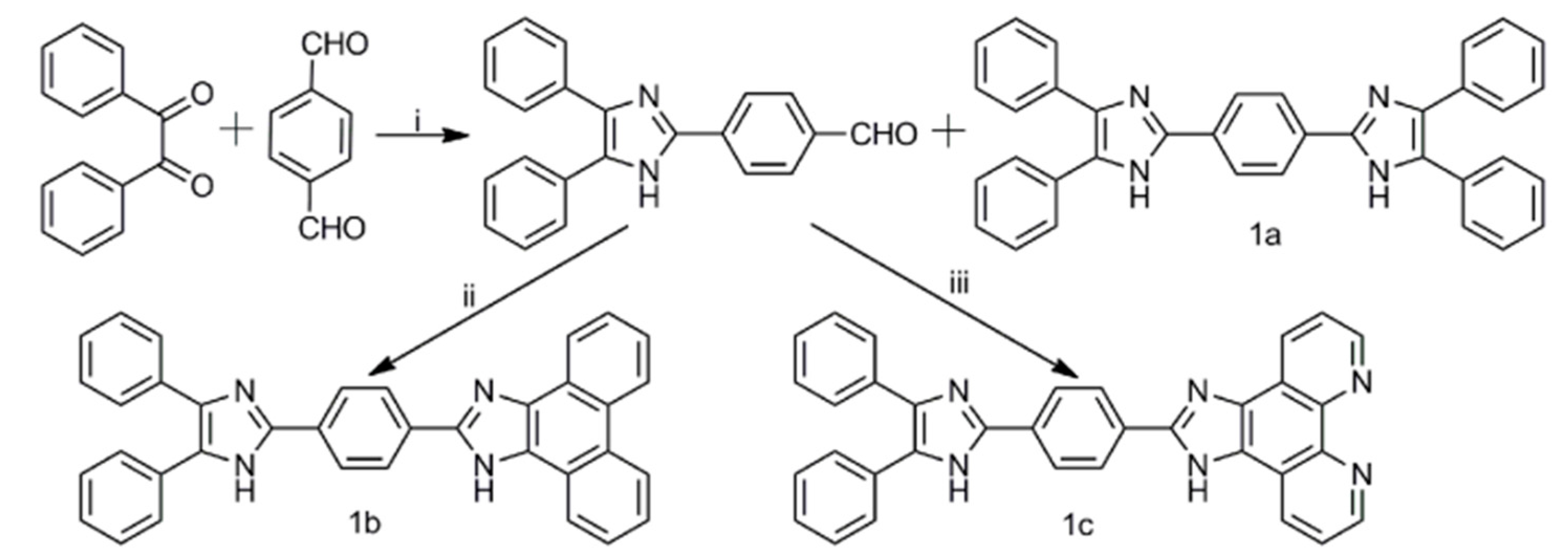
2.1. Synthesis of Compounds 1a1c

Scheme 1 depicts the synthetic procedure for the preparation of compounds 1a1c according to previous studies [42,43]. The two adjacent phenyl rings of compound 1a were connected through C–C bond to form compound 1b, which exhibited a rigid structure and high degree of coplanarity, to investigate the relationship between molecular structures and optical behaviors. Furthermore, two sp2-hybridized N atoms as electron-withdrawing substituents were introduced into compound 1b to form compound 1c to study the electronic effects of substituents. Consequently, compound 1a displayed a symmetrical structure, whereas compounds 1b and 1c had an asymmetrical structure.

2.2. Optical Properties of Compounds 1a1c

Figure 1a illustrates the normalized absorption spectra of compounds 1a1c in DMSO solution. The absorption spectrum of compound 1a exhibits two prominent bands at λmax 296 nm and 358 nm, which can be ascribed to a localized aromatic π-π* transition and the absorption of the entire molecule, respectively [3,33]. Compared with the absorption spectrum of compound 1a, the long peak of compound 1b is red shifted from 358 nm to 372 nm with two shoulder peaks at λmax 352 nm and 390 nm because the structure of compound 1b is more coplanar than that of compound 1a due to the inhibition of C–C single bond rotation of compound 1b. Although compound 1c possesses two sp2-hybridized N atoms as electron-withdrawing substituents, the long peak of compound 1c is slightly blue shifted from 372 nm (the absorption peak of compound 1b) to 367 nm with a shoulder peak at λmax 385 nm. This phenomenon is similar to that presented in a previous report regarding symmetrical bisimidazole systems [44]. Molar absorption coefficients of compounds 1a1c are 6.0 × 10⁴, 5.2 × 10⁴, and 5.2 × 10⁴ L mol1 cm1, respectively. Compounds 1a1c emit a strong blue light in the DMSO solution, and corresponding emission spectra are shown in Figure 1b. As shown in Figure 1b, two emission bands exist at λem = 406 nm and λem = 426 nm under excitation at λex 358 nm for compound 1a, λem = 416 nm and λem = 436 nm under excitation at λex 372 nm for compound 1b, and λem = 438 nm under excitation at λex 367 nm for compound 1c. The long emission wavelength is red shifted from compound 1a to compound 1b to compound 1c due to the increase in coplanarity, which can lead to the expansion of the π electron delocalization.
Although imidazole derivatives have been used as chemosensors for fluoride ion, systematic studies on the effects of coplanarity and electron-withdrawing substituents have rarely been reported. Figure 2a exhibits the absorption spectra of the DMSO solution of compound 1a in the presence of nine anions (F, Cl, Br, I, NO3, ClO4, HSO4, BF4, and PF6, 20 equiv.). The absorption intensity at λmax 358 nm slightly decreases, but a new band at λmax 414 nm appears, while no evident change occurred upon the addition of eight other anions. The new band at λmax 414 nm is attributed to the variations of the electronic transition due to the occurrence of a strong interaction between fluoride ion and N–H groups. As shown in Scheme 2, the imidazole group serves as an electron acceptor before the addition of Bu4N+F, but the imidazole group acts as an electron donor after the addition of Bu4N+F, which leads to the charge transfer enhancement. The titration absorption spectra of the DMSO solution of compound 1a with the addition of different amounts of Bu4N+F have been examined to verify this phenomenon, as shown in Figure 2d. The absorption intensity at λmax 358 nm decreases, and the absorption intensity at λmax 414 nm steadily increases with the increase in the amount of Bu4N+F. Meanwhile, the absorption peak at λmax 358 nm is red shifted to 378 nm when 100 equiv. Bu4N+F is added into the DMSO solution of compound 1a, suggesting that compound 1a can be a candidate as a chemosensor for fluoride ion. The absorption behavior of the DMSO solution of compound 1b in the presence of Bu4N+F is similar to that of the DMSO solution of compound 1a. However, a new band at λmax 420 nm can be easily observed. Compared with the absorption behaviors of the DMSO solutions of compounds 1a and 1b in the presence of Bu4N+F, the absorption behavior of the DMSO solution of compound 1c is different to some extent. The absorption peak at λmax 367 nm is red shifted with the increase in the amount of Bu4N+F.
The fluorescence emission behaviors of the DMSO solutions of compounds 1a1c in the presence of nine anions (F, Cl, Br, I, NO3, ClO4, HSO4, BF4, and PF6, 20 equiv.) are also investigated, and the corresponding emission spectra are shown in Figure 3. As shown in Figure 3a,d, the photoluminescence (PL) intensity of compound 1a is slightly quenched, and the fluorescence emission spectra are slightly broadened in the presence of fluoride ion (20 equiv.), while no evident fluorescence change occurred in the presence of eight other anions. Figure 3g depicts the titration emission spectra of the DMSO solution of compound 1a with the addition of different amounts of Bu4N+F. The PL intensity at λmax 426 nm is nearly quenched, and the fluorescence emission spectra are further broadened when 100 equiv. Bu4N+F are added into the DMSO solution of compound 1a. The UV/Vis absorption and fluorescence emission spectra of compound 1a shows a fact that firstly formation of N-HF hydrogen bond and subsequent deprotonation with adding excess Bu4N+F is responsible for the behaviour of compound 1a (Scheme 2). Figure S11 shows that the linear regression equation of compound 1a was y = 4.781 − 0.00347x, and the slope was −0.00347 (Supplementary Materials). The detection limit of compound 1a was calculated to be 5.47 × 10⁻⁶ M with the equation: detection limit = 3Sd/ρ, where Sd is the standard deviation of blank measurement, and ρ is the slope between the fluorescence intensity versus fluoride ion concentration [45,46]. The emission behavior of the DMSO solution of compound 1b in the presence of Bu4N+F is similar to that of the DMSO solution of compound 1a. However, the PL intensity of compound 1b is quenched, and a new band at λmax 470 nm appears in the presence of fluoride ion (20 equiv.). The detection limit of compound 1b was calculated to be 4.21 × 10⁻⁶ M according to Figure S12. Compared with the emission behaviors of the DMSO solutions of compounds 1a and 1b in the presence of Bu4N+F, the emission behavior of the DMSO solution of compound 1c is different. The PL intensity of compound 1c at λmax 438 nm is dramatically quenched, and a new band at λmax 513 nm is easily observed in the presence of fluoride ion (20 equiv.). Furthermore, the PL peak of compound 1c at λmax 438 nm disappears when 50 equiv. Bu4N+F are added into the DMSO solution of compound 1c. The detection limit of compound 1c was calculated to be 9.12 × 10⁻⁷ M according to Figure S13. As shown in Scheme 2, the imidazole group acts as as an electron acceptor before the addition of Bu4N+F, charge transfer can easily occur from diphenyl (for compound 1a) or phenanthrene (for compound 1b) group (donor) to imidazole group (acceptor) but charge transfer can not easily occur from 1,10-phenanthroline group (for compound 1c) to imidazole group because 1,10-phenanthroline group is an stronger electron accepor. While the imidazole group acts an electron donor after the addition of excess Bu4N+F, which leads to the charge transfer enhancement from the imidazole group to 1,10-phenanthroline. Therefore, the largest red shift (75 nm) of the DMSO solution of compound 1c is in the presence of Bu4N+F.

3. Conclusions

In summary, one symmetrical structure (1a) and two asymmetric structures (1b and 1c) were successfully synthesized. Interestingly, compounds 1a1c can act as efficient anion sensors for fluoride ion over a wide range of other anions (Cl, Br, I, NO3, ClO4, HSO4, BF4 and PF6) owing to the imidazole group in the main backbone. Compared with the optical behaviors of the DMSO solution of compound 1a in the presence of Bu4N+F, compounds 1b and 1c exhibit considerable sensitivity to fluoride ion due to the increase in the coplanarity. Furthermore, compared with the fluorescence emission behaviors of the DMSO solutions of compounds 1a and 1b in the presence of Bu4N+F, compound 1c exhibits the most significant sensitivity to fluoride ion due to charge transfer enhancement. Consequently, the detection limits of compounds 1a1c increase from 5.47 × 10⁶ M to 4.21 × 10⁶ M to 9.12 × 10⁷ M. Our results suggest that the increase in the coplanarity and the introduction of electron-withdrawing groups to the imidazole backbone can improve the performance in detecting fluoride ion. We will introduce poly(ethylene glycol) methyl ether into start materials to improve the solubily of target compounds in water in our future work.

Supplementary Materials

Supplementary Materials are available online. Figure S1. 1H NMR spectrum of compound 4-(4,5-diphenyl-1H-imidazol-2-yl)benzaldehyde in DMSO-d6. Figure S2. 1H NMR spectrum of compound 1a in DMSO-d6. Figure S3. 1H NMR spectrum of compound 1b in DMSO-d6. Figure S4. 1H NMR spectrum of compound 1c in DMSO-d6. Figure S5. MS spectrum of compound 1a. Figure S6. MS spectrum of compound 1b. Figure S7. MS spectrum of compound 1c. Figure S8. Correlation curves of compound 1a at 358 nm and 414 nm adding different equivalents of F. Figure S9. Correlation curves of compound 1b at 372 nm and 420 nm adding different equivalents of F. Figure S10. Correlation curves of compound 1c at 367 nm and 380 nm adding different equivalents of F. Figure S11. Change in PL intensity of compound 1a (10 uM) upon titration with F. Figure S12. Change in PL intensity of compound 1b (10 uM) upon titration with F. Figure S13. Change in PL intensity of compound 1c (10 uM) upon titration with F.

Author Contributions

Liang Zhang and Fang Liu conceived and designed the experiments and wrote the paper; Liang Zhang reviewed the paper.

Conflicts of Interest

The authors declare no conflict of interest.

References

  1. Descalzo, A.B.; Rurack, K.; Weisshoff, H.; Martínez-Máñez, R.; Marcos, M.D.; Amorós, P.; Hoffmann, K.; Soto, J. Rational design of a chromo- and fluorogenic hybrid chemosensor material for the detection of long-chain carboxylates. J. Am. Chem. Soc. 2005, 127, 184–200. [Google Scholar] [CrossRef] [PubMed]
  2. Vetrichelvan, M.; Nagarajan, R.; Valiyaveettil, S. Carbazole-containing conjugated copolymers as colorimetric/fluorimetric sensor for iodide anion. Macromolecules 2006, 39, 8303–8310. [Google Scholar] [CrossRef]
  3. Lin, T.-P.; Chen, C.-Y.; Wen, Y.-S.; Sun, S.-S. Synthesis, photophysical, and anion-sensing properties of quinoxalinebis (sulfonamide) functionalized receptors and their metal complexes. Inorg. Chem. 2007, 46, 9201–9212. [Google Scholar] [CrossRef] [PubMed]
  4. Ajayakumar, M.; Mukhopadhyay, P.; Yadav, S.; Ghosh, S. Single-electron transfer driven cyanide sensing: A new multimodal approach. Org. Lett. 2010, 12, 2646–2649. [Google Scholar] [CrossRef] [PubMed]
  5. Eun Jun, M.; Roy, B.; Han Ahn, K. “Turn-on” fluorescent sensing with “reactive” probes. Chem. Commun. 2011, 47, 7583–7601. [Google Scholar] [CrossRef] [PubMed]
  6. Li, X.; Gao, X.; Shi, W.; Ma, H. Design strategies for water-soluble small molecular chromogenic and fluorogenic probes. Chem. Rev. 2014, 114, 590–659. [Google Scholar] [CrossRef] [PubMed]
  7. Busschaert, N.; Caltagirone, C.; Van Rossom, W.; Gale, P.A. Applications of supramolecular anion recognition. Chem. Rev. 2015, 115, 8038–8155. [Google Scholar] [CrossRef] [PubMed]
  8. Ros-Lis, J.V.; Martinez-Manez, R.; Soto, J. Subphthalocyanines as fluoro-chromogenic probes for anions and their application to the highly selective and sensitive cyanide detection. Chem. Commun. 2005, 5260–5262. [Google Scholar] [CrossRef] [PubMed]
  9. Ma, G.; Müller, A.M.; Bardeen, C.J.; Cheng, Q. Self-assembly combined with photopolymerization for the fabrication of fluorescence “turn-on” vesicle sensors with reversible “on-off” switching properties. Adv. Mater. 2006, 18, 55–60. [Google Scholar] [CrossRef]
  10. Garcia, F.; Garcia, J.M.; Garcia-Acosta, B.; Martinez-Manez, R.; Sancenon, F.; Soto, J. Pyrylium-containing polymers as sensory materials for the colorimetric sensing of cyanide in water. Chem. Commun. 2005, 2790–2792. [Google Scholar] [CrossRef] [PubMed]
  11. Yang, C.; Xu, J.; Ma, J.; Zhu, D.; Zhang, Y.; Liang, L.; Lu, M. An efficient long fluorescence lifetime polymer-based sensor based on europium complex as chromophore for the specific detection of F, CH3COO, and H2PO4. Polym. Chem. 2012, 3, 2640–2648. [Google Scholar] [CrossRef]
  12. Isaad, J.; Perwuelz, A. New color chemosensors for cyanide based on water soluble azo dyes. Tetrahedron Lett. 2010, 51, 5810–5814. [Google Scholar] [CrossRef]
  13. Zhou, Y.; Zhang, J.F.; Yoon, J. Fluorescence and colorimetric chemosensors for fluoride-ion detection. Chem. Rev. 2014, 114, 5511–5571. [Google Scholar] [CrossRef] [PubMed]
  14. Hu, R.; Feng, J.; Hu, D.; Wang, S.; Li, S.; Li, Y.; Yang, G. A rapid aqueous fluoride ion sensor with dual output modes. Angew. Chem. Int. Ed. 2010, 49, 4915–4918. [Google Scholar] [CrossRef] [PubMed]
  15. Kim, T.H.; Swager, T.M. A fluorescent self-amplifying wavelength-responsive sensory polymer for fluoride ions. Angew. Chem. Int. Ed. 2003, 42, 4803–4806. [Google Scholar] [CrossRef] [PubMed]
  16. Ke, B.; Chen, W.; Ni, N.; Cheng, Y.; Dai, C.; Dinh, H.; Wang, B. A fluorescent probe for rapid aqueous fluoride detection and cell imaging. Chem. Commun. 2013, 49, 2494–2496. [Google Scholar] [CrossRef] [PubMed]
  17. Ambrosi, G.; Formica, M.; Fusi, V.; Giorgi, L.; Macedi, E.; Piersanti, G.; Retini, M.; Varrese, M.A.; Zappia, G. New coumarin-urea based receptor for anions: A selective off–on fluorescence response to fluoride. Tetrahedron 2012, 68, 3768–3775. [Google Scholar] [CrossRef]
  18. Martinez-Manez, R.; Sancenon, F. New advances in fluorogenic anion chemosensors. J. Fluoresc. 2005, 15, 267–285. [Google Scholar] [CrossRef] [PubMed]
  19. Yang, X.-F.; Qi, H.; Wang, L.; Su, Z.; Wang, G. A ratiometric fluorescent probe for fluoride ion employing the excited-state intramolecular proton transfer. Talanta 2009, 80, 92–97. [Google Scholar] [CrossRef] [PubMed]
  20. Bao, Y.; Liu, B.; Wang, H.; Tian, J.; Bai, R. A “naked eye” and ratiometric fluorescent chemosensor for rapid detection of F based on combination of desilylation reaction and excited-state proton transfer. Chem. Commun. 2011, 47, 3957–3959. [Google Scholar] [CrossRef] [PubMed]
  21. Sivaraman, G.; Chellappa, D. Rhodamine based sensor for naked-eye detection and live cell imaging of fluoride ions. J. Mater. Chem. B 2013, 1, 5768–5772. [Google Scholar] [CrossRef]
  22. Kim, S.Y.; Park, J.; Koh, M.; Park, S.B.; Hong, J.I. Fluorescent probe for detection of fluoride in water and bioimaging in A549 human lung carcinoma cells. Chem. Commun. 2009, 21, 4735–4737. [Google Scholar] [CrossRef] [PubMed]
  23. Amendola, V.; Bergamaschi, G.; Boiocchi, M.; Fabbrizzi, L.; Mosca, L. The interaction of fluoride with fluorogenic ureas: An ON1-OFF-ON2 response. J. Am. Chem. Soc. 2013, 135, 6345–6355. [Google Scholar] [CrossRef] [PubMed]
  24. Kim, D.; Singha, S.; Wang, T.; Seo, E.; Lee, J.H.; Lee, S.J.; Kim, K.H.; Ahn, K.H. In vivo two-photon fluorescent imaging of fluoride with a desilylation-based reactive probe. Chem. Commun. 2012, 48, 10243–10245. [Google Scholar] [CrossRef] [PubMed]
  25. Lin, Z.H.; Zhao, Y.G.; Duan, C.Y.; Zhang, B.G.; Bai, Z.P. A highly selective chromo- and fluorogenic dual responding fluoride sensor: Naked-eye detection of F- ion in natural water via a test paper. Dalton Trans. 2006, 30, 3678–3684. [Google Scholar] [CrossRef] [PubMed]
  26. Ren, J.; Wu, Z.; Zhou, Y.; Li, Y.; Xu, Z. Colorimetric fluoride sensor based on 1,8-naphthalimide derivatives. Dyes Pigments 2011, 91, 442–445. [Google Scholar] [CrossRef]
  27. Rao, M.R.; Mobin, S.M.; Ravikanth, M. Boron–dipyrromethene based specific chemodosimeter for fluoride ion. Tetrahedron 2010, 66, 1728–1734. [Google Scholar] [CrossRef]
  28. Hou, P.; Chen, S.; Wang, H.; Wang, J.; Voitchovsky, K.; Song, X. An aqueous red emitting fluorescent fluoride sensing probe exhibiting a large Stokes shift and its application in cell imaging. Chem. Commun. 2014, 50, 320–322. [Google Scholar] [CrossRef] [PubMed]
  29. Lu, H.; Wang, Q.; Li, Z.; Lai, G.; Jiang, J.; Shen, Z. A specific chemodosimeter for fluoride ion based on a pyrene derivative with trimethylsilylethynyl groups. Org. Biomol. Chem. 2011, 9, 4558–4562. [Google Scholar] [CrossRef] [PubMed]
  30. Goswami, S.; Das, A.K.; Manna, A.; Maity, A.K.; Fun, H.-K.; Quah, C.K.; Saha, P. A colorimetric and ratiometric fluorescent turn-on fluoride chemodosimeter and application in live cell imaging: High selectivity via specific SiO cleavage in semi aqueous media and prompt recovery of ESIPT along with the X-ray structures. Tetrahedron Lett. 2014, 55, 2633–2638. [Google Scholar] [CrossRef]
  31. Gu, P.-Y.; Wang, Z.; Zhang, Q. Azaacenes as active elements for sensing and bio applications. J. Mater. Chem. B 2016, 4, 7060–7074. [Google Scholar] [CrossRef]
  32. Gu, P.-Y.; Wang, Z.; Liu, G.; Nie, L.; Ganguly, R.; Li, Y.; Zhang, Q. Synthesis, physical properties, and sensing behaviour of a novel naphthalenediimide derivative. Dyes Pigments 2016, 131, 224–230. [Google Scholar] [CrossRef]
  33. Mahapatra, A.K.; Karmakar, P.; Roy, J.; Manna, S.; Maiti, K.; Sahoo, P.; Mandal, D. Colorimetric and ratiometric fluorescent chemosensor for fluoride ions based on phenanthroimidazole (PI): Spectroscopic, NMR and density functional studies. RSC Adv. 2015, 5, 37935–37942. [Google Scholar] [CrossRef]
  34. Batista, R.M.F.; Oliveira, E.; Costa, S.P.G.; Lodeiro, C.; Raposo, M.M.M. Cyanide and fluoride colorimetric sensing by novel imidazo-anthraquinones functionalised with indole and carbazole. Supramol. Chem. 2013, 26, 71–80. [Google Scholar] [CrossRef] [Green Version]
  35. Kumar, D.; Thomas, K.R.J. 2-Hydroxyarylimidazole-based colorimetric and ratiometric fluoride ion sensors. RSC Adv. 2014, 4, 56466–56474. [Google Scholar] [CrossRef]
  36. Gwon, S.Y.; Kim, S.H. Anion sensing and F(-)-induced reversible photoreaction of D-pi-A type dye containing imidazole moiety as donor. Spectrochim. Acta A 2014, 117, 810–813. [Google Scholar] [CrossRef] [PubMed]
  37. Li, R.; Wang, S.; Li, Q.; Lan, H.; Xiao, S.; Li, Y.; Tan, R.; Yi, T. A fluorescent non-conventional organogelator with gelation-assisted piezochromic and fluoride-sensing properties. Dyes Pigments 2017, 137, 111–116. [Google Scholar] [CrossRef]
  38. Manivannan, R.; Satheshkumar, A.; Elango, K.P. Tuning of the H-bonding ability of imidazole N–H towards the colorimetric sensing of fluoride and cyanide ions as their sodium salts in water. New J. Chem. 2013, 37, 3152–3160. [Google Scholar] [CrossRef]
  39. Jayasudha, P.; Manivannan, R.; Elango, K.P. Highly selective colorimetric receptors for detection of fluoride ion in aqueous solution based on quinone-imidazole ensemble—Influence of hydroxyl group. Sens. Actuator B Chem. 2016, 237, 230–238. [Google Scholar] [CrossRef]
  40. Jain, A.; Gupta, R.; Agarwal, M. Rationally designed tri-armed imidazole–indole hybrids as naked eye receptors for fluoride ion sensing. Synth. Commun. 2017, 47, 1307–1318. [Google Scholar] [CrossRef]
  41. Gu, P.-Y.; Gao, J.; Zhang, Q.; Liu, G.; Zhou, F.; Xu, Q.-F.; Lu, J.-M. Tuning optical properties of phenanthroline derivatives through varying excitation wavelength and pH values. J. Mater. Chem. C 2014, 2, 1539–1544. [Google Scholar] [CrossRef]
  42. Tian, M.; Wang, C.; Wang, L.; Luo, K.; Zhao, A.; Guo, C. Study on the synthesis and structure-effect relationship of multi-aryl imidazoles with their fluorescence properties. Luminescence 2014, 29, 540–548. [Google Scholar] [CrossRef] [PubMed]
  43. Gao, F.; Chen, X.; Zhou, F.; Weng, L.-P.; Guo, L.-T.; Chen, M.; Chao, H.; Ji, L.-N. pH responsive luminescent switches of ruthenium(II) complexes containing two imidazole groups: Synthesis, spectroscopy, electrochemistry and theoretical calculations. Inorg. Chim. Acta 2009, 362, 4960–4966. [Google Scholar] [CrossRef]
  44. Xie, N.; Chen, Y. Synthesis and photophysical properties of 1,4-bis(4,5-diarylimidazol) benzene dyes. J. Photochem. Photobiol. A 2007, 189, 253–257. [Google Scholar] [CrossRef]
  45. Hu, J.-Y.; Liu, R.; Zhu, X.-L.; Cai, X.; Zhu, H.-J. A highly efficient and selective probe for F detection based on 1H-imidazo[4,5-b]phenazine derivative. Chin. Chem. Lett. 2015, 26, 339–342. [Google Scholar] [CrossRef]
  46. Niu, H.T.; Su, D.; Jiang, X.; Yang, W.; Yin, Z.; He, J.; Cheng, J.P. A simple yet highly selective colorimetric sensor for cyanide anion in an aqueous environment. Org. Biomol. Chem. 2008, 6, 3038–3040. [Google Scholar] [CrossRef] [PubMed]
Sample Availability: Samples of the compounds 1a1c are available from the authors.
Scheme 1. Synthetic route of compounds 1a1c. (i) ammonium acetate, glacial acetic acid; (ii) ammonium acetate, glacial acetic acid, phenanthrene-9,10-dione; (iii) ammonium acetate, glacial acetic acid, 1,10-phenanthroline-5,6-dione.
Molecules 22 01519 sch001
Figure 1. (a) Normalized absorption and (b) emission spectra of compounds 1a1c in DMSO solution (λex = λabs, 10−5 mol L1).
Molecules 22 01519 g001
Figure 2. UV/Vis absorption spectra of DMSO solutions of (a) compound 1a; (b) compound 1b; and (c) compound 1c (105 mol L1) in the absence (blank) and presence of nine anions (20 equiv.). UV/Vis absorption spectra of DMSO solutions of (d) compound 1a; (e) compound 1b; and (f) compound 1c (105 mol L1) with different amounts of Bu4N+F.
Molecules 22 01519 g002
Scheme 2. Schematic diagram for electron transfer process of compounds 1a1c in the presence of Bu4N+F (A = electron acceptor; D = electron donor).
Molecules 22 01519 sch002
Figure 3. Photographs of the DMSO solutions of (a) compound 1a; (b) compound 1b; and (c) compound 1c (105 mol L1) in the absence (blank) and presence of nine anions (20 equiv.) taken under UV light at 365 nm. Emission spectra of DMSO solutions of (d) compound 1a; (e) compound 1b, and (f) compound 1c (105 mol L1) in the absence (blank) and presence of nine anions (20 equiv.). UV/Vis absorption spectra of DMSO solutions of (g) compound 1a; (h) compound 1b; and (i) compound 1c (105 mol L1) with different amounts of Bu4N+F.
Molecules 22 01519 g003

