Article

Characterization of Proanthocyanidin Oligomers of Ephedra sinica

Department of Natural Product Chemistry, Graduate School of Biomedical Sciences, Nagasaki University, 1-14 Bunkyo-machi, Nagasaki 852-8521, Japan
*
Author to whom correspondence should be addressed.
Molecules 2017, 22(8), 1308; https://doi.org/10.3390/molecules22081308
Submission received: 14 July 2017 / Revised: 27 July 2017 / Accepted: 3 August 2017 / Published: 6 August 2017

Abstract

:
Ephedra sinica, an important plant in Chinese traditional medicine, contains a complex mixture of proanthocyanidin oligomers as major constituents; however, only the minor components have been chemically characterized. In this study, oligomers with relatively large molecular weights, which form the main body of the proanthocyanidin fractions, were separated by adsorption and size-exclusion chromatography. Acid-catalyzed degradation in the presence of mercaptoethanol or phloroglucinol led to the isolation of 18 fragments, the structures of which were elucidated from their experimental and TDDFT-calculated ECD spectra. The results indicated that (−)-epigallocatechin was the main extension unit, while catechin, the A-type epigallocatechin–gallocatechin dimer, and the A-type epigallocatechin homodimer, were identified as the terminal units. Among the degradation products, thioethers of gallocatechin with 3,4-cis configurations, a B-type prodelphinidin dimer, a prodelphinidin trimer with both A- and B-type linkages, and a prodelphinidin dimer with an α-substituted A-type linkage were new compounds. In addition, a phloroglucinol adduct of an A-type prodelphinidin dimer, a doubly-linked phloroglucinol adduct of epigallocatechin, and a unique product with a flavan-3-ol skeleton generated by the rearrangement of the aromatic rings were also isolated.

1. Introduction

Ephedra sinica Stapf (Fam. Ephedraceae) is one of the most important plants in traditional medicine, and is used as a diuretic, antipyretic, diaphoretic, and for relieving a cough and asthma [1]. As the crude drug, it has an official monograph in both the Chinese and Japanese Pharmacopoeias, where it is standardized against the major alkaloids, ephedrine and pseudoephedrine [2]. Thus, the main emphasis is conventionally given to its alkaloidal content, despite the fact that this only constitutes about 0.7–0.8% of the whole plant [3,4]. Clearly, the motivation for this is the proven clinical effects of these alkaloids on the respiratory, central nervous, and cardiovascular systems [5]. However, many species of Ephedra have also been shown to contain significant amounts of proanthocyanidins [6]. Recently, many health benefits of foods and medicinal plants have been attributed to proanthocyanidins [7], and some of their biological activities, including hypotensive and vasorelaxant effects [8,9], improvement of the airway microenvironment in asthma [10], and the inhibition of inflammation and remodeling in murine models of chronic asthma [11], are responsible for the aforementioned activities of E. sinica, especially its respiratory and cardiovascular effects. A number of studies have shown that Ephedra spp. also display other biological activities that are not attributed to alkaloids, including antimicrobial [12,13], antioxidant [14], anti-inflammatory [15,16], immunosuppressive [17], antiviral [18], anti-invasive, antiangiogenic, antitumor [19], and cytotoxic [20] properties. The dimeric proanthocyanidins of E. sinica show cytotoxic activity against the tumor cell lines SGC-7901, HepG2, and HeLa [21]. In addition, a decrease in the uremic toxin parameters of rats was reportedly induced by the administration of proanthocyanidin oligomers of E. sinica [22,23].
As for the composition of the proanthocyanidins of E. sinica, monomeric flavan-3-ols [12,24] and dimeric proanthocyanidins with A-type linkages have been isolated [12,21,25,26,27,28]. The presence of prodelphinidin trimers and tetramers with A- and B-type linkages has also been shown [12]. However, these flavan-3-ols and proanthocyanidins are minor components of the total polyphenol content, and our preliminary HPLC and TLC analysis of the extract suggested that the main body of the polyphenols was a complex mixture of oligomers, detected as a broad hump on the HPLC baseline and at the origin of the TLC plate (Figure 1a). Thus, the present study aimed at characterizing these proanthocyanidin oligomers by acid-catalyzed degradation in the presence of nucleophilic agents, that is, 2-mercaptoethanol or phloroglucinol. The degradation involved the cleavage of the interflavan bonds under acidic conditions, generating flavan-3-ols from the terminal units and flavanyl-4 cations from the extension units, which were trapped by nucleophilic agents (Scheme 1) [29].

2. Results and Discussion

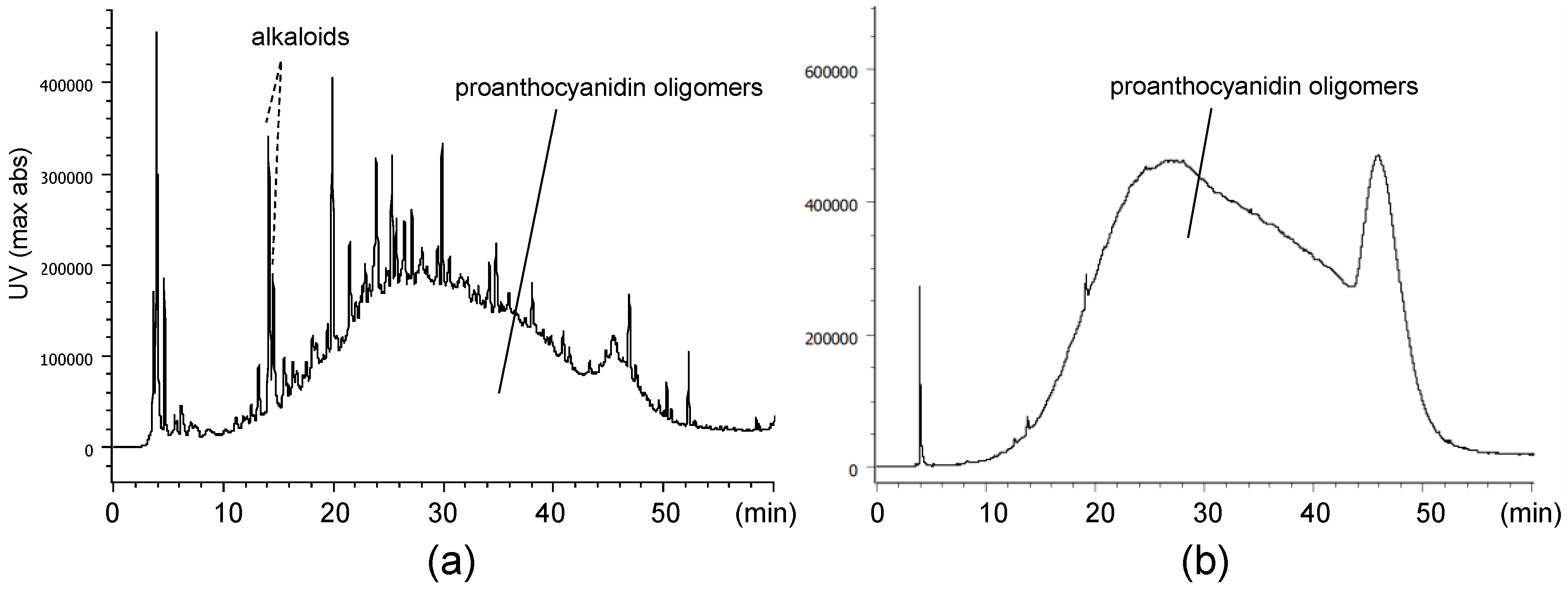
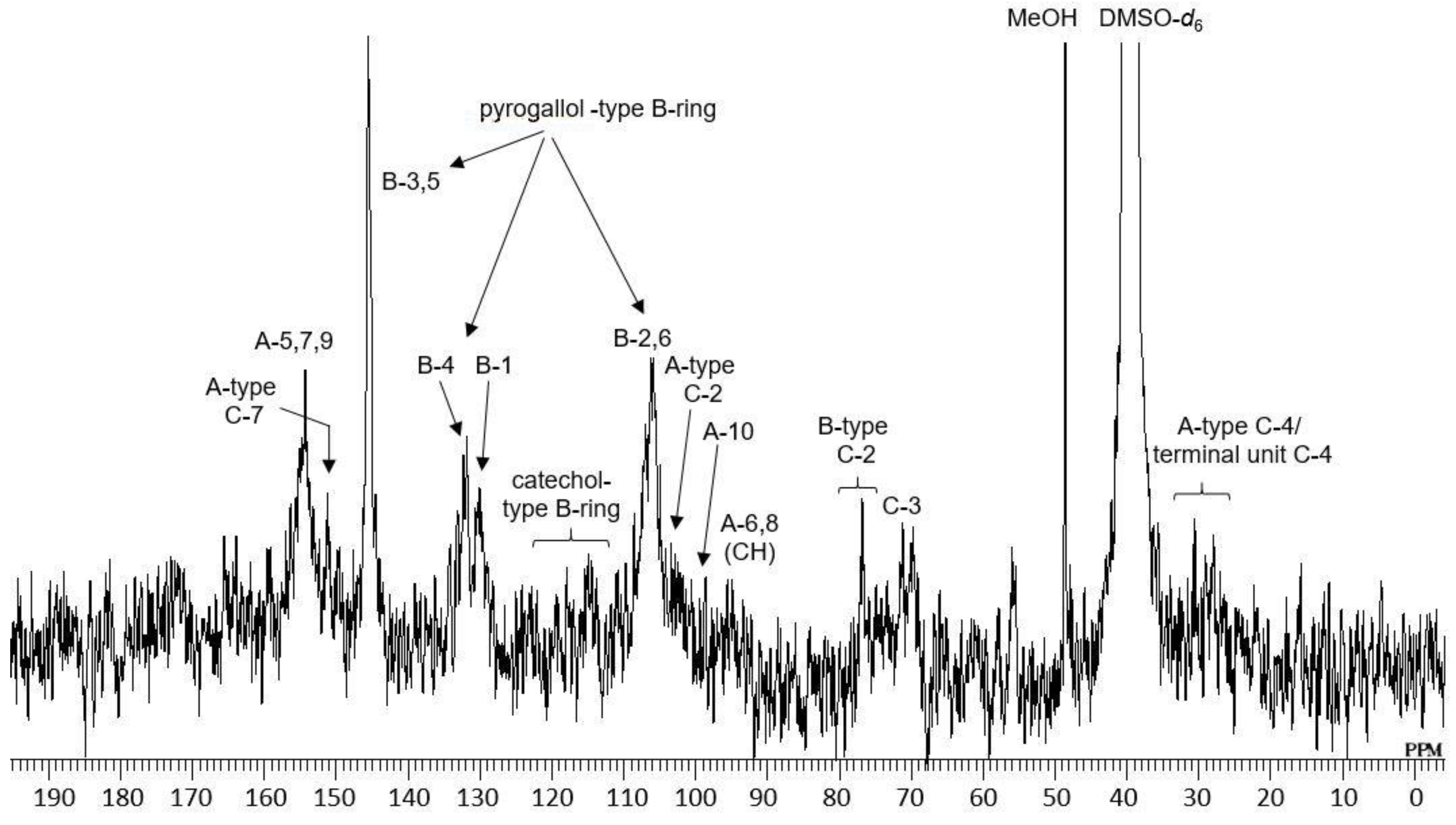
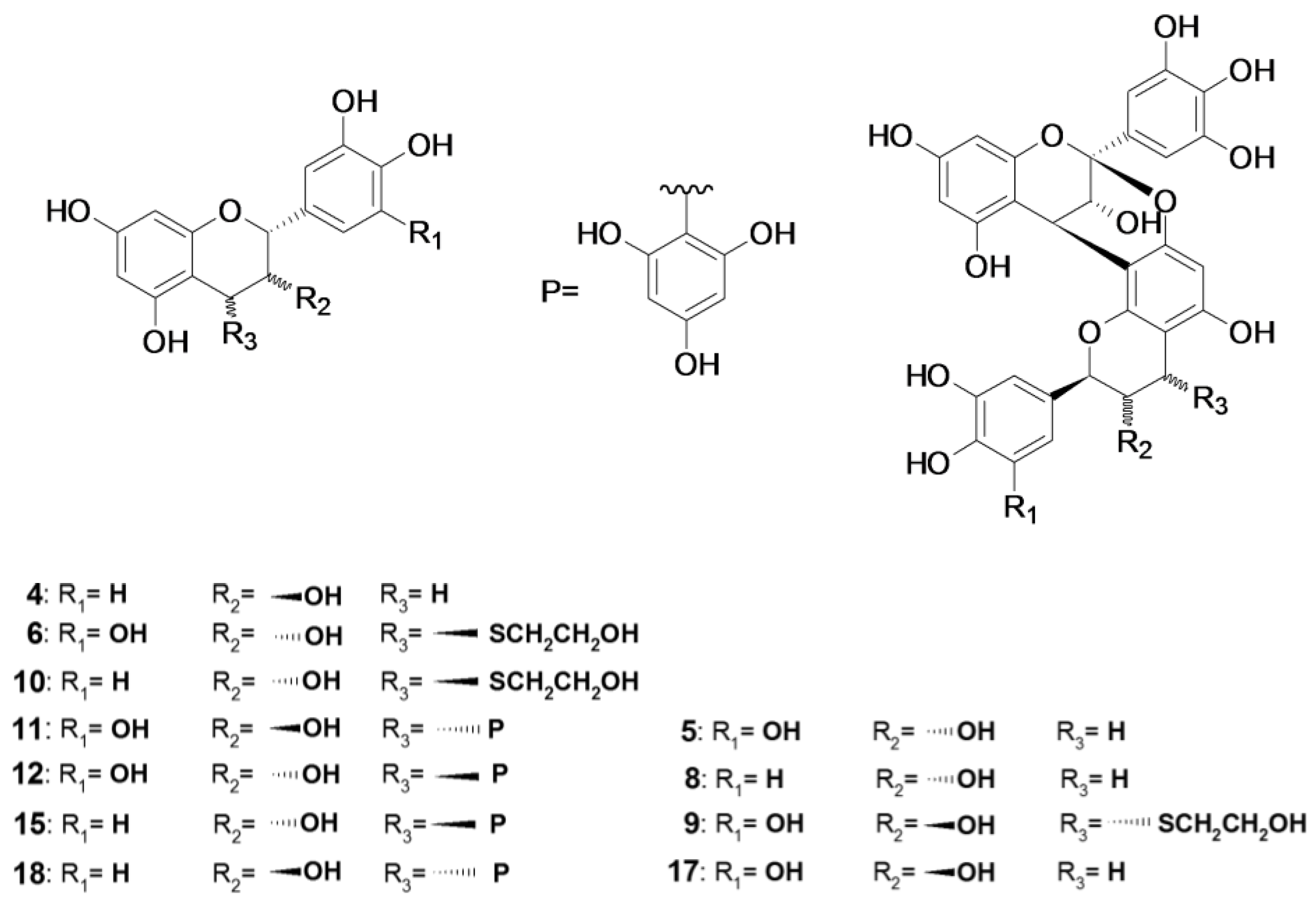
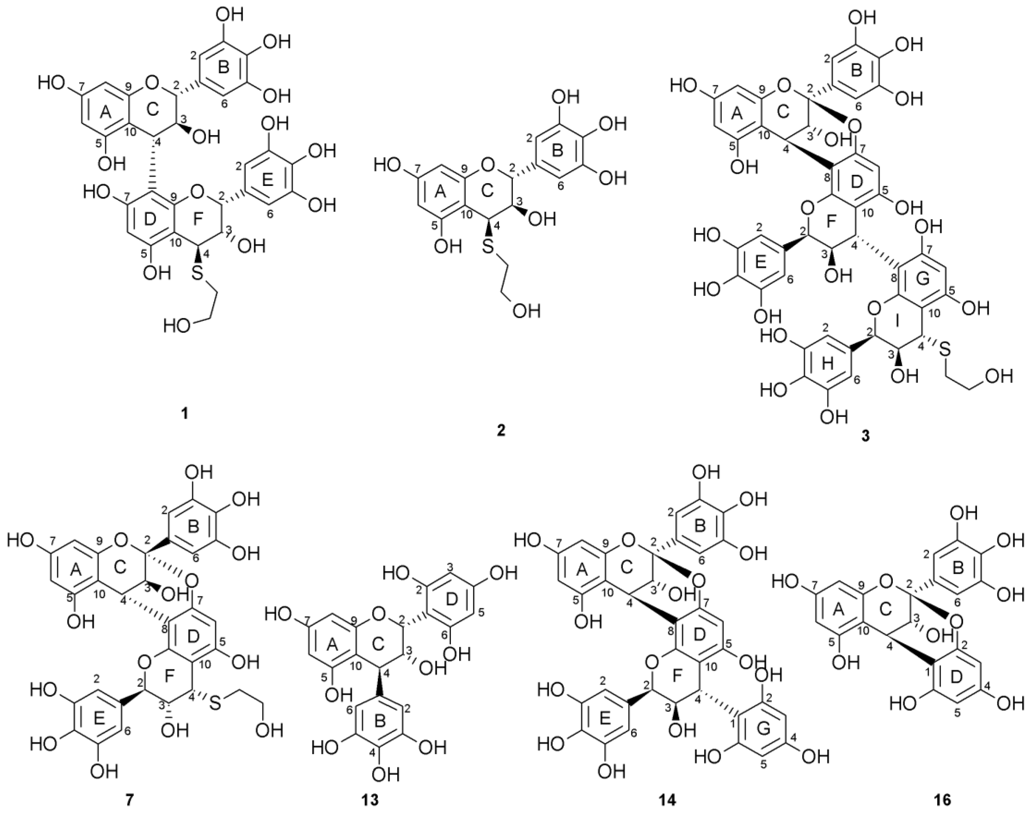
2.1. Composition of the Intact Proanthocyanidin Oligomer

The dried aerial parts of E. sinica were extracted with aqueous acetone and fractionated by a series of chromatographic separation methods, including size-exclusion chromatography [30]. The fractions containing only oligomeric proanthocyanidins accounted for 2.7% of the dried plant material, and the HPLC profile showed a broad hump on the baseline (Figure 1b). The 13C-NMR spectrum of the oligomer fraction in DMSO-d6 (Figure 2) showed signals characteristic of proanthocyanidins [31]. Based on a comparison with the literature data [11], the signals at δC 77 and δC 70–73, which were attributable to flavan C-ring C-2 and C-3 methine carbons, respectively, suggested the occurrence of B-type linkages. The chemical shifts also indicated that the 2,3-cis configuration was more abundant than 2,3-trans [32,33]. The signals in the range of δC 27–31 were attributable to the C-4 carbons of A-type proanthocyanidin extension units [12] and of terminal units [34,35]. The prominent aromatic signals observed at δC 106, 130, 132, and 145 suggested the predominance of pyrogallol-type B-rings over catechol-type B-rings (δC 115–120).

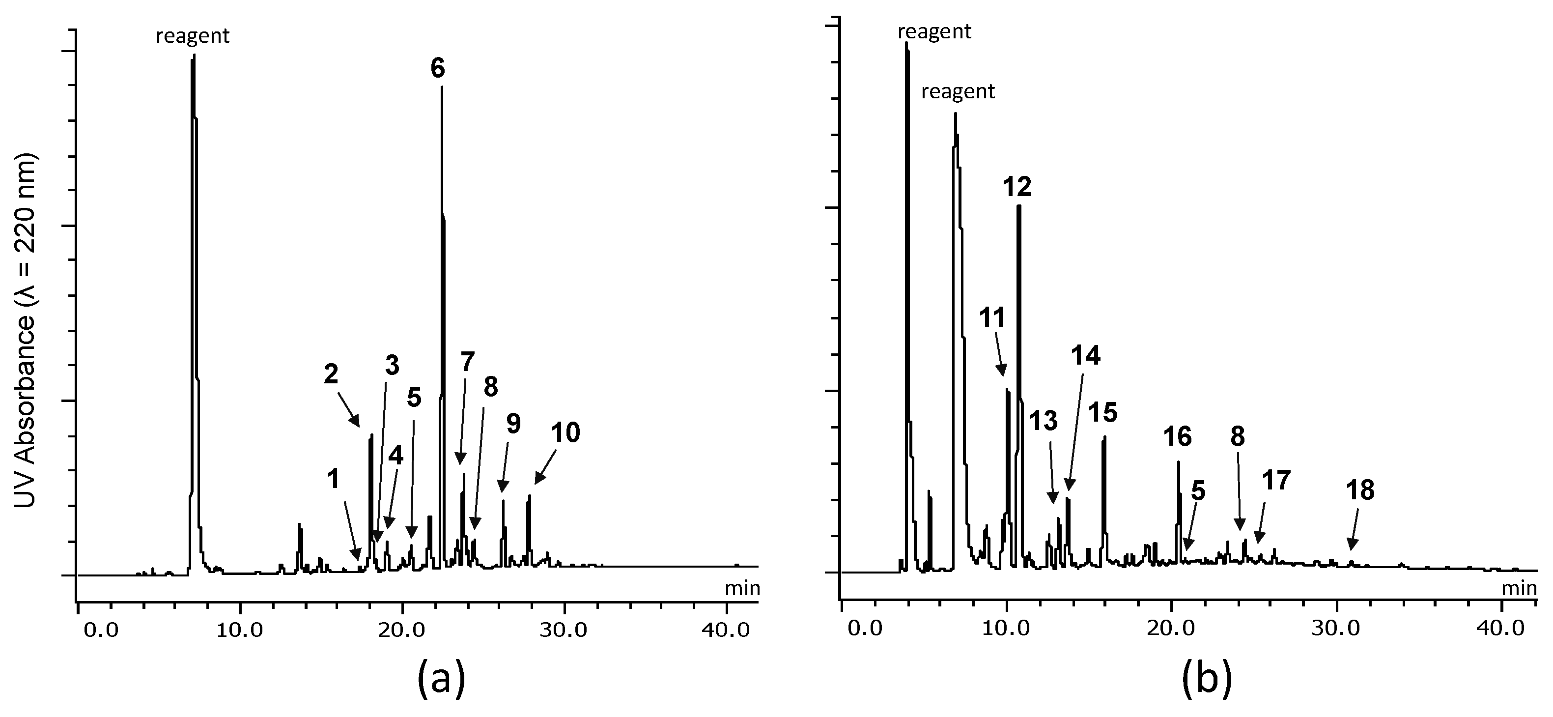
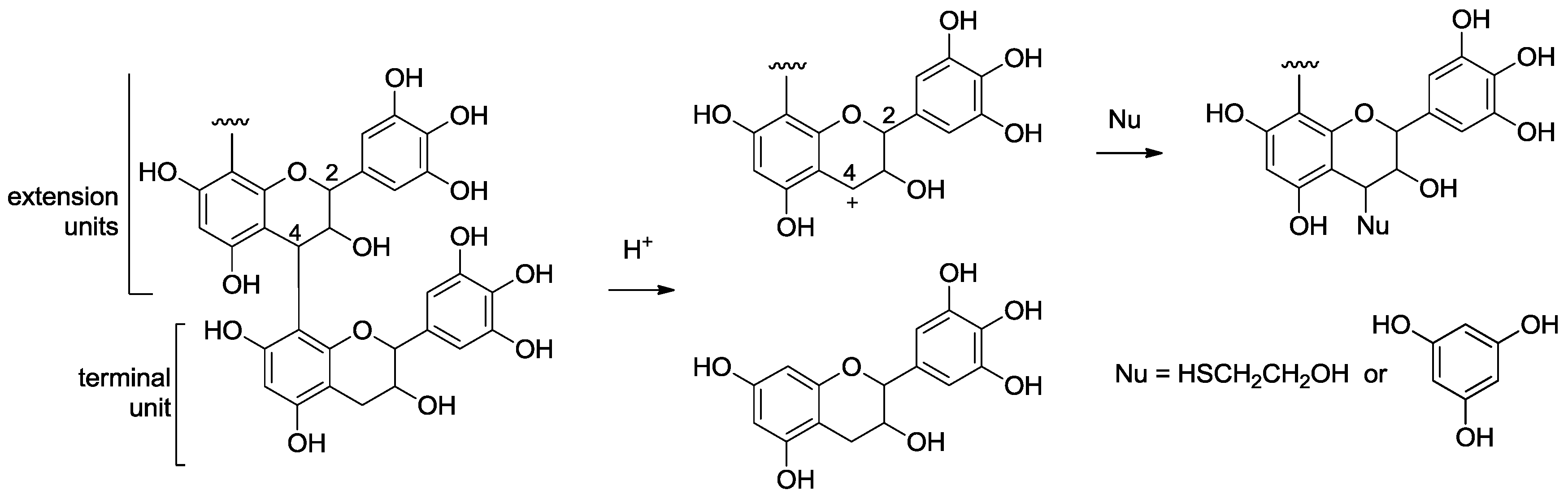
2.2. Acid-Catalyzed Degradation Products

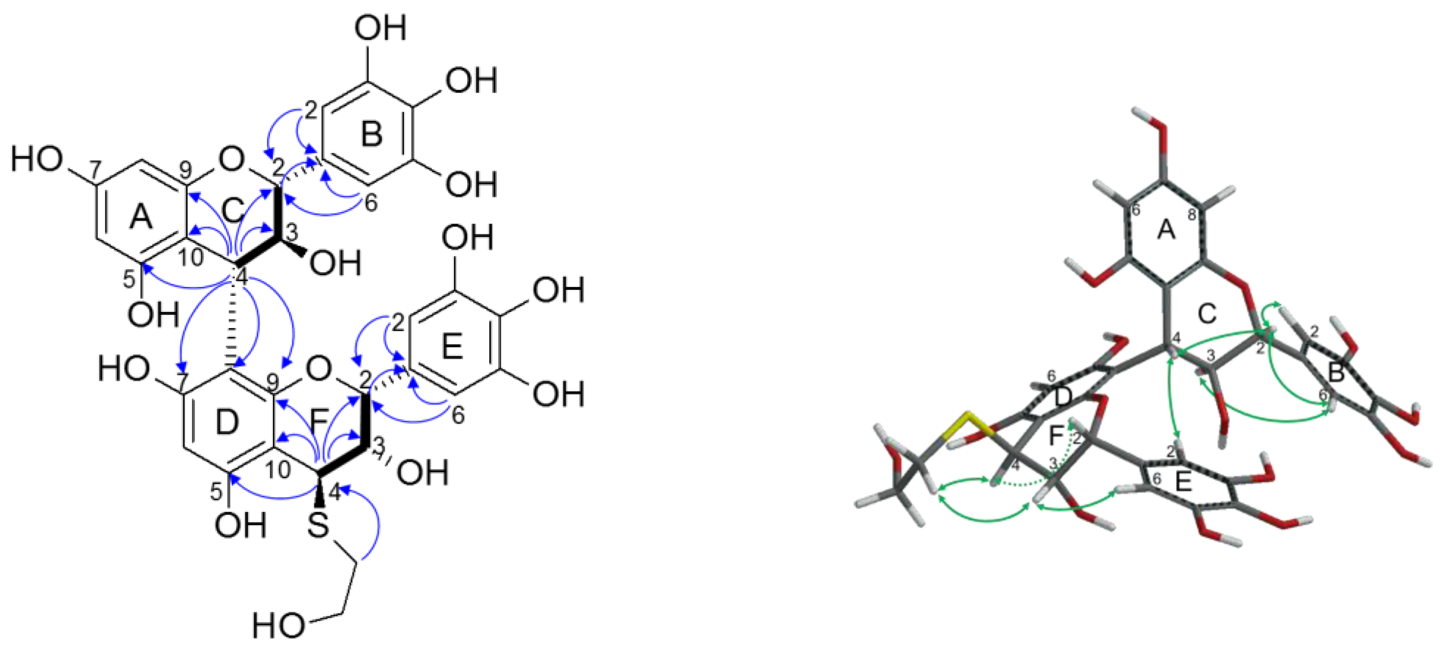
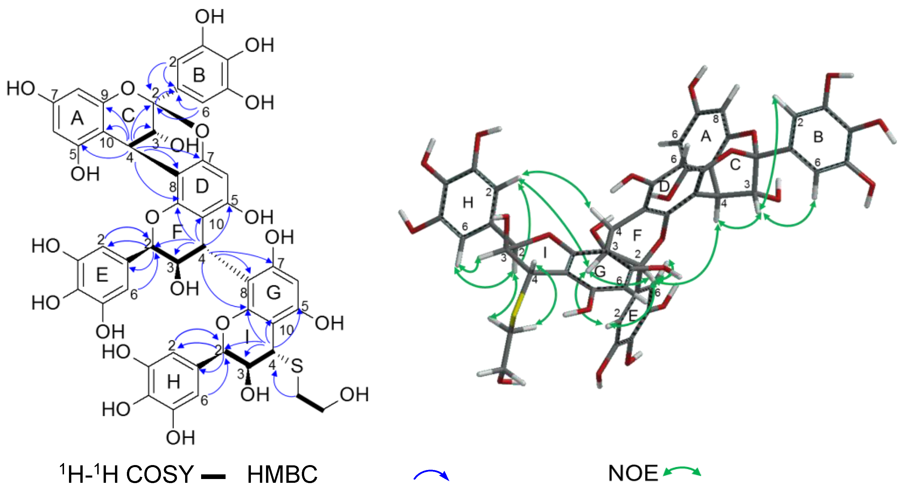
2.2.1. Identification of Known Products

Thiol degradation was performed according to the previously described method [35] with modifications of the reaction time and temperature, and 10 compounds (110) were isolated and characterized (Figure 3a). Acid-catalyzed degradation with phloroglucinol [29,36] yielded a different set of 10 products (5, 8, 1118), among which two products (5 and 8) were identical to those obtained by thiol degradation (Figure 3b).
Based on a comparison of the 1H- and 13C-NMR data with those published [12,33], four products were identified as (+)-catechin (4), (−)-epigallocatechin-(4β→8,2→O→7)-(+)-gallocatechin (5), (−)-epigallocatechin-(4β→8,2→O→7)-(+)-catechin (8), and (−)-epigallocatechin-(4→8,2→O→7)-(−)-epigallocatechin (17) (Figure 4). These products originated from the terminal units. As depicted in the HPLC profile of the reaction mixture (Figure 3), the peaks attributable to the terminal units were very small compared with those of the extension units, suggesting a high degree of polymerization. The major degradation products of thiolysis 6 and of phloroglucinolysis 12 were identified as epigallocatechin–nucleophile adducts [33,37], indicating that epigallocatechin was the major extension unit of the oligomer. The 1H- and 13C-NMR spectra of 9, 10, 11, 15, and 18 were found to be consistent with those previously reported for (−)-epigallocatechin-(4→8,2→O→7)-(−)-epigallocatechin-4-(2-hydroxyethyl)-thioether, (−)-epicatechin-4-(2-hydroxyethyl)-thioether, (+)-gallocatechin-4-phloroglucinol, (−)-epicatechin-4-phloroglucinol, and (+)-catechin-4-phloroglucinol, respectively [33,37,38].

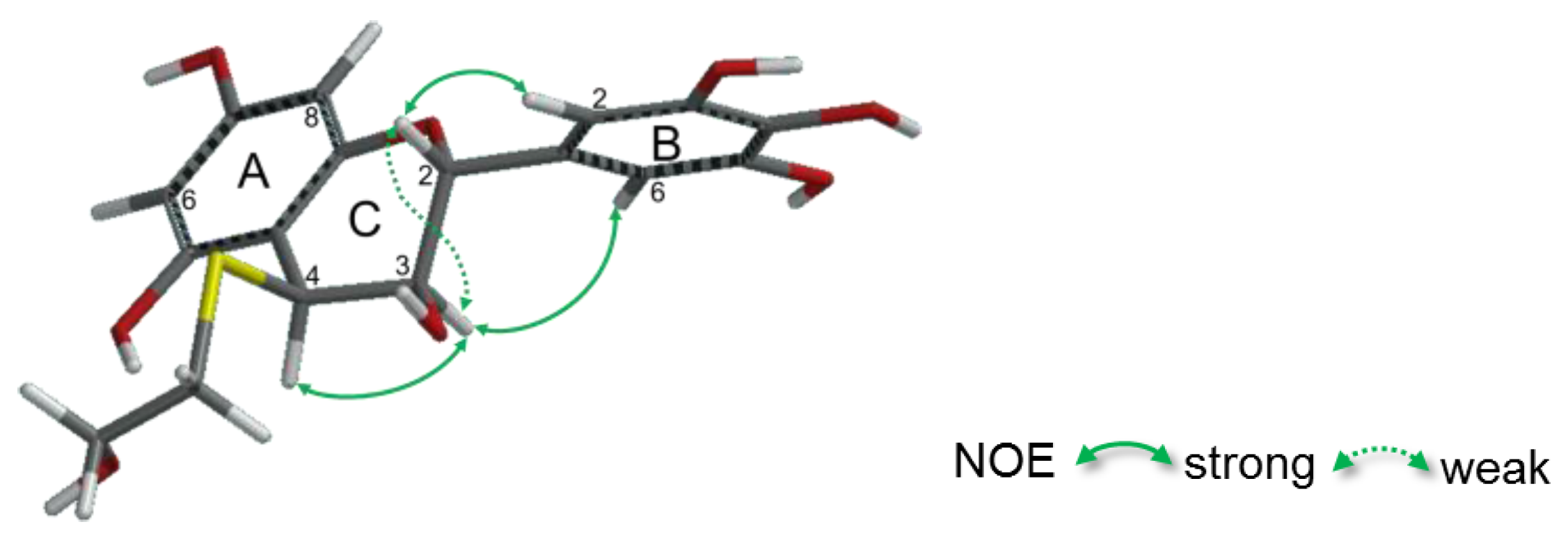
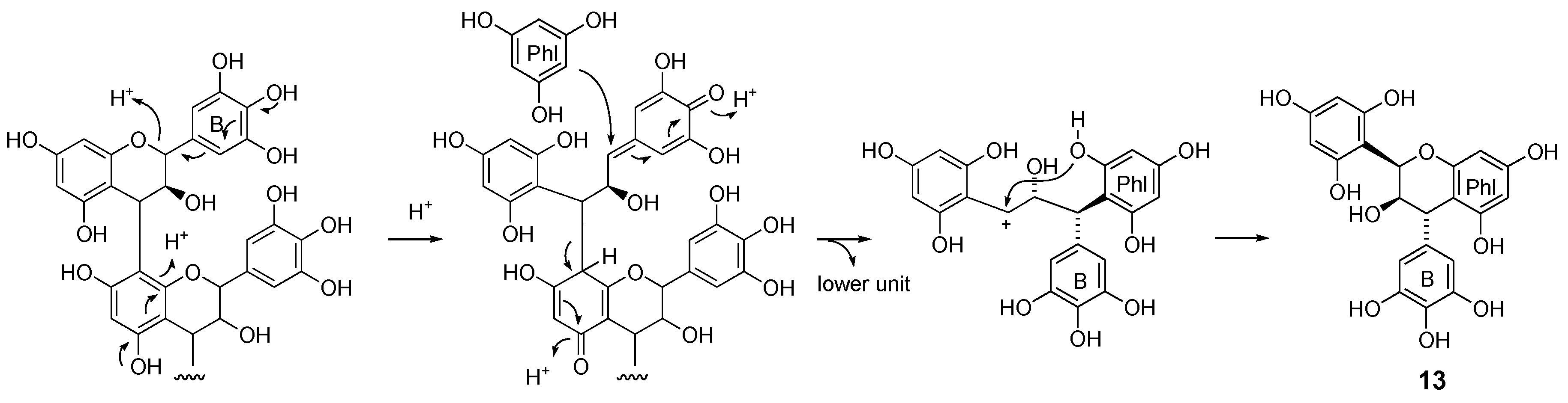
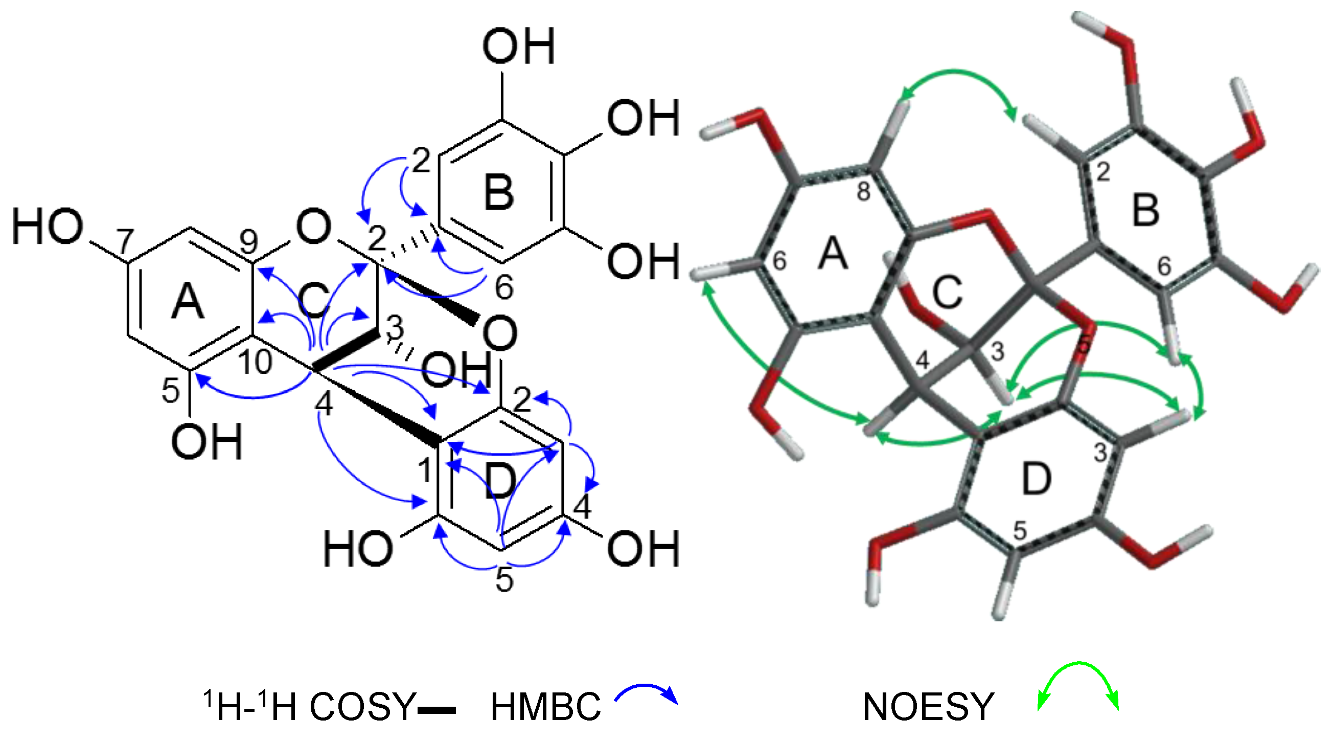
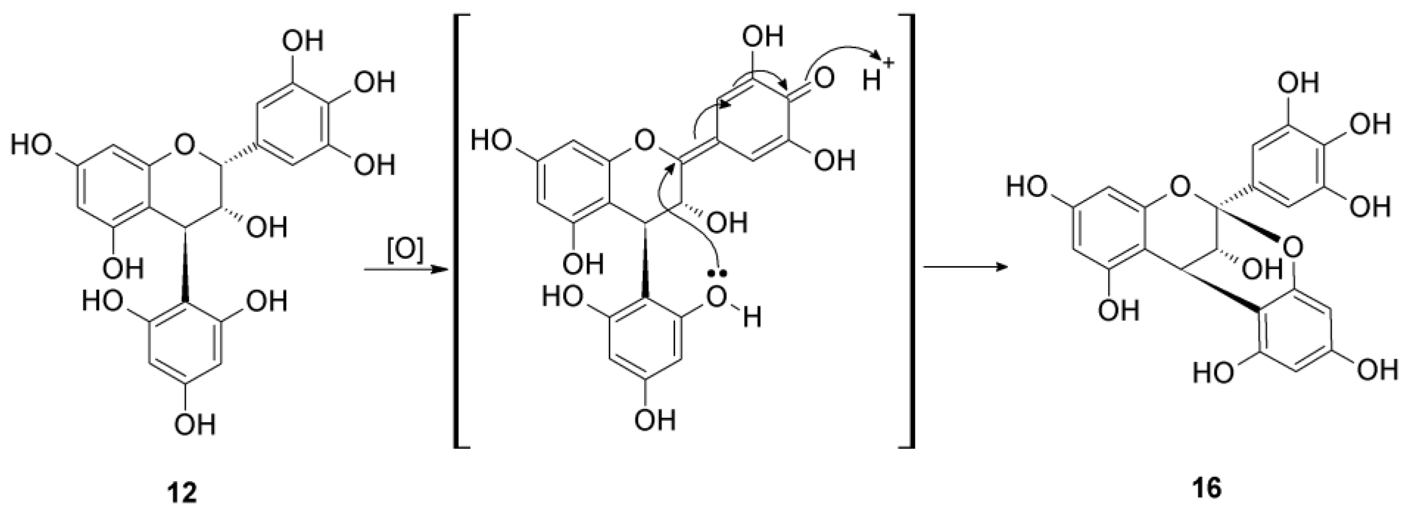
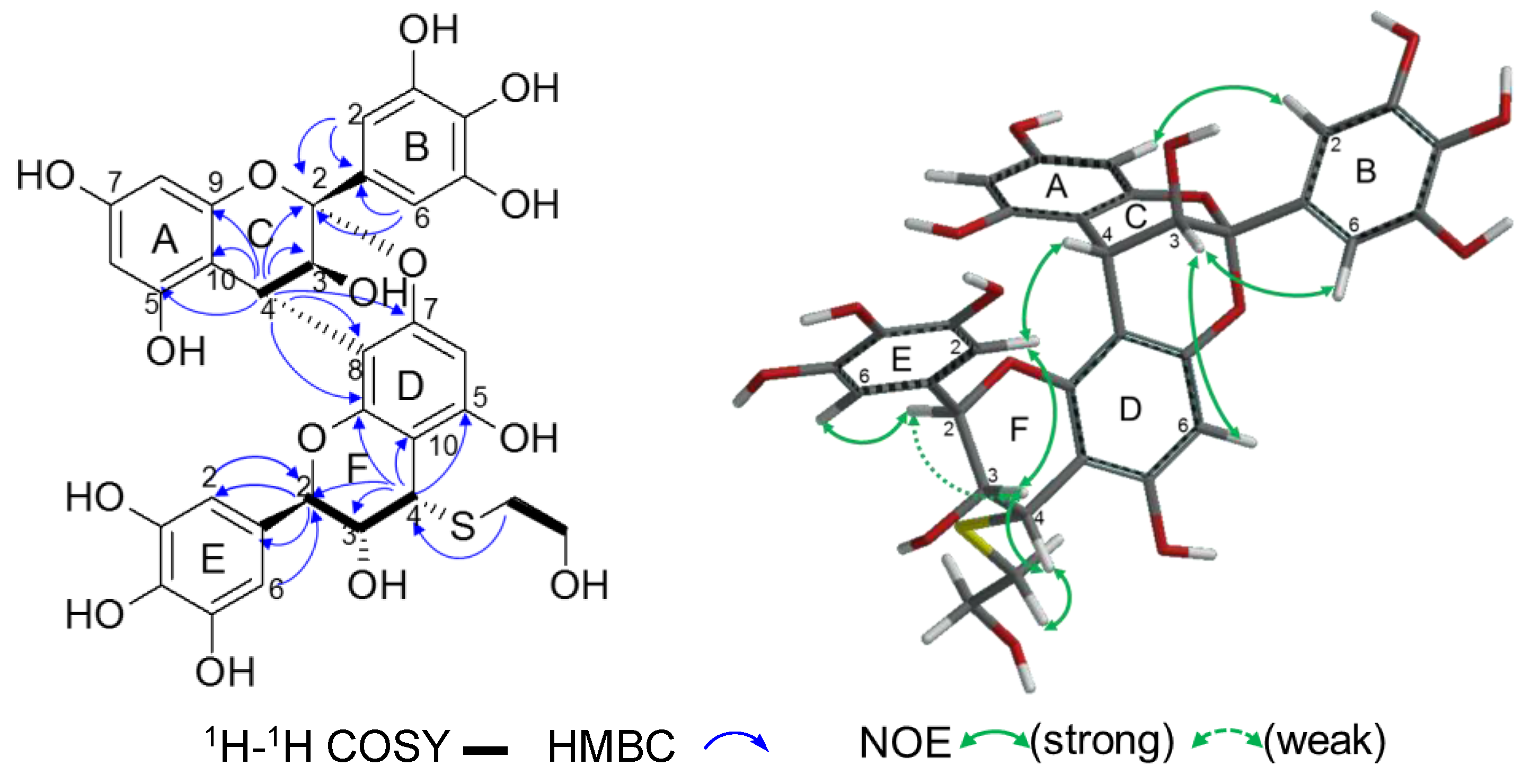
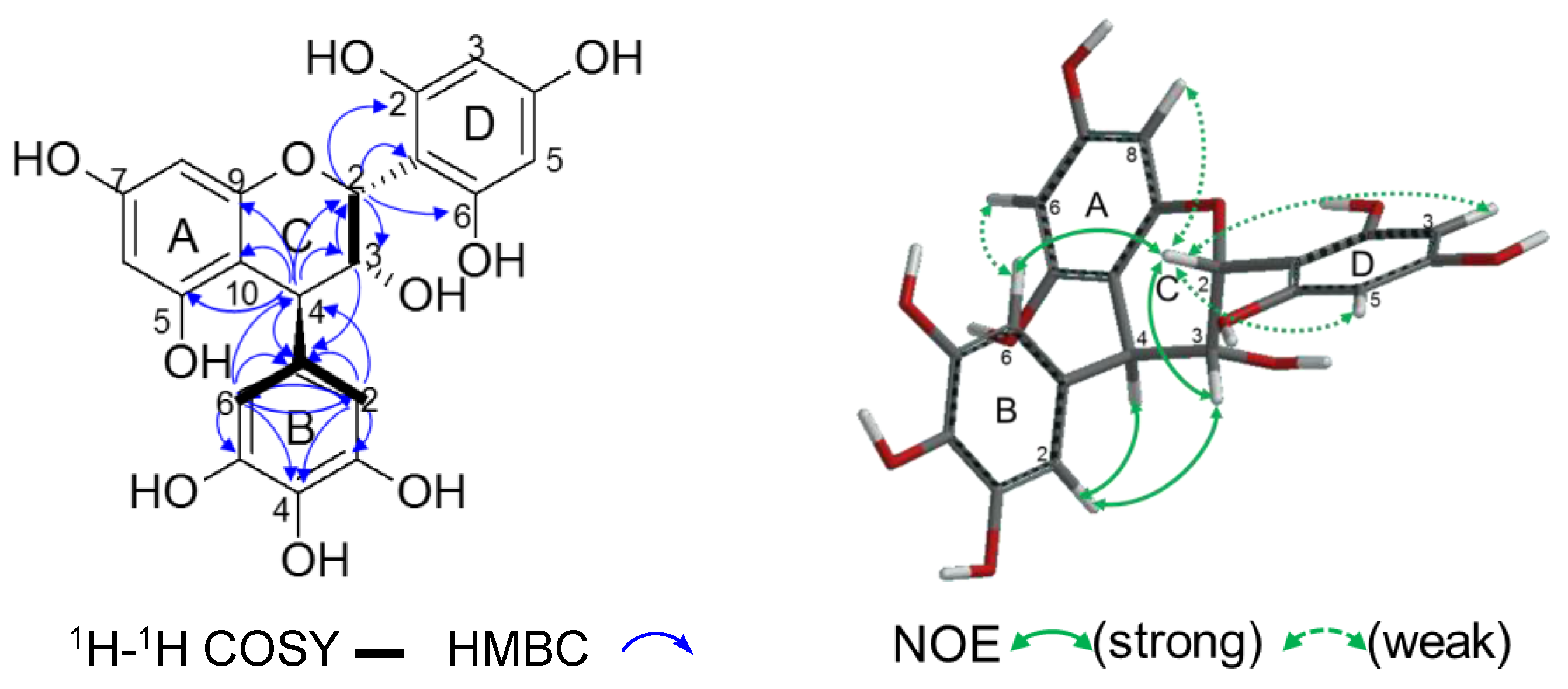
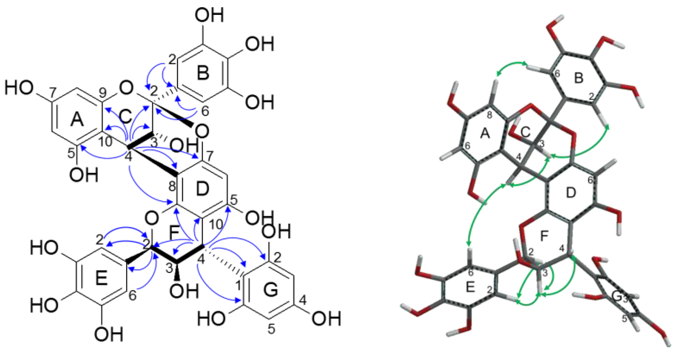
2.2.2. Structure Elucidation of New Degradation Products

Among the 18 isolated products, 1, 2, 3, 7, 13, 14, and 16 are reported here for the first time. Their structures are shown in Figure 5 and the 1H- and 13C-NMR spectroscopic data are summarized in Table 1 and Table 2.
The molecular formula of 1 was shown to be C32H30O15S based on the [M + H]+ peak at m/z 687.1382 in HRFABMS, indicating that 1 was a mercaptoethanol adduct of a prodelphinidin dimer with a B-type linkage. This was confirmed by the appearance of two intense signals at δH 6.57 and δH 6.67 (each 2H) arising from two pyrogallol-type B-rings and two methine proton signals attributable to C-ring H-2 in the 1H-NMR signals (Table 1). In the HSQC spectrum, the C-ring H-2 signal at δH 4.34 (J = 9.4 Hz) was correlated to a carbon signal at δC 83.2, while the other F-ring H-2 (Figure 6) at δH 5.31 (br s) was found to be connected to the carbon that resonated at δC 75.05. The former indicated a 2,3-trans configuration and the latter, a 2,3-cis configuration [33]; thus, the dimer was composed of gallocatechin and epigallocatechin. The 1H-1H COSY and HMBC correlations (Figure 6) allowed the determination of the connectivity of the two catechin units and hydroxyethylthiol group. The strong NOESY correlations between C-ring H-2 and H-4 (Figure 6), and between -SCH2- and F-ring H-3, confirmed the configuration of the C- and F-rings [39,40]. A linkage between C-ring C-4 and D-ring C-8 was deduced from the NOESY correlation between aromatic E-ring H-2, 6 and C-ring H-4 [41,42].
ECD spectroscopy allowed the determination of the absolute configuration at C-4. The 1H coupling constants of the C-ring indicated that the B-ring was in an equatorial position (E-conformer). Taking this observation into account, the negative Cotton effect at 218 nm implied an α-orientation of the terminal unit at C-4 [40]. Thus, the extension unit was concluded to be (+)-gallocatechin. The establishment of the absolute configuration of the epigallocatechin unit relied on the Cotton effect at the 1Lb band (280 nm) rather than the 1La band (220–240 nm) [40]. Here, the negative Cotton effect at 288 nm, in addition to the predominance of E-conformers, both led to the conclusion that the pyrogallol E-ring had an α-orientation relative to the F-2 carbon. The terminal unit was thereby designated as (−)-epigallocatechin. Furthermore, the ECD spectrum for 1 showed a close resemblance to those of procyanidins B-4 previously observed by Barrett and colleagues [43]. Accordingly, 1 was concluded to be (+)-gallocatechin-(4→8)-(−)-epigallocatechin-4-(2-hydroxyethyl)thioether.
Product 2 showed the [M + H]+ peak at m/z 383.0801 in HRFABMS, confirming the molecular formula as C17H18O8S. The 1H-NMR spectrum (Table 1) showed a doublet signal at δH 4.77 (J = 9.6 Hz), indicating the 2,3-trans configuration characteristic of gallocatechin. The C-ring H-4 resonated as a doublet at δH 4.36 (J = 4.3 Hz), which indicated the 3,4-cis configuration [31]. This was further confirmed by the appearance of a strong NOESY correlation between H-3 and H-4, and the absence of NOE between H-2 and H-4 (Figure 7). As for the absolute configuration, a negative Cotton effect at 284 nm in the ECD spectrum, which was similar to that of (+)-catechin [43], suggested P-helicity for the flavan A- and B-rings. Based on these results, 2 was concluded to be (+)-gallocatechin-4-(2-hydroxyethyl)thioether.
Compound 3 was found to have the molecular formula C47H40O22S based on the [M + Na]+ peak at m/z 1011.1639 in HRFABMS. This implied that 3 was a thioether of a prodelphinidin trimer involving both A-type and B-type linkages. In the 1H-NMR spectrum (Table 1), three intense aromatic singlets at δH 6.77, δH 6.70, and δH 6.56 (each 2H) indicated the presence of three pyrogallol-type B-rings. The presence of two B-type linkages was apparent from the two C-ring H-2 signals resonating at δH 5.31 and δH 5.29 with small J2,3 values (<2 Hz). This again indicated that the two units were epigallocatechin. These spectroscopic features suggested a close relationship between 3 and the epigallocatechin trimer isolated from E. sinica with A- and B-type linkages [12]. In the HMBC spectrum of 3 (Figure 8), the ketal carbon C-2 (δC 99.91) of the A-type linkage was correlated to C-ring H-4 (δH 4.38, J = 3.4 Hz), which was in turn correlated to a D-ring C-9 (δC 151.31) of the middle unit. Another benzylic methine H-4 of the middle unit F-ring (δH 4.79, J = 2.2 Hz) showed an HMBC correlation to the D-ring C-9 and terminal unit G-ring C-9 (δC 153.90). This indicated that an A-type linkage was involved between the top and middle units. The 13C-NMR chemical shift for F-ring C-4 at δC 36.21 was consistent with its involvement in a B-type linkage at this position [12,33,44], and the I-ring C-4 at a lower field (δC 43.83) was indicative of a thioether at this position [38]. The F-ring H-4 and I-ring H-4 were observed as doublet signals with coupling constants of J = 2.3 Hz and J = 2.4 Hz, respectively, indicating that both flavan rings adopted a 3,4-trans configuration [31]. This was further supported by the absence of a NOESY correlation between H-2 and H-4 in both the F-ring and the I-ring (Figure 8). The linkage between rings C and D was established as 4→8, 2→O→7 by the presence of a NOESY correlation between E-ring H-2,6 and C-ring H-4 [41]. The connection between the F-ring and the G-ring was also determined to be from C-4 to C-8 based on the NOESY cross peaks of H-ring H-2,6 with F-ring H-4 and H-3.
The ECD spectrum of 3 showed a strong positive Cotton effect at 233 nm, reflecting the configuration at C-ring C-4, thereby establishing the top extension unit as (−)-epigallocatechin; however, the configuration of the middle and bottom epigallocatechin units could not be determined from the ECD data. Prodelphinidin oligomers with (+)-epigallocatechin units were previously isolated from the same plant source [12]; therefore, the absolute configuration of 3 was established by TDDFT calculations of the ECD spectra for four stereostructures: (a) (−)-epigallocatechin-(−)-epigallocatechin-(−)-epigallocatechin, (b) (−)-epigallocatechin-(+)-epigallocatechin-(−)-epigallocatechin, (c) (−)-epigallocatechin-(−)-epigallocatechin-(+)-epigallocatechin, and (d) (−)-epigallocatechin-(+)-epigallocatechin-(+)-epigallocatechin (Figure 9). The experimental ECD spectrum of 3 (Figure 9e) showed a positive Cotton effect at 233 nm and a negative Cotton effect at 218 nm, similar to the Cotton effects observed in the calculated ECD spectra a and c. This comparison of calculated and experimental spectra revealed that the absolute structure of the upper and middle units in 3 was (−)-epigallocatechin. Moreover, the experimental spectrum e contained a weak negative Cotton effect at 250–300 nm, similar to the negative Cotton effect in the calculated ECD spectrum a. Therefore, 3 was established as (−)-epigallocatechin-(4β→8,2→O→7)-(−)-epigallocatechin-(4β→8)-(−)-epigallocatechin-4-(2-hydroxyethyl)thioether.
Compound 7 was characterized as an A-type prodelphinidin dimer with a mercaptoethanol substituent, and its molecular formula was determined as C32H28O15S from the [M + Na]+ peak at m/z 707.1043 in HRFABMS. The presence of an A-type linkage was apparent from the signal at δC 100.07, attributable to the C-ring C-2 ketal carbon [12,42]. The large coupling constant (J = 9.8 Hz) of the H-2 at δH 4.99 indicated the 2,3-trans configuration of the lower unit F-ring. The coupling constants of the C-ring H-4 at δH 4.17 (J = 3.6 Hz) and F-ring H-4 at δH 4.39 (J = 4.3 Hz), which were similar to the values observed in 2, suggested the 3,4-cis configuration of these rings. A comparison of the 1H- and 13C-NMR data with those in the literature suggested that the dimer was composed of epigallocatechin and gallocatechin [12,37]. This was supported by a strong NOE between F-3 and F-4, and weak NOE between F-2 and F-3 (Figure 10). The linkage between the C- and D-rings was established to be 4→8 by the observation of a NOESY correlation between C-ring H-4 and E-ring H-2,6. The absolute configuration at the C-ring C-4 was established by ECD spectroscopy, where the strong negative Cotton effect at 228 nm indicated that the extension unit was (+)-epigallocatechin. The terminal unit was designated as (+)-gallocatechin based on a comparison of the ECD spectrum with that of compound 2, which also had a negative Cotton effect, of a lesser amplitude, at 284 nm. Compound 7 was thereby established as (−)-epigallocatechin-(4α→8,2→O→7)-(+)-gallocatechin-4-(2-hydroxyethyl)thioether.
Compound 13 was obtained as a product of phloroglucinolysis, and the HRFABMS peak (m/z 431.0979 [M + H]+) confirmed the molecular formula as C21H18O10, the same as that of 11 and 12. Because of overlapping C-ring proton signals in the 1H-NMR spectrum measured in acetone-d6, the 2D NMR spectra were measured in methanol-d4 (Table 2). The resulting 1H-NMR spectrum showed signals attributable to pyrogallol (δH 6.19, 2H) and phloroglucinol (δH 5.79, 2H) rings, as well as mutually meta-coupled A-ring H-6 and H-8 (δH 5.94 and 5.95, J = 2.4 Hz), which were related to those observed in the spectra of 11 and 12 [12,37,42]. In the 1H-1H COSY spectrum (Figure 11), a broad aliphatic singlet signal at δH 5.53 was correlated to a methine signal at δH 4.05 (J = 2.4, 1.0 Hz), and these signals were attributed to C-ring H-2 and H-3, respectively. The small coupling constant suggested the 2,3-cis configuration [33]. Another aliphatic methine signal at δH 4.06 was assigned to C-ring H-4 based on its HMBC correlations to A-ring C-5, 9 and 10 (Figure 11). H-4 also showed HMBC correlations with pyrogallol H-2,6 (δH 6.19), indicating that the pyrogallol ring was attached to C-4. This was further supported by the long-range 1H-1H coupling between C-ring H-4 and pyrogallol B-ring H-2,6 in the 1H-1H COSY spectrum and the HMBC cross peak between C-ring H-3 and pyrogallol C-1 [45]. The remaining moiety, i.e., the phloroglucinol ring with a symmetrical structure, was shown to be located at C-ring C-2 by the HMBC correlation of C-ring H-2 to the phloroglucinol C-1, 2, and 6. The 2,3-cis-3,4-trans configuration was inferred by a comparison of the coupling constants with those in the literature [31], and this was further supported by the absence of NOE between H-2 and H-4 and occurrence of the strong NOE between C-ring H-2 and pyrogallol B-ring H-2,6 (Figure 11). The weak correlation observed between the C-ring H-2 and A-ring H-8 protons suggested that H-2 was at the axial position, thereby implying that the C-ring adopted the E-conformation. From the positive Cotton effect at 227 nm, the absolute configuration at C-4 was determined to be S [31]. Accordingly, 13 was concluded to be 2-(2,4,6-trihydroxyphenyl)-4-(3,4,5-trihydroxyphenyl)-3,4-dihydro-2H-1-benzopyran-3,5,7-triol (2R,3R,4S). This compound was a byproduct of phloroglucinolysis, and a plausible production mechanism is proposed in Scheme 2.
Compound 14 was determined to have the molecular formula C36H28O17 (m/z 733.1406, [M + H]+), identifying it as a phloroglucinol adduct of a prodelphinidin dimer involving an A-type linkage. The signals in the 1H- and 13C-NMR spectra were related to those of the epigallocatechin–epigallocatechin dimer [12] and the procyanidin A2–phloroglucinol adduct [46], and their assignments (Table 2) were based on a comparison with the reported data. The signal of F-ring H-2 (δH 5.32) was observed as a singlet, indicating the 2,3-cis configuration of the F-ring [33]. In addition, the coupling constant of the F-ring H-4 (δH 4.63, J = 2.3 Hz) was consistent with the 3,4-trans configuration [31]. This was confirmed by the NOESY spectrum, which displayed a strong correlation between F-ring H-3 and H-4, but no NOE between H-2 and H-4 (Figure 12). The 4→8 linkage between the C- and D-rings was established by the NOE between E-ring H-2,6 and C-ring H-4. Furthermore, the strong positive Cotton effect at 232 nm established that the upper unit was (−)-epigallocatechin [12]. On the basis of previous studies of compound 7, by Nam et al. [42] and Barrett et al. [43], and considering the weak positive Cotton effect at 220–240 nm of 12, the lower unit was deduced to be (−)-epigallocatechin. It was therefore concluded that compound 14 was (−)-epigallocatechin-(4β→8,2→O→7)-(−)-epigallocatechin-4-phloroglucinol.
Product 16 showed the [M + H]+ peak at m/z 429.0818 in HRFABMS, indicating the molecular formula C21H16O10. An unambiguous assignment of the 1H- and 13C-NMR signals was achieved by 1H-1H COSY, HSQC, HMBC, and NOESY spectroscopy. The absence of a C-2 proton signal and appearance of a C-2 carbon signal at δC 100.21 confirmed the presence of an A-type linkage [12,44,46]. The HMBC cross peaks (Figure 13) between C-ring H-4 and D-ring C-1,2,6 indicated the linkage of the phloroglucinol moiety to C-ring C-4. The NOE cross peaks between H-3 and B-ring H-2,6 indicated the 3,4-trans configuration [47]. Furthermore, the ECD spectrum showed a positive Cotton effect at 220–240 nm, indicating a β- configuration at C-4. Accordingly, 16 was established to be (−)-epigallocatechin-(4β→1,2→O→2)-phloroglucinol. Compound 16 was regarded as a byproduct of phloroglucinolysis involving the oxidation of the pyrogallol-type B-ring (Scheme 3) [45].

3. Materials and Methods

3.1. General

NMR spectra were recorded in acetone-d6 (Wako Pure Chem. Ind. Ltd., Osaka, Japan), methanol-d4 (Kanto Chem. Co., Inc., Tokyo, Japan), and DMSO-d6 (Kanto Chemical Co., Inc., Tokyo, Japan) with a Varian Unity Plus 500 spectrometer (Palo Alto, CA, USA) operating at 500 MHz for 1H and 125 MHz for 13C, and with a JEOL JNM-AL 400 spectrometer (JEOL Ltd, Tokyo, Japan) at 400 MHz for 1H and 100 MHz for 13C. HRFABMS spectra were recorded on a JMS 700N spectrometer (JEOL Ltd., Tokyo, Japan) in positive ion mode, with glycerol or m-nitrobenzyl alcohol, with or without NaCl, as the matrix. UV spectra were recorded in MeOH with a Jasco V-560 UV/Vis spectrometer (Jasco Co. Ltd., Tokyo, Japan). The same solvent was used for the ECD spectroscopic analysis using a Jasco-725N spectrometer (Jasco Co. Ltd., Tokyo, Japan), and optical rotation measurement using a Jasco P-1020 (Jasco Co. Ltd., Tokyo, Japan). IR spectra were recorded using a Jasco FT/IR-410K (Jasco Co. Ltd., Tokyo, Japan). Column chromatography was performed using a Sephadex LH-20 (25–100 mm, GE Healthcare UK Ltd., Buckinghamshire HP7 9NA, UK), a Diaion HP20SS (Mitsubishi Chemical Co., Tokyo, Japan), and a Chromatorex ODS (Fuji Silysia Chemical Ltd., Kasugai, Japan). TLC was performed on 0.25-mm thick, precoated silica gel 60 F254 (Merck, Darmstadt, Germany) with toluene–ethyl formate–formic acid (1:7:1, v/v) as the solvent system. Spots were detected by illumination under a short wavelength UV (254 nm) followed by spraying with 2% ethanolic FeCl3. Analytical HPLC was performed with gradient elution from 4–30% (39 min), 30–75% (15 min), 75–95% (6 min) acetonitrile (Kanto Chemical Co., Inc., Tokyo, Japan) in 50 mM phosphoric acid (Kishida Chemical Co., Osaka, Japan) on a Cosmosil 5C18-ARII 4.6 × 250 mm column (Nacalai Tesque, Inc., Kyoto, Japan) at a flow rate of 0.8 mL/min, using an HPLC system composed of a Jasco DG-2080-53 Plus degasser, Jasco PU-2080 Plus pump, Jasco AS-2055 Plus autosampler, Jasco CO-2065 Plus column oven (maintained at 35 °C), and Jasco MD-2018 Plus PDA detector (Jasco Co. Ltd., Tokyo, Japan).

3.2. Plant Material

Dried aerial parts of Ephedra sinica were purchased from Uchida Wakanyaku Ltd., Tokyo, Japan.

3.3. Extraction and Isolation

The dried aerial parts (500 g) of E. sinica were extracted with 70% acetone (3 L) at room temperature overnight, three times. The extracts were combined and concentrated by rotary evaporation under reduced pressure at 40 °C. The resulting aqueous solution was charged into a Sephadex LH-20 (5 cm × 19 cm) and eluted with H2O-MeOH (0–100%, 20% stepwise gradient) and 60% acetone to give two fractions: Fr. 1 and 2 (Figure S1). The first fraction eluted with H2O was acidified with trifluoroacetic acid and loaded into a Diaion HP20SS column (5 cm × 30 cm). After washing out sugars and inorganic substances with H2O, the column was eluted with 0–100% MeOH (10% stepwise gradient) and then 60% acetone to give Fr. 1-1 (9.16 g) containing proanthocyanidin oligomers and Fr. 1-2 (14.57 g) containing oligomers and low-molecular weight proanthocyanidins. A portion (5 g) of Fr. 1-2 was separated by size-exclusion column chromatography using a Sephadex LH-20 (4 cm × 45 cm) with a mixture of acetone and 7 M urea (3:2, v/v, containing conc. HCl 5 mL/L) to afford Fr. 1-2-1 containing oligomers and Fr. 1-2-2 containing low-molecular weight polyphenols. After the removal of acetone by evaporation, the resulting aqueous solution of Fr. 1-2-1 was subjected to Diaion HP20SS (3 cm × 19 cm) column chromatography, and urea and HCl were washed out by elution with H2O. Subsequent elution of the column with 0–100% MeOH (10% stepwise gradient) yielded Fr. 1-2-1-1 (1.11 g) containing oligomeric proanthocyanidins. Separately, Fr. 2 was subjected to size-exclusion chromatography in a manner similar to that described for Fr. 1-2 to give three fractions. The resulting Fr. 2-2 was loaded into a Diaion HP20SS column to remove urea and HCl, yielding oligomeric proanthocyanidins (Fr. 2-2-2, 3.36 g).

3.4. Thiolysis

Thiol degradation was performed according to the method of Kusano et al. [35] with modifications. Proanthocyanidin oligomers (Fr. 1-1, 1.0 g) were dissolved in 60% EtOH (200 mL) containing mercaptoethanol (10 mL) (Kanto Chemical Co. Inc., Tokyo, Japan) and concentrated HCl (0.5 mL) (Kishida Chemical Co., Osaka, Japan). The reaction mixture was then heated at 70 °C for 22 h. The reaction mixture was then analyzed by HPLC and was further fractionated. Fr. 2-2-2 (1.0 g) was also subjected to thiolysis in the same manner. The reaction mixture of Fr. 1-1 was first concentrated to remove EtOH. The resulting aqueous solution was subjected to Sephadex LH-20 chromatography (3 cm × 24 cm), and mercaptoethanol was washed out with H2O. Further elution of the column with increasing proportions of MeOH in H2O (0–50%, 5% stepwise gradient; 50–100%, 10% stepwise gradient) gave eight fractions. Fr 1-1-5 (0.35 g) was loaded into a Diaion HP20SS (3 cm × 22 cm) with H2O-MeOH to furnish five subfractions. Purification of Fr. 1-1-5-4 (41.9 mg) by Sephadex LH-20 chromatography (3 cm × 25 cm) with a systematic stepwise gradient of EtOH-H2O-acetone (1:0:0, 9:1:0, 8:2:0, 6:4:0, 54:36:10, 48:32:20, 36:24:40, 0:50:50, v/v) enabled the isolation of 10 (19.5 mg). Separation of Fr. 1-1-5-3 (178.7 mg) with the same chromatographic procedure afforded compounds 4 (4.9 mg) and 6 (60.8 mg). Fr. 1-1-5-2 (72.9 mg) was separated by Chromatorex ODS chromatography (3 cm × 17 cm) with H2O-MeOH and a Sephadex LH-20 (2.5 cm × 13 cm) with H2O-MeOH to afford 6 (21.0 mg), 1 (5.2 mg), and 2 (21.3 mg). Separation of Fr. 1-1-7 by Diaion HP20SS chromatography (2 × 17 cm) with H2O-MeOH afforded six subfractions, and Fr. 1-1-7-3 (104.3 mg) was further separated by Chromatorex ODS chromatography (2 cm × 16 cm) with H2O-MeOH to give 3 (21.2 mg), 7 (19.3 mg), and 9 (9.0 mg). The thiol degradation products of the other oligomers, Fr. 2-2-2, were also fractionated in the same manner as described for Fr. 1-1 to give seven subfractions. Fr. 2-2-2-6 (241.5 mg) was separated by Diaion HP20SS column chromatography (3 cm × 16 cm) with H2O-MeOH (0–100%, 20% stepwise gradient). Among the six subfractions obtained, the separation of Fr. 2-2-2-6-3 (74.5 mg) by a Chromatorex ODS (2.5 cm × 15 cm) with H2O-MeOH yielded 5 (0.6 mg), 7 (36.2 mg), and 9 (10.0 mg). The same chromatographic procedure using a Chromatorex ODS was applied to the separation of Fr. 2-2-2-6-4 (49.3 mg), which yielded 9 (21.0 mg) and a crude crop of 8. The latter was purified by Sephadex LH-20 (2 cm × 16 cm) and a system of increasing MeOH concentration in H2O (0–40%, 20% stepwise gradient; 40–100%, 5% stepwise gradient), which led to the isolation of 8 (8.8 mg).
(+)-Gallocatechin-(4α8)-(−)-epigallocatechin-4-(2-hydroxyethyl)thioether (1): yellowish brown amorphous powder; [ α ] D 16 −105.9 (c = 0.10, MeOH); UV (MeOH) λmax (log ε) 270 (3.61), 239 (4.41), 214 (5.00) nm; CD (MeOH) Δε218 −33.1, Δε288 −2.3; IR νmax 3311, 1609, 1449, 1541, 1449 cm−1; HRFABMS m/z 687.1382 [M + H]+ (calcd for C32H31O15S, 687.1378); 1H- and 13C-NMR data, see Table 1.
(+)-Gallocatechin-4-(2-hydroxyethyl)thioether (2): pale brown amorphous powder; [ α ] D 16 +29.9 (c = 0.13, MeOH); UV (MeOH) λmax (log ε) 273 (3.17), 235 (4.42), 209 (4.72) nm; CD (MeOH) Δε220 −2.8, Δε249 +2.2, Δε284 −0.4; IR νmax 3344, 1621, 1537, 1515, 1455, 1345 cm−1; HRFABMS m/z 383.0801 [M + H]+ (calcd for C17H19O8S, 383.0795); 1H- and 13C-NMR data, see Table 1.
(−)-Epigallocatechin-(4β8,2O7)-(−)-epigallocatechin-(4α8)-(−)-epigallocatechin-4-(2-hydroxyethyl) thioether (3): reddish brown amorphous powder; [ α ] D 20 −17.2 (c = 0.11, MeOH); UV (MeOH) λmax (log ε) 270 (3.83), 241 (4.65), 205 (5.31) nm; CD (MeOH) Δε218 −25.0, Δε233 +14.2, Δε284 −4.3; IR νmax 3276, 1615, 1541, 1445, 1348 cm−1; HRFABMS m/z 1011.1639 [M + Na]+ (calcd for C47H40O22SNa, 1011.1624); 1H- and 13C-NMR data, see Table 1.
(+)-Epigallocatechin-(4α8,2αO7)-(+)-gallocatechin-4-(2-hydroxyethyl)thioether (7): pale brown amorphous powder; [ α ] D 17 −50.1 (c = 0.15, MeOH); UV (MeOH) λmax (log ε) 270 (3.55), 247 (4.32), 208 (5.00) nm; CD (MeOH) Δε228 −25.6, Δε252 +3.7, Δε284 −3.0; IR νmax 3389, 1612, 1537, 1502, 1447, 1333 cm−1; HRFABMS m/z 707.1043 [M + Na]+ (calcd for C32H28O15SNa, 707.1041); 1H- and 13C-NMR data, see Table 1.

3.5. Phloroglucinolysis

Fr. 1-1 was subjected to phloroglucinolysis according to the method reported by Kennedy and Jones and Bautista-Ortin and colleagues [18,36], with a few modifications. Fr. 1-1 (2.0 g) was dissolved in MeOH (200 mL) and then mixed with phloroglucinol reagent (200 mL). This phloroglucinol reagent was a methanolic solution containing phloroglucinol (20 g) (Sigma Chemical Co., St. Louis, MO, USA), ascorbic acid (4 g) (Kishida Chemical Co., Osaka, Japan), and concentrated HCl (2.92 mL). The mixture was heated at 50 °C for 60 min, and the reaction was then terminated by the addition of 0.2 M sodium acetate (800 mL). The reaction mixture was concentrated in vacuo to remove MeOH and acidified to pH 4 prior to fractionation. The aqueous solution was loaded into a Sephadex LH-20 column (3 cm × 24 cm) and eluted with H2O containing increasing proportions of MeOH to give 10 fractions. Fr. 1-1-5 (810.7 mg) was separated by Diaion HP 20SS column chromatography (3 cm × 21 cm) with H2O-MeOH to yield 12 (84.2 mg), 16 (103.7 mg), and five subfractions. Fr. 1-1-5-3 (454.4 mg) was successively separated by a Sephadex LH-20 (H2O-MeOH) and a Chromatorex ODS (H2O-MeOH) to afford 11 (24.4 mg). From Fr. 1-1-5-5 (59.8 mg), 17 (6.8 mg) was isolated by Chromatorex ODS chromatography (H2O-MeOH). Purification of 1-1-6 (102.3 mg) by Diaion HP20SS column chromatography (3 cm × 26 cm) with 0–100% MeOH in H2O yielded 13 (10.7 mg). Fractionation of 1-1-7 (120 mg) on a Diaion HP20SS (2 cm × 17 cm) with H2O-MeOH gave 5 (9.3 mg) and nine subfractions. Purification of Fr. 1-1-7-2 (6.3 mg) and Fr. 1-1-7-7 (11.4 mg) by a Chromatorex ODS (H2O-MeOH) furnished 13 (1.2 mg) and 17 (3.2 mg), respectively. Fr. 1-1-8 (210.7 mg) was separated by Diaion HP20SS chromatography to yield 8 (16.3 mg) and five subfractions, and subfraction 1-1-8-1 (59.4 mg) was further subjected to purification using a Sephadex LH 20 (2 cm × 16 cm) with EtOH-H2O-acetone (1:0:0, 9:1:0, 8:2:0, 6:4:0, 54:36:10, 48:32:20, 36:24:40, 0:50:50, v/v) and then a Chromatorex ODS (H2O-MeOH) to give 14 (39.6 mg). Purification of Fr. 1-1-9 (204.8 mg) by Diaion HP 20SS chromatography (2 × 17 cm) with H2O-MeOH resulted in the isolation of 18 (11.4 mg).
2-(2,4,6-Trihydroxyphenyl)-4-(3,4,5-trihydroxyphenyl)-3,4-dihydro-2H-1-benzopyran-3,5,7-triol (2R,3R,4S) (13): pale brown amorphous powder; [ α ] D 20 −15.0 (c = 0.11, MeOH); UV (MeOH) λmax (log ε) 270 (3.50), 234 (4.40), 210 (4.85) nm; CD (MeOH) Δε227 +0.8, Δε267 +1.2; IR νmax 3333, 1614, 1522, 1462, 1328 cm−1; HRFABMS m/z 431.0979 [M + H]+ (calcd for C21H19O10, 431.0973); 1H- and 13C-NMR data, see Table 2.
(−)-Epigallocatechin-(8,2O7)-(−)-epigallocatechin-4-phloroglucinol (14): pale brown amorphous powder; [ α ] D 19 +53.1 (c = 0.14, MeOH); UV (MeOH) λmax (log ε) 270 (3.62), 235 (4.39), 214 (4.84) nm; CD (MeOH) Δε232 +18.9, Δε270 +1.5, Δε283 −1.2; IR νmax 3297, 1625, 1476, 1347 cm−1; HRFABMS m/z 733.1406 [M + H]+ (calcd for C36H29O17, 733.1399); 1H- and 13C-NMR data, see Table 2.
(−)-Epigallocatechin-(1,2O2)-phloroglucinol (16): pale yellow amorphous powder; [ α ] D 19 −6.6 (c = 0.14, MeOH); UV (MeOH) λmax (log ε) 270 (3.62), 235 (4.39), 214 (4.84) nm; CD (MeOH) Δε216 +4.2, Δε246 +1.2, Δε270 −1.6; IR νmax 3297, 1625, 1476, 1347 cm−1; HRFABMS m/z 429.0818 [M + H]+ (calcd for C21H17O10, 429.0816); 1H- and 13C-NMR data, see Table 2.

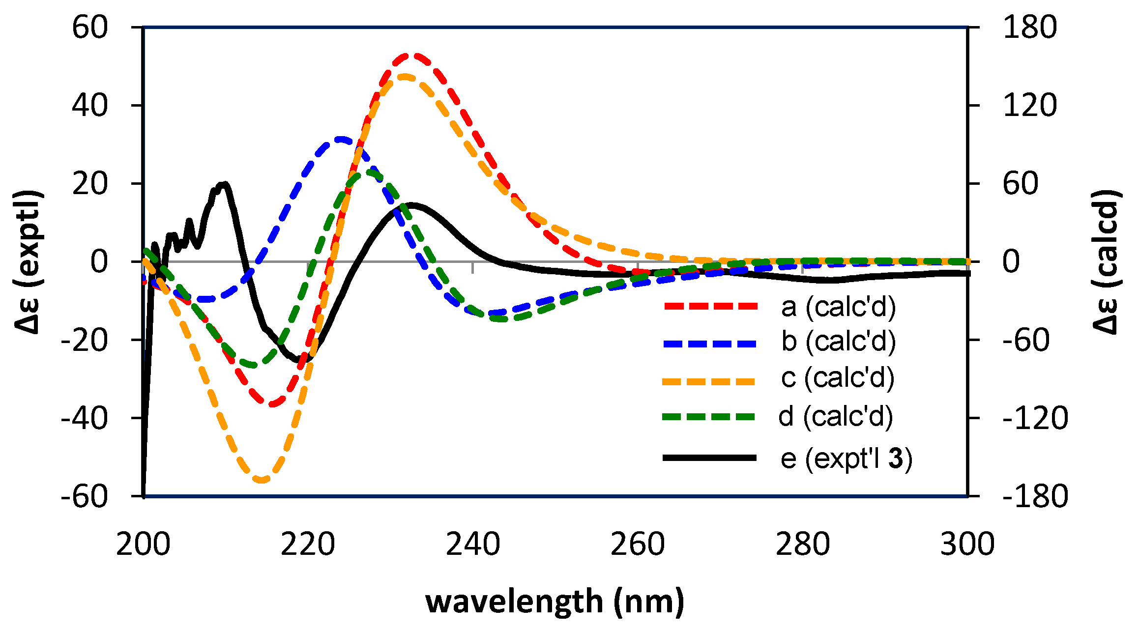
3.6. Calculations of ECD Spectra

A conformational search was performed using the Monte Carlo method with the MMFF94 force field using Spartan’14 (Wavefunction, Irvine, CA, USA). The resulting low-energy conformers within 6 kcal/mol of the global minimum were optimized at the B3LYP-SCRF/6-31G(d,p) level in MeOH (PCM). The vibrational frequencies were also calculated at the same level to confirm that they were true minima, and no imaginary frequencies were found. The energies, oscillator strengths, and rotational strengths of the low-energy conformers with Boltzmann populations greater than 1% were calculated using TDDFT at the CAM-B3LYP-SCRF/6-31G(d,p) level in MeOH (PCM). The ECD spectra were simulated by overlapping Gaussian functions with a 0.3 eV exponential half-width, and red-shifted by 25 nm. The calculated data for each conformer were averaged according to the Boltzmann distribution theory at 298 K based on their relative Gibbs free energies. All DFT calculations were performed using Gaussian 09 [48].

4. Conclusions

The proanthocyanidins of E. sinica are mainly composed of oligomers, and in this study, the oligomers were separated and chemically characterized for the first time. Acid-catalyzed degradation with mercaptoethanol and phloroglucinol afforded 18 products, among which seven were previously unreported compounds. Epigallocatechin was the major extension unit, and catechin and A-type prodelphinidin dimers were identified as terminal units. The new compounds were characterized by spectroscopic analyses, and the stereochemistry of the trimeric products was determined with the aid of TDDFT calculations of the ECD spectra. Since E. sinica is one of the most important crude drugs in East Asia, and proanthocyanidins are major constituents with a comparable abundance to alkaloids, our results provide important insights into the molecular basis of traditional medicine.

Supplementary Materials

The following are available online: Figure S1: Fractionation of proanthocyanidin oligomers of E. sinica, Figures S2–S4, Scheme for isolation of acid-catalyzed degradation products of E. sinica, Figures S5–S53, IR, 1H-NMR, 13C-NMR, 1H-1H COSY, HSQC, HMBC, and NOE spectra of compounds 1, 2, 3, 7, 13, 14, and 16.

Acknowledgments

This work was supported by the Japan Society for the Promotion of Science KAKENHI (Grant No. 17K08338 and 26460125). The authors are grateful to N. Tsuda, K. Chifuku, and H. Iwata at the Center for Industry, University and Government Cooperation, Nagasaki University, for recording the NMR and MS data. In addition, J.O. would like to express her sincere thanks to the Japanese Government (MEXT) Scholarships for doctoral scholarships. We thank Edanz Group (www.edanzediting.com/ac) for editing a draft of this manuscript. The computation was partly carried out using the computer facilities at the Research Institute for Information Technology, Kyushu University.

Author Contributions

T.T. and Y.M. conceived and designed the experiments, and analyzed the data; J.O. performed the experiments and analyzed the data. Y.M. performed the calculation and analyzed the ECD spectra of compound 3. J.O., Y.M., Y.S., and T.T. all wrote the paper.

Conflicts of Interest

The authors declare no conflict of interest.

References

  1. Zhu, Y. Chinese Materia Medica: Chemistry, Pharmacology and Applications; Harwood Academic Publisher: Amsterdam, The Netherlands, 1998; pp. 48–51. ISBN 90-5702-285-0. [Google Scholar]
  2. World Health Organization. WHO Monographs on Selected Medicinal Plants; World Health Organization: Geneva, Switzerland, 1999; Volume 1, pp. 145–153. ISBN 9241545178. [Google Scholar]
  3. Wang, L.; Kakiuchi, N.; Mikage, M. Studies of Ephedra plants in Asia. Part 6: Geographical changes of anatomical features and alkaloids content of Ephedra sinica. J. Nat. Med. 2009, 64, 63–69. [Google Scholar] [CrossRef] [PubMed]
  4. Liu, Y.; Sheu, S.; Chiou, S.; Chang, H.; Chen, Y. A comparative study of commercial samples of Ephedrae herba. Planta Med. 1993, 59, 376–378. [Google Scholar] [CrossRef] [PubMed]
  5. Westfall, T.; Westfall, D. Adrenergic agonists and antagonists. In Goodman & Gilman's the Pharmacological Basis of Therapeutics, 12th ed.; Brunton, L., Chabner, B., Knollman, B., Eds.; McGraw-Hill Medical: New York, NY, USA, 2011; pp. 277–333. ISBN 978-0-07-162442-8. [Google Scholar]
  6. Caveney, S.; Charlet, D.; Freitag, H.; Maier-Stolte, M.; Starratt, A. New observations on the secondary chemistry of world Ephedra (Ephedraceae). Am. J. Bot. 2001, 88, 1199–1208. [Google Scholar] [CrossRef] [PubMed]
  7. De Bruyne, T.; Pieters, L.; Delstra, H.; Vlietinck, A. Condensed vegetable tannins: Biodiversity in structure and biological activities. Biochem. Syst. Ecol. 1999, 27, 445–459. [Google Scholar] [CrossRef]
  8. Magos, G.; Mateos, J.; Páez, E.; Fernández, G.; Lobato, C.; Márquez, C.; Enríquez, R. Hypotensive and vasorelaxant effects of the procyanidin fraction from Guazuma ulmifolia bark in normotensive and hypertensive rats. J. Ethnopharm. 2008, 117, 58–68. [Google Scholar] [CrossRef] [PubMed]
  9. Sanz, M.; Terencio, M.; Paya, M. Isolation and hypotensive activity of a polymeric procyanidin fraction from Pistacia lentiscus L. Pharmazie 1992, 47, 466–467. [Google Scholar] [PubMed]
  10. Zeng, X.; Ma, Y.; Gu, H.; Gu, Y.; Huang, M. The effect of oligomeric proanthocyanidin on airway microenvironment in asthma. Eur. Respir. J. 2016, 48 (Suppl. S60). [Google Scholar] [CrossRef]
  11. Zhou, D.; Fang, S.; Zou, C.; Zhang, Q.; Gu, W. Proanthocyanidin from grape seed extract inhibits airway inflammation and remodeling in a murine model of chronic asthma. Nat. Prod. Commun. 2015, 10, 257–262. [Google Scholar] [PubMed]
  12. Zang, X.; Shang, M.; Xu, F.; Liang, J.; Wang, X.; Mikage, M.; Cai, S. A-Type proanthocyanidins from the stems of Ephedra sinica (Ephedraceae) and their antimicrobial activities. Molecules 2013, 18, 5172–5189. [Google Scholar] [CrossRef] [PubMed]
  13. Bagheri-Gavkosh, S.; Bigdeli, M.; Shams-Ghahfarokhi, M.; Razzaghi-Abyaneh, M. Inhibitory effects of Ephedra major host on Aspergillus parasiticus growth and aflatoxin production. Mycopathologia 2009, 168, 249–255. [Google Scholar] [CrossRef] [PubMed]
  14. Okawa, M.; Kinjo, J.; Nohara, T.; Ono, M. DPPH (1,1-diphenyl-2-picrylhydrazyl) radical scavenging activity of flavonoids obtained from some medicinal plants. Biol. Pharm. Bull. 2001, 24, 1202–1205. [Google Scholar] [CrossRef] [PubMed]
  15. Kim, I.; Park, Y.J.; Yoon, S.; Lee, H. Ephedrannin A and B from roots of Ephedra sinica inhibit lipopolysaccharide-induced inflammatory mediators by suppressing nuclear factor-kB activation in RAW 264.7 macrophages. Int. Immunopharmacol. 2010, 10, 1616–1625. [Google Scholar] [CrossRef] [PubMed]
  16. Kim, S.; Son, K.; Chang, H.; Kang, S.; Kim, H. Inhibitory effects of plant extracts on adjuvant-induced arthritis. Arch. Pharm. Res. 1997, 20, 313–317. [Google Scholar] [CrossRef] [PubMed]
  17. Chen, R.; Zhu, G.; Xu, Z. Effect of different extracts from Ephedra on cell immunity. J. Nanjing Univ. Tradit. Chin. Med. 2001, 4, 15. [Google Scholar]
  18. Li, L.; Yu, C.; Ying, H.; Yu, J. Antiviral effects of modified Dingchuan decoction against respiratory syncytial virus infection in vitro and in an immunosuppressive mouse model. J. Ethnopharm. 2013, 147, 238–244. [Google Scholar] [CrossRef] [PubMed]
  19. Nam, N.; Lee, C.; Hong, D.; Kim, H.; Bae, K.; Ahn, B. Antiinvasive, antiangiogenic and antitumor activity of Ephedra sinica extract. Phytother. Res. 2003, 17, 70–76. [Google Scholar] [CrossRef] [PubMed]
  20. Lee, M.; Cheng, W.; Che, C.; Hsieh, D. Cytotoxicity assessment of Ma-huang (Ephedra) under different conditions of preparation. Toxicol. Sci. 2000, 56, 424–430. [Google Scholar] [CrossRef] [PubMed]
  21. Tao, H.; Wang, L.; Cui, Z.; Zhao, D.; Liu, Y. Dimeric proanthocyanidins from the roots of Ephedra sinica. Planta Med. 2008, 74, 1823–1825. [Google Scholar] [CrossRef] [PubMed]
  22. Yokozawa, T.; Fujioka, K.; Oura, H.; Tanaka, T.; Nonaka, G.; Nishioka, I. Decrease in uremic toxins, a newly found beneficial effect of Ephedrae Herba. Phytother. Res. 1995, 9, 382–384. [Google Scholar] [CrossRef]
  23. Wang, G.Z.; Hikokichi, O. Experimental study in treating chronic renal failure with dry extract and tannins of Herba Ephedra. Zhongguo Zhong Xi Yi Jie He Za Zhi 1994, 14, 485–488. [Google Scholar] [PubMed]
  24. King, F.; Clark-Lewis, J.; Forbes, W. The chemistry of extractives from hardwoods. Part XXV. (—)-epiAfzelechin, a new member of the catechin series. J. Chem. Soc. 1955, 2948–2956. [Google Scholar] [CrossRef]
  25. Hikino, H.; Takahashi, M.; Konno, C. Structure of ephedrannin A, a hypotensive principle of Ephedra roots. Tetrahedron Lett. 1982, 23, 673–676. [Google Scholar] [CrossRef]
  26. Hikino, H.; Shimoyama, N.; Kasahara, Y.; Takahashi, M.; Konno, C. Structures of mahuannin A and B, hypotensive principles of Ephedra roots. Heterocycles 1982, 19, 1381–1384. [Google Scholar] [CrossRef]
  27. Kasahara, H.; Hikino, H. Structure of mahuannin D, a hypotensive principle of Ephedra roots. Heterocycles 1983, 20, 1953–1956. [Google Scholar] [CrossRef]
  28. Nonaka, G.; Morimoto, S.; Kinjo, J.; Nohara, T.; Nishioka, I. Tannins and related compounds L. Structures of proanthocyanidin A-1 and related compounds. Chem. Pharm. Bull. 1987, 35, 149–155. [Google Scholar] [CrossRef]
  29. Kennedy, J.; Jones, G. Analysis of proanthocyanidin cleavage products following acid-catalysis in the presence of excess phloroglucinol. J. Agric. Food Chem. 2001, 49, 1740–1746. [Google Scholar] [CrossRef] [PubMed]
  30. Yanagida, A.; Shoji, T.; Shibusawa, Y. Separation of proanthocyanidins by degree of polymerization by means of size-exclusion chromatography and related techniques. J. Biochem. Biophys. Methods 2003, 56, 311–322. [Google Scholar] [CrossRef]
  31. Kolodziej, H. 1H NMR spectral studies of procyanidin derivatives: Diagnostic 1H NMR parameters applicable to the structural elucidation of oligomeric procyanidins. In Plant Polyphenols-Synthesis, Properties, Significance; Hemingway, R., Laks, P.E., Eds.; Plenum Press: New York, NY, USA, 1992; pp. 295–319. ISBN 978-1-4615-3476-1. [Google Scholar]
  32. Porter, L.; Newman, R.; Foo, L.; Wong, H.; Hemingway, R. Polymeric proanthocyanidins. 13C-NMR studies of procyanidins. J. Chem. Soc. Perkin Trans. 1 1982, 1217–1221. [Google Scholar] [CrossRef]
  33. Foo, L.; Newman, R.; Waghorn, G.; McNabb, W.; Ulyatt, M. Proanthocyanidins from Lotus corniculatus. Phytochemistry 1996, 41, 617–624. [Google Scholar] [CrossRef]
  34. Chai, W.; Shi, Y.; Feng, H.; Qiu, L.; Zhou, H.; Deng, Z.; Yan, C.; Chen, X. NMR, HPLC-ESI-MS, and MALDI-TOF MS analysis of condensed tannins from Delonix regia (Bojer ex Hook.) Raf. and their bioactivities. J. Agric. Food Chem. 2012, 60, 5013–5022. [Google Scholar] [CrossRef] [PubMed]
  35. Kusano, R.; Ogawa, S.; Matsuo, Y.; Tanaka, T.; Yazaki, Y.; Kouno, I. α-amylase and lipase inhibitory activity and structural characterization of acacia bark proanthocyanidins. J. Nat. Prod. 2011, 74, 119–128. [Google Scholar] [CrossRef] [PubMed]
  36. Bautista-Ortin, A.; Molero, N.; Marín, F.; Ruiz-García, Y.; Gómez-Plaza, E. Reactivity of pure and commercial grape skin tannins with cell wall material. Eur. Food Res. Technol. 2015, 240, 645–654. [Google Scholar] [CrossRef]
  37. Tanaka, T.; Takahashi, R.; Kouno, I.; Nonaka, G. Chemical evidence for the de-astringency (insolubilization of tannins) of persimmon fruit. J. Chem. Soc. Perkin Trans. 1 1994, 3013–3022. [Google Scholar] [CrossRef]
  38. Tanaka, T.; Kondou, K.; Kouno, I. Oxidation and epimerization of epigallocatechin in banana fruits. Phytochemistry 2000, 53, 311–316. [Google Scholar] [CrossRef]
  39. Steynberg, J.; Brandt, E.; Hoffman, M.; Ferreira, D. Conformations of proanthocyanidins. In Plant Polyphenols-Synthesis, Properties, Significance; Hemingway, R., Laks, P.E., Eds.; Plenum Press: New York, NY, USA, 1992; pp. 501–519. ISBN 978-1-4615-3476-1. [Google Scholar]
  40. Slade, D.; Ferreira, D.; Marais, J.P.J. Circular dichroism, a powerful tool for the assessment of absolute configuration of flavonoids. Phytochemistry 2005, 66, 2177–2215. [Google Scholar] [CrossRef] [PubMed]
  41. Esatbeyoglu, T.; Jaschok-Kentner, B.; Wray, V.; Winterhalter, P. Structure elucidation of proanthocyanidin oligomers by low-temperature 1H NMR spectroscopy. J. Agric. Food Chem. 2011, 59, 62–69. [Google Scholar] [CrossRef] [PubMed]
  42. Nam, J.W.; Phansaklar, R.S.; Lankin, D.C.; McAlpine, J.B.; Leme-Kraus, A.A.; Vidal, C.M.; Gan, L.S.; Bedran-Russo, A.; Chen, S.N.; Pauli, G.F. Absolute configuration of native oligomeric proanthocyanidins with dentin biomodification potency. J. Org. Chem. 2017, 82, 1316–1329. [Google Scholar] [CrossRef] [PubMed]
  43. Barrett, M.; Klyne, W.; Scopes, P.; Fletcher, A.; Porter, L.; Haslam, E. Plant proanthocyanidins. Part 6. Chiroptical studies. Part 95. Circular dichroism of procyanidins. J. Chem. Soc. Perkin Trans. 1 1979, 2375–2377. [Google Scholar] [CrossRef]
  44. Yu, R.J.; Liu, H.B.; Yu, Y.; Liang, L.; Xu, R.; Liang, C.; Tang, J.S.; Yao, X.S. Anticancer activities of proanthocyanidins from the plant Urceola huaitingii and their synergistic effects in combination with chemotherapeutics. Fitoterapia 2016, 112, 175–182. [Google Scholar] [CrossRef] [PubMed]
  45. Burger, J.; Kolodziej, H.; Hemingway, R.; Steynberg, J.; Young, D.; Ferreira, D. Oligomeric flavonoids. Part 15a. Base-catalyzed pyran rearrangements of procyanidin B-2, and evidence for the oxidative transformation of B- to A-type procyanidins. Tetrahedron 1990, 46, 5733–5740. [Google Scholar] [CrossRef]
  46. Koerner, J.; Hsu, V.; Lee, J.K. Determination of proanthocyanidin A2 content in phenolic polymer isolates by reversed-phase high-performance liquid chromatography. J. Chromatogr. A 2009, 1216, 1403–1409. [Google Scholar] [CrossRef] [PubMed]
  47. Cronjé, A.; Burger, J.; Brandt, E.; Kolodziej, H.; Ferreira, D. Assessment of 3,4-trans and 3,4-cis relative configurations in the A-series of 4,8-linked proanthocyanidins. Tetrahedron Lett. 1990, 31, 3789–3792. [Google Scholar] [CrossRef]
  48. Frisch, M.J.; Trucks, G.W.; Schlegel, H.B.; Scuseria, G.E.; Robb, M.A.; Cheeseman, J.R.; Scalmani, G.; Barone, V.; Mennucci, B.; Petersson, G.A.; et al. Gaussian 09, Revision D.01; Gaussian, Inc.: Wallingford, CT, USA, 2013. [Google Scholar]
Sample Availability: Samples of compounds 118 are all available from the authors.
Figure 1. HPLC profiles of 60% EtOH extract of E. sinica (a) and proanthocyanidin oligomer fraction (b).
Molecules 22 01308 g001
Scheme 1. Reaction mechanism of the acid-catalyzed cleavage of the interflavan bond in the presence of nucleophiles (Nu).
Molecules 22 01308 sch001
Figure 2. 13C-NMR spectrum of the proanthocyanidin oligomers from E. sinica, measured at 100 MHz in DMSO-d6.
Molecules 22 01308 g002
Figure 3. HPLC profiles of thiol degradation products (a) and phloroglucinolysis products (b).
Molecules 22 01308 g003
Figure 4. Structures of known degradation products.
Molecules 22 01308 g004
Figure 5. Structures of the new products obtained by the thiolysis and phloroglucinolysis of E. sinica proanthocyanidin oligomers.
Molecules 22 01308 g005
Figure 6. HMBC, 1H-1H COSY, and NOESY correlations of 1.
Molecules 22 01308 g006aMolecules 22 01308 g006b
Figure 7. NOE correlations of 2.
Molecules 22 01308 g007
Figure 8. 1H-1H COSY and HMBC correlations (left) and NOESY correlations (right) of 3.
Molecules 22 01308 g008
Figure 9. Calculated (a–d) and experimental (e) ECD spectra of prodelphinidin trimer thioether (3).
Molecules 22 01308 g009
Figure 10. 1H-1H COSY, HMBC, and NOESY correlations of 7.
Molecules 22 01308 g010
Figure 11. 1H-1H COSY, HMBC, and NOESY correlations of 13.
Molecules 22 01308 g011
Scheme 2. A possible production mechanism of 13.
Molecules 22 01308 sch002
Figure 12. 1H-1H COSY, HMBC, and NOESY correlations of 14.
Molecules 22 01308 g012aMolecules 22 01308 g012b
Figure 13. 1H-1H COSY, HMBC, and NOESY correlations of 16.
Molecules 22 01308 g013
Scheme 3. Formation of 16 by oxidative hydride abstraction.
Molecules 22 01308 sch003
Table 1. 1H- (500 MHz) and 13C- (125 MHz) NMR data of 1, 2, 3, and 7 in acetone-d6.
1 2 3 7
positionδH( J in Hz)δC δH ( J in Hz)δC δH ( J in Hz)δC δH ( J in Hz)δC
C24.34d (9.5)83.19 4.77d(9.7)78.77 99.91 100.07
34.50dd (7.8, 9.5)73.26 4.07dd(9.7, 4.4)71.59 4.20d (3.4)67.31 4.15d (3.6)66.82
44.68d (7.8)38.11 4.36d(4.4)44.90 4.38d (3.4)28.68 4.17d (3.6)28.47
A5 158.27 157.51 156.59 156.04
65.82s97.04 6.00d(2.3)96.67 5.85d (2.4)97.58 5.89d (2.3)97.62
7 157.15 159.14 157.69 157.88
85.82d (1.7)95.82 5.78d(2.3)94.96 6.05s96.00 6.02d (2.3)96.12
9 156.93 155.88 151.65 153.76
10 106.09 102.14 103.92 103.38
B1 131.56 130.36 131.50 130.96
26.57s107.94 6.50s108.19 6.77s107.23 6.74s107.23
3 146.16 146.08 145.63 145.64
4 133.28 133.49 133.74 133.81
5 146.16 146.08 145.63 145.64
66.57s107.94 6.50s108.19 6.77s107.23 6.74s107.23
F25.31br s75.05 5.31br s78.02 4.99d (9.8)79.57
34.04d (1.1)71.95 3.95d (2.2)72.84 4.21dd (9.8, 4.3)71.02
44.05d (1.1)43.67 4.79d (2.2)36.21 4.39d (4.3)44.43
D5 156.49 155.79 155.74
66.04s97.69 6.05s96.00 6.13s96.92
7 156.49 153.78 153.02
8 108.15 106.13 105.98
9 154.34 151.31 149.62
10 98.91 104.48 104.33
E1 130.97 130.62 129.08
26.67s106.11 6.56s106.42 6.64s108.00
3 145.94 146.27 146.32
4 132.60 133.08 134.05
5 145.94 146.27 146.32
66.67s106.11 6.56s106.42 6.64s108.00
I2 5.29br s75.06
3 4.14d (2.3)71.18
4 4.12d (2.3)43.83
G5 156.77
6 5.96s97.45
7 157.05
8 106.91
9 153.90
10 99.96
H1 130.93
2 6.70s106.42
3 146.14
4 132.74
5 146.14
6 6.70s106.42
CH2OH-3.73-3.89m62.65 3.68-3.88m62.68 3.73-3.94m62.72 3.73-3.84m62.47
SCH2-2.75-2.96m34.95 2.75-3.11m37.22 2.78-2.99m35.14 2.77-3.09m37.54
Table 2. 1H- (500 MHz) and 13C- (125 MHz) NMR data of 13 in methanol-d4, 14 and 16 in acetone-d6.
13 14 16
position δ H(J in Hz)δC δH(J in Hz)δC δH(J in Hz)δC
C25.53br s70.22 99.97 100.21
34.05dd (2.4,1.0)74.27 4.22d (3.5)67.26 4.15d (3.6)66.99
44.06br s45.70 4.38d (3.5)28.66 4.25d (3.6)28.74
A5 158.59 156.60 155.32
65.95d (2.4)96.59 5.84d (2.4)97.58 6.04d (1.5)97.21
7 158.05 157.65 157.69
85.94d (2.4)95.24 6.04d (2.4)96.17 6.07br s96.21
9 157.76 153.79 153.96
10 102.49 104.06 104.26
B1 135.98 131.53 131.23
26.19s108.50 6.78s107.29 6.53s107.32
3 146.47 145.62 145.62
4 132.19 133.73 133.76
5 146.47 145.62 145.62
66.19s108.50 6.78s107.29 6.53s107.32
F2 5.32br s78.12
3 3.95d (2.3)72.61
4 4.63d (2.3)36.24
D1 104.59 106.96
2 158.59 153.81
35.79s95.95 5.96d (1.5)96.01
4 159.52 158.18
55.79s95.95 156.16 6.03d (1.5)97.19
6 158.59 6.02s95.63 154.23
7 151.54
8 105.79
9 157.65
10 104.58
E1 130.96
2 6.58s106.42
3 146.27
4 133.02
5 146.27
6 6.58s106.42
G1 106.90
2 151.31
3 6.05s95.96
4 155.43
5 6.05s95.96
6 151.31

