Article

Time-dependent Inhibition of CYP2C8 and CYP2C19 by Hedera helix Extracts, A Traditional Respiratory Herbal Medicine

1
Department of Pharmacy, COMSATS Institute of Information Technology, Abbottabad 22060, Pakistan
2
Institute of Pharmaceutical Science and Technology and College of Pharmacy, Hanyang University, Ansan, Gyeonggi-do 15588, Korea
3
College of Pharmacy, Yonsei Institute of Pharmaceutical Science, Yonsei University, Incheon 21983, Korea
4
School of Pharmacy, Tongji Medical College of Huazhong University of Science and Technology, Wuhan 430030, China
*
Author to whom correspondence should be addressed.
Molecules 2017, 22(7), 1241; https://doi.org/10.3390/molecules22071241
Submission received: 1 June 2017 / Revised: 17 July 2017 / Accepted: 20 July 2017 / Published: 24 July 2017

Abstract

:
The extract of Hedera helix L. (Araliaceae), a well-known folk medicine, has been popularly used to treat respiratory problems, worldwide. It is very likely that this herbal extract is taken in combination with conventional drugs. The present study aimed to evaluate the effects of H. helix extract on cytochrome P450 (CYP) enzyme-mediated metabolism to predict the potential for herb–drug interactions. A cocktail probe assay was used to measure the inhibitory effect of CYP. H. helix extracts were incubated with pooled human liver microsomes or CYP isozymes with CYP-specific substrates, and the formation of specific metabolites was investigated to measure the inhibitory effects. H. helix showed significant inhibitory effects on CYP2C8, CYP2C19 and CYP2D6 in a concentration-dependent manner. In recombinant CYP2C8, CYP2C19 and CYP2D6 isozymes, the IC50 values of the extract were 0.08 ± 0.01, 0.58 ± 0.03 and 6.72 ± 0.22 mg/mL, respectively. Further investigation showed that H. helix extract has a positive time-dependent inhibition property on both CYP2C8 and CYP2C19 with IC50 shift value of 2.77 ± 0.12 and 6.31 ± 0.25, respectively. Based on this in vitro investigation, consumption of herbal medicines or dietary supplements containing H. helix extracts requires careful attention to avoid any CYP-based interactions.

1. Introduction

Hedera helix L. (Araliaceae), also known as common ivy or English ivy, has been traditionally used for the treatment of respiratory disorders [1]. The pharmacological data of H. helix extracts, including its bronchodilator, antibacterial, bronchospasmolytic, and expectorant effects, have supported its traditional use as a natural remedy for respiratory illness [2,3]. Currently, it is one of the top-selling herbal respiratory medicines in many countries worldwide, and it is popularly used for the treatment of cough and cough-related problems [4,5]. Bronchospasmolytic activity was exerted by hederacoside C, α-hederin, aglycone hederagenin, kaempferol and quercetin of H. helix extract [6]. Apigenin, kaempferol and quercetin significantly reduced the contraction of guinea-pig isolated ileum induced by prostaglandin E2 and leukotriene D4 [7]. The saponin from H. helix inhibited the terbutaline-stimulated internalization of the β2-adrenergic receptor in alveolar epithelial type-II cell line to explain its spasmolytic and β-mimetic effects [8,9]. Hederacoside C (HDC) is known as one of the primary constituents responsible for the therapeutic efficacy of H. helix extracts [10].
Unlike conventional drugs, herbal products are a complex mixture of bioactive constituents. As a result, their co-administration with prescription drugs may produce unexpected toxic or adverse consequences [11]. The main mechanisms underlying such interactions are via pharmacokinetic modulations such as inhibition or induction of drug-metabolizing enzymes and transporters. Among them, the inhibition of cytochrome P450 (CYP), a representative drug-metabolizing enzyme, is considered as one of the most frequent causes for herb–drug interactions [11,12]. Therefore, evaluating the inhibition of CYP enzyme activity by herbal and herb-derived medicine is vital to predict any possible pharmacokinetic interactions with conventional drugs and to characterize their safety profile.
Due to its properties as a respiratory remedy and a traditional herbal medicine, H. helix extracts are very likely to be used as an adjuvant to conventional drugs in treating various diseases accompanied by respiratory disorders. In this context, it is necessary to investigate and characterize the drug interactions with H. helix extracts to ensure safe use. It has been reported that liver enzymes are the major metabolizing enzymes to convert the principal bioactive constituents of H. helix to the secondary metabolites [13,14]. In two in vivo interaction studies [15,16], subcutaneously administered α-hederin influenced P450 enzymes in a dose-dependent manner, but no clinical relevance was expected from the results, as the IC50 values are high in comparison with its bioavailability [14]. However, to our knowledge, no previous studies have investigated how H. helix whole extracts affect CYP enzyme activity. Here, we examined the inhibitory effects of H. helix extract (as a whole) and its major bioactive constituent HDC on CYP450-mediated drug metabolism using human liver microsomes and individual recombinant CYP isozymes.

2. Results

2.1. CYP Inhibition Assay in Pooled Human Liver Microsomes

We investigated the inhibitory effect of H. helix extract on CYP enzymes in pooled human liver microsomes. The CYP inhibition assay system was confirmed with the following well-known CYP-selective inhibitors: furafylline for CYP1A2, methoxsalen for CYP2A6, quercetin for CYP2C8, sulfaphenazole for CYP2C9, ticlopidine for CYP2C19, quinidine for CYP2D6, and ketoconazole for CYP3A4. Each of these inhibitors reduced the formation of each corresponding CYP-specific metabolite by >95%, indicating that the assay system was functioning well. The activities of seven CYP isozymes were tested with various concentrations of H. helix extracts, and the amount of metabolite produced at each concentration was measured. Figure 1 presents the representative multiple reaction monitoring (MRM) chromatograms of the control and H. helix extract/HDC-treated human liver microsome samples. Notably, H. helix extracts showed significant inhibitory activity against CYP2C8, CYP2C19, and CYP2D6 enzyme activity in a concentration-dependent manner (Figure 2A,C). The IC50 values of the extract against CYP2C8, CYP2C19 and CYP2D6 were 0.13 ± 0.01, 1.04 ± 0.06 and 7.41 ± 0.09 mg/mL, respectively. The inhibitory effects of the extracts on the other CYP isozymes were negligible at all the concentrations tested. As HDC is a known principal bioactive component of the H. helix extract, its effects on CYPs were also investigated. The resulting data showed slight inhibition of the CYP2C8 isozyme (18%) by HDC (Figure 2B,D), indicating that HDC is not primarily responsible for the CYP inhibition of H. helix extract.

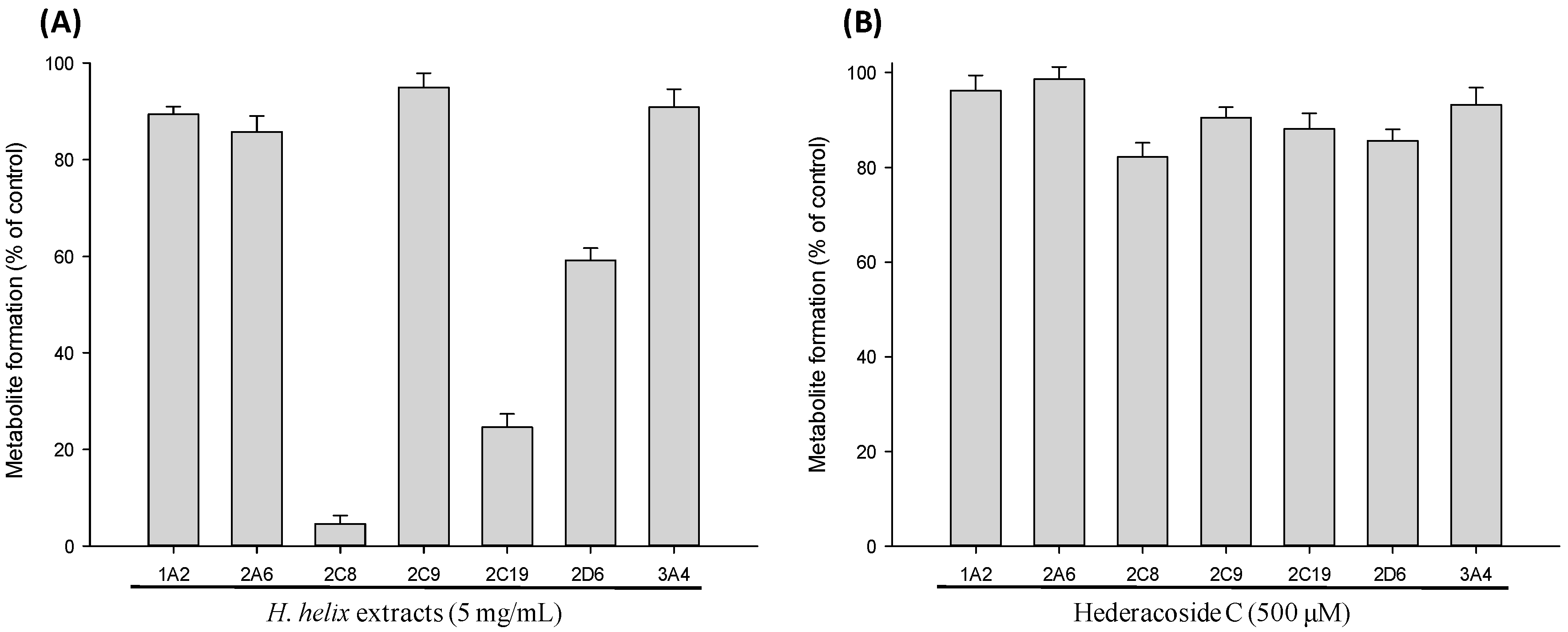
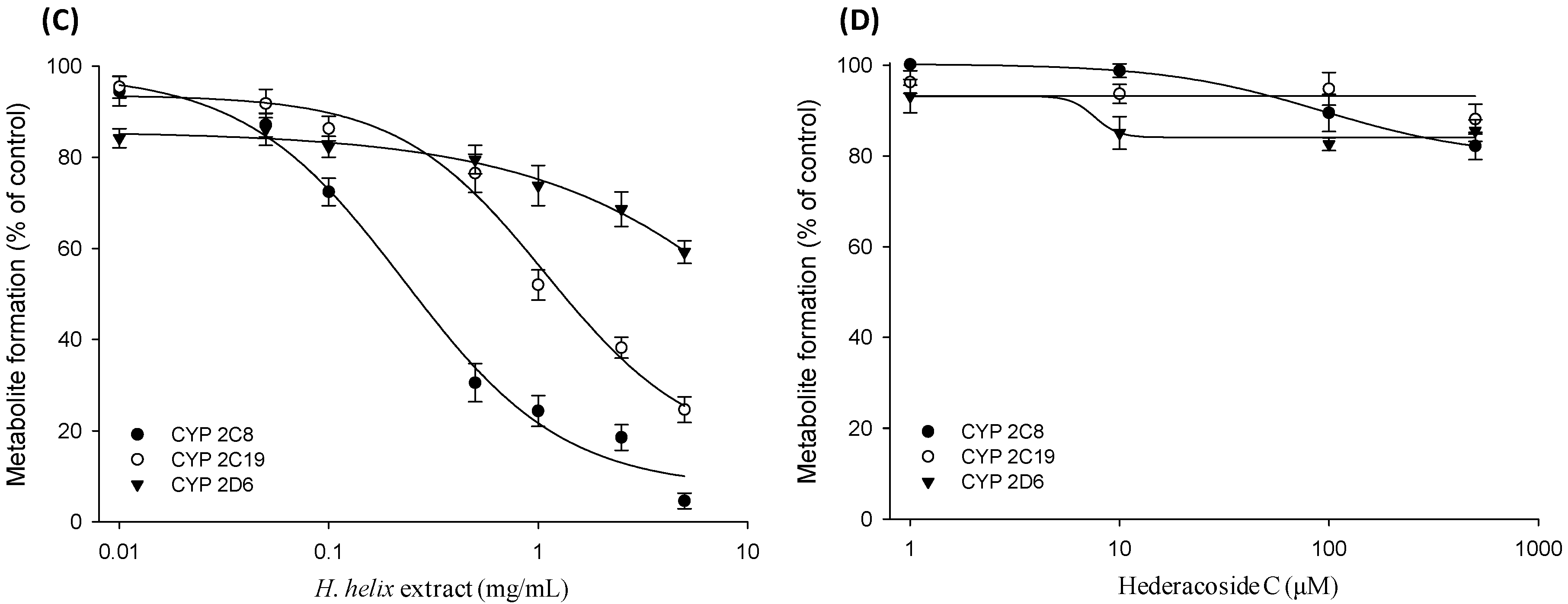
2.2. CYP Inhibition Assay in cDNA-Expressed Recombinant CYP Isozymes

The results of the initial assessment with human liver microsomes revealed that H. helix extract significantly inhibited the enzyme activities of all three enzymes. Accordingly, the CYP inhibitory effect of H. helix extract was further investigated using the cDNA-expressed recombinant CYP isozymes for CYP2C8, CYP2C19, and CYP2D6. As a result, the inhibitory effect of the H. helix extract on CYP2C8, CYP2C19 and CYP2D6 was concentration-dependent, as shown in the assay with human liver microsomes (Figure 3). The IC50 values of the extract against CYP2C8, CYP2C19 and CYP2D6 were 0.08 ± 0.01, 0.58 ± 0.03 and 6.72 ± 0.22 mg/mL, respectively, which are lower than the corresponding levels in human liver microsomes. Additionally, the inhibitory effect of HDC against recombinant CYP isozymes was found to be negligible (data not shown); this finding was consistent with the data of human liver microsomes, confirming that the CYP inhibitory effect of H. helix extract was not attributed to HDC.

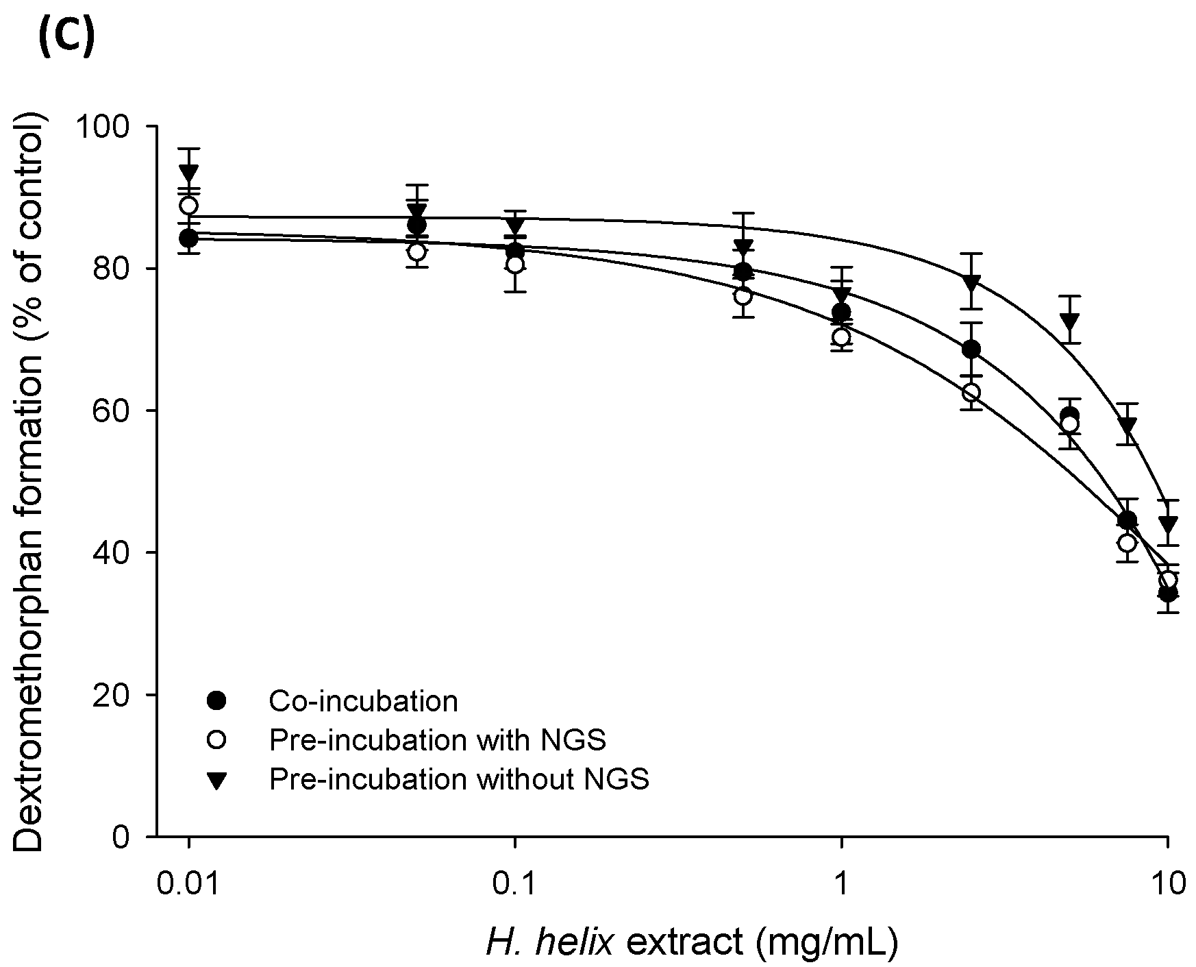
2.3. Time-Dependent Inhibition (TDI) and Mechanism-based Inactivation (MBI) Tests with Recombinant CYP2C8, CYP2C19 and CYP2D6 Isozymes

To elucidate whether the inhibition of CYP by H. helix extracts was time-dependent, the IC50 values derived from the co-incubation and pre-incubation assays were compared. Thus, the H. helix extract in recombinant CYP2C8, CYP2C19 and CYP2D6 isozymes was pre-incubated for 20 min with NADPH and the IC50 value shift for co-incubation was calculated. The inhibitory effects of H. helix extracts on CYP2C8 and CYP2C19 were more potent after pre-incubation (Figure 3), which suggested that the H. helix extract may act as a time-dependent inhibitor of CYP2C8 and CYP2C19. The IC50 shift for CYP2C8, CYP2C19 and CYP2D6 from the co-incubation assay to the pre-incubation assay was 3.01 ± 0.02, 1.88 ± 0.01 and 1.21 ± 0.06 mg/mL, respectively. The fold shift was calculated as the ratio of the IC50 of co-incubation (IC50-co) to that of the pre-incubation with NADPH IC50-pre as follows:
IC50 shift-fold = IC50-co/IC50-pre
Compounds showing an IC50 shift of ≥1.5 can be classified as positive for TDI [17,18]. Based on this criterion, H. helix extracts may have a time-dependent inhibitory effect on CYP2C8 and CYP2C19. Another IC50 shift approach [19,20] was performed to identify the potential of MBI. For this, the IC50 shift from the pre-incubation assay in the absence and presence of NADPH was investigated (Figure 3). As a result, the IC50 shift for CYP2C8, CYP2C19 and CYP2D6 was 2.77 ± 0.12, 6.31 ± 0.25, and 1.38 ± 0.08 mg/mL, respectively. Compounds showing a ratio of IC50 shift of ≥4 can be classified as positive for MBI [19]. Thus, H. helix extracts were considered to be a mechanism-based inactivator on CYP2C19.

3. Discussion

CYP enzymes are vital to the metabolism of many conventional and herbal medicines. Some of these enzymes can be induced or inhibited by xenobiotics, resulting in clinically significant drug-drug or herb–drug interactions and causing unanticipated adverse reactions and/or therapeutic failure [17]. There are no standardized methods for detecting in vitro TDI, but they generally have in common a two-step process of pre-incubation with a test compound followed by the quantification of enzyme activity with a probe substrate. The MBI represents the TDI, where the inhibitory effects are not only time-dependent but they also require metabolism by the enzyme that is ultimately inactivated [20]. Thus, time-dependent enzyme inhibitors display an increasing degree of enzyme inhibition with an increased pre-incubation time with the enzyme. For CYP enzymes, a source of NADPH cofactor is often required to generate MBI. The enzymatic process leading to MBI is irreversible, and catalytic activity cannot be restored. However, the enzyme inactivation occurring in the pre-incubation step must be distinguished from reversible inhibition by comparison to a suitable control (i.e., without NADPH) incubation, to predict NADPH-independent metabolism as well as enzyme degradation. The presence of NADPH in the absence of a substrate may accelerate enzyme activity loss (possibly due to generation of reactive oxygen species) or exert a stabilizing effect, depending on the enzyme and assay conditions [21]. Compared to reversible CYP inhibition, drug-induced MBI (a quasi-irreversible or irreversible inhibition) of CYP enzymes poses a greater safety concern because of the increased risk of pharmacokinetic interactions upon multiple dosing and the sustained duration of such interactions even after the termination of such entities [22]. Characterization of the inactivation property of CYP is essential for predictions of the drug–drug or herb–drug interaction potential of TDI-positive medicines [20]. Recently, guidelines from regulatory agencies such as the United States Food and Drug Administration [23] and pharmaceutical companies [24] have recognized the importance of mitigating drug interaction risks, particularly with respect to CYP TDI/MBI. In pre-clinical drug discovery, the in vitro assessment of PK interactions through TDI is routinely conducted for lead optimization [22].
As herbal medicines (extracts) are a complex mixture formed by various chemical entities [25], some of these constituents possibly contain the functional group responsible for the MBI of CYPs [20,26]. Many studies have reported the MBI of human CYPs by constituents isolated from traditional herbal medicines [27,28,29]. For the TDI assessment with H. helix extracts, IC50 shifts were calculated by comparing IC50 vales from the co-incubation assay to the pre-incubation assay [17], which showed positive results (≥1.5) for CYP2C8 and CYP2C19. As for MBI, the IC50 shift was demonstrated from the ratio of the IC50 value from the pre-incubation assay in the absence of NADPH to that from pre-incubation in its presence. Based on the resulting data, the H. helix extracts were considered positive for CYP2C19 (IC50 shift ≥4) [19]. Thus, our in vitro results indicated that H. helix shows TDI effects for CYP2C8 and CYP2C19, but it has a positive MBI response only for CYP2C19. CYP2D6 inhibition was slightly effected during this study, indicating that H. helix has minimal effects as TDI or MBI.
Based on our findings, there is a potential for herb–drug interactions between the H. helix extract and commonly prescribed conventional medicines that are CYP2C8, CYP2C19 and CYP2D6 substrates. The most sensitive in vivo substrates for CYP2C8 include repaglinide, rosiglitazone and paclitaxel, while quercetin and montelukast are the inhibitors and moderate sensitive substrates for CYP2C8 [23]. Lansoprazole, omeprazole, esoprazole, and S-mephenytoin are the in vivo sensitive substrates for CYP2C19, while dextromethorphan is a substrate for CYP2D6 [23]. In particular, paclitaxel and S-mephenytoin are known to have a narrow therapeutic range, while the drugs used for respiratory illness like dextromethorphan and montelokast are quite sensitive here. Therefore, the concomitant use of these drugs with H. helix extracts may result in clinically significant drug interactions and requires careful attention. The dosage of H. helix preparations is discussed contradictorily in the literature. In a controlled study, the efficacy was shown with low dosages (approximately 300 mg herbal substance), while in the market there are preparations with daily dosages up to approximately 1000 mg herbal substance (Committee on Herbal Medicinal Products, European Medicines Agency) [4,10,14]. The clinical pharmacokinetic data on H. helix extracts is unavailable at present. Although the absorption of H. helix extract from the gastrointestinal tract is shown to be somewhat inadequate for some constituents like hederacoside C and α-hederin [10,14], if the extract is assumed as a single compound, its plasma concentration could reach up around 1 mg/mL. Therefore, it might have clinically close relevance for CYP-mediated herb–drug interactions, at least for CYP2C8 and CYP2C19, considering its maximum daily dosages.

4. Materials and Methods

4.1. Chemicals and Reagents

HDC (>98.0%) and H. helix extract were obtained from the Lab. of Pharmacognosy, College of Pharmacy, Yonsei University (Incheon, Korea). The extract was prepared by extracting the pulverized ivy leaves with 30% ethanol for 1 h using sonication. A voucher specimen of H. helix extract (HY-2016-01-05) was deposited at the Herbarium of the College of Pharmacy, Hanyang University, Ansan, Korea. H. helix extract contains 8.2% of HDC. The content of HDC was determined by liquid chromatography–tandem mass spectrometry (LC-MS/MS) [10] and the representative chromatogram is shown in Figure 4. Pooled human liver microsomes and recombinant CYP2C8, CYP2C19, and CYP2D6 isozymes were purchased from BD Gentest (Woburn, MA, USA). Glucose-6-phosphate, β-NADP+, glucose-6-phosphate dehydrogenase, coumarin, phenacetin, diclofenac, midazolam, mephenytoin, dextromethorphan, ketoconazole, and terfenadine were purchased from Sigma Chemical Co. (Saint Louis, MO, USA). All other solvents used were of HPLC grade and were obtained from J. T. Baker (Phillipsburg, NJ, USA). Distilled water was prepared using a Milli-Q purification system (Millipore, Billerica, MA, USA). All standard solutions and mobile phases were passed through a 0.22 µm membrane filter before use.

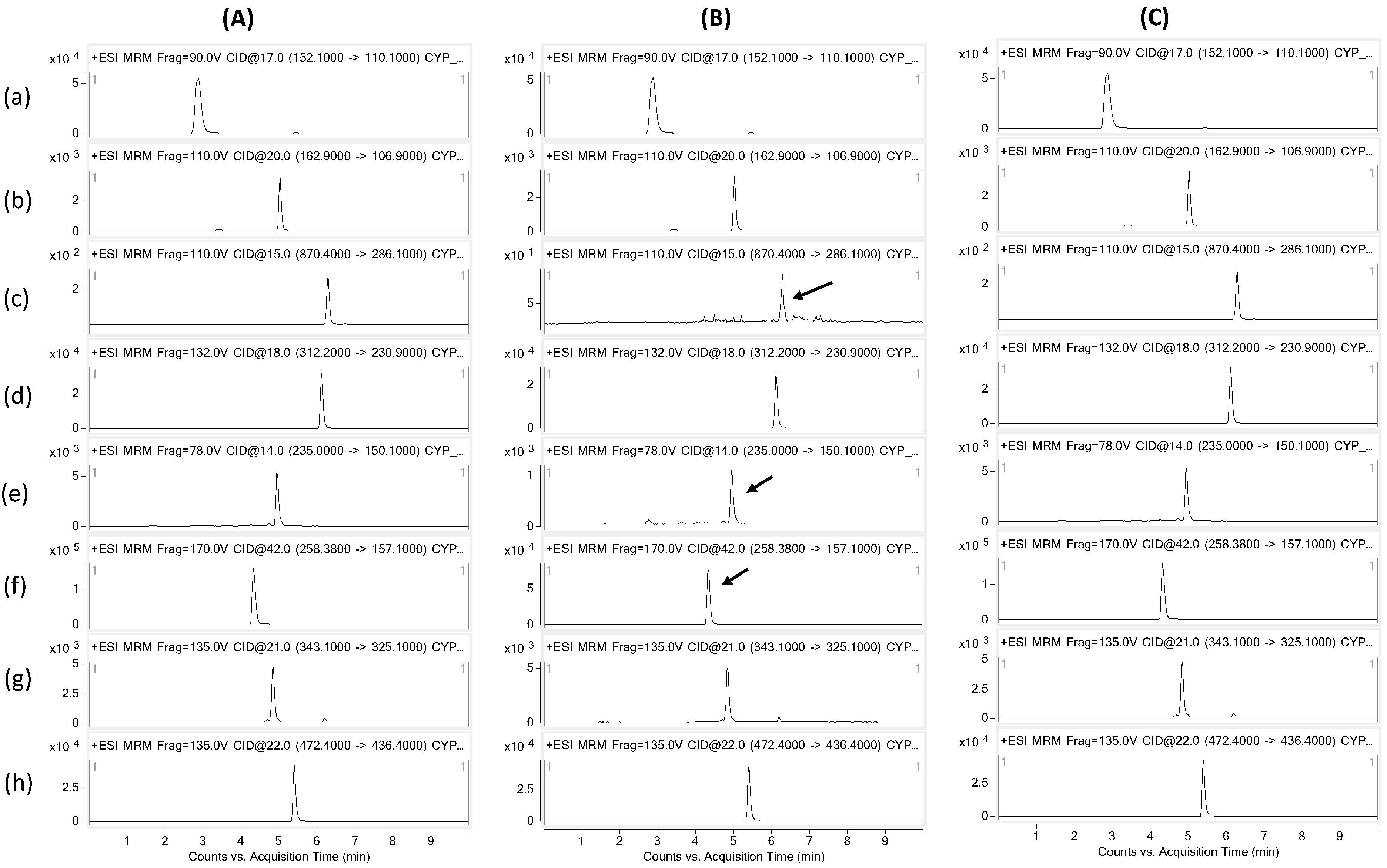
4.2. CYP Inhibition Assay

CYP inhibition assays with human liver microsomes were performed for H. helix extract at various concentrations (0.01, 0.05, 0.1, 0.5, 1.0, 2.5 and 5 mg/mL) and HDC (1, 10, 100, and 500 µM) according to the method used in our previous study [30] (For the determination of the IC50 value for CYP2D6, the concentration range of H. helix extract was extended up to 10 mg/mL). The reaction mixtures consisted of human liver microsomes at a concentration of 0.5 mg/mL; H. helix extract or HDC standard solution at various concentrations; an NADPH-generating system (NGS, containing 0.1 M glucose-6-phosphate, 10 mg/mL β-NADP+, and glucose-6-phosphate dehydrogenase) at 1.0 U/mL; and probe substrates (the final concentrations were at ~Km, which were 40 µM phenacetin, 2.5 µM coumarin, 5 µM dextromethorphan, 10 µM diclofenac, 160 µM mephenytoin, 10 µM paclitaxel, and 2.5 µM midazolam) in 200 µL of 0.1 M potassium phosphate buffer (pH 7.4). The reaction mixture was pre-incubated at 37 °C for 5 min without NGS and then further incubated for 30 min with NGS in a water bath. Ketoconazole (5 µM), furafylline (10 µM), methoxsalen (10 µM), sulfaphenazole (50 µM), ticlopidine (20 µM), quercetin (30 µM), and quinidine (50 µM), which are all CYP specific inhibitors, were tested as positive controls to confirm substrate selectivity for various CYP isoforms. After the incubation, the reaction was stopped by adding 400 µL of 0.1% acetic acid containing internal standard (0.16 µM terfenadine). For the further investigation of specific CYP isozyme inhibition, 12.5 pmol isozyme (CYP 2C8, 2C19, and 2D6) were used instead of human liver microsomes, and the corresponding specific substrates were added to the reaction mixture (10 µM paclitaxel, 160 µM mephenytoin, and 5 µM dextromethorphan, respectively), following the procedure as described before. For time-dependent inhibition, the reaction mixtures consisting of recombinant CYP2C8/CYP2C19/CYP2D6, and NGS in a 0.1 M potassium phosphate buffer, was pre-incubated with H. helix extract for zero (co-incubation) and 20 min, followed by the addition of the corresponding substrates (i.e., paclitaxel (CYP2C8) or mephenytoin (CYP2C19) or dextromethorphan (CYP2D6)), and further incubated for 30 min in water bath (followed by rest of the procedure, as described). For the determination of IC50 values for mechanism-based inactivation, the recombinant CYP isozyme was pre-incubated for 30 min in the presence and absence of NGS [18].

4.3. Sample Preparation

For sample preparation, the incubation mixtures were passed through activated Sep-Pak C18 cartridges (96-well OASIS HLB Extraction Cartridge, Waters, Milford, MA, USA). The cartridges were first eluted with methanol (1 mL) and 0.1% acetic acid (1 mL). After loading the sample, the cartridges were washed twice with 0.1% acetic acid (2 mL) and finally eluted with 1 mL of methanol. The eluate was evaporated under nitrogen gas, and the residue was reconstituted in the mobile phase (100 µL, 0.1% formic acid in an 85/15 mixture of water/acetonitrile, v/v). A 5 µL aliquot was injected into the LC-MS/MS system.

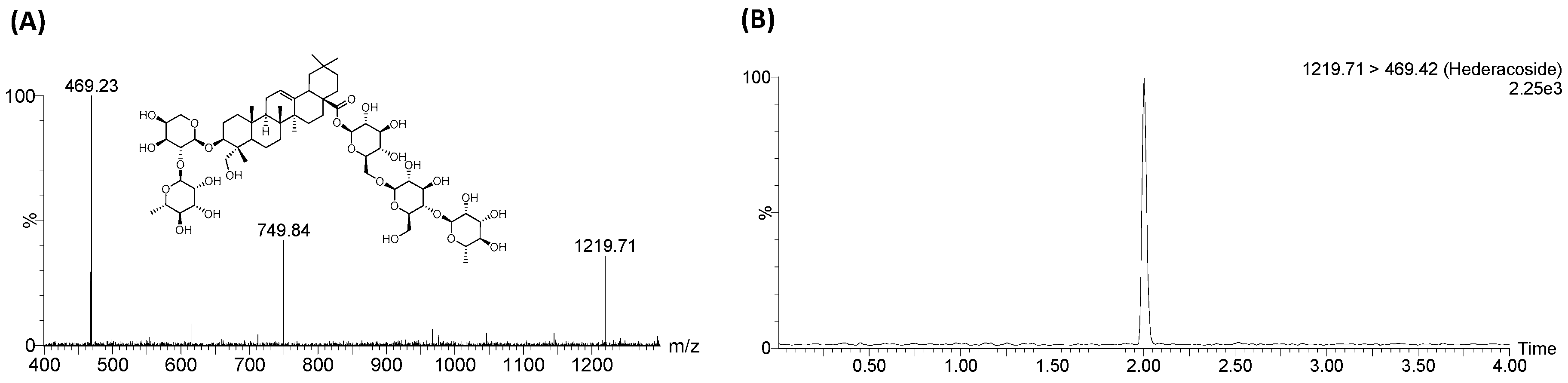
4.4. LC-MS/MS Analysis

The LC-MS/MS system consisted of an Agilent 1260 series binary pump HPLC system and an Agilent 6460 Triple Quadrupole mass spectrometer (Agilent Technologies, Palo Alto, CA, USA) which was equipped with the source of an electrospray ionization (ESI). Chromatographic separation was conducted by using a Fortis-C8 column (2.1 mm × 100 mm, 5 µm; Fortis Technologies Ltd., Cheshire, UK), and the column temperature was maintained at 40 °C. The composition of the HPLC mobile phases were (A) 0.1% formic acid in distilled water and (B) 90% acetonitrile in the solvent A. A gradient program was used at a flow rate of 0.2 mL/min; the composition of solvent B was initially set at 15%, then gradually increased to 85% (15−85%) over 3 min, and maintained for 1.5 min (85%), and finally re-equilibrium for 3.5 min (85−15%). The mass spectrometer was operated in the positive ion mode, with MRM. The precursor–product ion pairs (Q1/Q3) used in MRM mode are shown in Table 1.

4.5. Statistics

Data was presented as the mean ± S.D. of the data obtained from two independent experiments. The IC50 value was calculated with the metabolite-formation values based on the four parameters logistic regression using Sigmaplot v12.5 (Systat Software, Inc., WPCubed GmbH, Munich, Germany) and GraphPad Prism v7.1 (GraphPad Software, Inc., La Jolla, CA, USA).

5. Conclusions

The H. helix extract is a popularly used traditional medicine as well as one of the top-selling herbal medicines for respiratory disorders in many countries. Despite its high possibility of co-administration with other prescribed drugs, no studies have investigated its inhibitory potential on CYP enzyme activities. In the present study, we reported CYP enzyme inhibition by H. helix extracts for the first time. H. helix extract inhibited the enzyme activities of CYP2C8, CYP2C19 and CYP2D6 in a concentration-dependent manner, while it showed time-dependent inhibition for CYP2C8 and CYP2C19. Furthermore, our results suggested that H. helix reacts as a mechanism-based inactivator for CYP2C19 (IC50 shift ≥4). Based on these findings, consuming herbal medicines or dietary supplements containing H. helix extracts requires careful attention and further in vivo studies should investigate CYP-mediated interactions with H. helix to verify the potential of herb–drug interactions and to establish proper recommendations for the safe therapeutic use of H. helix.

Acknowledgments

This work was supported by the research grant through the National Research Foundation of Korea (NRF-2016K1A3A1A20005958).

Author Contributions

H.H.Y., S.H.K. and Y.Z. conceived and designed the experiments; S.U.R. performed the experiments; M.S.C. and Y.Z. analyzed the data; I.S.K. and M.S.C. contributed analysis tools; S.U.R. and H.H.Y. wrote the paper.

Conflicts of Interest

The authors declare no conflict of interest.

References

  1. Natural Medicines Comprehensive Database. Available online: http://naturaldatabase.therapeuticresearch.com/home.aspx?cs=&s=ND (accessed on 2 July 2017).
  2. Cioaca, C.; Margineanu, C.; Cucu, V. The saponins of Hedera helix with antibacterial activity. Pharmazie 1978, 33, 609–610. [Google Scholar] [PubMed]
  3. Wolf, A.; Gosens, R.; Meurs, H.; Haberlein, H. Pre-treatment with α-hederin increases β-adrenoceptor mediated relaxation of airway smooth muscle. Phytomedicine 2011, 18, 214–218. [Google Scholar] [CrossRef] [PubMed]
  4. Stauss-Grabo, M.; Atiye, S.; Warnke, A.; Wedemeyer, R.S.; Donath, F.; Blume, H.H. Observational study on the tolerability and safety of film-coated tablets containing ivy extract (Prospan® Cough Tablets) in the treatment of colds accompanied by coughing. Phytomedicine 2011, 18, 433–436. [Google Scholar] [CrossRef] [PubMed]
  5. Fazio, S.; Pouso, J.; Dolinsky, D.; Fernandez, A.; Hernandez, M.; Clavier, G.; Hecker, M. Tolerance, safety and efficacy of Hedera helix extract in inflammatory bronchial diseases under clinical practice conditions: a prospective, open, multicentre postmarketing study in 9657 patients. Phytomedicine 2009, 16, 17–24. [Google Scholar] [CrossRef] [PubMed]
  6. Trute, A.; Gross, J.; Mutschler, E.; Nahrstedt, A. In vitro antispasmodic compounds of the dry extract obtained from Hedera helix. Planta Med. 1997, 63, 125–129. [Google Scholar] [CrossRef] [PubMed]
  7. Capasso, A.; Pinto, A.; Mascolo, N.; Capasso, F. Reduction of agonist-induced contractions of guinea-pig isolated ileum by flavonoids. Phytother. Res. 1991, 5, 85–87. [Google Scholar] [CrossRef]
  8. Hegener, O.; Prenner, L.; Runkel, F.; Baader, S.L.; Kappler, J.; Haberlein, H. Dynamics of β2-adrenergic receptor−ligand complexes on living cells. Biochemistry 2004, 43, 6190–6199. [Google Scholar] [CrossRef] [PubMed]
  9. Sieben, A.; Prenner, L.; Sorkalla, T.; Wolf, A.; Jakobs, D.; Runkel, F.; Haberlein, H. α-Hederin, but not hederacoside c and hederagenin from hedera helix, affects the binding behavior, dynamics, and regulation of β2-adrenergic receptors. Biochemistry 2009, 48, 3477–3482. [Google Scholar] [CrossRef] [PubMed]
  10. Rehman, S.U.; Choi, M.S.; Kim, I.S.; Kim, S.H.; Yoo, H.H. An ultra-high-performance liquid chromatography-tandem mass spectrometric method for the determination of hederacoside C, a drug candidate for respiratory disorder, in rat plasma. J. Pharmaceut. Biomed. 2016, 129, 90–95. [Google Scholar] [CrossRef] [PubMed]
  11. Rehman, S.U.; Choi, M.S.; Choe, K.; Yoo, H.H. Interactions between herbs and antidiabetics: an overview of the mechanisms, evidence, importance, and management. Arch. Pharm. Res. 2015, 37, 1281–1298. [Google Scholar] [CrossRef] [PubMed]
  12. Rehman, S.U.; Choi, M.S.; Kim, I.S.; Luo, Z.; Xue, Y.; Yao, G.; Zhang, Y.; Yoo, H.H. In Vitro Assessment of CYP-Mediated Drug Interactions for Kinsenoside, an Antihyperlipidemic Candidate. Molecules 2016, 21, 800. [Google Scholar] [CrossRef] [PubMed]
  13. Arimura, G.I.; Maffei, M. Plant Specialized Metabolism: Genomics, Biochemistry, and Biological Functions, 1st ed.; CRC Press: Boca Raton, FL, USA, 2016. [Google Scholar]
  14. Assessment report on Hedera helix, L., folium, Committee on Herbal Medicinal Products (HMPC), European Medicines Agency (EMA). EMA/HMPC/586887/2014. Available online: http://www.ema.europa.eu/docs/en_GB/document_library/Herbal_HMPC_assessment_report/2016/01/WC500199887.pdf (accessed on 24 November 2015).
  15. Liu, J.; Liu, Y.P.; Bullock, P.; KlaasseN, C.D. Suppression of liver cytochrome P450 by α-hederin: relevance to hepatoprotection. Toxicol. Appl. Pharm. 1995, 134, 124–131. [Google Scholar] [CrossRef]
  16. Jeong, H.G. Suppression of constitutive and inducible cytochrome P450 gene expression by α-hederin in mice. IUBMB Life 1998, 46, 1019–1026. [Google Scholar] [CrossRef]
  17. Nebert, D.W.; Russell, D.W. Clinical importance of the cytochromes P450. Lancet 2002, 360, 1155–1162. [Google Scholar] [CrossRef]
  18. Nomeir, A.A.; Palamanda, J.R.; Favreau, L. Identification of CYP mechanism-based inhibitors. In Optimization in Drug Discovery: In Vitro Methods, 1st ed.; Humana Press: Totowa, NJ, USA, 2004; pp. 245–262. ISBN 978-1-62703-742-6. [Google Scholar]
  19. Obach, R.S.; Walsky, R.L.; Venkatakrishnan, K. Mechanism-based inactivation of human cytochrome p450 enzymes and the prediction of drug-drug interactions. Drug Metab. Dispos. 2007, 35, 246–255. [Google Scholar] [CrossRef] [PubMed]
  20. Riley, R.J.; Grime, K.; Weaver, R. Time-dependent CYP inhibition. Expert Opin. Drug Met. 2007, 3, 51–66. [Google Scholar] [CrossRef] [PubMed]
  21. Stresser, D.M.; Mao, J.; Kenny, J.R.; Jones, B.C.; Grime, K. Exploring concepts of in vitro time-dependent CYP inhibition assays. Expert Opin. Drug Met. 2014, 10, 157–174. [Google Scholar] [CrossRef] [PubMed]
  22. Orr, S.T.; Ripp, S.L.; Ballard, T.E.; Henderson, J.L.; Scott, D.O.; Obach, R.S.; Sun, H.; Kalgutkar, A.S. Mechanism-based inactivation (MBI) of cytochrome P450 enzymes: structure–activity relationships and discovery strategies to mitigate drug–drug interaction risks. J. Med. Chem. 2012, 55, 4896–4933. [Google Scholar] [CrossRef] [PubMed]
  23. U.S. Food and Drug Administration (FDA), Drug Development and Drug Interactions: Table of Substrates, Inhibitors and Inducers. Available online: http://www.fda.gov/Drugs/DevelopmentApprovalProcess/DevelopmentResources/DrugInteractionsLabeling/ucm093664.htm (accessed on 11 July 2017).
  24. Grimm, S.W.; Einolf, H.J.; Hall, S.D.; He, K.; Lim, H.K.; Ling, K.H.J.; Lu, C.; Nomeir, A.A.; Seibert, E.; Skordos, K.W.; et al. The conduct of in vitro studies to address time-dependent inhibition of drug-metabolizing enzymes: a perspective of the pharmaceutical research and manufacturers of America. Drug Metab. Dispos. 2009, 37, 1355–1370. [Google Scholar] [CrossRef] [PubMed]
  25. Rehman, S.U.; Choe, K.; Yoo, H.H. Review on a traditional herbal medicine, Eurycoma longifolia jack (Tongkat Ali): its traditional uses, chemistry, evidence-based pharmacology and toxicology. Molecules 2016, 21, 331. [Google Scholar] [CrossRef] [PubMed]
  26. Kamel, A.; Harriman, S. Inhibition of cytochrome P450 enzymes and biochemical aspects of mechanism-based inactivation (MBI). Drug Discov. Today Technol. 2013, 10, e177–e189. [Google Scholar] [CrossRef] [PubMed]
  27. Brahmi, Z.; Katho, T.; Hatsumata, R.; Hiroi, A.; Miyakawa, N.; Yakou, E.; Sugaya, K.; Onose, J.I.; Naoki, A.B.E. Effective Cytochrome P450 (CYP) Inhibitors Isolated from Tarragon (Artemisia dracunculus). Biosci. Biotechnol. Biochem. 2012, 76, 1028–1031. [Google Scholar] [CrossRef] [PubMed]
  28. Taesotikul, T.; Dumrongsakulchai, W.; Wattanachai, N.; Navinpipat, V.; Somanabandhu, A.; Tassaneeyakul, W.; Tassaneeyakul, W. Inhibitory effects of Phyllanthus amarus and its major lignans on human microsomal cytochrome P450 activities: evidence for CYP3A4 mechanism-based inhibition. Drug Metab. Pharmacokinet. 2011, 26, 154–161. [Google Scholar] [CrossRef] [PubMed]
  29. Zhao, Y.; Hellum, B.H.; Liang, A.; Nilsen, O.G. Inhibitory mechanisms of human CYPs by three alkaloids isolated from traditional chinese herbs. Phytother. Res. 2015, 29, 825–834. [Google Scholar] [CrossRef] [PubMed]
  30. Han, Y.M.; Kim, I.S.; Rehman, S.U.; Choe, K.; Yoo, H.H. In Vitro Evaluation of the Effects of Eurycoma longifolia Extract on CYP-Mediated Drug Metabolism. Evid. Based Complement. Alternat. Med. 2015, 631329. [Google Scholar] [CrossRef]
Sample Availability: Not available.
Figure 1. Representative multiple reaction monitoring (MRM) chromatograms of human liver microsome samples of, (A) control; (B) H. helix extract-treated, and (C) Hederacoside C (HDC)-treated. A fraction of human liver microsomal was incubated with the substrate mixture, in an NADPH-generating system, and H. helix extract (2.5 mg/mL) or HDC (100 µM) for 30 min and the cytochromes P450 (CYP)-specific metabolite formation was determined by LC-MS/MS. (a) Acetaminophen; (b) 7-OH coumarin; (c) 6-OH-paclitaxel (d) 4-OH-diclofenac; (e) 4-OH-mephenytoin; (f) dextrorphan; (g) 1-OH-madazolam; and (h) terfenadine (IS).
Molecules 22 01241 g001
Figure 2. Effects of H. helix extract and hederacoside C on the CYP-specific metabolite formation in human liver microsomes. (A) H. helix extract (5 mg/mL); (B) Hederacoside C (500 μM); (C) Effects of H. helix extract on the metabolic activities of CYP2C8, CYP2C19, and CYP2D6; and (D) Effects of hederacoside C on the metabolic activities of CYP2C8, CYP2C19, and CYP2D6. Data was presented as the mean ± S.D. of the data obtained from two independent experiments.
Molecules 22 01241 g002aMolecules 22 01241 g002b
Figure 3. CYP-specific metabolite formation as the percent of control after co-incubation and pre-incubation of H. helix extracts with and without NADPH in c-DNA expressed CYP isozymes; (A) CYP2C8; (B) CYP2C19, and (C) CYP2D6. Data was presented as the mean ± S.D. of the data obtained from two independent experiments.
Molecules 22 01241 g003aMolecules 22 01241 g003b
Figure 4. Hederacoside C, (A) product ion mass spectra of the [M − H] ions of HDC, and (B) MRM chromatograms of HDC in H. helix extracts.
Molecules 22 01241 g004
Table 1. Information on the probe substrates and their corresponding CYP-specific metabolites used in this study.
P450 IsozymePROBE SubstrateSubstrate Conc. (µM)Metabolite MonitoredPrecursor-Ion (m/z)Daughter-Ion (m/z)
CYP1A2Phenacetin40Acetaminophen152.1110.1
CYP2A6Coumarin2.57-OH-coumarin162.9106.9
CYP2C8Paclitaxel106-OH-paclitaxel870.4286.1
CYP2C9Diclofenac104-OH-diclofenac312.2230.9
CYP2C19Mephenytoin1604-OH-mephenytoin235.0150.1
CYP2D6Dextromethorphan5Dextrorphan258.3157.1
CYP3A4Midazolam2.51-OH-midazolam343.1325.1
Internal Standard -Terfenadine472.4436.4

