Article

The Phenolic Fraction of Mentha haplocalyx and Its Constituent Linarin Ameliorate Inflammatory Response through Inactivation of NF-κB and MAPKs in Lipopolysaccharide-Induced RAW264.7 Cells

1
Department of Traditional Chinese Medicine Chemistry, School of Chinese Materia Medica, Beijing University of Chinese Medicine, Beijing 100102, China
2
Department of Scientific Research Center, School of Chinese Medicine, Beijing University of Chinese Medicine, Beijing 100029, China
3
Department of Microbiology and Immunology, School of Life Science, Beijing University of Chinese Medicine, Beijing 100029, China
4
Department of Pathology, School of Chinese Medicine, Beijing University of Chinese Medicine, Beijing 100029, China
*
Authors to whom correspondence should be addressed.
Molecules 2017, 22(5), 811; https://doi.org/10.3390/molecules22050811
Submission received: 15 March 2017 / Revised: 1 May 2017 / Accepted: 9 May 2017 / Published: 16 May 2017
(This article belongs to the Section Natural Products Chemistry)

Abstract

:
Mentha haplocalyx has been widely used for its flavoring and medicinal properties and as a traditional Chinese medicine with its anti-inflammation properties. The present study was designed to investigate the anti-inflammatory effects and potential molecular mechanisms of the phenolic fraction of M. haplocalyx (MHP) and its constituent linarin in lipopolysaccharide (LPS)-induced RAW264.7 cells. The high-performance liquid chromatography coupled with linear ion trap-orbitrap mass spectrometry (HPLC-LTQ-Orbitrap MS) was used to analyze the chemical composition of MHP. Using the enzyme-linked immunosorbent assay (ELISA) and quantitative realtime polymerase chain reaction (qRT-PCR), the expression of pro-inflammatory meditators and cytokines was measured at the transcriptional and translational levels. Western blot analysis was used to further investigate changes in the nuclear factor kappa B (NF-κB), mitogen-activated protein kinase (MAPK), and Akt signaling pathways. Fourteen phenolic constituents were identified from MHP based on the data of the mass spectrometry (MS)/MS analysis. MHP and linarin decreased the production of NO, tumor necrosis factor-α (TNF-α), interlenkin-1β (IL-1β), and IL-6. The messenger ribonucleic acid (mRNA) expression levels of inducible NO synthase (iNOS), TNF-α, IL-1β, and IL-6 were also suppressed by MHP and linarin. Further investigation showed that MHP and linarin down-regulated LPS-induced phosphorylation content of NF-κB p65, inhibitor kappa B α (IκBα), extracellular signal-regulated kinase (ERK), c-Jun NH2-terminal kinase (JNK), and p38. However, MHP and linarin showed no inhibitory effect on the phosphorylated Akt. These results suggested that MHP and linarin exerted a potent inhibitory effect on pro-inflammatory meditator and cytokines production via the inactivation of NF-κB and MAPKs, and they may serve as potential modulatory agents for the prevention and treatment of inflammatory diseases.

1. Introduction

Inflammation is an important defense mechanism of the host against invasive stimuli [1,2]. However, excessive inflammatory responses may give rise to various pathological changes related to human diseases, including asthma, rheumatoid arthritis, atherosclerosis, and cancer [3]. Many risk factors are responsible for inflammation, such as pathogens, impaired cells and tissues, and physical and chemical stimuli [4]. Macrophages are phagocytic white blood cells that play an important role in innate immune inflammatory responses. Activated macrophages secrete various cytokines such as tumor necrosis factor-α (TNF-α), interlenkin-1β (IL-1β), interlenkin-6 (IL-6), and other inflammatory cytokines [5], which show pleiotropic effects via activating adaptive immune responses and worsening inflammatory responses [6]. The nuclear factor kappa-B (NF-κB) signaling pathway is involved in regulating diverse gene expression during inflammatory responses [7]. When activated with extracellular stimuli, the NF-κB released from the cytoplasmic NF-κB-inhibitor kappa B (IκB) complex by the ubiquitination and degradation of IκB translocates into the nucleus [8,9], combining with specific DNA sequences and then regulating the transcription of inflammatory cytokines and mediators [10,11]. Moreover, phosphoinositide 3-kinase (PI3K)/Akt and mitogen-activated protein kinases (MAPKs) including extracellular signal-regulated kinase (ERK), p38, and c-Jun NH2-terminal kinase (JNK) subfamilies could activate NF-κB and regulate inflammatory responses [12,13].
Mentha species are widely used throughout the world as flavoring agents and medicinal plants, particularly for the treatment of common fever, cold, flu, and motion sickness [14,15]. Although the genus Mentha comprises more than 20 species, only the fresh aerial part of Mentha haplocalyx Briq. is described and used as a traditional Chinese herb in Chinese pharmacopoeia. The leaves of M. haplocalyx Briq. are also used in teas, beverages, jellies, syrups, candies, and other products. Modern pharmacological studies have revealed that M. haplocalyx shows various biological activities, such as antiallergenic, antimicrobial, anti-inflammatory, antioxidant, antitumor, antiviral, gastrointestinal protective, hepatoprotective, and chemopreventive activities [16]. Other well-known Mentha species, including M. spicata, M. longifolia, M. piperita, M. pulegium, and M. arvensis, have been reported to express similar biological activities. For instance, gastrointestinal protective properties have been reported for M. longifolia, M. pulegium, and M. arvensis [17,18,19], chemopreventive activities for M. piperita and M. spicata [20,21], and antiviral effects for M. piperita, M. spicata, and M. longifolia [22,23]. Mentha species are characterized by two important classes of secondary metabolites: essential oils and phenolic compounds [24]. Numerous researchers have proved that these compounds contribute to the multiple biological activities of Mentha species [24,25].
In traditional Chinese medicine, M. haplocalyx is used frequently as a herb that releases the “exterior” in the common cold of wind-heat type, headache, red eye, measles, and thoracic and abdominal oppression [26]. Pharmacological studies have revealed that “exterior releasing herbs” are effective against viral and bacterial infections that usually induce inflammation [27]. Based on the fact that heat is related to inflammation in Chinese medicine [28] and that M. haplocalyx has cooling properties, it can be assumed that M. haplocalyx could exert an inhibitory effect on inflammation. Indeed, some studies have proved that flavonoids and phenolic acids from the aqueous extract of M. haplocalyx [29,30,31] express high anti-inflammatory activity [16,32]. In our previous work, we have optimized a method for the purification of the phenolic fraction of M. haplocalyx (MHP), mainly consisting of flavonoids and phenolic acids [33]. In the present study, our particular interest was to investigate the mechanism of the anti-inflammatory effect of M. haplocalyx extract, as well as to analyze the chemical composition of MHP by high-performance liquid chromatography coupled with linear ion trap-orbitrap mass spectrometry (HPLC-LTQ-Orbitrap MS). We explored the mechanism of inflammatory activity by evaluating the impact of MHP on the production of pro-inflammatory meditators and the activation of NF-κB, Akt, and MAPKs in lipopolysaccharide (LPS)-induced RAW264.7 cells. Since the linarin was the main flavonoid from MHP, the mechanism of its activity was also investigated for the first time in this study.

2. Results

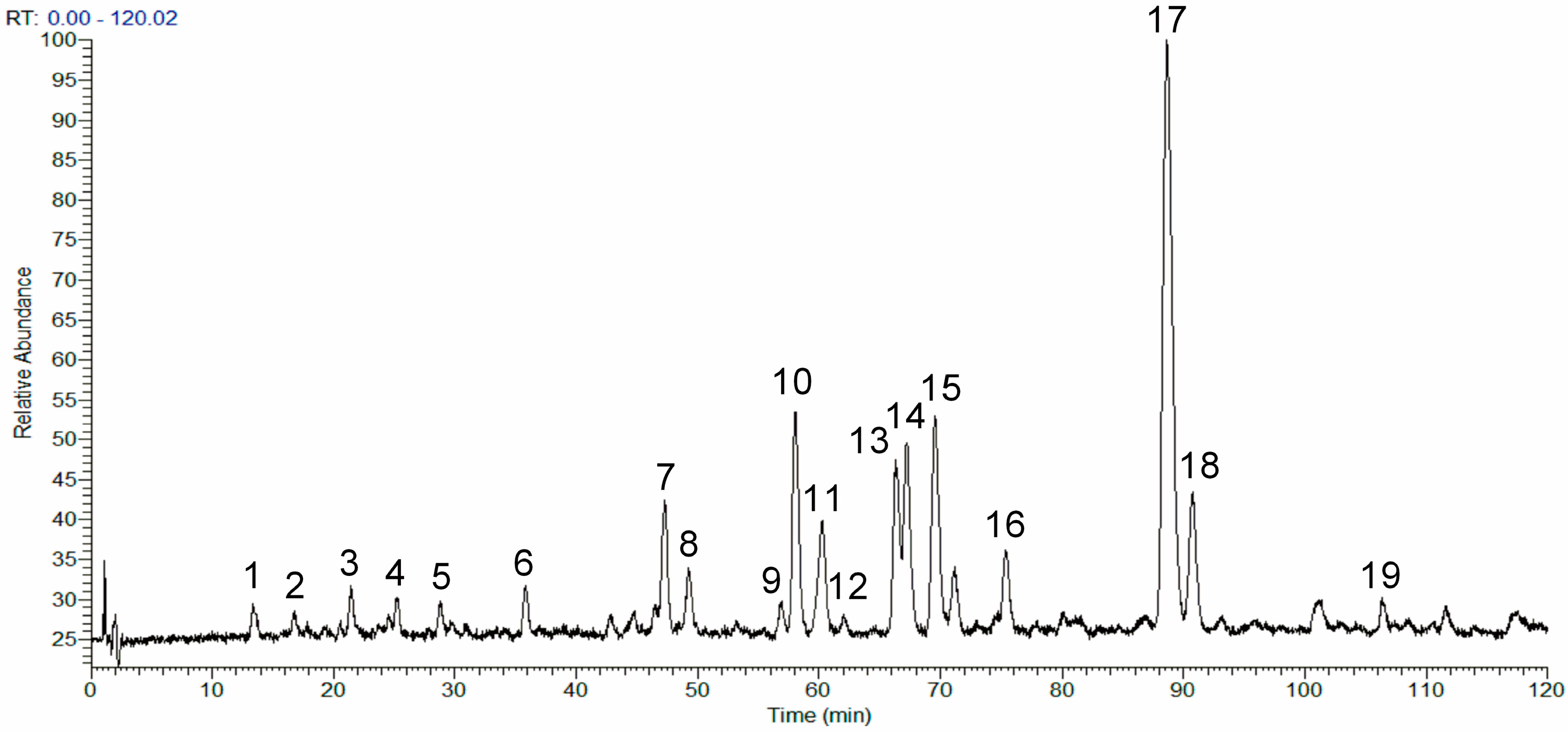
2.1. Analysis of the Chemical Composition of the Phenolic Fraction of Mentha haplocalyx by HPLC-LTQ-Orbitrap MS

Structural elucidation of the MHP constituents was performed according to the obtained chromatographic retention times and mass spectrometry (MS)/MS data by comparison with the data of available reference compounds including rosmarinic acid, linarin, or literature data. As a result, 14 phenolic compounds were identified in the MHP sample. The total ion chromatogram (TIC) profile of MHP is shown in Figure 1 and the data for compound identification are summarized in Table 1.

2.2. Effects of the Phenolic Fraction of Mentha haplocalyx and Linarin on NO Production and iNOS Expression Level in LPS-Induced RAW264.7 Cells

The toxicity of drugs could have an influence on the proliferation and function of cells, misleading the judgement of their efficacy. Thus, we firstly evaluated the cell cytotoxicity of MHP and linarin. The RAW264.7 cells were treated with increased concentrations of MHP and linarin for 24 h and cell cytotoxicity was examined by the 3-(4,5-dimethylthiazol-2-yl)-2,5-diphenyltetrazolium bromide (MTT) assay. As shown in Figure 2, MHP and linarin did not exhibit toxic effects on RAW264.7 cells at 0–200 μg/mL and 0–20 μM, respectively.
To investigate the anti-inflammatory activities of MHP and linarin, we studied the inhibitory effect of MHP and linarin on LPS-induced nitric oxide (NO) production in RAW264.7 cells. As shown in Figure 3A and Figure 4A, the NO production level significantly increased after LPS-stimulation for 24 h in RAW264.7 cells. MHP at 50–200 μg/mL and linarin at 5–20 μM showed markedly inhibitory effects on the increase of NO in a dose-dependent manner. Subsequently, we investigated the cause of reduced NO production by measuring the expression of inducible NO synthase (iNOS) mRNA. Quantitative real-time polymerase chain reaction (qRT-PCR) analysis showed that both MHP and linarin were able to inhibit the expression of iNOS mRNA (Figure 5A,B). The results suggested that MHP and linarin inhibited NO production by down-regulating the expression of iNOS mRNA.

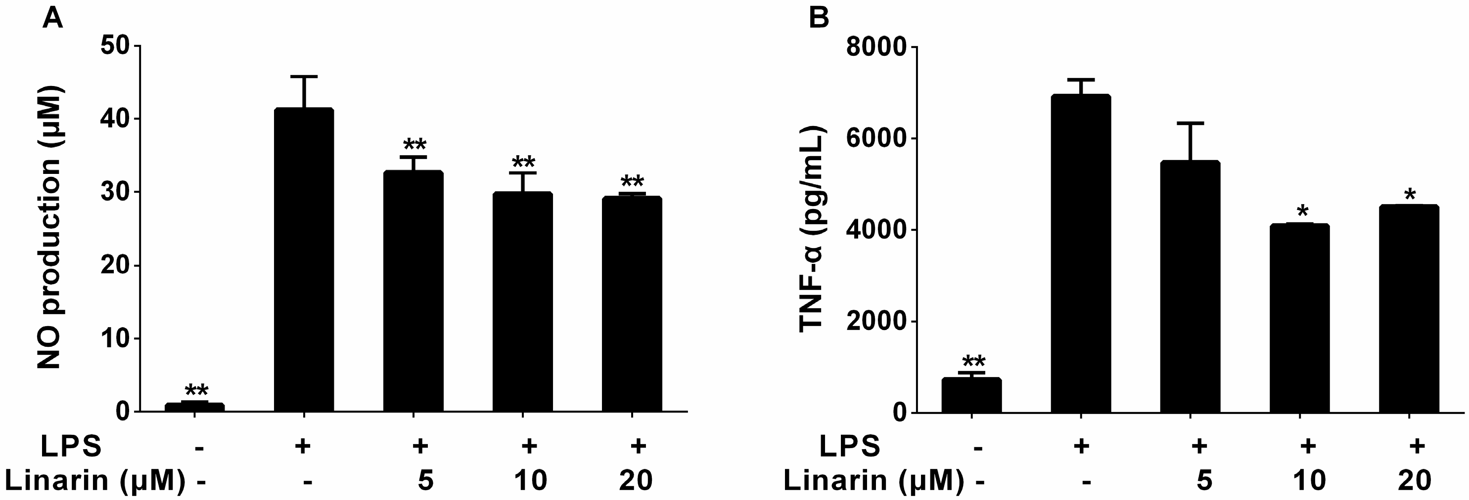
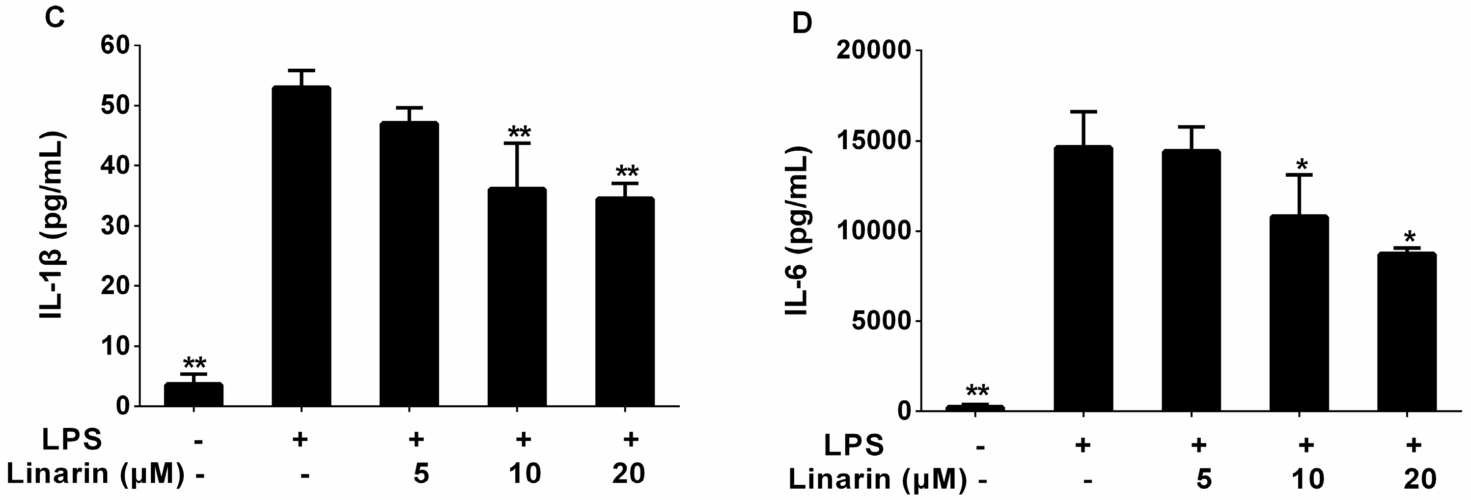
2.3. Effects of the Phenolic Fraction of Mentha haplocalyx and Linarin on the Production of TNF-α, IL-1β, and IL-6 in LPS-Induced RAW264.7 Cells

To determine the effects of MHP and linarin on the production of inflammatory cytokines including TNF-α, IL-1β, and IL-6, the RAW264.7 cells were pretreated with LPS in the presence or absence of screened concentrations of MHP and linarin. The results of enzyme-linked immunosorbent assay (ELISA) showed that the amount of TNF-α, IL-1β, and IL-6 significantly increased in RAW264.7 cells induced by LPS alone. As shown in Figure 3B–D, the level of TNF-α was significantly inhibited by MHP at 100 and 200 μg/mL, while the levels of IL-1β and IL-6 were markedly suppressed by MHP at 50, 100, and 200 μg/mL. Meanwhile, linarin remarkably suppressed the secretion of TNF-α, IL-1β, and IL-6 at concentrations of 10 and 20 μM (Figure 4B–D).

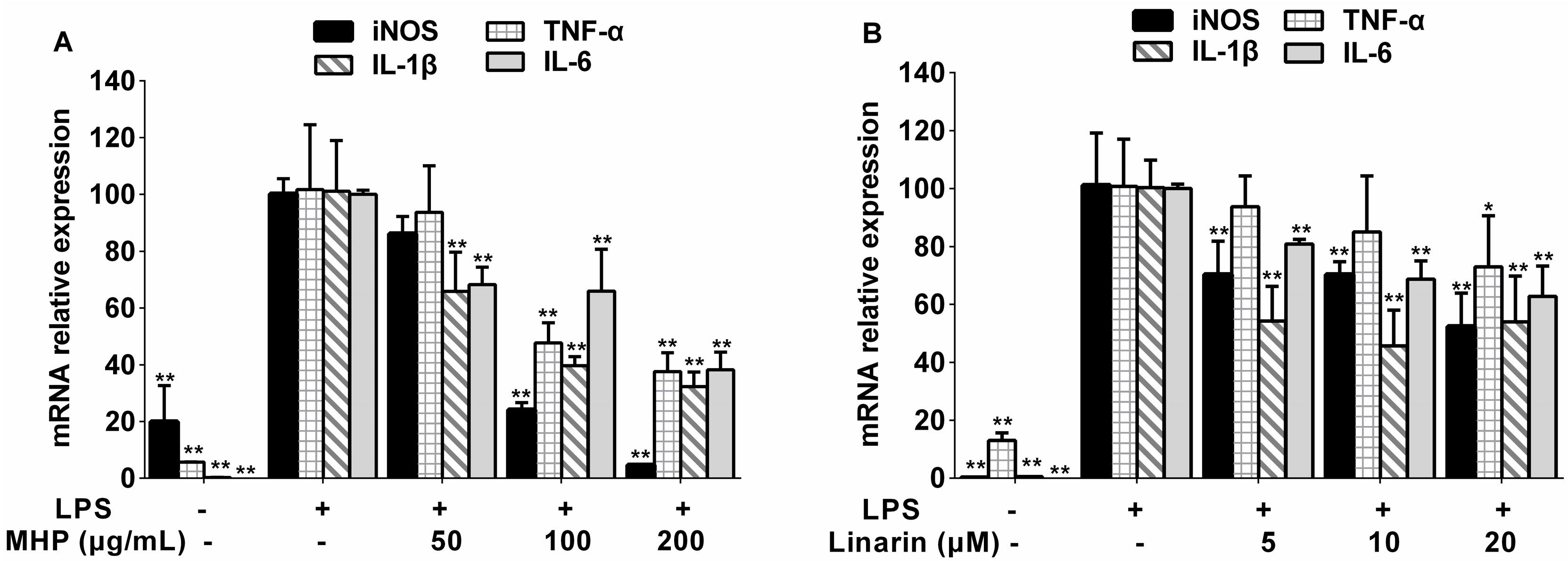
2.4. Effects of the Phenolic Fraction of Mentha haplocalyx and Linarin on TNF-α, IL-1β, and IL-6 mRNA Expression in LPS-Induced RAW264.7 Cells

In order to investigate whether MHP and linarin inhibited the expression of pro-inflammatory cytokines at the transcriptional level, we examined TNF-α, IL-1β, and IL-6 mRNA expression levels in RAW264.7 cells. As expected in Figure 5A, 100 and 200 μg/mL of MHP significantly inhibited the LPS-induced increase of the TNF-α mRNA expression while MHP at 50, 100, and 200 μg/mL decreased the expression of IL-1β and IL-6 mRNA induced by LPS. In addition, linarin significantly repressed IL-1β and IL-6 mRNA expression at 5–20 μM and the mRNA expression level of TNF-α was significantly down-regulated by linarin at 20 μM (Figure 5B).

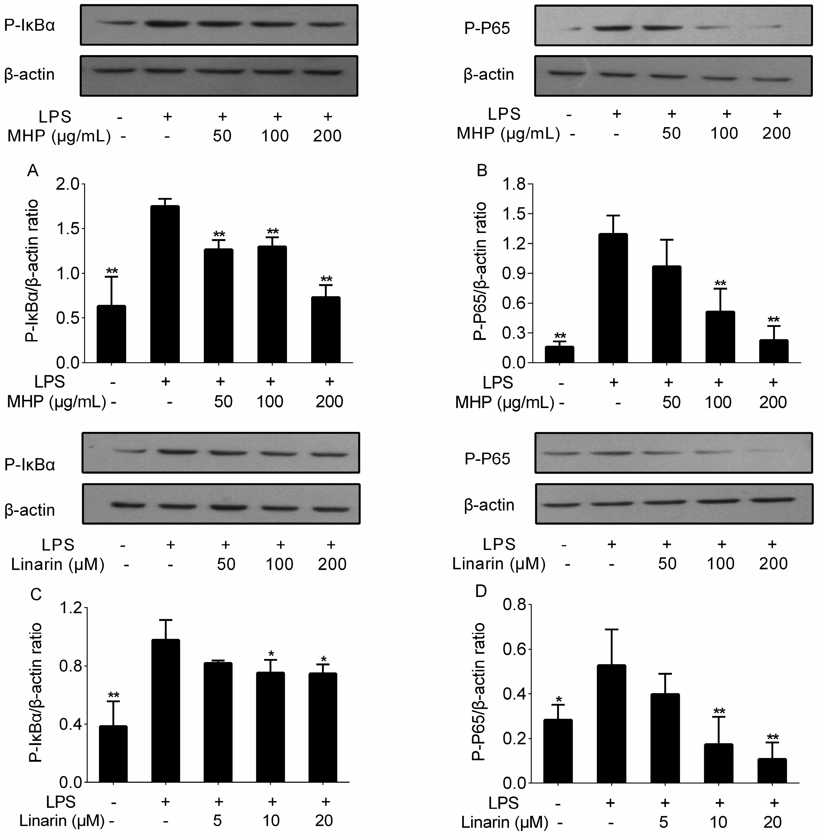
2.5. Effects of the Phenolic Fraction of Mentha haplocalyx and Linarin on NF-κB p65 and IκBα in LPS-Induced RAW264.7 Cells

The activation of NF-κB in response to LPS is a vital step in inducing the expression of inflammatory cytokines, such as TNF-α, IL-1β, and IL-6 [34]. Here, we investigated whether MHP and linarin inhibited the phosphorylation of inhibitor kappa B α (IκBα) and p65. As depicted in Figure 6, the relative content of phosphorylated (p-)IκBα and p-p65 significantly increased after LPS stimulation, which suggested that LPS stimulation triggered the phosphorylation of IκBα and p65. However, these increases were inhibited by MHP and linarin. MHP treatment significantly restrained the level of p-IκBα at 50, 100, and 200 μg/mL (Figure 6A), and at 100 and 200 μg/mL for p-p65 (Figure 6B). The increased levels of p-p65 and p-IκBα were also repressed by linarin at 10 and 20 μM (Figure 6C,D). These results indicated that MHP and linarin exerted anti-inflammatory effects through inhibition of LPS-induced activation of the NF-κB signaling pathway.

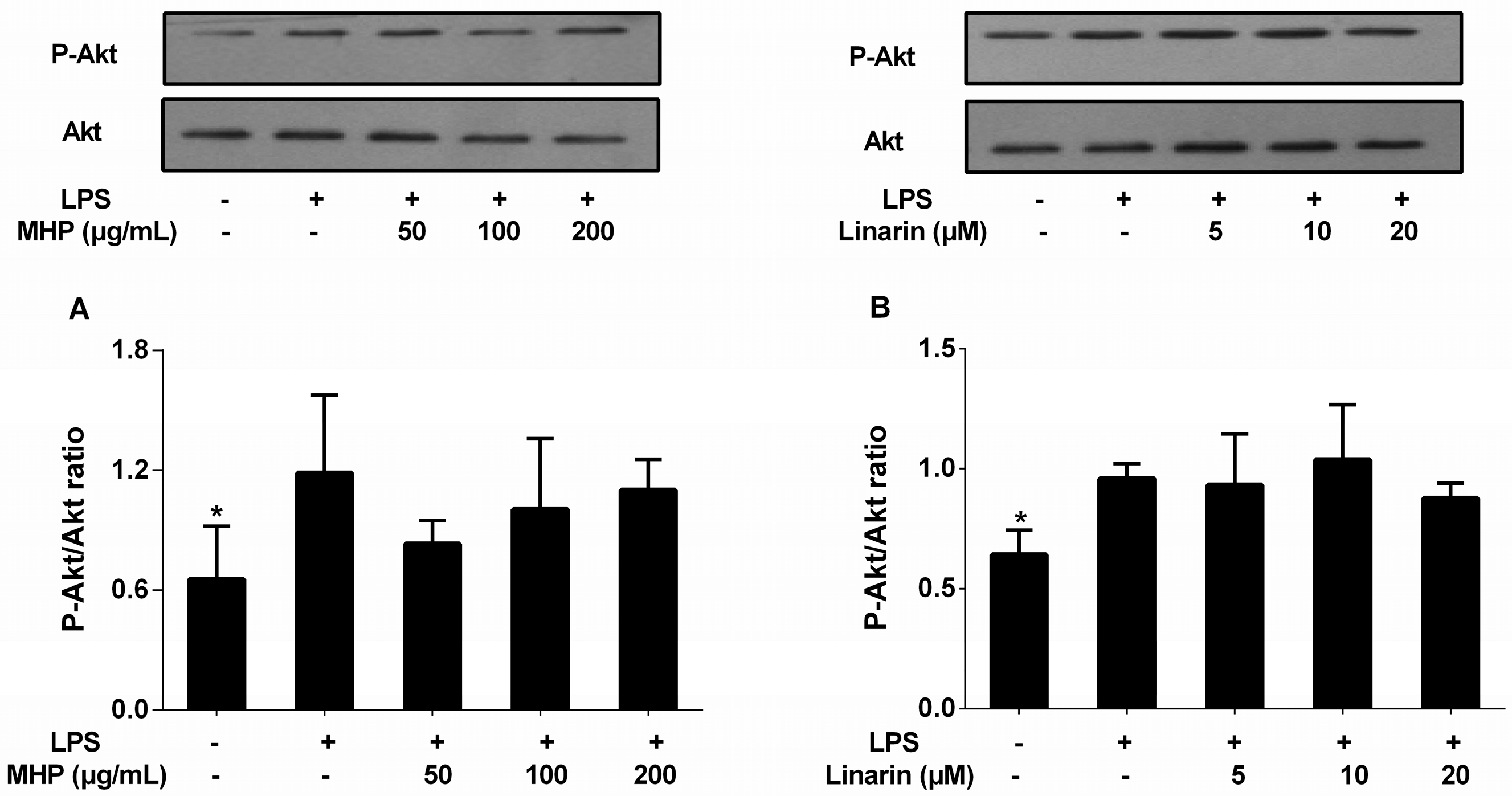
2.6. Effects of the Phenolic Fraction of Mentha haplocalyx and Linarin on Akt in LPS-Induced RAW264.7 Cells

A previous study has reported that activated Akt, a key protein of the PI3K signaling pathway, could turn on the activation of NF-κB [12]. Thus, we investigated the inhibitory effects of MHP and linarin on LPS-induced expression of phosphorylated Akt protein in RAW264.7 cells. However, the results shown in Figure 7A,B indicate that MHP and linarin exerted no inhibitory effect on the LPS-induced increase of the phosphorylation level of Akt.

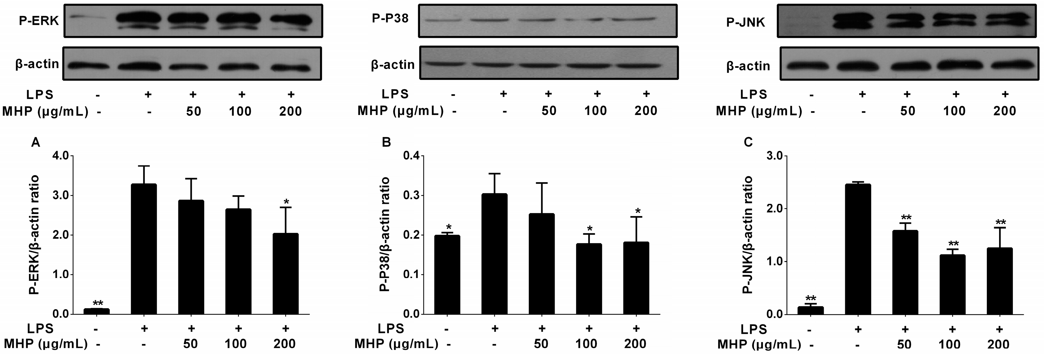
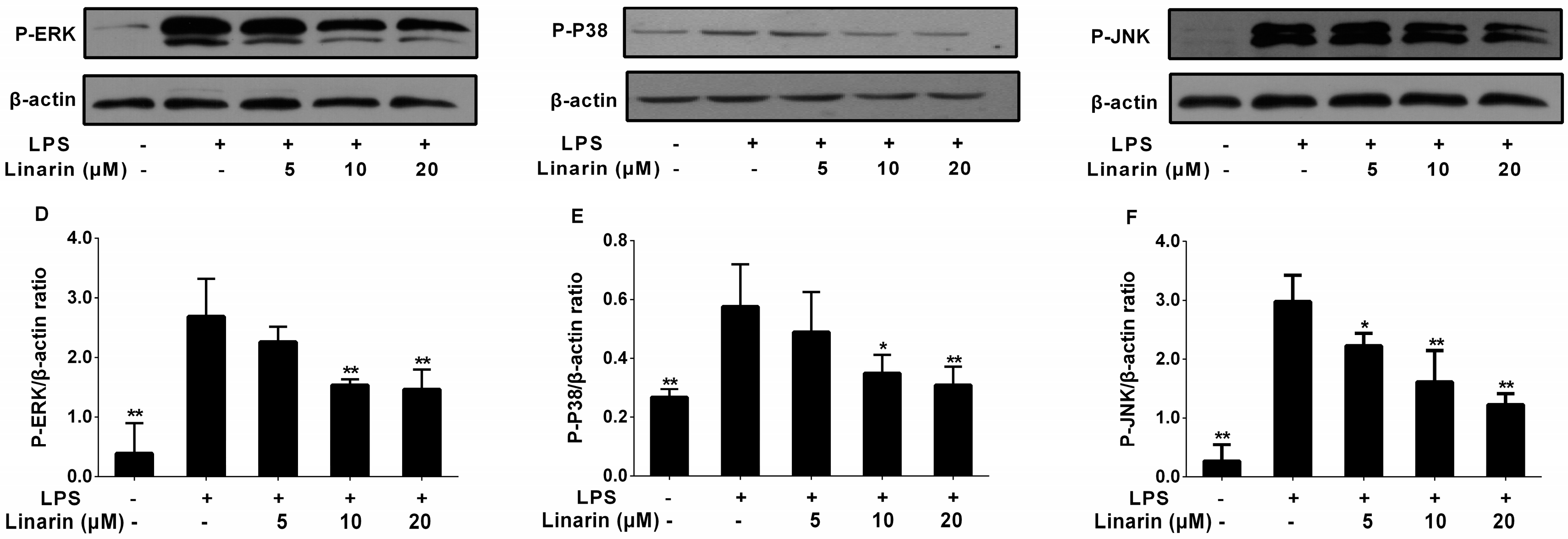
2.7. Effects of the Phenolic Fraction of Mentha haplocalyx and Linarin on MAPKs in LPS-Induced RAW264.7 Cells
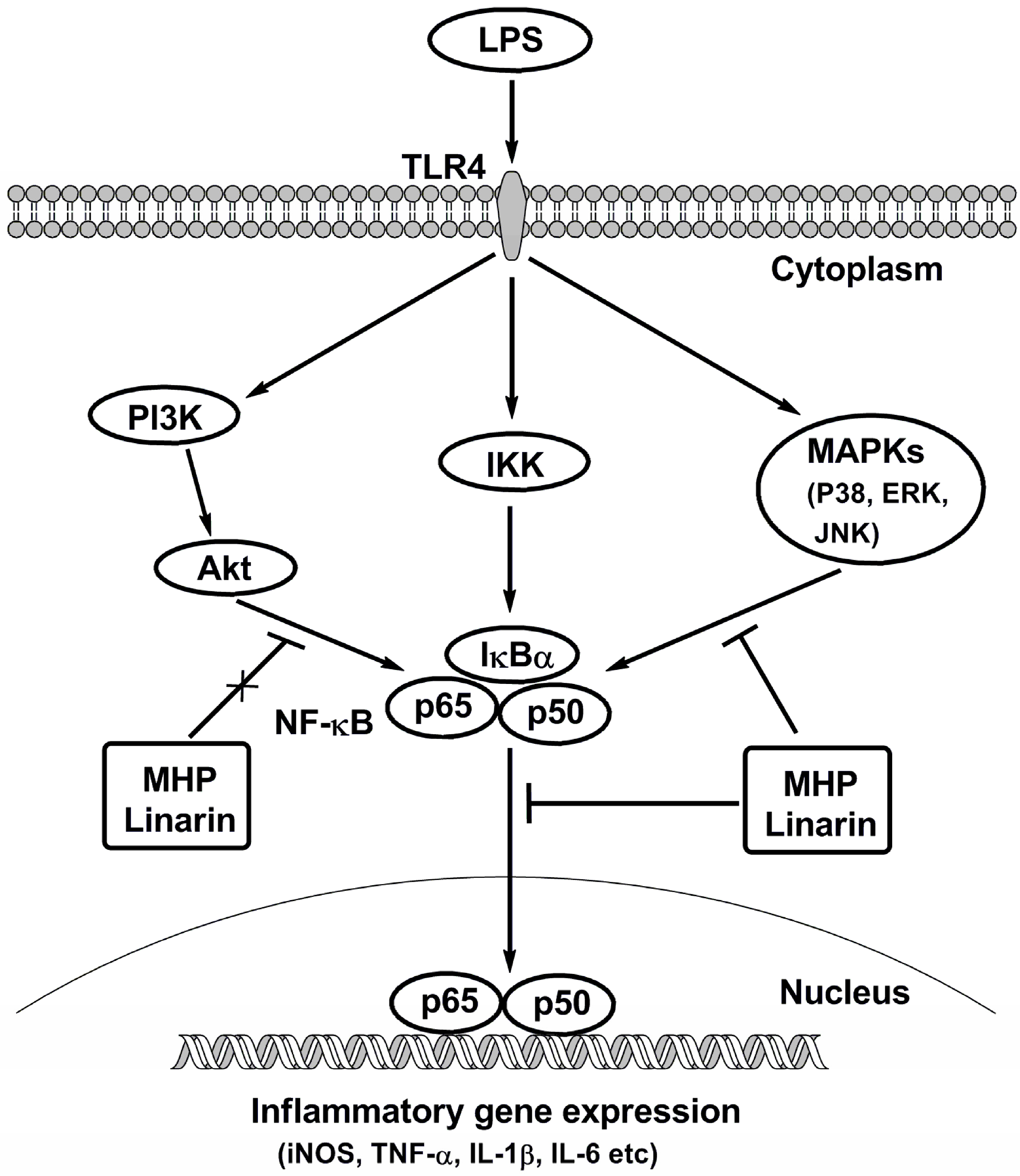
To further explore whether MHP and linarin exerted inhibitory effects on the inflammatory mediators and cytokines through inactivation of the MAPK signaling pathway, we investigated the phosphorylation levels of MAPKs including ERK1/2, JNK, and p38. As observed in Figure 8, LPS stimulation increased the relative content of p-p38, p-JNK, and p-ERK1/2. MHP treatment remarkably inhibited the phosphorylation level of JNK at 50, 100, and 200 μg/mL (Figure 8C) and suppressed the phosphorylation level of p38 at 100 and 200 μg/mL (Figure 8B), while at 200 μg/mL for the phosphorylated ERK (Figure 8A). Furthermore, linarin significantly inhibited the phosphorylation of JNK at 5, 10, and 20 μM (Figure 8F), but at 10 and 20 μM for the phosphorylation of ERK and p38 (Figure 8D,E). Analytical data indicated that MHP and linarin exerted an anti-inflammation effect via the inhibition of the LPS-induced MAPK signaling pathway.

3. Discussion

Pharmacological investigations have shown that anti-inflammation is an essential feature of many Mentha species. M. haplocalyx ethanol extract significantly lessened the severity of airway inflammation by inhibiting the production of immunoglobulin E, IL-4, and IL-6 in bronchoalveolar lavage fluid and lung tissue [35]. Ethyl acetate and aqueous fractions of M. spicata effectively reduced the inflammation induced by carrageenan and cotton pellet [36]. The aqueous ethanol (70%) extract of M. arvensis at 100 μg/mL showed a strong inhibitory effect on IL-8 secretion [37], and the aqueous methanol (80%) extract of M. arvensis, the main constituents of which were linarin and rosmarinic acid, showed inhibitory activity against the strain Chlamydia pneumoniae CWL-029 [38]. The hexane fraction of M. longifolia, mainly containing phenolic and flavonoid compounds, was able to significantly reduce NO production, iNOS, and TNF-α mRNA expression at 0.05–0.20 mg/mL in LPS-induced J774A.1 cells [39]. The results of all the studies mentioned above suggest that Mentha species could serve as an important source of anti-inflammatory agents.
In this paper, we investigated the chemical composition and anti-inflammatory activity of the phenolic fraction from M. haplocalyx. The chemical profile of the MHP sample was analyzed by HPLC-LTQ-Orbitrap MS and showed that its main constituents were phenolic acids and flavonoids, and some of them, such as protocatechuic aldehyde, caffeic acid, rosmarinic acid, salvianolic acid B, and diosmin, have been reported to display inhibitory effects on inflammation [40,41,42,43,44]. These bioactive constituents probably contribute to the anti-inflammatory activity of MHP. Flavonoid linarin is also one of the main constituents of the MHP extract. Previous research has shown that linarin from M. arvensis exhibited a selective inhibitory effect on acetylcholinesterase in a concentration-dependent manner [45]. It has also been reported that linarin possesses analgesic, antipyretic [46], and neuroprotective activities [47], and showed a protective effect against d-glactosamine/LPS-induced fulminant hepatic failure [48]. Furthermore, a tissue distribution study of linarin has revealed that it was rapidly and widely distributed in the heart, liver, spleen, lung, kidney, brain, muscle, stomach, small intestine, epididymis, pancreas, and bladder of rats after intragastric administration [49]. Since there was little data on the anti-inflammatory mechanism of linarin [50], the present study explored its involvement in the anti-inflammatory action of the MHP extract.
NO is a small diffusible molecule, which exerts a regulating effect on various biological functions including apoptosis, neurotransmission, inflammation, and blood vessel tone [51]. NO is generated from l-arginine by three types of NO synthases including neuronal NOS (nNOS), iNOS, and endothelial NOS (eNOS) [51]. Among them, iNOS is the main enzyme to catalyze NO production in acute and chronic inflammation, and inflammatory stimuli such as LPS can cause its expression [7,52]. Our data indicated that both MHP and linarin significantly suppressed the LPS-induced NO production in RAW264.7 cells with a concentration-dependent manner via down-regulating the transcriptional level of iNOS.
Apart from NO, many pro-inflammatory cytokines including TNF-α, IL-6, and IL-1β are produced and take part in the process of inflammatory responses. TNF-α acts as a principal endogenous mediator during inflammatory responses and is also capable of stimulating the secretion of IL-6 and IL-1β, which could exert a synergistic effect with TNF-α [2,53], causing further damage to the body. Inhibiting the synthesis or release of these inflammatory cytokines could emerge as a potential therapeutic approach for inflammatory diseases. In the present study, MHP significantly blocked LPS-induced overproduction of TNF-α, IL-1β, and IL-6. Linarin attenuated TNF-α, IL-1β, and IL-6 production induced by LPS as well, which was consistent with the reported article [50]. Additionally, further investigation demonstrated that MHP and linarin treatment down-regulated the expression levels of TNF-α, IL-1β, and IL-6 mRNA. The above results indicated that MHP and linarin exerted an inhibitory effect on LPS-induced inflammation by suppressing the expression of inflammatory cytokines at the transcriptional and translational levels.
By modulating the secretion of inflammatory cytokines, NF-κB exerts a critical effect on regulating inflammatory responses to extracellular stimuli [34,54]. The major forms of NF-κB are composed of p50 and p65, which are bound to the cytoplasmic inhibitory protein IκB in the resting state [54]. Once activated by external stimuli, IκB is phosphorylated by IκB kinase (IKK) which leads to the degradation of IκBα [55], and p65 is then released and translocates to the nucleus where it may trigger the expression of multiple inflammatory genes. Among the identified constituents of MHP, protocatechuic aldehyde exerted an anti-inflammatory effect on myocardial ischemia/reperfusion injury through inhibiting the NF-κB pathway [40]. Caffeic acid exhibited the anti-inflammatory property via reducing the activation of NF-κB targeting genes [41]. Both salvianolic acid B and diosmin showed a protective effect on lung inflammation by suppressing the expression of TNF-α, IL-1β, and IL-6 through inactivation of the NF-κB pathway [42,43]. Therefore, MHP was expected to exert an anti-inflammatory effect by inhibiting the NF-κB pathway. Data in this paper showed that treatment with definite concentrations of MHP effectively blocked LPS-induced phosphorylation of IκBα and p65. Meanwhile, the increased phosphorylation levels of IκBα and p65 were also down-regulated by linarin. These suggested that MHP and linarin were able to repress the activation of the NF-κB pathway in LPS-induced RAW264.7 cells.
In addition to the NF-κB pathway, the PI3K/Akt and MAPK pathways play essential roles in regulating inflammatory cytokine production in macrophages. It has been reported that Akt and MAPKs were involved in the activation of NF-κB [56,57]. Akt is a key downsteam protein of PI3K and shows diverse roles in cellular growth, adhesion, and inflammation responses [58]. However, our finding showed that LPS-induced phosphorylated Akt was not blocked by MHP. Rosmarinic acid and linarin were two main constituents of MHP. In the previous study of the molecular mechanism of melanogenesis, rosmarinic acid did not reduce Akt phosphorylation in B16 melanoma cells [59]. The results of the present study also demonstrated that linarin displayed no significant inhibitory effect on the phosphorylated Akt. These results suggest that other signaling pathways may be involved in the anti-inflammatory mechanisms of MHP and linarin. Therefore, the impacts of MHP and linarin on MAPKs were further investigated. MAPKs including JNK, p38, and ERK are reported to be closely associated with diverse cellular processes, such as differentiation, proliferation, apoptosis, and immune responses [60]. In general, mitogen and differentiation signals are able to activate the ERK while stress stimuli activate p38 and JNK [61]. Many studies have illustrated that the up-regulation of TNF-α, IL-1β, and IL-6 is diminished by phytochemicals through the inhibition of the activation of ERK, JNK, and p38 [6,62]. In this paper, MHP and linarin down-regulated the phosphorylation levels of ERK, JNK, and p38. Taken together, these findings show that MHP and linarin could inhibit the activation of the NF-κB pathway via suppressing MAPKs activities.
In conclusion, the present study revealed that the phenolic fraction of M. haplocalyx and linarin showed potent inhibitory effects on the secretion of NO, TNF-α, IL-1β, and IL-6 and the mRNA expression of iNOS, TNF-α, IL-1β, and IL-6 in LPS-induced RAW264.7 cells. As shown in Figure 9, these effects were closely related to the inhibition of LPS-induced activation of NF-κB and MAPKs signals. The results not only clarified the anti-inflammatory molecular mechanisms of MHP and linarin, but also pointed out the potential value of MHP and linarin as anti-inflammatory agents for the prevention and treatment of inflammatory diseases.

4. Materials and Methods

4.1. Reagents

Linarin and rosmarinic acid were purchased from Must Bio-technology Co. (Chengdu, China). Lipopolysaccharides and 3-(4,5-dimethylthiazol-2-yl)-2,5-diphenyl tetrazolium bromide (MTT) were purchased from Sigma-Aldrich, Inc. (St. Louis, MO, USA). Dulbecco’s modified eagle’s medium (DMEM) and fetal bovine serum (FBS) were purchased from Corning Inc. (Corning, NY, USA). Nitric oxide assay kit was purchased from Beyotime Biotech. (Jiangsu, China). Mouse TNF-α, IL-1β, and IL-6 ELISA kits were purchased from eBioscience, Inc. (San Diego, CA, USA). Trizol, RevertAid™ first strand cDNA synthesis kit, power sybr green pcr master mix, and Super SignalTM West Pico Chemiluminescent Substrate were purchased from Thermo Fisher Scientific Inc. (Waltham, MA, USA). Oligonucleotide primers were synthesized by Sangon Biotech Co., Ltd. (Shanghai, China). Horse-radish peroxidase-conjugated anti-rabbit or anti-mouse IgG secondary antibodies were obtained from Cell Signaling Technology (Danvers, CO, USA). Fluorescein-conjugated affinipure goat anti-rabbit IgG was purchased from Zhongshan Golden Bridge Biotechnology Co., Ltd. (Beijing, China). Antibodies against phospho (p)-ERK, p-p38, p-JNK, p-Akt, Akt, p-p65, and p-IκBα were obtained from Cell Signaling Technology. All other reagents were of analytical grade.

4.2. Preparation of the Phenolic Fraction of Mentha haplocalyx Extract

The sample (300 g) of M. haplocalyx was extracted three times by refluxing with 4800 mL of 30% ethanol for 1.5 h each time. Then the extracted solution was filtered and the procedure was repeated another two times. The filtered extracts were mixed together and then concentrated until there was no alcoholic odour. The concentrate was centrifuged at 3000 rpm for 30 min. The pH of the supernatant collected was adjusted to 3 by adding acetic acid. The sample solution was applied onto a column of treated HPD-400 macroporous resin (Cangzhou Baoen Chemical Co., Ltd., Cangzhou, Hebei, China). The elution conditions were as follows: 20% ethanol for two column volume, 70% ethanol for three column volume. The eluent of 70% ethanol was collected and then concentrated to dryness. MHP was obtained after the residue was dried under reduced pressure. For in vitro experiments, the phenolic fraction was dissolved in dimethyl sulfoxide with a final concentration of less than 0.1%.

4.3. HPLC-MS/MS Analysis of MHP

MHP (10 mg) was dissolved using 70% methanol in a 25 mL volumetric flask, and the solution was filtered by a 0.45 μM microporous membrane.
HPLC-MS/MS analysis was performed using a LTQ-Orbitrap XL mass spectrometer (Thermo Scientific, Bremen, Germany). Analysis was carried out at 30 °C on an Inersil C18 column (4.6 mm × 150 mm, Waters, Milford, MA, USA). The mobile phase system was composed of acetonitrile (A) and 0.3% formic acid aqueous solution (B). A gradient program was used as follows: 0–40 min, 5%–15% A; 40–70 min, 15%–19.5% A; 70–90 min, 19.5%–22% A; 90–120 min, 22%–30% A. The flow rate was kept at 1 mL/min with a post-column splitting ratio of 1:4 and the sample volume injected was set at 5 μL.
The operating conditions of mass spectrometry were as follows: electrospray ionization (ESI) source in positive or negative mode, capillary temperature of 350 °C; sheath gas flow, 30 arb; auxiliary gas flow, 10 arb; positive mode, 4 kV of spray voltage, 25 V of capillary voltage; 110 V of tube lens negative mode, 3 kV of spray voltage, −35 V of capillary voltage; −110 V of tube lens. The range of full-scan mass was from m/z 100 to 1000. The resolution of the Orbitrap analyzer was set at 30,000. The three most intense ions from the one-stage mass spectrometry scanning were selected for further MS2 analysis. The other specific parameters were as follows: collision-induced dissociation (CID) of activation type, 2 amu of isolation width, 30 ms of activation time, 35% of normalized collision energy.

4.4. Cell Culture Conditions and Treatment

The RAW264.7 macrophage cell line was purchased from China Infrastructure of Cell Line Resource (Beijing, China). RAW264.7 cells were cultured in DMEM supplemented with 10% FBS and 1% penicillin-streptomycin in a humidified incubator with 5% CO2 at 37 °C. The cells were treated with MHP and linarin at different concentrations and then stimulated with LPS (1 μg/mL) for the incubation time.

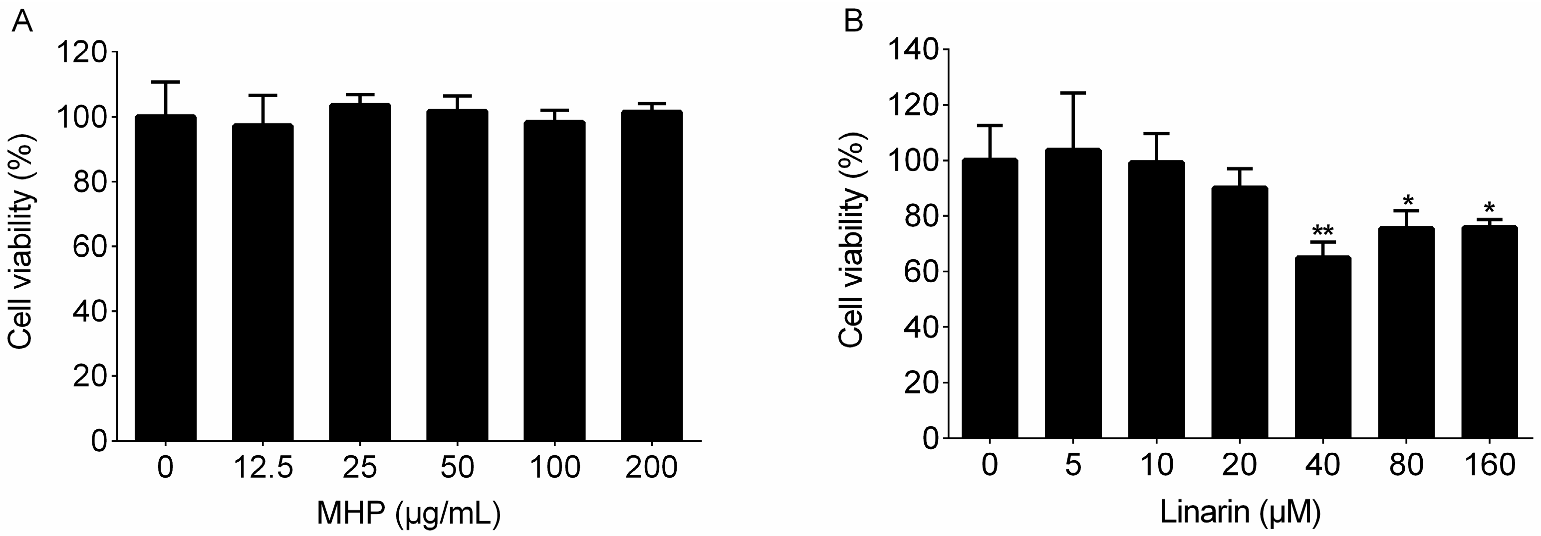
4.5. Cell Viability Assay

The RAW264.7 cells were seeded in 96-well plates overnight, followed by incubation with MHP (0, 12.5, 25, 50, 100, and 200 μg/mL) and linarin (0, 5, 10, 20, 40, 80, and 160 μM) for 24 h. After treatment for 24 h, the supernatant was removed, and the MTT solution (10 μL) and fresh culture medium (90 μL) were added to each well. The cells were incubated for 4 h, after which the culture medium was removed and DMSO was added to dissolve the crystals for an additional 10 min. The value of optical density was read on a microplate reader (MK3, Thermoelectric Technology Instrument Co., Ltd., Shanghai, China) at 492 nm.

4.6. Effect on NO Production

The RAW264.7 cells were seeded in 96-well plates overnight and then treated with MHP (50, 100, and 200 μg/mL) and linarin (5, 10, and 20 μM) for 1 h, followed by incubation with LPS (1 μg/mL) for an additional 24 h. Griess reagent was used to determine the content of nitric oxide in the cell culture supernatant. Briefly, the culture supernatant (50 μL) was reacted with an equal volume of Griess reagent (50 μL), and the optical density of each well was measured on the microplate reader at 570 nm.

4.7. Cytokines Measurement

For the measurement of TNF-α, IL-1β, and IL-6, the RAW264.7 cells were seeded in 24-well plates overnight and then treated with MHP (50, 100, and 200 μg/mL) and linarin (5, 10, and 20 μM) for 1 h, followed by incubation with LPS (1 μg/mL) for an additional 24 h. The content of the pro-inflammatory cytokines including TNF-α, IL-1β, and IL-6 in the supernatant were assayed using the ELISA kits according to the manufacturer’s instructions.

4.8. RNA Extraction and qRT-PCR

Total RNA was isolated using the Trizol reagent according to the manufacturer’s protocol. Reverse transcription of total RNA (3.0 μg) was performed by using the RevertAid™ first strand cDNA synthesis kit on a MyCycler Thermal Cycler (Bio-Rad, Hercules, CA, USA). qRT-PCR was implemented using the SYBR qPCR kit with 2 μL cDNA on a StepOnePlus™ Real-Time PCR System (Applied Biosystems, Foster City, CA, USA). The thermal cycling conditions were as follows: 95 °C for 10 min, and then 40 cycles of 95 °C for 15 s, 60 °C for 1 min, and finally 95 °C for 15 s and 60 °C for 1 min. The mRNA expression levels of iNOS, TNF-α, IL-1β, and IL-6 were normalized to GAPDH. The following primers were used for PCR amplification: iNOS, 5′-GGTGAAGGGACTGAGCTGTT-3′ (forward) and 5′-ACGTTCTCCGTTCTCTTGCAG-3′ (reverse); TNF-α, 5′-TATGGCTCAGGGTCCAACTC-3′ (forward) and 5′-GGAAAGCCCATTTGAGTCCT-3′ (reverse); IL-1β, 5′-CTCACAAGCAGAGCACAAGC-3′ (forward) and 5′-CAGTCCAGCCCATACTTTAGG-3′ (reverse); IL-6, 5′-CGGAGAGGAGACTTCACAGAG-3′ (forward) and 5′-CATTTCCACGATTTCCCAGA-3′ (reverse); GAPDH, 5′-GGTTGTCTCCTGCGACTTCA-3′ (forward) and 5′-TGGTCCAGGGTTTCTTACTCC-3′ (reverse).

4.9. Western Blot Analysis

The RAW264.7 cells were seeded in 100-mm dishes overnight, after which they were treated with MHP (50, 100, and 200 μg/mL) and linarin (5, 10, and 20 μM) for 4 h, and further incubated with LPS (1 μg/mL) for an additional 30 min. Subsequently, the cells were rinsed two times with cold phosphate-buffered saline (PBS), lysed using ice-cold radioimmunoprecipitation assay (RIPA) buffer supplemented with a cocktail of protease and phosphatase inhibitors, and placed on ice for 30 min. The supernatants were collected after centrifuging at 10,000 g for 15 min at 4 °C. The bicinchoninic acid (BCA) protein assay kit was used to measure protein content for each sample. Protein samples were separated on a 10% SDS-PAGE using PowerPac™ Basic Power Supply (Bio-Rad, Hercules, CA, USA), blotted onto PVDF membranes and then blocked for 1 h at room temperature with 5% nonfat dry milk in Tris-buffered saline with Tween 20 (TBST) solution. The membranes were then incubated at 4 °C overnight with 1:1000 dilutions of the primary antibodies. After washed with TBST solution three times for 10 min, the membranes were incubated with HRP-conjugated secondary antibody for 1 h at room temperature. Finally, the membranes were washed with the TBST solution three times for 10 min and chemiluminescent signals of the protein bands were developed using the Super SignalTM West Pico Chemiluminescent Substrate (Thermo Fisher Scientific Inc.).

4.10. Statistical Analysis

The results were expressed as the means ± standard deviation (SD). Multiple group comparisons were performed using one-way ANOVA test using the SPSS 17.0 software (SPSS Inc., Chicago, IL, USA); * p < 0.05 or ** p < 0.01 were considered statistically significant.

Acknowledgments

This work was supported by the National Natural Science Foundation of China (No. 81173520).

Author Contributions

Bin Liu and Ying Wu designed the experiments; Bin Liu and Xiangyang Chen contributed to the isolation and identification of MHP. Xiangyang Chen, Shujing Zhang, and Zinan Xuan performed the experiments related to the anti-inflammatory evaluation of the MHP and linarin; Xiaoming Chen and Junjie Zhang contributed to the data analysis; Dongyu Ge and Qian Wang contributed reagents/materials/analysis tools; Xiangyang Chen, Bin Liu, and Ying Wu wrote the paper.

Conflicts of Interest

The authors declare no conflict of interest.

References

  1. Kim, D.H.; Yun, C.H.; Kim, M.H.; Naveen Kumar, C.; Yun, B.H.; Shin, J.S.; An, H.J.; Lee, Y.H.; Yun, Y.D.; Rim, H.K.; et al. 4′-Bromo-5,6,7-trimethoxyflavone represses lipopolysaccharide-induced iNOS and COX-2 expressions by suppressing the NF-κB signaling pathway in RAW 264.7 macrophages. Bioorg. Med. Chem. Lett. 2012, 22, 700–705. [Google Scholar] [CrossRef] [PubMed]
  2. Ying, X.; Yu, K.; Chen, X.; Chen, H.; Hong, J.; Cheng, S.; Peng, L. Piperine inhibits LPS induced expression of inflammatory mediators in RAW 264.7 cells. Cell. Immunol. 2013, 285, 49–54. [Google Scholar] [CrossRef] [PubMed]
  3. Liu, X.H.; Pan, L.L.; Jia, Y.L.; Wu, D.; Xiong, Q.H.; Wang, Y.; Zhu, Y.Z. A novel compound DSC suppresses lipopolysaccharide-induced inflammatory responses by inhibition of Akt/NF-κB signalling in macrophages. Eur. J. Pharmacol. 2013, 708, 8–13. [Google Scholar] [CrossRef] [PubMed]
  4. Jungbauer, A.; Medjakovic, S. Anti-inflammatory properties of culinary herbs and spices that ameliorate the effects of metabolic syndrome. Maturitas 2012, 71, 227–239. [Google Scholar] [CrossRef] [PubMed]
  5. Adams, D.O.; Hamilton, T.A. The cell biology of macrophage activation. Annu. Rev. Immunol. 1984, 2, 283–318. [Google Scholar] [CrossRef] [PubMed]
  6. Park, J.W.; Kwon, O.K.; Yuniato, P.; Marwoto, B.; Lee, J.; Oh, S.R.; Kim, J.H.; Ahn, K.S. Amelioration of an LPS-induced inflammatory response using a methanolic extract of Lagerstroemia ovalifolia to suppress the activation of NF-κB in RAW264.7 macrophages. Int. J. Mol. Med. 2016, 38, 482–490. [Google Scholar] [CrossRef] [PubMed]
  7. Jin, M.; Moon, T.C.; Quan, Z.; Lee, E.; Kim, Y.K.; Yang, J.H.; Suh, S.J.; Jeong, T.C.; Lee, S.H.; Kim, C.H.; et al. The naturally occurring flavolignan, deoxypodophyllotoxin, inhibits lipopolysaccharide-induced iNOS expression through the NF-κB activation in RAW264. 7 macrophage cells. Biol. Pharm. Bull. 2008, 31, 1312–1315. [Google Scholar] [CrossRef] [PubMed]
  8. Lee, T.H.; Jung, M.; Bang, M.H.; Chung, D.K.; Kim, J. Inhibitory effects of a spinasterol glycoside on lipopolysaccharide-induced production of nitric oxide and proinflammatory cytokines via down-regulating MAP kinase pathways and NF-κB activation in RAW264.7 macrophage cells. Int. Immunopharmacol. 2012, 13, 264–270. [Google Scholar] [CrossRef]
  9. May, M.J.; Ghosh, S. Signal transduction through NF-κB. Immunol. Today 1998, 19, 80–88. [Google Scholar] [CrossRef]
  10. Fan, G.W.; Zhang, Y.; Jiang, X.; Zhu, Y.; Wang, B.; Su, L.; Cao, W.; Zhang, H.; Gao, X. Anti-inflammatory activity of baicalein in LPS-stimulated RAW264.7 macrophages via estrogen receptor and NF-κB-dependent pathways. Inflammation 2013, 36, 1584–1591. [Google Scholar] [CrossRef] [PubMed]
  11. Ma, J.; Shi, H.; Mi, C.; Li, H.L.; Lee, J.J.; Jin, X. Malloapelta B suppresses LPS-induced NF-κB activation and NF-κB-regulated target gene products. Int. Immunopharmacol. 2015, 24, 147–152. [Google Scholar] [CrossRef] [PubMed]
  12. Lee, H.-S.; Bilehal, D.; Lee, G.-S.; Ryu, D.-S.; Kim, H.-K.; Suk, D.-H.; Lee, D.-S. Anti-inflammatory effect of the hexane fraction from Orostachys japonicus in RAW 264.7 cells by suppression of NF-κB and PI3K-Akt signaling. J. Funct. Foods 2013, 5, 1217–1225. [Google Scholar] [CrossRef]
  13. Pan, X.; Cao, X.; Li, N.; Xu, Y.; Wu, Q.; Bai, J.; Yin, Z.; Luo, L.; Lan, L. Forsythin inhibits lipopolysaccharide-induced inflammation by suppressing JAK-STAT and p38 MAPK signalings and ROS production. Inflamm. Res. 2014, 63, 597–608. [Google Scholar] [CrossRef] [PubMed]
  14. She, G.M.; Xu, C.; Liu, B. New monocyclic monoterpenoid glycoside from Mentha haplocalyx Briq. Chem. Cent. J. 2012, 6, 37. [Google Scholar] [CrossRef] [PubMed]
  15. Park, Y.J.; Baskar, T.B.; Yeo, S.K.; Arasu, M.V.; Al-Dhabi, N.A.; Lim, S.S.; Park, S.U. Composition of volatile compounds and in vitro antimicrobial activity of nine Mentha spp. SpringerPlus 2016, 5, 1628. [Google Scholar] [CrossRef] [PubMed]
  16. Cao, G.; Shan, Q.; Li, X.; Cong, X.; Zhang, Y.; Cai, H.; Cai, B. Analysis of fresh Mentha haplocalyx volatile components by comprehensive two-dimensional gas chromatography and high-resolution time-of-flight mass spectrometry. Analyst 2011, 136, 4653–4661. [Google Scholar] [CrossRef] [PubMed]
  17. Gul, H.; Abbas, K.; Qadir, M.I. Gastro-protective effect of ethanolic extract of Mentha longifolia in alcohol- and aspirin-induced gastric ulcer models. Bangladesh J. Pharmacol. 2015, 10. [Google Scholar] [CrossRef]
  18. Estrada-Soto, S.; Gonzalez-Maldonado, D.; Castillo-Espana, P.; Aguirre-Crespo, F.; Sanchez-Salgado, J.C. Spasmolytic effect of Mentha pulegium L. involves ionic flux regulation in rat ileum strips. J. Smooth Muscle Res. 2010, 46, 107–117. [Google Scholar] [CrossRef] [PubMed]
  19. Jagetia, G.C.; Baliga, M.S. Influence of the leaf extract of Mentha arvensis linn. (mint) on the survival of mice exposed to different doses of gamma radiation. Strahlenther. Onkol. 2002, 178, 91–98. [Google Scholar] [CrossRef] [PubMed]
  20. Samarth, R.M.; Panwar, M.; Kumar, A. Modulatory effects of Mentha piperita on lung tumor incidence, genotoxicity, and oxidative stress in benzo[a]pyrene-treated Swiss albino mice. Environ. Mol. Mutagen. 2006, 47, 192–198. [Google Scholar] [CrossRef] [PubMed]
  21. Saleem, M.; Alam, A.; Sultana, S. Attenuation of benzoyl peroxide-mediated cutaneous oxidative stress and hyperproliferative response by the prophylactic treatment of mice with spearmint (Mentha spicata). Food Chem. Toxicol. 2000, 38, 939–948. [Google Scholar] [CrossRef]
  22. Orhan, I.E.; Ozcelik, B.; Kartal, M.; Kan, Y. Antimicrobial and antiviral effects of essential oils from selected Umbelliferae and Labiatae plants and individual essential oil components. Turk. J. Biol. 2012, 36, 239–246. [Google Scholar] [CrossRef]
  23. Amzazi, S.; Ghoulami, S.; Bakri, Y.; Idrissi, A.; Fkih-Tétouani, S.; Benjouad, A. Human immunodeficiency virus type 1 inhibitory activity of Mentha longifolia. Thérapie 2003, 58, 531–534. [Google Scholar] [CrossRef] [PubMed]
  24. Mimica-Dukic, N.; Bozin, B.; Mentha, L. Species (Lamiaceae) as promising sources of bioactive secondary metabolites. Curr. Pharm. Des. 2008, 14, 3141–3150. [Google Scholar] [CrossRef] [PubMed]
  25. Dong, W.; Ni, Y.; Kokot, S. Differentiation of mint (Mentha haplocalyx Briq.) from different regions in China using gas and liquid chromatography. J. Sep. Sci. 2015, 38, 402–409. [Google Scholar] [CrossRef] [PubMed]
  26. Liu, Y.; Zhang, Y.H.; Shi, R.B. Studies on the chemixcal constituents in herb of Mentha haplocalyx. China J. Chin. Mater. Med. 2005, 30, 1086–1088. [Google Scholar] [CrossRef]
  27. Hao, L.L.; Li, X.R.; Cao, L.; Chen, S.H. The interaction between exterior releasing herbs and anti-infection. Mod. Med. J. China 2006, 8, 84–85. [Google Scholar] [CrossRef]
  28. Liao, H.; Banbury, L.K.; Leach, D.N. Elucidation of Danzhixiaoyao Wan and its constituent herbs on antioxidant activity and inhibition of nitric oxide production. Evid. Based Complement. Altern. Med. 2007, 4, 425–430. [Google Scholar] [CrossRef] [PubMed]
  29. Li, M.L.; Xu, L.Y.; Li, Z.L.; Qian, S.H.; Qin, M.J. Flavonoids from Mentha haplocalyx. Chem. Nat. Compd. 2014, 50, 124–125. [Google Scholar] [CrossRef]
  30. She, G.M.; Xu, C.; Liu, B.; Shi, R.B. Polyphenolic acids from mint (the aerial of Mentha haplocalyx Briq.) with DPPH radical scavenging activity. J. Food Sci. 2010, 75, C359–C362. [Google Scholar] [CrossRef] [PubMed]
  31. She, G.M.; Xu, C.; Liu, B. Phenylpropanoids from Mentha haplocalyx. Chem. Nat. Compd. 2013, 48, 1083–1084. [Google Scholar] [CrossRef]
  32. Farnad, N.; Heidari, R.; Aslanipour, B. Phenolic composition and comparison of antioxidant activity of alcoholic extracts of peppermint (Mentha piperita). J. Food Meas. Charact. 2014, 8, 113–121. [Google Scholar] [CrossRef]
  33. Jiang, Y.Y.; Zhang, F.; Liu, B. Optimization of purification technology for total phenolic acid from Mentha haplocalyx by macroporous resin. Chin. J. Exp. Tradit. Med. Form. 2012, 18, 9–11. [Google Scholar]
  34. Li, Q.; Verma, I.M. NF-κB regulation in the immune system. Nat. Rev. Immunol. 2002, 2, 725–734. [Google Scholar] [CrossRef] [PubMed]
  35. Lee, M.Y.; Lee, J.A.; Seo, C.S.; Ha, H.; Lee, N.H.; Shin, H.K. Protective effects of Mentha haplocalyx ethanol extract (MH) in a mouse model of allergic asthma. Phytother. Res. 2011, 25, 863–869. [Google Scholar] [CrossRef] [PubMed]
  36. Arumugam, P.; Priya, N.G.; Subathra, M.; Ramesh, A. Anti-inflammatory activity of four solvent fractions of ethanol extract of Mentha spicata L. investigated on acute and chronic inflammation induced rats. Environ. Toxicol. Pharmacol. 2008, 26, 92–95. [Google Scholar] [CrossRef] [PubMed]
  37. Zaidi, S.F.; Muhammad, J.S.; Shahryar, S.; Usmanghani, K.; Gilani, A.H.; Jafri, W.; Sugiyama, T. Anti-inflammatory and cytoprotective effects of selected Pakistani medicinal plants in Helicobacter pylori-infected gastric epithelial cells. J. Ethnopharmacol. 2012, 141, 403–410. [Google Scholar] [CrossRef] [PubMed]
  38. Salin, O.; Tormakangas, L.; Leinonen, M.; Saario, E.; Hagstrom, M.; Ketola, R.A.; Saikku, P.; Vuorela, H.; Vuorela, P.M. Corn mint (Mentha arvensis) extract diminishes acute chlamydia pneumoniae infection in vitro and in vivo. J. Agric. Food Chem. 2011, 59, 12836–12842. [Google Scholar] [CrossRef] [PubMed]
  39. Karimian, P.; Kavoosi, G.; Amirghofran, Z. Anti-inflammatory effect of Mentha longifolia in lipopolysaccharide-stimulated macrophages reduction of nitric oxide production through inhibition of inducible nitric oxide synthase. J. Immunotoxicol. 2013, 10, 393–400. [Google Scholar] [CrossRef] [PubMed]
  40. Wei, G.; Guan, Y.; Yin, Y.; Duan, J.; Zhou, D.; Zhu, Y.; Quan, W.; Xi, M.; Wen, A. Anti-inflammatory effect of protocatechuic aldehyde on myocardial ischemia/reperfusion injury in vivo and in vitro. Inflammation 2013, 36, 592–602. [Google Scholar] [CrossRef] [PubMed]
  41. Kim, S.R.; Jung, Y.R.; Kim, D.H.; An, H.J.; Kim, M.K.; Kim, N.D.; Chung, H.Y. Caffeic acid regulates LPS-induced NF-κB activation through NIK/IKK and c-Src/ERK signaling pathways in endothelial cells. Arch. Pharm. Res. 2014, 37, 539–547. [Google Scholar] [CrossRef] [PubMed]
  42. Imam, F.; Al-Harbi, N.O.; Al-Harbi, M.M.; Ansari, M.A.; Zoheir, K.M.; Iqbal, M.; Anwer, M.K.; Al Hoshani, A.R.; Attia, S.M.; Ahmad, S.F. Diosmin downregulates the expression of T cell receptors, pro-inflammatory cytokines and NF-κB activation against LPS-induced acute lung injury in mice. Pharmacol. Res. 2015, 102, 1–11. [Google Scholar] [CrossRef] [PubMed]
  43. Zhang, D.F.; Zhang, J.; Li, R. Salvianolic acid B attenuates lung inflammation induced by cigarette smoke in mice. Eur. J. Pharmacol. 2015, 761, 174–179. [Google Scholar] [CrossRef] [PubMed]
  44. Jiang, W.L.; Chen, X.G.; Qu, G.W.; Yue, X.D.; Zhu, H.B.; Tian, J.W.; Fu, F.H. Rosmarinic acid protects against experimental sepsis by inhibiting proinflammatory factor release and ameliorating hemodynamics. Shock 2009, 32, 608–613. [Google Scholar] [CrossRef] [PubMed]
  45. Oinonen, P.P.; Jokela, J.K.; Hatakka, A.I.; Vuorela, P.M. Linarin, a selective acetylcholinesterase inhibitor from Mentha arvensis. Fitoterapia 2006, 77, 429–434. [Google Scholar] [CrossRef] [PubMed]
  46. Martinez-Vazquez, M.; Ramirez Apan, T.O.; Aguilar, H.; Bye, R. Analgesic and antipyretic activities of an aqueous extract and of the flavone linarin of Buddleia cordata. Planta Med. 1996, 62, 137–140. [Google Scholar] [CrossRef] [PubMed]
  47. Lou, H.; Fan, P.; Perez, R.G.; Lou, H. Neuroprotective effects of linarin through activation of the PI3K/Akt pathway in amyloid-β-induced neuronal cell death. Bioorg. Med. Chem. 2011, 19, 4021–4027. [Google Scholar] [CrossRef] [PubMed]
  48. Kim, S.J.; Cho, H.I.; Kim, S.J.; Park, J.H.; Kim, J.S.; Kim, Y.H.; Lee, S.K.; Kwak, J.H.; Lee, S.M. Protective effect of linarin against d-galactosamine and lipopolysaccharide-induced fulminant hepatic failure. Eur. J. Pharmacol. 2014, 738, 66–73. [Google Scholar] [CrossRef] [PubMed]
  49. Feng, X.; Liu, Y.; Wang, X.; Di, X. A rapid and sensitive LC-MS/MS method for the determination of linarin in small-volume rat plasma and tissue samples and its application to pharmacokinetic and tissue distribution study. Biomed. Chromatogr. 2016, 30, 618–624. [Google Scholar] [CrossRef] [PubMed]
  50. Kim, B.; Lee, J.H.; Seo, M.-J.; Eom, S.H.; Kim, W. Linarin down-regulates phagocytosis, pro-inflammatory cytokine production, and activation marker expression in RAW264.7 macrophages. Food Sci. Biotechnol. 2016, 25, 1437–1442. [Google Scholar] [CrossRef]
  51. Chen, G.; Li, K.K.; Fung, C.H.; Liu, C.L.; Wong, H.L.; Leung, P.C.; Ko, C.H. Er-Miao-San, a traditional herbal formula containing Rhizoma Atractylodis and Cortex Phellodendri inhibits inflammatory mediators in LPS-stimulated RAW264.7 macrophages through inhibition of NF-κB pathway and MAPKs activation. J. Ethnopharmacol. 2014, 154, 711–718. [Google Scholar] [CrossRef] [PubMed]
  52. Murphy, S. Production of nitric oxide by glial cells regulation and potential roles in the CNS. Glia 2000, 29, 1–13. [Google Scholar] [CrossRef]
  53. Sclavons, C.; Burtea, C.; Boutry, S.; Laurent, S.; Vander Elst, L.; Muller, R.N. Phage display screening for tumor necrosis factor-α-binding peptides: Detection of inflammation in a mouse model of hepatitis. Int. J. Pept. 2013, 2013, 348409. [Google Scholar] [CrossRef] [PubMed]
  54. Suh, S.J.; Chung, T.W.; Son, M.J.; Kim, S.H.; Moon, T.C.; Son, K.H.; Kim, H.P.; Chang, H.W.; Kim, C.H. The naturally occurring biflavonoid, ochnaflavone, inhibits LPS-induced iNOS expression, which is mediated by ERK1/2 via NF-κB regulation, in RAW264.7 cells. Arch. Biochem. Biophys. 2006, 447, 136–146. [Google Scholar] [CrossRef] [PubMed]
  55. Zhang, W.; Wei, Q. Calcineurin stimulates the expression of inflammatory factors in RAW 264.7 cells by interacting with proteasome subunit alpha type 6. Biochem. Biophys. Res. Commun. 2011, 407, 668–673. [Google Scholar] [CrossRef] [PubMed]
  56. Hu, B.; Zhang, H.; Meng, X.; Wang, F.; Wang, P. Aloe-emodin from rhubarb (Rheum rhabarbarum) inhibits lipopolysaccharide-induced inflammatory responses in RAW264.7 macrophages. J. Ethnopharmacol. 2014, 153, 846–853. [Google Scholar] [CrossRef] [PubMed]
  57. Kim, H.G.; Yoon, D.H.; Lee, W.H.; Han, S.K.; Shrestha, B.; Kim, C.H.; Lim, M.H.; Chang, W.; Lim, S.; Choi, S.; et al. Phellinus linteus inhibits inflammatory mediators by suppressing redox-based NF-κB and MAPKs activation in lipopolysaccharide-induced RAW 264.7 macrophage. J. Ethnopharmacol. 2007, 114, 307–315. [Google Scholar] [CrossRef] [PubMed]
  58. Pan, M.H.; Lai, C.S.; Wang, Y.J.; Ho, C.T. Acacetin suppressed LPS-induced up-expression of iNOS and COX-2 in murine macrophages and TPA-induced tumor promotion in mice. Biochem. Pharmacol. 2006, 72, 1293–1303. [Google Scholar] [CrossRef] [PubMed]
  59. Lee, J.; Kim, Y.S.; Park, D. Rosmarinic acid induces melanogenesis through protein kinase a activation signaling. Biochem. Pharmacol. 2007, 74, 960–968. [Google Scholar] [CrossRef] [PubMed]
  60. Kim, K.S.; Cui, X.; Lee, D.S.; Sohn, J.H.; Yim, J.H.; Kim, Y.C.; Oh, H. Anti-inflammatory effect of neoechinulin a from the marine fungus Eurotium sp. SF-5989 through the suppression of NF-κB and p38 MAPK pathways in lipopolysaccharide-stimulated RAW264.7 macrophages. Molecules 2013, 18, 13245–13259. [Google Scholar] [CrossRef] [PubMed]
  61. Sabio, G.; Davis, R.J. TNF and MAP kinase signalling pathways. Semin. Immunol. 2014, 26, 237–245. [Google Scholar] [CrossRef] [PubMed]
  62. Kwon, D.-J.; Bae, Y.-S.; Ju, S.M.; Youn, G.S.; Choi, S.Y.; Park, J. Salicortin suppresses lipopolysaccharide-stimulated inflammatory responses via blockade of NF-κB and JNK activation in RAW 264.7 macrophages. BMB Rep. 2014, 47, 318–323. [Google Scholar] [CrossRef] [PubMed]
Sample Availability: Samples of the phenolic fraction from Mentha haplocalyx are available from the authors.
Figure 1. Total ion chromatogram profile of the phenolic fraction of Mentha haplocalyx (MHP).
Molecules 22 00811 g001
Figure 2. Effects of MHP and linarin on the viability of RAW264.7 cells. (A) The cells were treated with MHP (0, 12.5, 25, 50, 100, and 200 μg/mL) for 24 h; (B) The cells were treated with linarin (0, 5, 10, 20, 40, 80, and 160 μM) for 24 h. Cell viability was determined by the 3-(4,5-dimethylthiazol-2-yl)-2,5-diphenyltetrazolium bromide (MTT) assay. The data shown are representative of three experiments and expressed as mean ± SD. * p < 0.05 and ** p < 0.01 versus the normal control group.
Molecules 22 00811 g002
Figure 3. Effect of MHP on the production of nitric oxide (NO), tumor necrosis factor-α (TNF-α), interlenkin-1β (IL-1β), and IL-6 in LPS-induced RAW264.7 cells. The cells were pretreated with MHP (50, 100, and 200 μg/mL) for 1 h, and then were stimulated with LPS (1.0 μg/mL) for 24 h. The level of NO (A) in the supernatant was determined using the nitric oxide assay kit. The levels of TNF-α (B), IL-1β (C), and IL-6 (D) in the supernatant were assayed using enzyme-linked immunosorbent assay (ELISA) kits. The data shown are representative of three experiments and expressed as mean ± SD. ** p < 0.01 versus the group treated with LPS alone.
Molecules 22 00811 g003
Figure 4. Effect of linarin on the production of NO, TNF-α, IL-1β and IL-6 in LPS-induced RAW264.7 cells. The cells were pretreated with linarin (5, 10, and 20 μM) for 1 h, and then were stimulated with LPS (1.0 μg/mL) for 24 h. The level of NO (A) in the supernatant was determined using the nitric oxide assay kit. The levels of TNF-α (B), IL-1β (C), and IL-6 (D) in the supernatant were assayed using ELISA kits. The data shown are representative of three experiments and expressed as mean ± SD. * p < 0.05 and ** p < 0.01 versus the group treated with LPS alone.
Molecules 22 00811 g004aMolecules 22 00811 g004b
Figure 5. Effects of MHP and linarin on the mRNA expression of inducible NO synthase (iNOS), TNF-α, IL-1β, and IL-6 in LPS-induced RAW264.7 cells. (A) The cells were pretreated with MHP (50, 100, and 200 μg/mL) for 1 h, and then were stimulated with LPS (1.0 μg/mL) for 24 h. (B) The cells were pretreated with linarin (5, 10, and 20 μM) for 1 h, and then were stimulated with LPS (1.0 μg/mL) for 24 h. The mRNA expression levels of iNOS, TNF-α, IL-1β, and IL-6 were measured by quantitative real time polymerase chain reaction (qRT-PCR). Glyceraldehyde 3-phosphate dehydrogenase (GAPDH) was used as the internal control. The data shown are representative of three experiments and expressed as mean ± SD. * p < 0.05 and ** p < 0.01 versus the group treated with LPS alone.
Molecules 22 00811 g005
Figure 6. Effects of MHP and linarin on the nuclear factor kappa B (NF-κB) signaling pathway in LPS-induced RAW264.7 cells. (A,B) The cells were pretreated with MHP (50, 100, and 200 μg/mL) for 4 h, and then were stimulated with LPS (1.0 μg/mL) for 30 min; (C,D) The cells were pretreated with linarin (5, 10, and 20 μM) for 4 h, and then were stimulated with LPS (1.0 μg/mL) for 30 min. The levels of phosphorylated inhibitor kappa B α (p-IκBα) and p-p65 were detected by Western blot analysis. β-actin was used as the internal control. The data shown are representative of three experiments and expressed as mean ± SD. * p <0.05 and ** p < 0.01 versus the group treated with LPS alone.
Molecules 22 00811 g006
Figure 7. Effects of MHP and linarin on the Akt signaling pathway in LPS-induced RAW264.7 cells. (A) The cells were pretreated with MHP (50, 100, and 200 μg/mL) for 4 h, and then were stimulated with LPS (1.0 μg/mL) for 30 min; (B) The cells were pretreated with linarin (5, 10 and 20 μM) for 4 h, and then were stimulated with LPS (1.0 μg/mL) for 30 min. The levels of p-Akt and Akt were detected by Western blot analysis. The data shown are representative of three experiments and expressed as mean ± SD. * p < 0.05 versus the group treated with LPS alone.
Molecules 22 00811 g007
Figure 8. Effects of MHP and linarin on the mitogen-activated protein kinase (MAPK) signaling pathway in LPS-induced RAW264.7 cells. (AC) The cells were pretreated with MHP (50, 100, and 200 μg/mL) for 4 h, and then were stimulated with LPS (1.0 μg/mL) for 30 min. (DF) The cells were pretreated with linarin (5, 10, and 20 μM) for 4 h, and then were stimulated with LPS (1.0 μg/mL) for 30 min. The levels of the phosphorylated extracellular signal-regulated kinase (ERK), p38, and c-Jun NH2-terminal kinase (JNK) were detected by Western blot analysis. β-actin was used as the internal control. The data shown are representative of three experiments and expressed as mean ± SD. * p < 0.05 and ** p < 0.01 versus the group treated with LPS alone.
Molecules 22 00811 g008aMolecules 22 00811 g008b
Figure 9. Possible roles of MHP and linarin in LPS-induced inflammatory responses in RAW264.7 cells. TLR4: Toll-like receptor 4; IKK: IκB kinase; PI3K: phosphoinositide 3-kinase.
Molecules 22 00811 g009
Table 1. Identification of the compounds in MHP.
PeaktR (min)[M + H]+/[M + Na]+ (m/z)[M − H] (m/z)FormulaMS2 Ions (m/z)Identification
113.38137.0246C7H6O3118.7616, 108.8020, 91.9109protocatechuic aldehyde
216.63163.0401C9H8O3163.0300, 118.9575, 94.9441p-coumaric acid
321.38179.0350C9H8O4160.8768, 134.9199, 82.7767caffeic acid
425.19387.1655C20H20O8369.2120, 340.9894, 207.0837, 163.0436, 119.0394ethyl rosmarinate
528.81337.0923C16H18O8172.9905, 162.8953, 154.9699, 136.9224p-coumaroyl quinic acid
635.79514.3244466.0504, 412.9174, 378.0275, 251.9538unknown
747.24588.4072570.5060, 475.3133, 362.3077, 249.0771unknown
849.10537.1034C27H22O12493.1606, 383.1935, 313.1821, 295.0162, 202.9409lithospermic acid
956.90577.1559C27H30O14311.0969, 269.0196, 241.0736apigenin-7-O-rutinoside
1058.02740.4911717.5318, 693.6151, 603.9049, 536.1751unknown
1160.17633.1774, 611.1957C28H34O15615.2299, 483.1602, 331.0648hesperidin
1261.90631.1624, 609.1804C28H32O15463.1456, 447.0624, 301.0494, 286.0661, 191.0885diosmin
1366.44359.0767C18H16O8341.1331, 160.9566, 132.9843rosmarinic acid
1466.91418.9493C20H18O10400.2057, 362.4225, 344.5225, 220.0517salvianolic acid D
1570.48741.1419C36H30O16561.0992, 543.1505, 451.1976, 363.1163, 319.0901salvianolic acid B
1675.29475.3232 457.2657, 317.2458, 249.2445, 167.2896unknown
1788.55593.1849, 615.1667C28H32O14489.1708, 447.1117, 285.0378, 270.0775, 242.1294linarin
1890.61797.2488651.1746, 447.0908, 285.0492, 24.9773unknown
19107.36361.0909C18H16O8346.0471, 328.0377, 300.1852, 213.0013, 150.7952thymonin

