
Screening of Peruvian Medicinal Plants for Tyrosinase Inhibitory Properties: Identification of Tyrosinase Inhibitors in Hypericum laricifolium Juss


Abstract
:1. Introduction
2. Results and Discussion
2.1. Ethnopharmacological Data
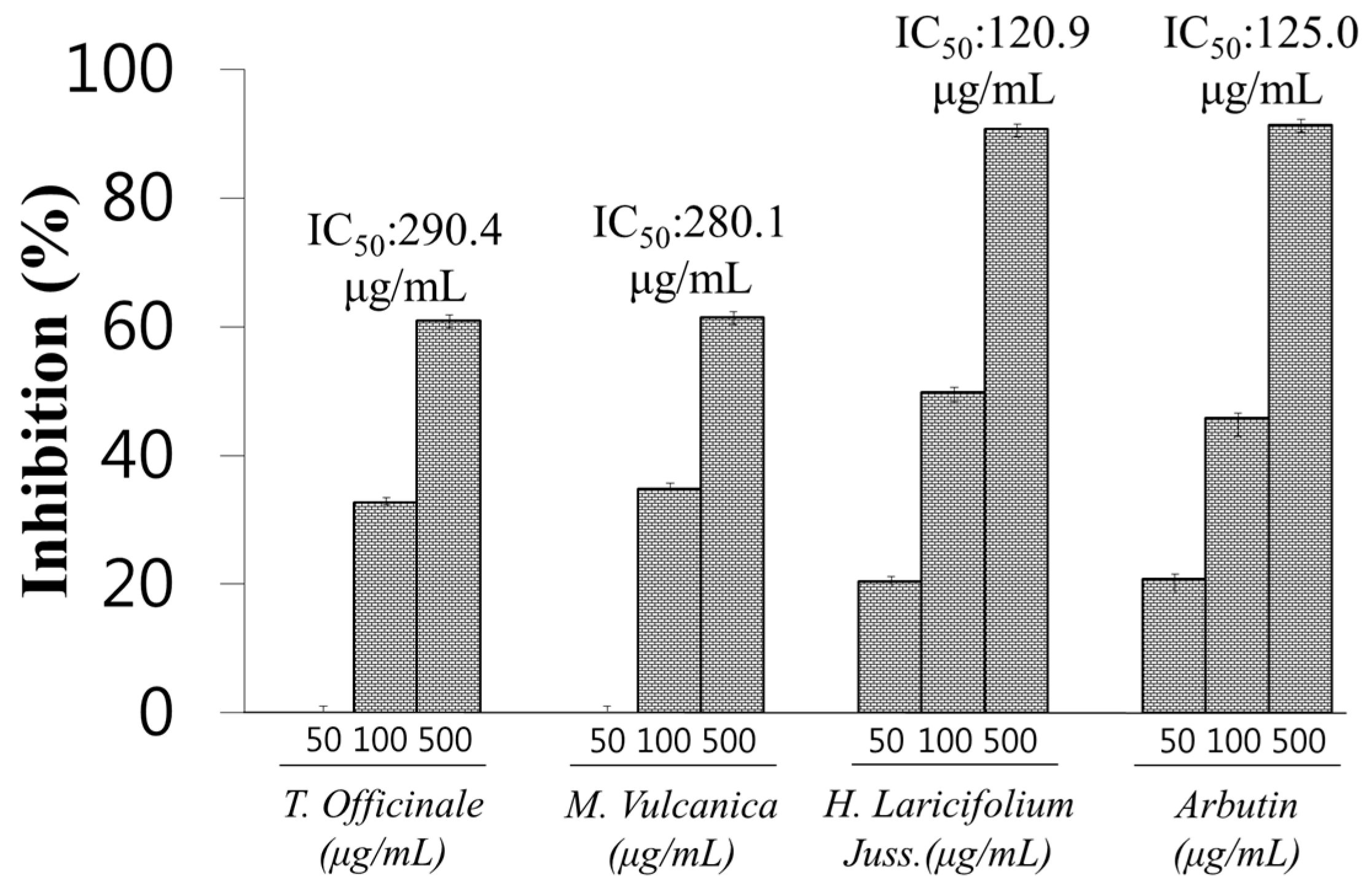
2.2. Screening of Tyrosinase Inhibitory Activities of Methanol Extracts
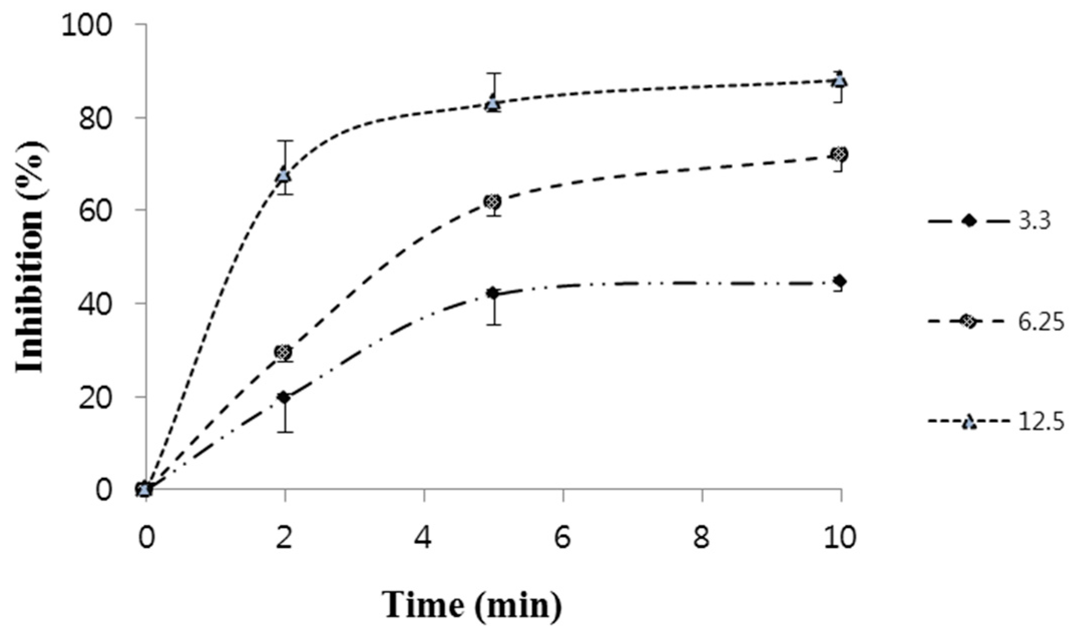
2.3. Effect of H. laricifolium Juss. on Tyrosinase Inhibition
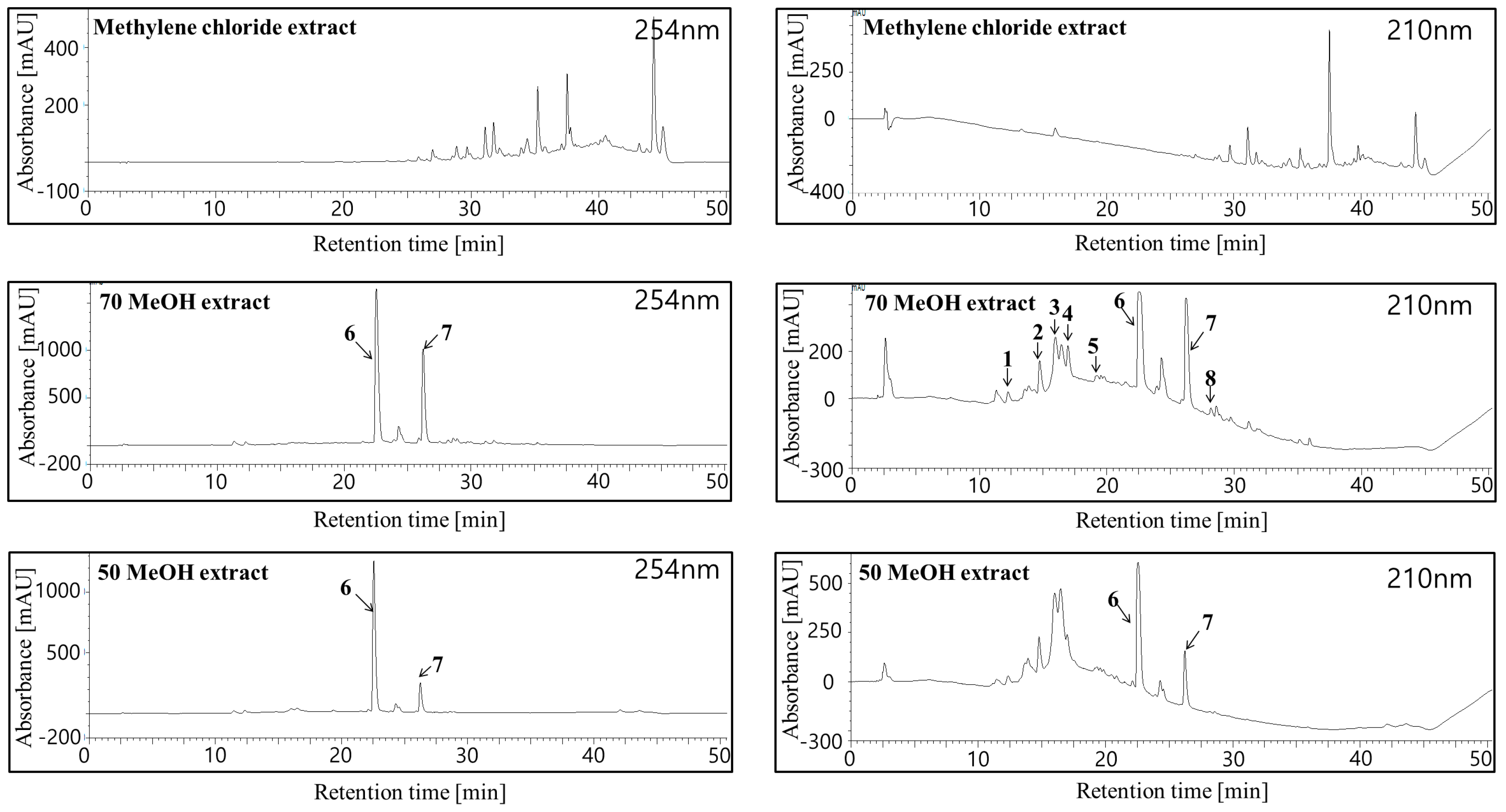
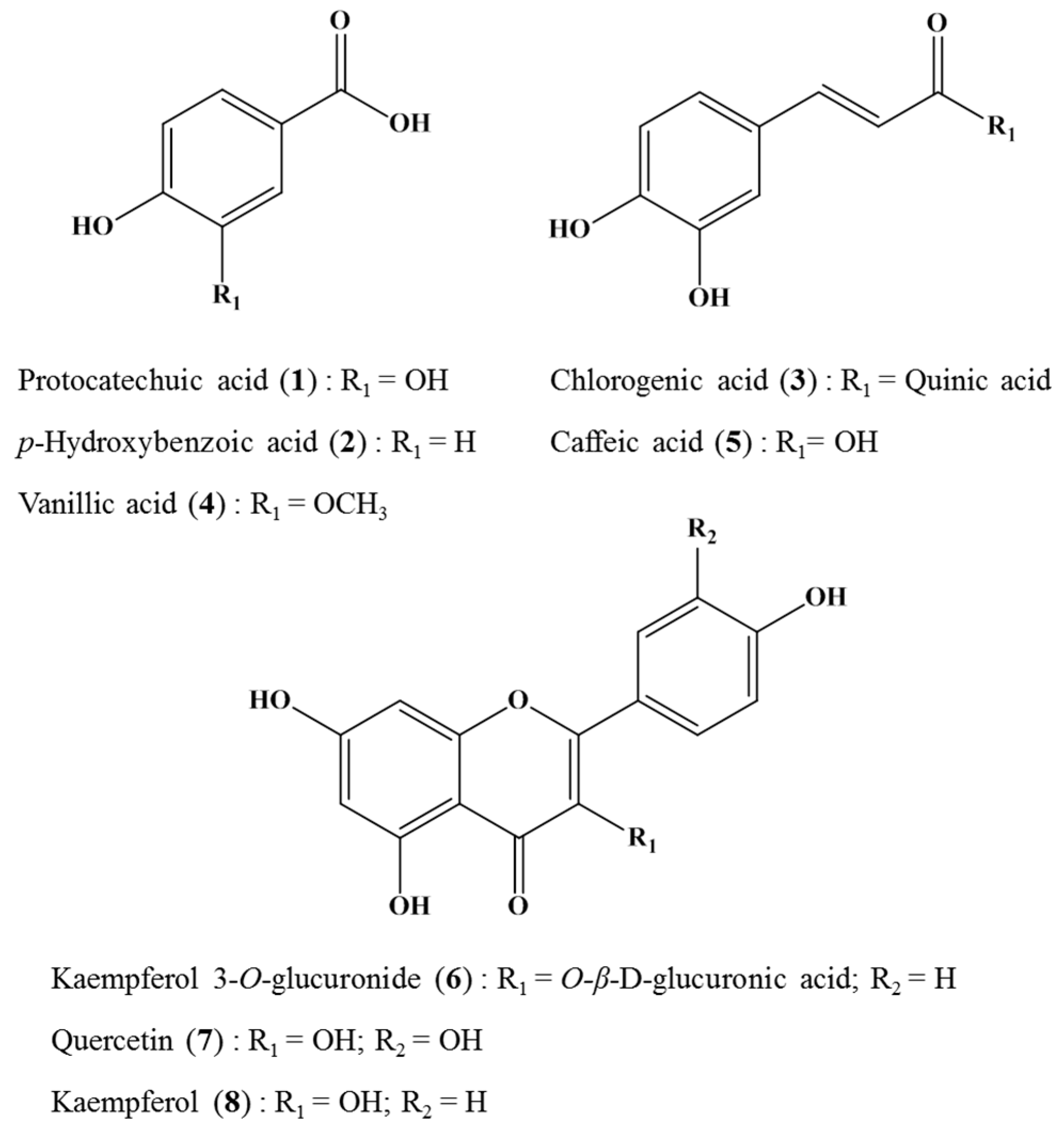
2.4. Identification of Major Bioactive Components of H. laricifolium Juss.
2.5. Effect of Bioactive Compounds on Tyrosinase Inhibition
3. Materials and Methods
3.1. Ethnobotanical Search
3.2. Chemicals
3.3. Plant Materials
3.4. Preparation of Extracts and Isolation of Plant Samples
3.5. HPLC Analysis
3.6. NMR Analysis
3.7. Tyrosinase Assay
3.8. Statistical Analysis
Acknowledgments
Author Contributions
Conflicts of Interest
References
- Garcia, P.; Furlan, R. Multiresponse Optimisation applied to the developement of a TLC Autography for the detection of tyrosinase inhibitors. Phytochem. Anal. 2015, 26, 287–292. [Google Scholar] [CrossRef] [PubMed]
- Tang, L.; Zhang, W.; Zhao, H.; Chen, Z. Tyrosinase inhibitor screening in traditional Chinese medicines by electrophoretically, mediated microanalysis. J. Sep. Sci. 2015, 38, 2887–9892. [Google Scholar] [CrossRef] [PubMed]
- Chiari, M.E.; Joray, M.B.; Ruiz, G.; Palacios, S.M.; Capinella, M.C. Tyrosinase inhibitory activity of native plants from central Argentina: Isolation of an active principle from Lithrea molleoides. Food Chem. 2010, 120, 10–14. [Google Scholar] [CrossRef]
- Hasegawa, T. Tyrosinase-Expressing Neuronal Cell Line as in Vitro Model of Parkinson’s Disease. Int. J. Mol. Sci. 2010, 11, 1082–1089. [Google Scholar] [CrossRef] [PubMed]
- Masuda, T.; Yamashita, D.; Takeda, Y.; Yonemori, S. Screening for tyrosinase inhibitors among extracts of seashore plants and identification of potent Inhibitors from Garcinia subelliptica. Biosci. Biotechnol. Biochem. 2005, 69, 197–201. [Google Scholar] [CrossRef] [PubMed]
- Adhikari, A.; Devkota, H.P.; Takano, A.; Masuda, K.; Nakane, T.; Basnet, P.; Skaiko-Basnet, N. Screening of Nepalese crude drugs traditionally used to treat hyperpigmentation: in vitro tyrosinase inhibition. Int. J. Cosmet. Sci. 2008, 30, 353–360. [Google Scholar] [CrossRef] [PubMed]
- Brack, A. Enciclopédico de Plantas Utiles del Perú; CBC: Cuzco, Perú, 1999; p. 550. [Google Scholar]
- Carraz, M.; Lavergne, C.; Jullian, V.; Wright, M.; Gairin, JE.; Gonzales de la Cruz, M.; Bourdy, G. Antiproliferative activity and phenotypic modification induced by selected Peruvian medicinal plants on human hepatocellular carcinoma Hep3B cells. J. Ethnopharmacol. 2015, 166, 185–199. [Google Scholar] [CrossRef] [PubMed]
- Monigatti, M.; Bussmann, R.W.; Weckerle, C. Medicinal plant use in two Andean communities located at different altitudes in the Bolivar Province, Peru. J. Ethnopharmacol. 2013, 145, 450–464. [Google Scholar] [CrossRef] [PubMed]
- Sanz-Bizet, J.; Campos de la Cruz, J.; Epiquien-Rivera, M.A.; Canigueral, S. A first survey on the medicinal plants of the Chazuta valley (Peruvian Amazon). J. Etnopharmacol. 2009, 122, 333–362. [Google Scholar] [CrossRef] [PubMed]
- Williams, J.E. OMD. Review of Antiviral and Immunomodulating Properties of Plants of the Peruvian Rainforest with a Particular Emphasis on Una de Gato and Sangre de Grado. Altern. Med. Rev. 2001, 6, 6. [Google Scholar]
- Neto, C.C.; Owens, C.W.; Langfield, R.D.; Comeau, A.B.; Onge, J.St.; Vaisberg, A.J.; Hammond, G.B. Antibacterial activity of some Peruvian medicinal plants from the Callejon de Huaylas. J. Ethnopharmacol. 2002, 79, 133–138. [Google Scholar] [CrossRef]
- Bussmann, R.W.; Malca, G.; Glenn, A.; Sharon, D.; Nilsen, B.; Parris, B.; Dubose, D.; Ruiz, D.; Saleda, J.; Martinez, M.; et al. Toxicity of medicinal plants used in traditional medicine in Northern Peru. J. Ethnopharmacol. 2011, 137, 121–140. [Google Scholar] [CrossRef] [PubMed]
- Chirinos, R.; Pedreschi, R.; Rogez, H.; Larondelle, Y.; Campos, D. Phenolic compound contents and antioxidant activity in plants with nutritional and/or medicinal properties from the Peruvian Andean region. Ind. Crop Prod. 2013, 47, 145–152. [Google Scholar] [CrossRef]
- Bussmann, R.W.; Glenn, A.; Sharon, D. Antibacterial activity plants of Northern Peru – can traditional applications provide leads for modern science? Ind. J. Tradit. Knowl. 2010, 9, 742–753. [Google Scholar]
- Berlowski, A.; Zawada, K.; Wawer, I.; Paradowska, K. Antioxidants Properties of Medicinal Plants from Peru. Food Nutr. Sci. 2013, 4, 71–77. [Google Scholar] [CrossRef]
- Huamani, M.E.; Ruiz, J. Determinacion de la Actividad Antifungica Contra Candida Albicans y Aspergillus Niger de 10 Plantas Medicinales de 3 Departamentos del Peru; Universidad Nacional Mayor de San Marcos: Lima, Peru, 2005. [Google Scholar]
- Duke, J.A.; Vasquez, R. Amazonian Ethnobotanical Dictionary; CRC Press: Boca Raton, FL, USA, 1994; p. 215. [Google Scholar]
- Solis, M.A. Vademecum de plantas medicinales del Ecuador. Available online: http://bases.bireme.br/cgi-bin/wxislind.exe/iah/online/?IsisScript=iah/iah.xis&src=google&base=LILACS&lang=p&nextAction=lnk&exprSearch=389748&indexSearch=ID (Accessed on 6 July 2016).
- Ramírez-Gonzáilez, I.; Amaro-Luis, J.M.; Bahsas, A. Xanthones from aerial parts of Hypericum laricifolium Juss. Nat. Prod. Commun. 2013, 8, 1731–1732. [Google Scholar] [PubMed]
- El-Seedi, H.R.; Ringbom, T.; Torssell, K.; Bohlin, L. Constituents of Hypericum laricifolium and their cyclooxygenase (COX) enzyme activities. Chem. Pharm. Bull. 2003, 51, 1439–1440. [Google Scholar] [CrossRef] [PubMed]
- Ccana-Ccapatinta, G.V.; Barros, F.M.C.; Bridi, H.; Von Poser, G.L. Dimeric acylphloroglucinols in Hypericum species from Brathys and Trigynobrathys. Phytochem. Rev. 2015, 14, 25–50. [Google Scholar] [CrossRef]
- Ccana-Ccapatinta, G.V.; Serrano, C.F.; Urrunaga, E.J.S.; Choquenaira, J.P.; Galiano, W.S.; Crockett, S.L.; Von Poser, G.L.; del Carpio, C.J. Assessing the phytochemical profiles and antidepressant-like activity of four Peruvian Hypericum species using the murine forced swimming test. Phytochem. Lett. 2014, 10, 107–112. [Google Scholar] [CrossRef]
- Ccana-Ccapatinta, G.V.; Von Poser, G.L. Acylphoroglucinol derivates from Hypericum laricifolium Juss. Phytochem. Rev. 2015, 12, 63–66. [Google Scholar]
- Rojas, J.; Buitrago, A.; Rojas, L.B.; Morales, A. Chemical composition of Hypericum laricifolium Juss. essential oil collected from Merida-Venezuela. Med. Aromat. Plants 2013, 2, 132–134. [Google Scholar]
- Chen, Y.S.; Lee, S.M.; Lin, C.C.; Liu, C.Y.; Wu, M.C.; Shi, W.L. Kinetic study on the tyrosinase and melanin formation inhibitory activities of carthamus yellow isolated from Carthamus tinctorius L. J. Biosci. Bioeng. 2013, 115, 242–245. [Google Scholar] [CrossRef] [PubMed]
- Huang, K.F.; Chen, Y.W.; Chang, C.T.; Chou, S.T. Studies on the inhitory effect of Graptopetalum paraguayense E. Walter extracts on mushroom tyrosinase. Food Chem. 2005, 89, 583–587. [Google Scholar] [CrossRef]
- Chen, Q.X.; Kubo, I. Kinetics of mushroom tyrosinase inhibition by quercetin. J. Agric. Food Chem. 2002, 50, 4108–4112. [Google Scholar] [CrossRef] [PubMed]
- Repo-Carrasco, R.; Acevedo de la Cruz, A.; Icochea, J. Chemical and Functional Characterization of Kañiwa (Chenopodium pallidicaule) Grain, Extrudate and Bran. Plant Foods Hum. Nutr. 2009, 64, 94–101. [Google Scholar] [CrossRef] [PubMed]
- Kaul Chahal, K.; Bhardwaj, U.; Kaushal, S.; Kaur Sandhu, A. Chemical composition and biological properties of Chrysopogon zizanioides (L.) Roberty syn. Vetiveria zizanioides (L.) Nash. Ind. J. Nat. Prod. Resour. 2015, 6, 251–260. [Google Scholar]
- Bussmann, R.W.; Glenn, A.; Sharon, D.; Meyer, K.; kuhlman, A.; Townesmith, A.; Pourmand, K.; Jonat, B.; Guardado, C.G.; Aguirre, R.; et al. Proving that traditional knowledge works: The antibacterial acitivity of Northern Peruvian medicinal plants. Ethnobot. J. 2011, 9, 67–96. [Google Scholar] [CrossRef]
- Han, E.; Lee, J.; Jung, E.; Jin, Y.; Chung, Ch. Antioxidative Activities of Water Extracts from Different Parts of Taraxacum officinale. J. Korean Soc. Food Sci. Nutr. 2010, 39, 1580–1586. [Google Scholar] [CrossRef]
- Mellado, M.; Madrid, A.; Pena-Cortes, H.; Lopez, R.; Jara, C.; Espinoza, L. Antioxidant Activity of Anthraquinones isolated from leaves of Muehlenbeckia Hastulata (J.E. SM) Johnst. (Polygonaceae). J. Chil. Chem. Soc. 2013, 58, 2. [Google Scholar] [CrossRef]
- Lu, T.; Ko, H. A new anthraquinone glycoside from Rhamnus nakaharai and anti-tyrosinase effect of 6-methoxysorigenin. Nat. Prod. Res. 2016, 30, 1–7. [Google Scholar] [CrossRef] [PubMed]
- Norlaily, A.S.; Keong, Y.; Wan, Y.H. The promising future of chia, salvia hispanica L. J. Biomed. Biotechnol. 2012, 17, 1956. [Google Scholar]
- Kubo, I.; Kinst-Hori, I.; Yokokawa, Y. Tyrosinase inhibitors from Anacardium occidentale fruits. J. Nat. Prod. 1994, 57, 545–551. [Google Scholar] [CrossRef] [PubMed]
- Raja, K.; Sivamani, J.J.R.; Howard, P.; Maibach, I. Cosmeceuticals and Active Cosmetics, 3rd ed.; Taylor & Francis Group: London, UK, 2015; p. 458. [Google Scholar]
- Crockett, S.; Eberhardt, M.; Kunert, O.; Schuhly, W. Hypericum species in the Paramos of Central and South America: a special focus upon H. irazuense Kuntze ex N. Robson. Phytochem. Rev. 2010, 9, 255–269. [Google Scholar] [CrossRef] [PubMed]
- Levent, M.A.; Sever, Y.B.; Erdogan, O.I.; Saltan, C.G. Assessment of cholinesterase and tyrosinase inhibitory and antioxidant effects of Hypericum perforatum L. (Jhon’s wort). Ind. Crops Prod. 2013, 43, 87–92. [Google Scholar]
- Bejaoui, A.; Ben Salem, I.; Rokbeni, N.; M’rabet, Y.; Boussaid, M.; Boulila, A. Bioactive compounds from Hypericum humifusum and Hypericum perfoliatum: Inhibition potential of polyphenols with acetylcholinesterase and key enzymes linked to type- 2 diabetes. Pharm. Biol. 2017, 1, 906–911. [Google Scholar] [CrossRef] [PubMed]
- Hosni, K.; Msaada, K.; Taarit, M.B.; Hammami, M.; Marzouk, B. Bioactive components of three Hypericum species from Tunisia: A comparative study. Ind. Crops Prod. 2010, 31, 158–163. [Google Scholar] [CrossRef]
- Germanò, M.P.; Cacciola, F.; Donato, P.; Dugo, P.; Certo, G.; D’Angelo, V.; Mondello, L.; Rapisarda, A. Betula pendula leaves: Polyphenolic characterization and potential innovative use in skin whitening products. Fitoterapia 2012, 83, 877–882. [Google Scholar] [CrossRef] [PubMed]
- Kwiecien, I.; Szydlowska, A.; Kawka, B.; Beerhues, L.; Ekiert, H. Accumulation of biologically active phenolic acids in agitated shoot cultures of three Hy Hypericum perforatum cultivars: Elixir, Helos, and Topas. Plant Cell Tiss. Organ Cult. 2015, 123, 273–281. [Google Scholar] [CrossRef]
- Filipiak-Szok, A.; Kurzawa, M.; Szlyk, E. Optimazation of extraction procedure and determination by high performance liquid chromstography of flavonols and phenolic acids from Hypericum Perforatum L. Copern. Lett. 2010, 1, 62–73. [Google Scholar] [CrossRef]
- Taherkhani, N.; Nematollah, G. Inhibitory Effects of Quercetin and Kaempferol as two Propolis Derived Flavonoids on Tyrosinase Enzyme. Biotech. Health Sci. 2014, 1, 22242. [Google Scholar] [CrossRef]
- Badria, F.; Ameen, M.R.; Akl, M. Evaluation of Cytotoxic Compounds from Calligonum comosum L. Growing in Egypt. Z. Naturforsch. 2007, 62, 656–660. [Google Scholar] [CrossRef]
- Huang, W.; Wan, C.; Shouran, Z. Quercetin—A Flavonoid Compound from Sarcopyramis bodinieri var delicate with Potential Apoptotic Activity in HepG2.Liver Cancer Cells. Trop. J. Pharm. Res. 2013, 12, 529–533. [Google Scholar] [CrossRef]
- Selvara, K.; Chowdhury, R.; Bhattacharjee, C. Isolation and structural elucidation of flavonoids from aquatic fern Azolla Microphyla and evaluation of free radical scavenging activity. Int. J. Pharm. Pharm. Sci. 2013, 5, 743–749. [Google Scholar]
- Sample Availability: Samples of the compounds from Hypericum laricifolium Juss. are available from the authors.


| N° | Scientific Name | Common Name a [9,10,11,12,13,14,15,16,17,29,30,31] b | Family Name | Traditional Uses and Ethnopharmacological Activity [9,10,11,12,13,14,15,16,17,29,30,31] b |
|---|---|---|---|---|
| 1 | Adiantum cf. poiretii Wikstr. | Culantrillo (S) | PTERIDACEAE | Excessive menstrual bleeding, menstrual cramps and vaginal inflammation |
| 2 | Alchornea castaneifolia (Humb. & Bonpl. ex Willd.) A. Juss. | Iporuro (S) | EUPHORBIACEAE | Rheumatism, arthritis, ulcer, gastritis and muscular pains |
| 3 | Anacardium occidentale L. | Casho (S), Marañón (S), Castaña de cajú (S), Pepa de la selva (S) | ANACARDIACEAE | Stomach discomfort, antidiarrheal and used as food |
| 4 | Annona muricata L. | Hojas de Graviola (S), Soursop (E) | ANNONACEAE | For coughs, asthma, and hypertension. Used as antibacterial, antifungal, antioxidant, and anti-inflammatory agents |
| 5 | Baccharis genistelloides (Lam.) Pers. | Carqueja (S), Cuchu Cuchu (Q), Tres esquinas (S), kimsacucho (Q), Carceja (S), Cadillo (S) | ASTERACEAE | Diabetes/cholesterol; used as an anti-inflammatory agent (liver, kidneys, biliar) and in intestinal disorders |
| 6 | Buddleja americana L. | Flor Blanca (S) | SCROPHULARIACEAE | Inflammation of womb, ovarian cysts and uterus |
| 7 | Caiophora cf. cirsiifolia C. Presl | Ortiga colorada (S), Ckora-quisa (Q), Pucahitana (Q), Puca-lalay (Q), Puca-Sasay (Q), Pucasique (Q) | LOASACEAE | Used as an antitussive, expectorant, and antipyretic to relieve cold, flu and bronchitis |
| 8 | Capsicum baccatum | Aji Amarillo (S) | SOLANACEAE | Rheumatism, arthritis, treatment of problems with skin and wounds |
| 9 | Cheilanthes pilosa Goldm. | Cuti Cuti (Q) | PTERIDACEAE | Diabetes and liver |
| 10 | Chenopodium pallidicaule | Cañihua (Q) | AMARANTHACEAE | Used in food, such as bread, and for drinks on long trips |
| 11 | Chrysopogon zizanioides (L.) Roberty | Pachuli (Q) | POACEAE | Depression, insomnia, anxiety, stress, tension, nervousness, inflamed skin and wounds |
| 12 | Chuquiraga spinosa Less. | Huamanpinta (Q), Care Sirve (Q), Pucacasha (Q), Chuquiraga (Q) | ASTERACEAE | Treatment of kidney disorders; used as an anti-inflammatory (renal), and for gonorrhea, as well as bladder and prostate problems |
| 13 | Clinopodium brevicalyx (Epling) Harley & A. Granda | Inka muña (Q) | LAMIACEAE | Treatment of diarrhea, gastritis and colic. Antitussive to relieve cold and flu |
| 14 | Clinopodium pulchellum (Kunth) Govaerts | Panisara (S) | LAMIACEAE | Spiritual cleansing |
| 15 | Cordia Lutea Lam. | Flor de Overo (S), Overo (S), Overal (S) | BORAGINACEAE | Used as an anti-inflammatory (liver, kidney, bladder, ovaries), and for hepatitis |
| 16 | Cymbopogon citratus (DC.) Stapf. | Hierba Luisa (S) | POACEAE | Cold, cough, flu and cancer |
| 17 | Desmodium molliculum (Kunth) DC. | Manayupa (Q), Pie de perro (S), Pata de perro (S), Chancas de comida (S) | FABACEAE | Used in gastritis, wound cleansing; as an anti-inflammatory (kidneys, ovaries) and in wound healing and diarrhea |
| 18 | Dianthus caryophyllus L. | Claveles (S), Clavelina (S), Clavel de la costa (S) | CARYOPHYLLACEAE | Insomnia, nerves and heart |
| 19 | cf. Endlicheria | Spingo (S) | LAURACEAE | No reports |
| 20 | Equisetum giganteum L. | Cola de caballo (S), Shawinco (Q) | EQUISETACEAE | Treatment of prostatitis, used as an anti-inflammatory (bladder, and renal calculous) and to cicatrize infectious injuries and wounds. Also used in cancer |
| 21 | Eucalyptus globolus L. | Eucalipto (S), Alcanfor Serrano (S) | MYRTACEAE | Antitussive, descongestant, analgesic and anti-spasmodic to relieve cold, cough, bronchitis, flu, asthma and rheuma |
| 22 | Flaveria bidentis (L.) Kuntze | Mata gusano (S) | ASTERACEAE | Treatment of prostatitis; used as an anti-inflammatory (bladder, renal calculous) and to cicatrize infectious injuries. Used in wounds, cancer, and for cough and bronchitis |
| 23 | Gentianella tristicha (Gilg) J.S. Pringle | Hercampure (Q) | GENTIANACEAE | Diabetes, diuretic and cholesterol |
| 24 | Gnaphalium dombeyanum DC. | Arnica (Q), Shymaicho (Q) | ASTERACEAE | Treatment of indigestion, also used as an anti-inflammatory and to cicatrize injuries and skin ulcers |
| 25 | Huperzia crassa (Humb. & Bonpl. ex Willd.) Rothm. | Trensilla o enredadera (S) | LYCOPODIACEAE | Spiritual cleansing |
| 26 | Hypericum laricifolium Juss. | Hierba de la fortuna (S), Solitario (S), Chinchango (Q), Abrecaminos (S), Romerillo (S) | CLUSIACEAAE | Luck in love, good fortune, good health, and paludism |
| 27 | Jatropha curcas L. | Piñones (S), Piñol (S) | EUPHORBIACEAE | Depurative-emetic, wound disinfectant, vaginal infection and sedative |
| 28 | Jatropha macrantha Müll. Arg | Male Huanarpo (E), Huanarpo macho (S) | EUPHORBIACEAE | Fertility, sexual potency, male impotence and tension |
| 29 | Lupinus mutabilis | Tarwi (Q) | FABACEAE | Used as food |
| 30 | Matricaria recutita L. | Labanda (S), Manzanillon (S) | ASTERACEAE | Infections of wounds, vaginal cleansing; used for blood purification, stomach pain, cold and flu. Also used as a laxative, for digestion, and as a sedative |
| 31 | Malesherbia splendens Ricardi | Veronica (S) | PASSIFLORACEAE | Bronchitis |
| 32 | Ocimum basilicum L. | Albahaca de olor (S) | LAMIACEAE | For better sleep, headaches and nerves |
| 33 | Otholobium mexicanum (L. f.) J.W. Grimes | Culen negro (S) | FABACEAE | Diarrhea and cold of the stomach |
| 34 | Otholobium pubescens (Poir.) J.W. Grimes | Culen Blanco (S) | FABACEAE | Diabetes, colic, constipation, indigestion, laxative and stomach purification |
| 35 | Oreobolus obtusangulus Gaudich./Eleocharis albibracteata Nees & Meyen ex Kunth | Hierba del caballero (S) | CYPERACEAE | Spiritual cleansing |
| 36 | Peumus boldus Molina | Boldo (S) | MONIMIACEAE | Anti-inflammatory (liver and kidney) |
| 37 | Phoradendron sp. | Suelda con suelda (S), Tullma tullma (Q) | LORANTHACEAE | Spiritual cleansing |
| 38 | Phyllanthus niruri L. | Chanca Piedra (S) | EUPHORBIACEAE | Cleansing (of the stomach, blood); anti-inflammatory (liver, kidneys, gallbladder) |
| 39 | Muehlenbeckia vulcanica Meisn. | Mullaka (Q), Viruta (S) | POLYGONACEAE | Treatment of diarrhea, bronchitis, asthma, pain, flu, throat and infections |
| 40 | Piper aduncum L. | Matico (S) | PIPERACEAE | Anti-inflammatory, and treatment of diarrhea |
| 41 | Puya sp. | Hierba del Carnero (S), Lana de carnero (S), Hierba de Borrego (S), Solitario (S), Abrecaminos (S) | BROMELIACEAE | Tumors and infections |
| 42 | Rosmarinus officinalis | Romero (S) | LAMIACEAE | Treatment of liver and bladder disorders, anti-inflammatory to relieve rheumatism and peripheral vascular diseases |
| 43 | Salvia hispanica L. | Chia (S) | LAMIACEAE | Used as food |
| 44 | Sambucus peruviana H. B. K. | Sauco (S), Tilo (S), Saucotillo (S) | CAPRIFOLIAEAE | Bronchitis, yellow fever, inflammation of the kidneys and cough |
| 45 | Senna sp. | Hojas de sen (S) | FABACEAE | Purgative, constipation, and cleansing of the stomach |
| 46 | Smallanthus sonchifolius (Poepp.) H. Rob. | Hojas de Yacon (S), Yacon (S), Llacon(Q) | ASTERACEAE | Diabetes, cholesterol, kidney and inflammation of the prostate |
| 47 | Taraxacum officinale F.H. Wigg. | Diente de leon (S), Amargon (S), Lengua de Leon (S) | ASTERACEAE | Liver, stomach, inflammation, (ovaries). Used for depurative and diuretic effects |
| 48 | Tiquilia Paronychioides (Phil.) Rich. | Flor de arena (S) | BORAGINACEAE | Anti-inflammatory (ovaries, kidneys) and used in urinary infections |
| 49 | Valeriana sp. | Raiz de valeriana (S) | CAPRIFOLIACEAE | Treatment of sleep disorders and sedative properties |
| 50 | Werneria nubigena Kunth | Condor (S) | ASTERACEAE | Calmative effects |
| No | Scientific Name | Voucher Specimen | Part Used | Yield (%) | Inhibition % (500 μg/mL) |
|---|---|---|---|---|---|
| 1 | A.cf. poiretii Wikstr. | A9 | Ar | 14.1 | <1 ± 1.9 |
| 2 | A. castaneifolia (Humb. & Bonpl. ex Willd.) A. Juss. | P19 | Lv | 16.1 | <1 ± 0.8 |
| 3 | A. occidentale L. | A41 | Fr | 25.6 | 42.51 ± 7.2 |
| 4 | A. muricata L. | A5 | Lv | 19.5 | <1 ± 2.9 |
| 5 | B. genistelloides (Lam.) Pers. | P78 | Ar | 19.7 | 18.43 ± 2.5 |
| 6 | B. americana L. | A19 | F | 8.5 | 8.09 ± 0.7 |
| 7 | C. cf. cirsiifolia C. Presl | A25 | Ar | 7.0 | 22.54 ± 3.4 |
| 8 | C. baccatum | A46 | Fr | 50.9 | 8.91 ± 1.1 |
| 9 | Ch. pilosa Goldm | P17 | Ar | 14.3 | 40.35 ± 2.0 |
| 10 | Ch. pallidicaule | A50 | S | 1.1 | 2.83 ± 3.4 |
| 11 | Ch. zizanioides (L.) Roberty | A13 | Lv | 9.1 | 4.99 ± 1.8 |
| 12 | Ch. spinosa Less | P77 | Ar | 28.7 | <1 ± 2.6 |
| 13 | Cl. brevicalyx (Epling) Harley & A. Granda | P11 | Lv | 26.8 | <1 ± 2.0 |
| 14 | Cl. pulchellum (Kunth) Govaerts | A15 | Lv | 4.1 | 12.55 ± 0.9 |
| 15 | C. lutea Lam | P79 | F | 12.3 | 10.6 ± 3.1 |
| 16 | C. citratus (DC.) Stapf | A18 | Lv. | 6.8 | 29.82 ± 0.9 |
| 17 | D. molliculum (Kunth) DC. | P80 | Lv | 23.8 | 20.65 ± 2.9 |
| 18 | D. caryophyllus L. | A20 | F | 2.5 | 30.36 ± 3.4 |
| 19 | Cf. endlicheria | A43 | S | 10.1 | 6.88 ±0.7 |
| 20 | E. giganteum L. | P55 | Ar | 11.2 | <1 ± 5.2 |
| 21 | E. globolus L. | P39 | Lv | 18.0 | 10.93 ± 1.9 |
| 22 | F. bidentis (L.) Kuntze | A31 | Lv | 7.9 | <1 ± 2.4 |
| 23 | G. tristicha (Gilg) J.S. Pringle | P7 | Ar | 30.3 | 22.54 ± 1.3 |
| 24 | G. dombeyanum DC. | A29 | Lv | 3.7 | 5.67 ± 4.0 |
| 25 | H. crassa (Humb. & Bonpl. ex Willd.) Rothm. | A27 | Lv. | 12.3 | <1 ± 0.3 |
| 26 | H. laricifolium Juss. | A10 | Lv | 15.9 | 74.00 ± 2.1 |
| 27 | J. curcas L. | A39 | S | 1.9 | <1 ± 2.9 |
| 28 | J. macrantha Müll. Arg. | P2 | R | 23.6 | 37.11 ± 5.7 |
| 29 | L. mutabilis | A47 | S | 10.2 | <1 ± 3.6 |
| 30 | M. recutita L. | A12 | Lv | 7.9 | 40.49 ± 2.1 |
| 31 | M. splendens Ricardi | A22 | Lv | 7.7 | 38.06 ± 1.8 |
| 32 | O. basilicum L. | A11 | Lv | 5.6 | 28.10 ± 4.1 |
| 33 | O. mexicanum (L. f.) J.W. Grimes | A3 | Ar | 6.1 | 19.57 ± 2.1 |
| 34 | O. pubescens (Poir.) J.W. Grimes | A4 | Ar | 19.6 | 31.58 ± 0.8 |
| 35 | O. obtusangulus Gaudich /E. albibracteata Nees & Meyen ex Kunth | A28 | Lv | 3.9 | 17.41 ± 1.1 |
| 36 | P. boldus Molina | P40 | Lv | 32.5 | <1 ± 2.6 |
| 37 | Phoradendron sp. | P83 | Lv | 57.1 | <1 ± 1.2 |
| 38 | P. niruri L. | P5 | Lv | 12.9 | 11.34 ± 6.3 |
| 39 | M. vulcanica Meisn | P82 | Lv | 20.1 | 57.1 ± 3.0 |
| 40 | P. aduncum L. | P44 | Lv | 13.4 | <1 ± 1.1 |
| 41 | Puya sp. | A38 | Ar | 1.9 | 12.55 ± 10.3 |
| 42 | R. officinalis | P81 | Lv | 20.4 | 8.22 ± 3.1 |
| 43 | S. hispanica L. | P4 | S | 3.5 | 44.31 ± 3.4 |
| 44 | S. peruviana H. B. K. | A24 | Lv | 8.1 | 18.49 ± 2.4 |
| 45 | Senna sp. | A7 | Lv | 8.1 | 44.13 ± 2.3 |
| 46 | S. sonchifolius (Poepp.) H. Rob. | A6 | Lv | 5.3 | <1 ± 1.1 |
| 47 | T. officinale F. H. Wigg | P49 | Ar + F | 4.8 | 60.8 ± 4.1 |
| 48 | T. paronychioides (Phil.) Rich. | P36 | Ar | 18.5 | 10.93 ±1.3 |
| 49 | Valeriana sp. | P42 | R | 52.7 | 17.04 ± 3.7 |
| 50 | W. nubigena Kunth | A33 | Lv | 8.5 | 1.21 ± 1.5 |
| Arbutin | 92.3 ± 2.2 | ||||
| Extracts | Concentration (μg/mL) | Inhibition (%) | IC50 (μg/mL) |
|---|---|---|---|
| H. laricifolium Juss. Methylene chloride | 1000 | <1 ± 4.3 | - |
| H. laricifolium Juss. 70% MeoH | 1000 | 80.9 ± 1.1 | 122.1± 4.1 |
| 500 | 77.7 ± 2.8 | ||
| 100 | 46.1 ± 2.5 | ||
| H. laricifolium Juss. 50% MeoH | 1000 | 22.3 ± 5.4 | - |
| Arbutin | 500 | 98.2 ± 0.9 | 42.0 ± 0.8 |
| 50 | 63.8 ± 0.1 | ||
| 10 | 13.7 ± 1.8 |
| Compounds | Inhibition (%) | IC50 (μM) | Regression Equation | Correlation Coefficient (R2) | Active Compounds (μg) |
|---|---|---|---|---|---|
| Protocatechuic acid (1) | NI | - | Y = 0.6294x + 2.0882 | 0.9829 | 8.4 |
| p-Hydroxybenzoic acid (2) | 8.3 ± 4.03 | - | Y = 0.1963x + 17.276 | 0.9479 | 5.62 |
| Chlorogenic acid (3) | NI | - | Y = 0.3199x + 10.849 | 0.9889 | 17 |
| Vanilic acid (4) | NI | - | Y = 0.1824x + 13.326 | 0.9198 | 5.07 |
| Caffeic acid (5) | NI | - | Y = 0.1641x + 1.1179 | 0.9994 | 2.39 |
| Kaempferol 3-O-glucuronide (6) | NI | - | Y = 0.0948x + 9.0847 | 0.9407 | 3787.9 |
| Quercetin (7) | 99.7 ± 0.28 | 14.29 ± 0.3 | Y = 0.1392x + 35.232 | 0.9018 | 720.5 |
| Kaempferol (8) | 30 ± 1.97 | - | Y = 0.2578x + 2.0698 | 0.9992 | 0.82 |
| Arbutin | 86.01 ± 1.6 | 110.4 ± 1.9 | - | ||
| Kojic acid | 99.8 ± 0.5 | 8.0 ± 0.5 | |||
© 2017 by the authors. Licensee MDPI, Basel, Switzerland. This article is an open access article distributed under the terms and conditions of the Creative Commons Attribution (CC BY) license ( http://creativecommons.org/licenses/by/4.0/).
Share and Cite
Quispe, Y.N.G.; Hwang, S.H.; Wang, Z.; Lim, S.S. Screening of Peruvian Medicinal Plants for Tyrosinase Inhibitory Properties: Identification of Tyrosinase Inhibitors in Hypericum laricifolium Juss. Molecules 2017, 22, 402. https://doi.org/10.3390/molecules22030402
Quispe YNG, Hwang SH, Wang Z, Lim SS. Screening of Peruvian Medicinal Plants for Tyrosinase Inhibitory Properties: Identification of Tyrosinase Inhibitors in Hypericum laricifolium Juss. Molecules. 2017; 22(3):402. https://doi.org/10.3390/molecules22030402
Chicago/Turabian StyleQuispe, Yanymee Nimesia Guillen, Seung Hwan Hwang, Zhiqiang Wang, and Soon Sung Lim. 2017. "Screening of Peruvian Medicinal Plants for Tyrosinase Inhibitory Properties: Identification of Tyrosinase Inhibitors in Hypericum laricifolium Juss" Molecules 22, no. 3: 402. https://doi.org/10.3390/molecules22030402
APA StyleQuispe, Y. N. G., Hwang, S. H., Wang, Z., & Lim, S. S. (2017). Screening of Peruvian Medicinal Plants for Tyrosinase Inhibitory Properties: Identification of Tyrosinase Inhibitors in Hypericum laricifolium Juss. Molecules, 22(3), 402. https://doi.org/10.3390/molecules22030402