Article

Chemical Constituents of Phaius mishmensis

1
Department of Chemistry, National Cheng Kung University, Tainan 701, Taiwan
2
School of Pharmacy, China Medical University, Taichung 40402, Taiwan
3
Department of Anatomy, School of Medicine, China Medical University, Taichung 40402, Taiwan
4
Research Center for Chinese Herbal Medicine, China Medical University, Taichung 40402, Taiwan
*
Authors to whom correspondence should be addressed.
Molecules 2016, 21(11), 1605; https://doi.org/10.3390/molecules21111605
Submission received: 5 October 2016 / Revised: 18 November 2016 / Accepted: 21 November 2016 / Published: 23 November 2016

Abstract

:
The partitioned n-hexane, CHCl3, and EtOAc extracts from the crude MeOH extract of Phaius mishmensis showed considerable cytotoxicities against the human breast carcinoma (MCF-7), lung carcinoma (NCI-H460), and central nervous system carcinoma (SF-268) cell lines. Four new compounds, phaindole (1), (7′R,8′R)-phaithrene (2), methyl 3-hydroxy-4,5-dimethoxypropiophenone (3), and methyl hematinate (4), as well as 44 known compounds were isolated from the MeOH extract of Phaius mishmensis. The structures of the compounds were determined using spectroscopic methods.

Graphical Abstract

1. Introduction

In our search for novel anticancer compounds from Taiwanese plants, the crude MeOH extract of Phaius mishmensis (Orchidaceae), a native orchid of Taiwan [1], showed considerable cytotoxicity against the human breast carcinoma (MCF-7), lung carcinoma (NCI-H460), and central nervous system carcinoma (SF-268) cell lines. Thus, the plant P. mishmensis was selected for purification based on the preliminary results. We isolated eight indoloquinazolinones, phaitanthrins A–E, methylisatoid, tryptanthrin, and candidine from the CHCl3-soluble extract of the plant [2]. After extensive column and preparative thin-layer chromatographic separations, four new compounds, phaindole (1), (7′R,8′R)-phaithrene (2), 3-hydroxy-4,5-dimethoxypropiophenone (3), and methyl hematinate (4), as well as 44 known compounds were isolated from the MeOH extract of P. mishmensis. Herein we describe the isolation, substance elucidation, and cytotoxic properties of the isolated compounds.

2. Results and Discussion

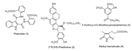
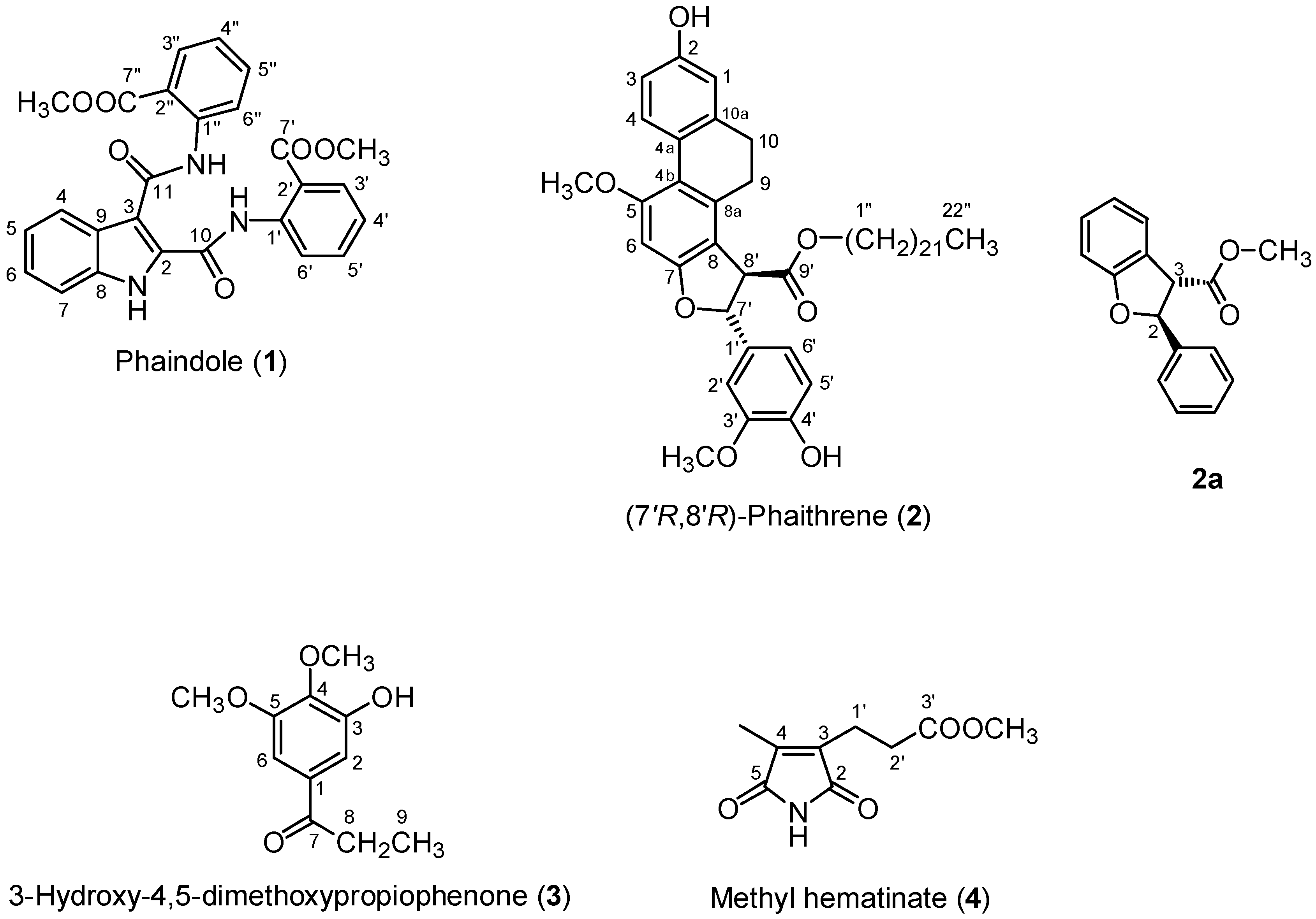
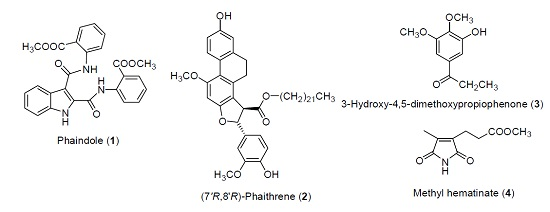
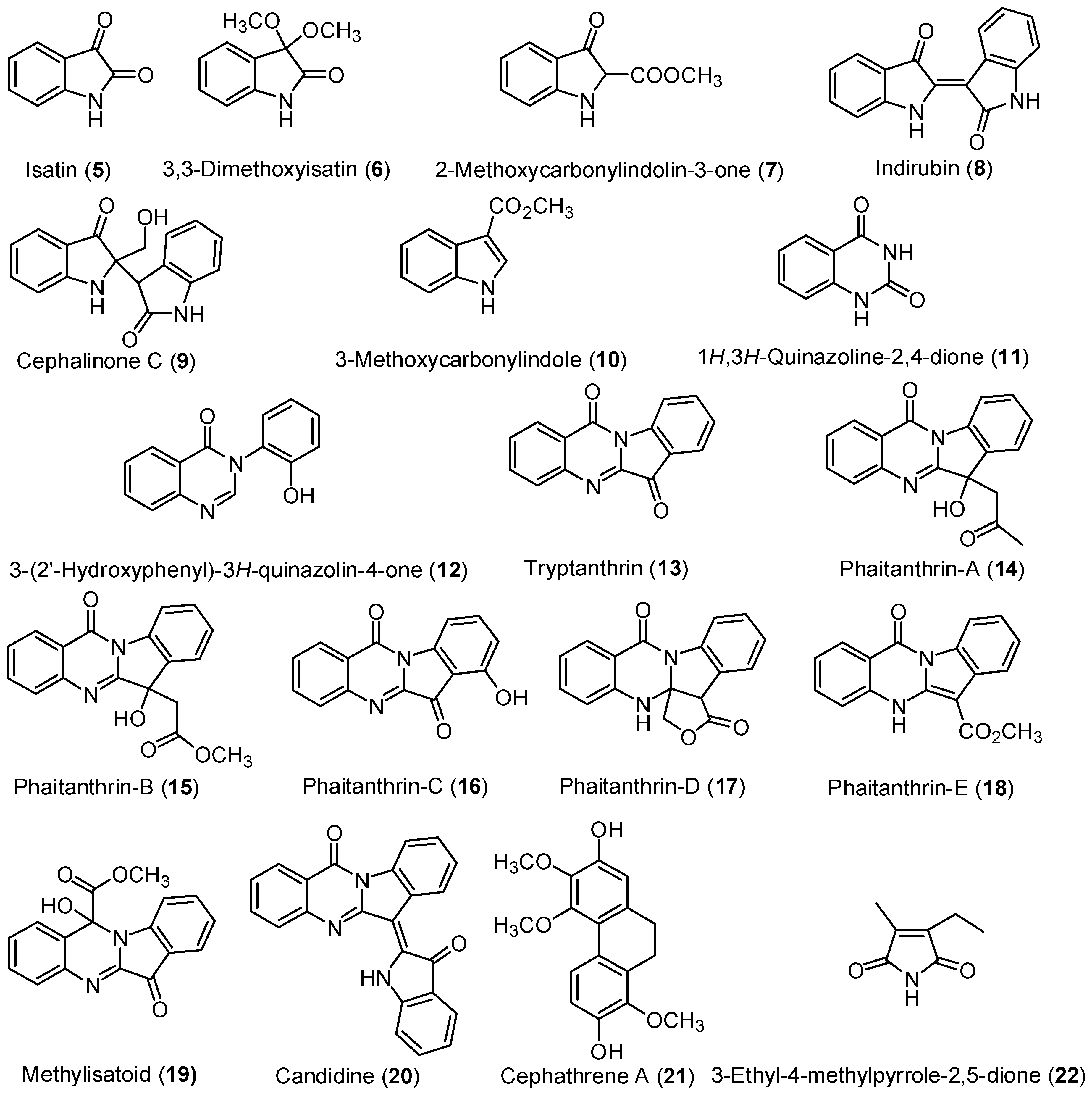
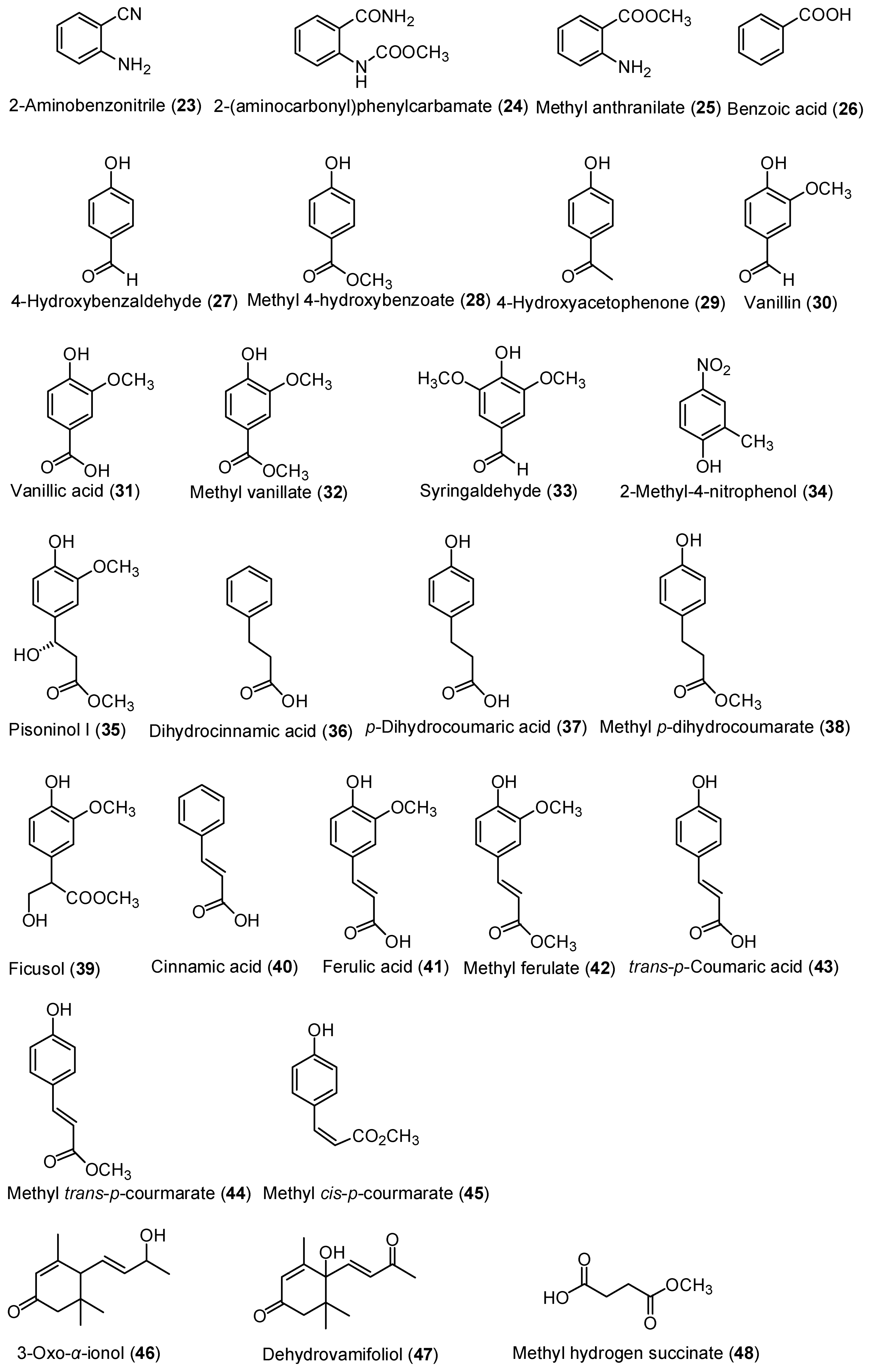
2.1. Isolation of Chemical Constituents of P. mishmensis

Compound 1, isolated as a yellowish powder, was determined to have the molecular formula C26H22N3O6 as noted by the pseudo-molecular ion peak at m/z 472.1506 in high resolution electrospray ionization mass spectroscopy (HR-ESIMS). A broad infrared (IR) absorption at 3264 cm−1 indicated the presence of a hydroxyl or amino functionality. Furthermore, four strong IR absorptions at 1702, 1697, 1659, and 1650 cm−1 might indicate the presence of two carbonyl and two amidic functionalities. The 1H-NMR (500 MHz) and 13C-NMR (125 MHz) spectral data of compound 1 were shown in Table 1, and all chemical shifts (δ) were given in ppm. In the aromatic region of the 1H- and COSY spectra, three sets of four mutually coupled proton signals at δ 7.34 ppm (t, J = 8.0 Hz, H-5), 7.40 ppm (t, J = 8.0 Hz, H-6), 7.52 ppm (d, J = 8.0 Hz, H-7), 8.14 ppm (d, J = 8.0 Hz, H-4), 7.22 ppm (t, J = 8.0 Hz, H-4′), 7.56 ppm (t, J = 8.0 Hz, H-5′), 7.98 ppm (d, J = 8.0 Hz, H-3′), 8.14 ppm (d, J = 8.0 Hz, H-6′), 7.16 ppm (t, J = 8.0 Hz, H-4′′), 7.62 ppm (t, J = 8.0 Hz, H-5′′), 8.07 ppm (d, J = 8.0 Hz, H-3′′), and 8.86 ppm (d, J = 8.0 Hz, H-6′′) were observed. Combined with 13C-, HMQC, and HMBC data, we determined that the first o-disubstituted benzene was fused with a pyrrole ring to form an indole unit, as noted by the HMBC correlations of H-1 (δ 9.98 ppm, br s, NH) with C-3 (δ 112.3 ppm), C-7 (δ 112.5 ppm), and C-9 (δ 125.7 ppm), and H-4 with C-3, whereas the latter two signals belonged to two methyl 2-aminobenzoate moieties, as determined by the HMBC correlations of H-3′ and 7′-OCH3 (δ 3.83 ppm) with C-7′ (δ 167.1 ppm), H-3′′ and 7′′-OCH3 (δ 3.85 ppm) with C-7′′ (δ 168.4 ppm), as well as 1′-NH (δ 12.55 ppm) with C-6′ (δ 124.8 ppm) and 1′′-NH (δ 11.85 ppm) with C-2′′ (δ 116.0 ppm) and C-6′′ (δ 121.6 ppm). The remaining two carbonyl signals at δ 159.2 ppm (C-10) and 164.7 ppm (C-11) were attributed to C-2 and C-3 of the indole ring, respectively, which formed an amidic linkage with two methyl 2-aminobenzoate groups as determined by the HMBC correlations of 1′-NH with δ 159.2 ppm (C-10) and 1′′-NH with δ 164.7 ppm (C-11). The NOE correlations between H-1 and H-7, 1′-NH and H-6′, 1′′-NH and H-4, and H-6′′ revealed that the structure of 1 was 2,3-di(2-methoxycarbonylphenyl)carbamoylindole; this compound was named phaindole (Figure 1).
The optically active compound 2 ([α]D −18.5°) was obtained as a colorless powder and determined to have the molecular formula C47H67O7, owing to the pseudo-molecular ion peak at m/z 743.4883 in HR-FABMS. The IR spectrum showed a broad absorption at 3399 cm−1 corresponding to a hydroxyl group and a strong absorption at 1728 cm−1 corresponding to a carbonyl group. The 1H-NMR (300 MHz) and 13C-NMR (75 MHz) spectral data of compound 2 were shown in Table 1. In the aromatic region of the 1H-NMR and COSY spectra, three mutually coupled proton signals at δ 6.69 ppm (d, J = 2.6 Hz, H-1), 6.71 ppm (dd, J = 8.2, 2.6 Hz, H-3), and 8.09 ppm (d, J = 8.2 Hz, H-4) corresponding to a trisubstituted benzene ring and singlet proton signal at δ 6.53 ppm (s, H-6) for a pentasubstituted benzene ring were observed. In the aliphatic region of the 1H-NMR spectrum, an ethylene proton signal at δ 2.66 ppm (4H, m, H-9, and H-10) was observed. Using HMQC, HMBC, and NOESY, key long-range 1H-13C correlations of H-1 with C-10 (δ 29.7 ppm); H-4 with C-4b (δ 116.8 ppm); H-9 with C-4b , C-8 (δ 113.9 ppm), C-10a (δ 139.3 ppm); H-10 with C-1 (δ 114.1 ppm) and C-8a (δ 136.8 ppm), as well as a key NOE correlation between H-1 and H-10 revealed a 2,5,7,8-tetrasubstituted 9,10-dihydrophenanthrene nucleus. Three very down-field–shifted 13C- signals at C-2 (δ 153.4 ppm), C-5 (δ 158.5 ppm), and C-7 (δ 159.0 ppm) together with the NOE correlations between a hydroxyl group (δ 4.76 ppm) and H-1, -3, a methoxyl group (δ 3.87 ppm) and H-4, -6, revealed a hydroxyl (2-OH), a methoxyl (5-OCH3), and oxygenated substituents (7-OR). The remaining three unresolved aromatic proton signals at δ 6.88 ppm (3H, m, H-2′, H-5′, and H-6′), a hydroxyl signal at δ 5.63 ppm (4′-OH), a methoxyl signal at δ 3.88 ppm (3′-OCH3), -CHCH- proton signals at δ 4.21 ppm (d, J = 5.4 Hz, H-8′) and 5.93 ppm (d, J = 5.4 Hz, H-7′), together with a carbonyl signal at δ 172.4 ppm (C-9'), revealed a 7′-oxygenated-8′-alkyl dihydroferulate moiety. The HMBC correlations of 4′-OH with C-3′ (δ 146.7 ppm) and C-5′ (δ 114.5 ppm); 3′-OCH3 with C-3′ (δ 146.7 ppm); H-7′ with C-2′ (δ 108.0 ppm), C-6′ (δ 118.6 ppm), and C-9′; H-8′ with C-1′ (δ 132.8 ppm) and C-9 confirmed the presence of a ferulate moiety. A 22-long chain alcohol at δ 0.88 ppm (3H, t, J = 6.8 Hz, H-22″), 1.25 ppm (38H, m, H-3″–H-21″), 1.62 ppm (2H, quintet, J = 6.9 Hz, H-2″), and 4.15 ppm (2H, t, J = 6.9 Hz, H-1″) formed docosyl ferulate, as noted by the HMBC correlation of H-1′′ with C-9′. Finally, the HMBC correlation of H-7′ with C-7, -8; H-8′ with C-7; and the NOE correlation between H-8′ and H-9 revealed that the ferulate was fused to the phenanthrene ring to form a furanophenanthrene. Based on a report by Juhasz [3], the absolute configuration was determined as follows: first, the smaller coupling constant 5.5 Hz between H-7′ and H-8′ suggested that the substituents on the dihydrofuran ring were in the trans configuration. Second, a similar compound, (2S,3S)-methyl 2,3-dihydro-2-phenylbenzofuran-3-carboxylate (2a), with the S configuration at C-2 showed a negative Cotton effect at 280 nm by circular dichroism (CD) spectrometry. Compound 2 presented a positive Cotton effect at 280 nm by CD spectrometry, indicating the 7′R configuration. Consequently, the absolute configuration of (7′R,8′R) was deduced for 2 and the compound was named (7′R,8′R)-phaithrene.
Compound 3 was isolated as yellowish needles, and was determined to have the molecular formula C11H14O4 by HR-EIMS at m/z 210.0894. A broad IR absorption at 3390 cm−1 and a strong IR absorption at 1680 cm−1 revealed the existence of hydroxyl and carbonyl groups, respectively. The 1H-NMR (300 MHz) and 13C-NMR (75 MHz) spectral data of compound 3 are shown in Table 1. The only two 1H-NMR signals at δ 7.16 ppm (d, J = 1.9 Hz, H-6) and 7.22 ppm (d, J = 1.9 Hz, H-2) indicated a 1,3,4,5-tetrasubstituted benzene nucleus. Three oxygenated substituents, namely a hydroxyl group at δ 5.87 ppm and two methoxyl groups at δ 3.91 ppm and 3.97 ppm, were observed. The fourth substituent was determined to be a propanoyl group due to an ethyl signal at δ 1.21 ppm (3H, t, H-9) and 2.94 ppm (2H, q, H-8) with a coupling constant of 7.2 Hz, which showed HMBC correlations with a carbonyl carbon at δ 199.8 ppm (C-7). The attachments were confirmed by the HMBC correlations between H-2, -6, and C-7, indicating that the propanoyl group was present on C-1; the HMBC correlations between the hydroxyl group at δ 5.87 ppm and H-2 indicated that the hydroxyl group was on C-3. Two methoxyl groups were present on C-4 and -5. Finally, the NOE correlation between a methoxyl group at δ 3.91 ppm (5-OCH3) and H-6 and a methylene group at δ 2.94 ppm (H-8) and H-2, -6 confirmed that the structure of 3 was 3-hydroxy-4,5-dimethoxypropiophenone.
Compound 4 was isolated as a white solid and was confirmed to have the molecular formula C9H12NO4 by the HR-FABMS signal at m/z 198.0767. From the 1H NMR spectrum (Table 1), the structure of 4 was determined to possess a mutually coupled ethylene group at δ 2.61 ppm (H-2′) and 2.71 ppm (H-1′), a methyl group at δ 2.00 ppm (4-CH3), and a methoxyl group at δ 3.67 ppm (3′-OCH3). The HMBC correlation of the 3′-OCH3 with a carbonyl C-3′ (δ 172.5 ppm); H-1′, H-2′, and 4-CH3 with accidently coinciding olefinic carbons C-3 and C-4 (δ 139.7 ppm); as well as a NOE correlation between 4-CH3 and H-1′ indicated a cis CH3C=CCH2CH2COOCH3 fragment. The remaining two carbonyl carbon signals at δ 171.4 ppm (C-2) and 171.5 ppm (C-5) and a broad NH proton signal at δ 7.52 ppm as well as the HMBC correlations between H-1′ and C-2 and 4-CH3 and C-5 formed an imido -O=CNHC=O- fragment. By combining these two fragments, the structure of 3-(methoxycarbonyl)ethyl-4-methyl-2,5-pyrroledione was thus deduced as methyl hematinate (4). This compound has been obtained from the photooxygenation of biliverdin [4]. However, this is the first time it has been isolated naturally.
Other known compounds were also isolated from P. mishmensis, including 44 known compounds. The compounds were six indoles: isatin (5) [5], 3,3-dimethoxyisatin (6) [6], 2-methoxycarbonylindolin-3-one (7) [7], indirubin (8) [8], cephalinone C (9) [9,10], 3-methoxycarbonylindole (10) [11]; 10 quinazolines: 1H,3H-quinazoline-2,4-dione (11) [12], 3-(2'-hydroxyphenyl)-3H-quinazolin-4-one (12) [13], tryptanthrin (13) [2], phaitanthrin-A (14) [2], phaitanthrin-B (15) [2], phaitanthrin-C (16) [2], phaitanthrin-D (17) [2], phaitanthrin-E (18) [2], methylisatoid (19) [2], candidine (20) [2]; one phenanthrene: cephathrene A (21) [10]; one imide: 3-ethyl-4-methylpyrrole-2,5-dione (22) [14] (Figure 2).
Moreover, 23 monocyclic aromatic hydrocarbons, 2-aminobenzonitrile (23) [15], 2-(aminocarbonyl)phenylcarbamate (24) [10], methyl anthranilate (25) [16], benzoic acid (26) [17], 4-hydroxybenzaldehyde (27) [18], methyl 4-hydroxybenzoate (28) [18], 4-hydroxyacetophenone (29) [18], vanillin (30) [10], vanillic acid (31) [10], methyl vanillate (32) [17], syringaldehyde (33) [10], 2-methyl-4-nitrophenol (34) [19], pisoninol I (35) [20], dihydrocinnamic acid (36) [21], p-dihydrocoumaric acid (37) [10], methyl p-dihydrocoumarate (38) [22], ficusol (39) [17], cinnamic acid (40) [18], ferulic acid (41) [10], methyl ferulate (42) [10], trans-p-coumaric acid (43) [23], methyl trans-p-courmarate (44) [18], and methyl cis-p-courmarate (45) [24], as well as 3-oxo-α-ionol (46) [25], dehydrovamifoliol (47) [26], and methyl hydrogen succinate (48) [27] were also isolated (Figure 3).

2.2. Cytotoxicity of Chemical Constituents of P. mishmensis

The partitioned n-hexane, CHCl3, and EtOAc extracts from the crude MeOH extract of P. mishmensis showed considerable cytotoxicities against the MCF-7, NCI-H460, and SF-268 cell lines (Table 2). Unfortunately, most of the isolated compounds, except tryptanthrin (13) and phaitanthrin-A (14) [28], did not exhibit significant cytotoxicity against the tested cell lines. This result suggested that the practically insoluble tryptanthrin (13) could disperse in organic layers during extraction.

3. Materials and Methods

3.1. General

Optical rotations were measured on a Jasco DIP-370 digital polarimeter (JASCO, Tokyo, Japan). CD spectra were determined on a Jasco J-715 spectropolarimeter (JASCO, Tokyo, Japan). UV spectra were recorded on an Agilent 8453 spectrophotometer (Agilent Technologies, Palo Alto, CA, USA). The IR spectra measured using a Nicolet Magna FT-IR spectrophotometer (Nicolet Instrument, Inc., Madison, WI, USA). The 1H-, 13C-, and 2D NMR spectra were recorded on Bruker Avance 300, AMX 400, and Avance-500 FT-NMR spectrometers (Bruker, Karlsruhe, Germany) at room temperature. All chemical shifts (δ) are given in ppm using tetramethylsilane as an internal standard. Mass spectra were obtained on a VG 70-250S spectrometer by a direct inlet system (Micromass Corp., Manchester, UK).

3.2. Plant Material

Whole plants of P. mishmensis were collected from Nanto Hsien, Taiwan, in October 2003. The collection was authenticated by Professor Chang-Sheng Kuoh, Department of Life Sciences, National Cheng Kung University, Tainan, Taiwan. A voucher specimen (No. PLW-0304) was deposited in the Herbarium of National Cheng Kung University, Tainan, Taiwan.

3.3. Extraction and Isolation

The air-dried P. mishmensis plants (3.5 kg) were extracted with MeOH (7 × 8 L) under reflux. The combined extracts were concentrated under reduced pressure to give a dark brown syrup. The syrup was suspended in H2O and then partitioned successively with n-hexane, CHCl3, and EtOAc. These concentrated layers were stored in a refrigerator at −20 °C before they were purified.
The concentrated n-hexane layer (81 g) was fractionated on a silica gel column by eluting with a gradient of n-hexane and Me2CO (9:1, v/v to 100% Me2CO) to obtain eight fractions. Fractions 1–5 were included fatty acids, chlorophylls, sitosterol, and stigmasterol. Fraction 6 was chromatographed on a silica gel column with n-hexane–EtOAc (4:1, v/v) to obtain 3-methoxycarbonylindole (10) (3.9 mg), phaindole (1) (3.4 mg), tryptanthrin (13) (total 350 mg), cephathrene A (21) (2.4 mg), (7′R,8′R)-phaithrene (2) (18.1 mg), 3-hydroxy-4,5-dimethoxypropiophenone (3) (4.8 mg), and methyl ferulate (42) (6.8 mg).
The concentrated CHCl3 layer (15 g) was chromatographed on a silica gel column by eluting with a gradient of CHCl3 and MeOH (20:1, v/v to 100% MeOH) to yield seven fractions. After repeated chromatography on silica gel followed by preparative TLC, fraction 2 gave 2-methoxycarbonylindolin-3-one (7) (3.0 mg), tryptanthrin (13) (total 350 mg), phaitanthrin-A (14) (30.0 mg), candidine (20) (9.8 mg), 2-aminobenzonitrile (23) (2.9 mg), methyl anthranilate (25) (3.1 mg), and methyl vanillate (32) (1.2 mg). Fraction 3 yielded isatin (5) (2.7 mg), 3,3-dimethoxyisatin (6) (1.5 mg), tryptanthrin (13) (total 350 mg), methylisatoid (19) (1.8 mg), phaitanthrin-D (17) (11.8 mg), phaitanthrin-E (18) (2.0 mg), 3-ethyl-4-methylpyrrole-2,5-dione (22) (1.0 mg), methyl hematinate (4) (8.6 mg), 4-hydroxybenzaldehyde (27) (11.1 mg), methyl 4-hydroxybenzoate (28) (7.8 mg), 4-hydroxyacetophenone (29) (4.2 mg), vanillin (30) (31.3 mg), syringaldehyde (33) (12.2 mg), 2-methyl-4-nitrophenol (34) (0.5 mg), and methyl p-dihydrocoumarate (38) (54.4 mg) by eluting with n-hexane and Me2CO (4:1, v/v to 100% Me2CO). Fraction 4 was chromatographed on a silica gel column by eluting with a gradient of CHCl3 and MeOH (50:1 to 20:1, v/v) to yield 3-(2′-hydroxyphenyl)-3H-quinazolin-4-one (12) (57.0 mg), tryptanthrin (13) (total 350 mg), phaitanthrin-C (16) (3.1 mg), 2-(aminocarbonyl)phenylcarbamate (24) (285.3 mg), benzoic acid (26) (1.3 mg), pisoninol I (35) (5.2 mg), dihydrocinnamic acid (36) (6.1 mg), ficusol (39) (9.0 mg), cinnamic acid (40) (1.2 mg), 3-oxo-α-ionol (46) (2.4 mg), and dehydrovamifoliol (47) (2.5 mg). Fraction 5 was chromatographed on a silica gel column by eluting with a gradient of CHCl3 and MeOH (50:1 to 4:1, v/v) to obtain indirubin (8) (36.8 mg), cephalinone C (9) (19.8 mg), tryptanthrin (13) (total 350 mg), phaitanthrin-B (15) (3.6 mg), methyl trans-p-courmarate (44) (0.7 mg), and methyl cis-p-courmarate (45) (0.5 mg).
The concentrated EtOAc layer (17 g) was chromatographed on a silica gel column by eluting with a gradient of CHCl3 and MeOH (10:1, v/v) and 2% H2O to yield six fractions. Fraction 2 was chromatographed on a silica gel column by eluting with a gradient of CHCl3 and MeOH (50:1, v/v) to give tryptanthrin (13) (total 350 mg). Fraction 2 was separated on a silica gel column by eluting with a gradient of CHCl3 and MeOH (50:1, v/v) to obtain tryptanthrin (13) (total 350 mg), vanillic acid (31) (55.3 mg), and methyl hydrogen succinate (48) (261.1 mg). Fraction 3 was chromatographed on a silica gel column by eluting with a gradient of CHCl3 and MeOH (50:1, v/v) to yield p-dihydrocoumaric acid (37) (8.4 mg), trans-p-coumaric acid (43) (1.6 mg), and ferulic acid (41) (0.7 mg). In addition, a solid (345.5 mg) insoluble in CHCl3 and MeOH was identified as 1H,3H-quinazoline-2,4-dione (11).
Phaindole (1): yellowish powder, UV (CHCl3) λmax (log ε) 254 (3.89), 320 (3.90), 412 (2.68) nm; IR (KBr) νmax 3264, 1702, 1697, 1659, 1650, 1605 cm−1; EIMS m/z (rel. int.) 471 (2, [M]+), 86 (100); HR-ESIMS m/z 472.1506 [M + H]+ (calcd for C26H22N3O6, 472.1506).
(7'R,8'R)-Phaithrene (2): colorless amorphous powders; [α]D − 18.5° (c 0.08, CHCl3); UV (CHCl3) λmax (log ε) 240 (3.94), 282 (4.01) nm; IR (KBr) νmax 3399, 1728, 1609 cm−1; FABMS m/z (rel. int.) 743 (25, [M + H]+), 389 (100); HR-FABMS m/z 743.4883 [M + H]+ (calcd for C47H67O7, 743.4886).
3-Hydroxy-4,5-dimethoxypropiophenone (3): colorless needles, UV (CHCl3) λmax (log ε) 238 (3.47), 274 (3.82) nm; IR (KBr) νmax 3390, 1680 cm−1; EIMS m/z (rel. int.) 210 (49, [M]+), 181 (100); HR-EIMS m/z 210.0894 [M]+ (calcd for C11H14O4, 210.0892).
Methyl hematinate (4): white powder; UV (MeOH) λmax (log ε) 230 (3.80), 270 (3.06) nm; IR (film) νmax 3295, 2955, 1776, 1731, 1714 cm−1; FABMS m/z (rel. int.) 198 ([M + H]+, 22), 149 (100); HR-FABMS m/z 198.0767 [M + H]+ (calcd for C9H12NO4, 198.0766).

3.4. Cytotoxicity Assay

The cytotoxicity assay was carried out according to a procedure described previously [29]. Carcinoma cells MCF-7 and SF-268 were maintained in DMEM (Dulbecco’s Modified Eagle Medium, Fisher Scientific, HyClone, Logan, UT, USA) and NCI-H460 were maintained in RPMI (Roswell Park Memorial Institute, MP Biomedicals, Inc., Solon, OH, USA) medium supplemented with 10% fetal bovine serum (Biological Industries Inc., Cromwell, CT, USA). Firstly, the MCF-7, NCI-H460, and SF-268 cells were plated at a density of 5 × 10³ cells per well in 96-well plates overnight and then treated with different concentrations of the isolated compounds. After 48 h, MTS cell proliferation assay kit (Promega, Madison, WI, USA) was added to each well; then, the experiment was performed as the manufacturer recommended (Promega). The absorbance was measured at 490 nm on a MQX200R microplate reader (BioTek, Winooski, VT, USA).

4. Conclusions

Four new compounds, phaindole (1), (7′R,8′R)-phaithrene (2), methyl 3-hydroxy-4,5-dimethoxypropiophenone (3), and methyl hematinate (4), and 44 known compounds were isolated from P. mishmensis. Most of the isolated compounds, except tryptanthrin (13) and phaitanthrin-A (14), did not exhibit any significant cytotoxicity against the MCF-7, NCI-H460, and SF-268 cell lines. Phaitanthrin-A (14), an aldol adduct of tryptanthrin with acetone, exhibited better solubility than compound 13 in commonly used solvents (e.g., chloroform, ethyl acetate, and methanol). Derivatives using tryptanthrin as a template are being produced in our laboratories to obtain tryptanthrin derivatives with better solubilities and stronger tumor-selective toxicities.

Acknowledgments

The authors would like to thank the National Science Council, Taiwan (NSC 93-2113-M-006-006), the Ministry of Science and Technology, Taiwan (MOST 105-2113-M-039-002), China Medical University, Taiwan (CMU105-S-21), and in part, the grant from the Chinese Medicine Research Center, China Medical University, Taiwan (the Ministry of Education, the Aim for the Top University Plan) for their financial support. We also thank Pei-Lin Wu (retired from National Cheng Kung University, Taiwan) for useful discussion and suggestions.

Author Contributions

C.W.J. performed experiments, and collected the data; T.-H.H. collected the data; C.-F.C. analyzed data, and wrote the paper; T.-H.C. performed experiments, analyzed data, and wrote the paper.

Conflicts of Interest

The authors declare no conflict of interest. The founding sponsors had no role in the design of the study; in the collection, analyses, or interpretation of data; in the writing of the manuscript, and in the decision to publish the results.

References and Notes

  1. Su, H.J. Flora of Taiwan, 2nd ed.; Editorial Committee of the Flora of Taiwan: Taipei, Taiwan, 2000; Volume 5, pp. 999–1001. [Google Scholar]
  2. Jao, C.W.; Lin, W.C.; Wu, Y.T.; Wu, P.L. Isolation, structure elucidation and synthesis of cytotoxic tryptanthrin analogues from Phaius mishmensis. J. Nat. Prod. 2008, 71, 1275–1279. [Google Scholar] [CrossRef] [PubMed]
  3. Juhasz, L.; Szilagyi, L.; Antus, S.; Visy, J.; Zsila, F.; Simonyi, M. New insight into the mechanism of hypervalent iodine oxidation of flavanones. Tetrahedron 2002, 58, 4261–4265. [Google Scholar] [CrossRef]
  4. Lightner, D.A.; Crandall, D.C. The photooxygenation of biliverdin. Tetrahedron Lett. 1973, 14, 953–956. [Google Scholar] [CrossRef]
  5. Zi, Y.; Cai, Z.J.; Wang, S.Y.; Ji, S.J. Synthesis of isatins by I2/TBHP mediated oxidation of indoles. Org. Lett. 2014, 16, 3094–3097. [Google Scholar] [CrossRef] [PubMed]
  6. Dou, X.; Yao, W.; Jiang, C.; Lu, Y. Enantioselective N-alkylation of isatins and synthesis of chiral N-alkylated indoles. Chem. Commun. 2014, 50, 11354–11357. [Google Scholar] [CrossRef] [PubMed]
  7. Danieli, R.; Maccagnani, G.; Taddei, F. Synthesis, reduction, and proton magnetic resonance spectra of isatogen esters. Boll. Sci. Fac. Chim. Ind. Bologna 1968, 26, 45–60. [Google Scholar]
  8. Klock, C.; Jin, X.; Choi, K.; Khosla, C.; Madrid, P.B.; Spencer, A.; Raimundo, B.C.; Boardman, P.; Lanza, G.; Griffin, J.H. Acylideneoxoindoles: A new class of reversible inhibitors of human transglutaminase 2. Bioorg. Med. Chem. Lett. 2011, 21, 2692–2696. [Google Scholar] [CrossRef] [PubMed]
  9. Wu, P.L.; Hsu, Y.L.; Jao, C.W. Indole Alkaloids from Cephalanceropsis gracilis. J. Nat. Prod. 2006, 69, 1467–1470. [Google Scholar] [CrossRef] [PubMed]
  10. Chang, C.F.; Hsu, Y.L.; Lee, C.Y.; Wu, C.H.; Wu, Y.C.; Chuang, T.H. Isolation and cytotoxicity evaluation of the chemical constituents from Cephalantheropsis gracilis. Int. J. Mol. Sci. 2015, 16, 3980–3989. [Google Scholar] [CrossRef] [PubMed]
  11. Jansen, R.; Sood, S.; Mohr, K.I.; Kunze, B.; Irschik, H.; Stadler, M.; Muller, R. Nannozinones and sorazinones, unprecedented pyrazinones from myxobacteria. J. Nat. Prod. 2014, 77, 2545–2552. [Google Scholar] [CrossRef] [PubMed]
  12. Lu, W.; Ma, J.; Hu, J.; Song, J.; Zhang, Z.; Yang, G.; Han, B. Efficient synthesis of quinazoline-2,4(1H,3H)-diones from CO2 using ionic liquids as a dual solvent-catalyst at atmospheric pressure. Green Chem. 2014, 16, 221–225. [Google Scholar] [CrossRef]
  13. Liu, Y.; Qin, G.; Ding, S.; Wu, X. Studies on chemical constituents in root of Isatis indigotica III. Zhongcaoyao 2002, 33, 97–99. [Google Scholar]
  14. Bishop, J.E.; Dagam, S.A.; Rapoport, H. Synthesis and characterization of monothiosuccinimides. J. Org. Chem. 1989, 54, 1876–1883. [Google Scholar] [CrossRef]
  15. Lu, G.P.; Bu, M.J.; Cai, C. Palladium-catalyzed cyanation of aryl bromides with malononitrile through carbon-nitrile bond cleavage mediated by copper. Synlett 2014, 25, 547–550. [Google Scholar]
  16. Baron, M.; Metay, E.; Lemaire, M.; Popowycz, F. Reduction of aromatic and aliphatic nitro groups to anilines and amines with hypophosphite associated with Pd/C. Green Chem. 2013, 156, 1006–1015. [Google Scholar] [CrossRef]
  17. Wu, P.L.; Wu, T.S.; He, C.X.; Su, C.H.; Lee, K.S. Constituents from the stems of Hibiscus taiwanensis. Chem. Pharm. Bull. 2005, 53, 56–59. [Google Scholar] [CrossRef] [PubMed]
  18. Chuang, T.H.; Chan, H.H.; Wu, T.S.; Li, C.F. Chemical constituents and biological studies of the leaves of Ggrevillea robusta. Molecules 2011, 16, 9331–9339. [Google Scholar] [CrossRef] [PubMed]
  19. Imoto, M.; Matsui, Y.; Takeda, M.; Tamaki, A.; Taniguchi, H.; Mizuno, K.; Ikeda, H. A probable hydrogen-bonded meisenheimer complex: An unusually high SNAr reactivity of nitroaniline derivatives with hydroxide ion in aqueous media. J. Org. Chem. 2011, 76, 6356–6361. [Google Scholar] [CrossRef] [PubMed]
  20. Kuo, H.T.; Peng, C.F.; Huang, H.Y.; Lin, C.H.; Chen, I.S.; Tsai, I.L. Chemical constituents and antitubercular activity of Formosan Pisonia umbellifera. Planta Med. 2011, 77, 736–741. [Google Scholar] [CrossRef] [PubMed]
  21. Kang, B. R.; Wang, J.; Li, H.; Li, Y.; Mei, Q.B.; Zhang, S.Q. Synthesis and antitumor activity evaluation of 2-arylisoquinoline-1,3(2H,4H)-diones in vitro and in vivo. Med. Chem. Res. 2014, 23, 1340–1349. [Google Scholar] [CrossRef]
  22. Jones, K.M.; Hillringhaus, T.; Klussmann, M. A singlet oxygen approach to oxaspirocycles. Tetrahedron Lett. 2013, 54, 3294–3297. [Google Scholar] [CrossRef]
  23. Lee, C.K.; Lu, C.K.; Kuo, Y.H.; Chen, J.Z.; Sun, G.Z. New prenylated flavones from the roots of Ficus Beecheyana. J. Chin. Chem. Soc. 2004, 51, 437–441. [Google Scholar] [CrossRef]
  24. Pirrung, M.C.; Chen, J.; Rowley, E.G.; McPhail, A.T. Mechanistic and stereochemical study of phenylpyruvate tautomerase. J. Am. Chem. Soc. 1993, 115, 7103–7110. [Google Scholar] [CrossRef]
  25. Oelschlaegel, S.; Pieper, L.; Staufenbiel, R.; Gruner, M.; Zeippert, L.; Pieper, B.; Koelling-Speer, I.; Speer, K. Floral markers of cornflower (Centaurea cyanus) honey and its peroxide antibacterial activity for an alternative treatment of digital dermatitis. J. Agric. Food Chem. 2012, 60, 11811–11820. [Google Scholar] [CrossRef] [PubMed]
  26. Schievano, E.; Morelato, E.; Facchin, C.; Mammi, S. Characterization of markers of botanical origin and other compounds extracted from unifloral honeys. J. Agric. Food Chem. 2013, 61, 1747–1755. [Google Scholar] [CrossRef] [PubMed]
  27. Niwayama, S. Highly efficient selective monohydrolysis of symmetric diesters. J. Org. Chem. 2000, 65, 5834–5836. [Google Scholar] [CrossRef] [PubMed]
  28. Tryptanthrin (13) and phaitanthrin-A (14) showed significant cytotoxicity against MCF-7, NCI-H460, and SF-268 cell lines with IC50 values ranging from 9.0–43.9 μM that had been published in reference [2].
  29. Chuang, T.H.; Lee, S.J.; Yang, C.W.; Wu, P.L. Expedient synthesis and structure-activity relationships of phenanthroindolizidine and phenanthroquinolizidine alkaloids. Org. Biomol. Chem. 2006, 4, 860–867. [Google Scholar] [CrossRef] [PubMed]
  • Sample Availability: Samples of the compounds 117, 1921, 2334, 3645, 47, and 48 are available from the authors.
Figure 1. Structures of (2S,3S)-methyl 2,3-dihydro-2-phenylbenzofuran-3-carboxylate (2a) and four new compounds 14.
Molecules 21 01605 g001
Figure 2. Structures of these known compounds 522.
Molecules 21 01605 g002
Figure 3. Structures of monocyclic aromatic hydrocarbons 2345 and compounds 4648.
Molecules 21 01605 g003
Table 1. 13C- and 1H-NMR spectroscopic data for 14.
Position1 in CDCl3 (125 MHz/500 MHz)2 in CDCl3 (75 MHz/300 MHz)3 in CDCl3 (75 MHz/300 MHz)4 in CDCl3 (75 MHz/300 MHz)
δC ppmδH ppm (J in Hz)δC ppmδH ppm (J in Hz)δC ppmδH ppm (J in Hz)δC ppmδH ppm (J in Hz)
1 9.98 br s114.16.69 d (2.6)132.6 7.52 br s
2133.4 153.4 108.87.22 d (1.9)171.4
3112.3 112.86.71 dd (8.2, 2.6)148.9 139.7
4121.48.14 d (8.0)129.28.09 d (8.2)139.4 139.7
4a 125.8
4b 116.8
5122.57.34 t (8.0)158.5 152.2 171.5
6125.57.40 t (8.0)92.76.53 s103.77.16 d (1.9)
7112.57.52 d (8.0)159.0 199.8
8134.4 113.9 31.62.94 q (7.2)
8a 136.8
9125.7 27.02.66 m8.41.21 t (7.2)
10159.2 29.72.66 m
10a 139.3
11164.7
1′137.9 132.8 19.32.71 m
2′122.0 108.06.88 m 31.72.61 m
3′130.97.98 d (8.0)146.7 172.5
4′124.67.22 t (8.0)145.8
5′133.07.56 t (8.0)114.56.88 m
6′124.88.14 d (8.0)118.66.88 m
7′167.1 87.45.93 d (5.4)
8′ 56.04.21 d (5.4)
9′ 172.4
1″141.2 65.74.15 t (6.9)
2″116.0 28.61.62 quintet (6.9)
3″131.08.07 d (8.0)25.81.25 m
4″123.17.16 t (8.0)29.5
5″134.47.62 t (8.0)
6″121.68.86 d (8.0)
7″168.4
8″–19″
20″ 31.9
21″ 22.7
22″ 14.10.88 t (6.8)
2-OH 4.76 br s
3-OH 5.87 br s
4-CH3 8.72.00 s
4-OCH3 61.03.97 s
5-OCH3 55.63.87 s *56.03.91 s
1′-NH 12.55 s
3′-OCH3 55.83.88 s * 51.93.67 s
4′-OH 5.63 br s
7′-OCH352.23.83 s
1″-NH 11.85 s
7″-OCH352.53.85 s
* Assignments may be interchangeable.
Table 2. Percentage inhibition of four partitioned extracts from the MeOH extract of P. mishmensis toward three cancer cell lines.
Extracts% Inhibition at 20 μg/mL
MCF-7 1NCI-H460 2SF-268 3
n-hexane3125
CHCl3214
EtOAc111
H2O13490114
1 MCF-7 = human breast tumor cell line. 2 NCI-H460 = human lung tumor cell line. 3 SF-268 = human central nervous system tumor cell line.

