Article

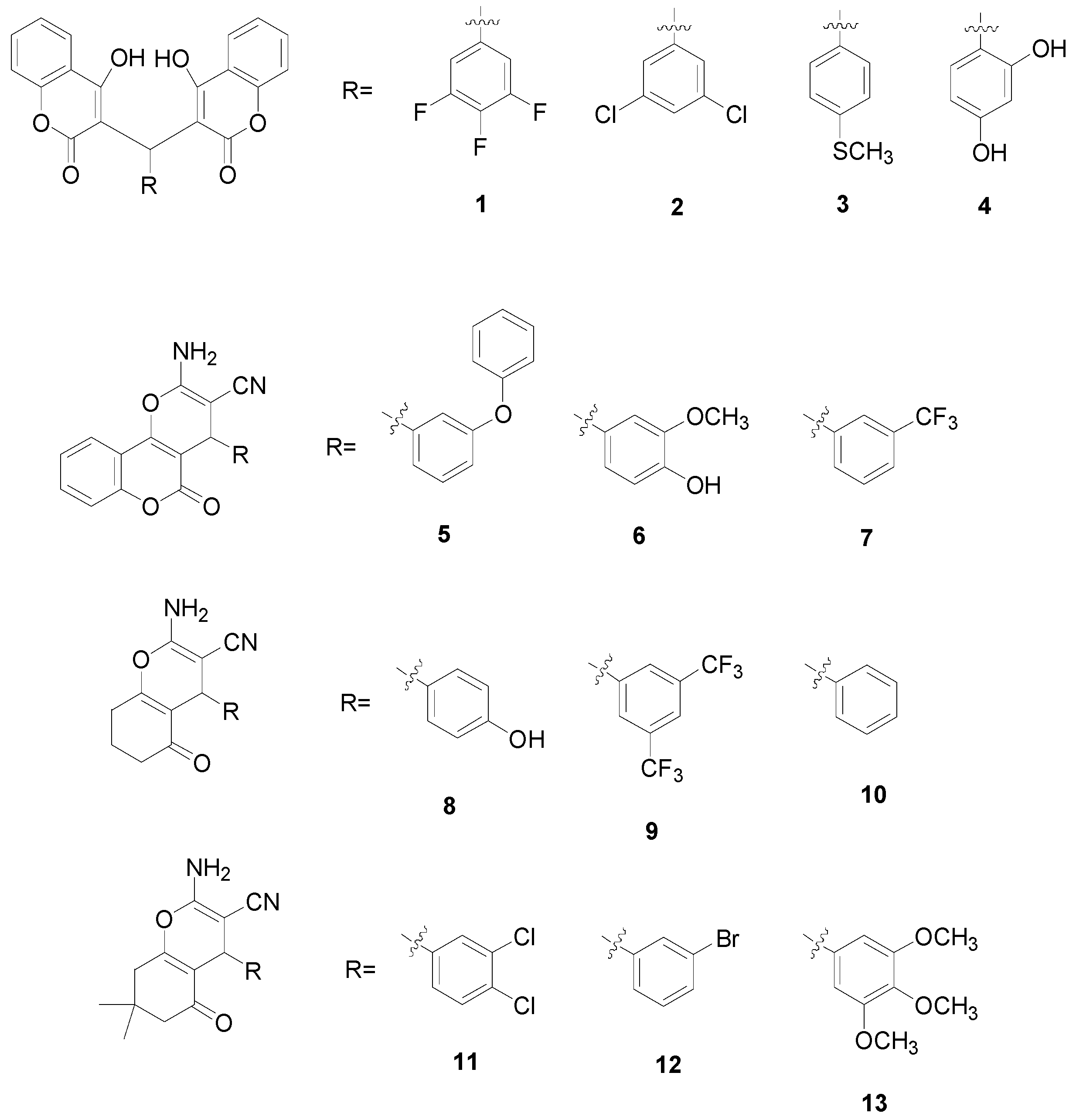
Antibacterial and Antitumor Activities of Biscoumarin and Dihydropyran Derivatives

1
Institute of Chinese Materia Medica, China Academy of Chinese Medical Sciences, Beijing 100700, China
2
Beijing Chao-Yang Hospital, Capital Medical University, Beijing 100020, China
3
Department of Blood Transfusion, Xijing Hospital, the Fourth Military Medical University, Xi’an 710032, China
4
The Key Laboratory for Surface Engineering and Remanufacturing in Shaanxi Province, School of Chemical Engineering, Xi’an University, Xi’an 710065, China
5
Graduate School of China Academy of Chinese Medical Sciences, Beijing 100700, China
6
Department of Pharmacology, School of Pharmacy, the Fourth Military Medical University, Xi’an 710032, China
*
Authors to whom correspondence should be addressed.
These authors contributed equally to this work.
Molecules 2015, 20(9), 17614-17626; https://doi.org/10.3390/molecules200917614
Submission received: 7 August 2015 / Revised: 11 September 2015 / Accepted: 14 September 2015 / Published: 23 September 2015
(This article belongs to the Section Medicinal Chemistry)

Abstract

:
A novel series of biscoumarin (14) and dihydropyran (513) derivatives were synthesized via a one-pot multicomponent condensation reaction and evaluated for antibacterial and antitumor activity in vitro. The X-ray crystal structure analysis of four representative compounds, 3, 7, 9 and 11, confirmed the structures of these compounds. Compounds 14 showed the most potent antitumor activity among the total 13 derivatives; especially for compounds 1 and 2, they also emerged as promising antibacterial members with better antibacterial activity. In addition, the results of density functional theory (DFT) showed that compared with compounds 3 and 4, biscoumarins 1 and 2 had lower intramolecular hydrogen bonds (HB) energy in their structures.

1. Introduction

Natural products have a profound impact on both chemical biology and drug discovery, and the great structural diversity of natural products with various interesting biological characteristics has always provided medicinal chemists an important source of inspiration in their search for new molecular entities with pharmacological activity [1,2]. Among them, biscoumarin and dihydropyran derivatives are two important groups of compounds covering a wide range of biological properties, including anti-oxidant, anti-inflammatory [3] and anti-microbial [4] as well as anticancer activities [5,6]. However, many of them are not suitable for therapeutic application due to their relatively lower activity or evident side-effect properties; and for the already marketable antibacterial and antitumor drugs [7,8], resistance has become one of main reasons for their failure in chemotherapy. Hence the search for high-quality novel antibacterial and anticancer agents has always been advisable and emergent [9].
Figure 1. Chemical structures of compounds 113.
Molecules 20 17614 g001
In order to get more effective antibacterial and antitumor agents, it is possible to make modifications on active chemical structures of title compounds. In the present study, a novel series of biscoumarin (14) and dihydropyran (513) derivatives were firstly synthesized (Figure 1), their antibacterial activities were then measured in vitro against Staphylococcus aureus (S. aureus ATCC 29213), methicillin-resistant S. aureus (MRSA XJ 75302), vancomycin-intermediate S. aureus (Mu50 ATCC 700699), and USA 300 (Los Angeles County clone, LAC), and finally their antitumor activities on intestinal epithelial adenocarcinoma cell line (HuTu80), mammary adenocarcinoma cell line (4T1) and pancreatic cancer cell line (PANC1) in vitro were then evaluated.

2. Results and Discussion

2.1. Molecular Structure

The crystal structures of compounds 3, 7, 9 and 11 are given in Figure 2. In the crystal structure of compound 3, two 4-hydroxycoumarin moieties are linked through a methylene bridge, wherein one hydrogen atom has been replaced with a 4-methylthiophenyl group; and two classical intramolecular hydrogen bonds (O3–H3···O4 and O6–H6···O1) between a hydroxyl group of one coumarin fragment and a lacton carbonyl group of another coumarin fragment further stabilize the whole structure.
Figure 2. Crystal structures of compounds 3, 7, 9 and 11.
Molecules 20 17614 g002
In the crystal structures of compounds 7, 9 and 11, the new formed pyran ring and the adjacent ketone (coumarin) ring are both basically planar, and the two planes are also essentially parallel to each other. However, the aromatic ring makes a torsion angle to the pyran ring in the three compounds.

2.2. Hydrogen Bonds Energies in Biscoumarins 14

We only used compound 1 as an example to estimate single and total intramolecular hydrogen bonds (HB) energies. The global minimum structure is stabilized by two HBs (1ab); two higher energy structures are stabilized by one HB (1a and 1b, respectively). The corresponding values are listed in Table 1.
Based on our previous calculation results [10], B3LYP/6-31G* exhibited sufficient agreement with experimental data and lower computational cost, so further theoretical study was performed at this level.
The O6–H6 O1 HB energy was calculated to be −51.3364015 kJ/mol by the equation E(O6–H6···O1) = E lab coor E la coor , from the energy difference between 1ab and 1a, where 1a is a global minimum structure with O3–H3···O4 HB. Similarly, the O3–H3···O4 HB energy was calculated to be −64.8682285 kJ/mol from the energy difference between 1ab and 1b by the equation E(O3–H3···O4) = E lab coor E lb coor , in which 1b was obtained from the global minimum structure 1ab, but H3 was rotated around the C3–O3 bond until O3–H3···O4 HB rupture occurred [11,12]. The total HB energy was calculated to be −116.20463 kJ/mol by the equation E(O3–H3···O4)+ E(O6–H6···O1). For compounds 24, the total HB energies are −115.7031595, −118.0346035 and −121.970228 kJ/mol, respectively.
Table 1. Single and total intramolecular hydrogen bonds (HB) energies in biscoumarins 14.
SystemTotal Electronic Energies a,bE(O6–H6···O1)E(O3–H3···O4)E(Total HB) c
1ab−1711.038294 −116.20463
1a−1711.018741−51.3364015
1b−1711.013587 −64.8682285
2ab−2332.538245 −115.7031595
2a−2332.518779−51.107983
2b−2332.513642 −64.5951765
3ab−2025.68685 −118.0346035
3a−2025.666896−52.389227
3b−2025.661847 −65.6453765
4ab−1625.791083 −121.970228
4a−1625.770804−53.2425145
4b−1625.764906 −68.7277135
a ZP corrected; b hartree; c kJ/mol.

2.3. Minimal Inhibitory Concentration (MIC) Assay

For compounds 113, one drug-sensitive S. aureus (S. aureus ATCC 29213) strain and three MRSA strains (MRSA XJ 75302, Mu50, and USA 300 LAC) were used in the systematic analysis of their antibacterial activities in vitro. Because of the liposolubility of these compounds, they were dissolved into the solution with 1% dimethyl sulfoxide (DMSO) at final concentration. From Table 2, we can see that, among these compounds, compounds 1 and 2 exerted more potent anti-bacterial activity against the tested S. aureus with minimum inhibitory concentration (MIC) values in the range of 2–16 μg/mL. Compared with compounds 113, the MIC values of levofloxacin, ceftazidime, ceftriaxone, gentamicin and piperacillin against S. aureus (ATCC 29213) strains were lower (less than 8 μg/mL) but were higher against other three strains at varying degrees.
Table 2. Minimum inhibitory concentration (MIC) of compounds 113 and antibiotics in Mueller–Hinton Broth Culture.
DrugsMIC (µg/mL)
S. aureas (ATCC 29213)MRSA (XJ 75302)Mu50 (ATCC 700699)LAC (USA 300)
Compound 1161688
Compound 24422
Compound 364646464
Compound 4>256>256>256>256
Compound 5>256>256>256>256
Compound 6>256>256>256>256
Compound 7>256>256>256>256
Compound 8>256>256>256>256
Compound 9>256>256>256>256
Compound 10>256>256>256>256
Compound 11>256>256>256>256
Compound 12>256>256>256>256
Compound 13>256>256>256>256
Levofloxacin<0.125 (S)4 (R)4 (R)8 (R)
Ceftazidime8 (S)>256 (R)256 (R)64 (R)
Ceftriaxone2 (S)>256 (R)256 (R)32 (R)
Gentamicin0.12 (S)64 (R)32 (R)0.25 (S)
Piperacillin2 (S)>128 (R)>128 (R)8 (R)
S means drug susceptibility, R means drug resistance.

2.4. In Vitro Antitumor Activity

Intestinal epithelial adenocarcinoma cell line (HuTu80), mammary adenocarcinoma cell line (4T1) and pancreatic cancer cell line (PANC1) representing three different tumor types were used in the systematic analysis of the antitumor activities of the newly synthesized compounds 113 in vitro. For comparison purpose, the cytotoxicity of carboplatin, a standard antitumor drug, was evaluated under the same condition.
The results showed that all the tested compounds possessed a certain degree of antitumor activities against the three tumor cell lines and their inhibitory action get stronger with the corresponding higher concentration. The related half maximal inhibitory concentration (IC50) and IC90 values (dose of the compound which cause a 50% and 90% reduction of survival values, respectively) are shown in Table 3. As can be seen in Table 3, there is great difference in the antitumor activity between the four different groups of the tested compounds. Biscoumarins 14 from the first group showed more potent antitumor activity against the three tested tumor cells (HuTu80, 4T1 and PANC1) with IC50 and IC90 values of 18.78–32.63 μg/mL and 36.05–64.55 μg/mL, respectively, which is much lower than the IC50 and IC90 values (45.85–65.62 μg/mL and 102.14–126.24 μg/mL) of the positive control drug carboplatin. However, the compounds in other three groups demonstrated lower antitumor activity with relatively higher IC50 and IC90 values.
Table 3. IC50 and IC90 values of compounds 113 and carboplatin against three tumor cell lines (μg/mL).
DrugsHUTU 804T1PANC1
IC50IC90IC50IC90IC50IC90
Compound 132.6364.5522.0939.5827.5251.33
Compound 228.9455.8718.7836.0525.0546.67
Compound 328.4255.3120.0838.2128.0151.75
Compound 428.5555.6119.3336.9226.1148.56
Compound 5116.00216.00138.00248.0779.23154.62
Compound 6130.67264.00163.67304.96149.42302.09
Compound 7209.00409.0091.44186.16174.71339.00
Compound 8266.79483.83214.87330.92172.99317.39
Compound 9582.881079.16333.93638.81744.591340.72
Compound 10493.58942.01303.98601.82566.141055.13
Compound 11268.72490.94316.54569.07305.25592.40
Compound 12509.78985.97621.341117.62604.451143.73
Compound 13374.50707.83481.83900.82205.17410.09
Carboplatin65.62126.2445.85102.1352.94109.94
The IC50 (dose of the compound which caused a 50% reduction of survival) and IC90 (dose of the compound which caused a 90% reduction of survival) values were calculated from dose-response curves done in triplicate for each compound. Carboplatin was used as positive control.

3. Experimental Section

3.1. Apparatus and Materials

IR spectra (400–4000 cm−1) were obtained using a Brucker Equinox-55 spectrophotometer (Bruker Optics, Ettlingen, Germany). 1H-NMR (Nuclear Magnetic Resonance) spectra, 13C-NMR spectra and mass spectra were tested using the Varian Inova-400 spectrometer (Varian Inc., Palo Alto, CA, USA), Bruker Avance III (Bruker Optics) spectrometer and micrOTOF-Q II (Bruker Optics) mass spectrometer, respectively. The melting points were taken on a XT-4 micro melting apparatus (Ledon, Suzhou, China), and the thermometer was uncorrected.
All antibiotics used were purchased from the National Institute for the Control of Pharmaceutical and Biological Products (Beijing, China). MRSA (XJ 75302) was isolated from cultures of sputum samples from patients in Xijing Hospital (Xi’an, China). S. aureus strain (ATCC 29213) was purchased from the Chinese National Center for Surveillance of Antimicrobial Resistance (Beijing, China). Mu50 (ATCC 700699) and USA 300 (LAC) were purchased from MicroBiologics (Saint Cloud, MN, USA).
RPMI 1640 medium, trypsin, and heat inactivated fetal bovine serum (HIFBS) were obtained from Gibco (New York, NY, USA). 5-Fluorouracil 99% HPLC grade, phosphate-buffered saline (PBS), penicillin/streptomycin (PS) solution, and 3-(4,5-dimethylthiazol-2-yl)-2-5-diphenyltetrazolium bromide (MTT) reagent were purchased from Sigma-Aldrich (St. Louis, MO, USA). The MTT assay was performed by using FlexStation 3 benchtop multi-mode microplate reader (Molecular Devices, San Jose, CA, USA). Human intestinal epithelial adenocarcinoma cell line (HuTu80) was purchased from Institute of Basic Medical Sciences (IBMS) of Chinese Academy of Medical Sciences (CAMS) (Beijing, China). The cells were cultured in RPMI supplemented with 10% HIFBS and 1% PS. Cells were cultured in a 5% CO2 in a humidified atmosphere at 37 °C.

3.2. Synthesis and Characterization of Compounds 113

Biscoumarins 14 were synthesized according to the methods of a previous report [13]. A mixture of 3,4,5-trifluorobenzaldehyde (3,5-dichlorobenzaldehyde, 4-methylthiobenzaldehyde or 2,4-dihydroxybenzaldehyde) (10 mmol) and 4-hydroxycoumarin (20 mmol) was dissolved in 100 mL of EtOH. A few drops of piperidine were added, and the mixture was stirred for 3 h at room temperature. After reaction completion as determined by TLC, water was added until precipitation occurred. After filtering the precipitates, they were sequentially washed with ice-cooled water and ethanol and then dried in a vacuum.
3,3′-(3,4,5-Trifluorobenzylidene)-bis-(4-hydroxycoumarin) (1): Yield: 57%. 230–231 °C. IR (KBr pellet cm−1): 3446, 2360, 1675, 1512, 1265, 1139, 767 cm−1. 1H-NMR (CDCl3, δ, ppm): 6.020 (s, 1H, CH), 6.854–6.890 (q, 2H), 7.434–7.464 (q, 4H), 7.669–7.708 (t, 2H), 8.031–8.108 (q, 2H), 11.337 (s, 1H, OH), 11.665 (s, 1H, OH). 13C-NMR (DMSO-d6) δ: 36.462, 103.773, 111.689, 111.729, 111.895, 116.431, 118.642, 124.072, 124.439, 132.355, 152.868, 164.907, 166.325. HRMS (ESI+): m/z: calcd for C25H13F3O6: 489.0556 [M + Na]+; found: 489.0533.
3,3′-(3,5-Dichlorobenzylidene)-bis-(4-hydroxycoumarin) (2): Yield: 55%. 239–240 °C. IR (KBr pellet cm−1): 3446, 2360, 1666, 1546, 1353, 1091, 761 cm−1. 1H-NMR (CDCl3, δ, ppm): 6.036 (s, 1H, CH), 7.106–7.113 (t, 2H), 7.307–7.309 (d, 1H), 7.432–7.468 (t, 4H), 7.666–7.705 (q, 2H), 8.042–8.114 (m, 2H), 11.314 (s, 1H, OH), 11.630 (s, 1H, OH). 13C-NMR (DMSO-d6) δ: 36.672, 103.422, 116.339, 119.160, 123.933, 124.530, 125.667, 126.059, 132.169, 134.153, 146.825, 152.896, 164.774, 167.058. HRMS (ESI+): m/z: calcd for C25H14Cl2O6: 503.0060 [M + Na]+; found: 503.0069.
3,3′-(4-Methylthiobenzylidene)-bis-(4-hydroxycoumarin) (3): Yield: 50%. 232–233 °C. IR (KBr pellet cm−1): 2605, 1668, 1614, 1349, 1211, 1100, 1029, 910, 767 cm−1. 1H-NMR (CDCl3, δ, ppm): 2.493 (s, 3H, SCH3), 6.073 (s, 1H, CH), 7.149–7.170 (d, 2H), 7.223–7.244 (d, 2H), 7.427–7.447 (d, 4H), 7.634–7.677 (m, 2H), 8.015–8.104 (q, 2H), 11.323 (s, 1H, OH), 11.545 (s, 1H, OH). 13C-NMR (DMSO-d6) δ: 15.408, 36.064, 104.671, 116.528, 118.068, 124.354, 124.373, 126.513, 127.932, 132.557, 135.395, 136.867, 152.635, 165.316, 165.392. HRMS (ESI+): m/z: calcd for C26H18O6S: 481.0716 [M + Na]+; found: 481.0779.
3,3′-(2,4-Dihydroxybenzylidene)-bis-(4-hydroxycoumarin) (4): Yield: 55%. m.p. 262–263 °C. IR (KBr): 3260, 2230, 1681, 1611, 1539, 1392, 1184, 910, 757 cm−1. 1H-NMR (DMSO-d6, δ, ppm): 5.635 (s, 1H), 6.568–6.595 (q, 1H), 6.713–6.718 (d, 1H), 6.978–6.999 (d, 1H), 7.318–7.373 (m, 1H), 7.445–7.505 (q, 2H), 7.582–7.624 (m, 1H), 7.683–7.726 (m, 1H), 8.086–8.108 (q, 2H), 9.792 (s, 1H). 13C-NMR (DMSO-d6) δ: 103.214, 114.327, 116.652, 116.936, 123.182, 124.434, 125.018, 129.643, 132.579, 132.912, 152.422, 157.792, 160.985. HRMS (ESI+): m/z: calcd for C25H16O8: 467.0737 [M + Na]+; found: 467.0779.
Dihydropyran derivatives (513) were also synthesized according to a reported procedure [14]. A mixture of 4-hydroxycoumarin (3,5-cyclohexanedione, or 1,1-dimethyl-3,5-cyclohexanedione) (10 mmol), aromatic aldehydes (10 mmol), malononitrile (10 mmol) and 4-(dimethylamino)pyridine (DMAP) (1 mmol) in ethanol (100 mL) was refluxed for 2–3 h and then cooled to room temperature. After filtering the precipitates, they were sequentially washed with ice-cooled water and ethanol and then dried under a vacuum.
2-Amino-4-(3-phenoxyphenyl)-3-cyano-5-oxo-4H,5H-pyrano[3,2c]chromene (5): Yield: 60%. 233–234 °C. IR (KBr pellet cm−1): 3688, 2198, 1710, 1670, 1574, 1486, 1374, 1238, 1062, 821 cm−1. 1H-NMR (DMSO-d6, δ, ppm): 4.477 (s, 1H, CH), 6.821–6.841 (d, 1H), 6.978–7.041 (m, 4H), 7.112–7.149 (t, 1H), 7.300–7.392 (m, 3H), 7.451–7.519 (m, 4H), 7.707–7.747 (t, 1H), 7.879–7.899 (d, 1H). 13C-NMR (DMSO-d6) δ: 37.241, 58.105, 104.086, 113.430, 117.067, 117.293, 118.560, 119.068, 119.655, 122.981, 123.034, 123.984, 125.159, 130.488, 130.648, 133.463, 146.071, 152.645, 154.038, 156.800, 157.033, 158.476, 160.047. HRMS (ESI+): m/z: calcd for C25H16N2O4: 431.1002 [M + Na]+; found: 431.1033.
2-Amino-4-(3-methoxy-4-hydroxyphenyl)-3-cyano-5-oxo-4H,5H-pyrano[3,2c]chromene (6): Yield: 60%. 236–237 °C. IR (KBr pellet cm−1): 3415, 3303, 2191, 1686, 1598, 1518, 1374, 1262, 1085, 749 cm−1. 1H-NMR (DMSO-d6, δ, ppm): 3.728 (s, 3H, OCH3), 4.359 (s, 1H, CH), 6.594–6.619 (q, 1H), 6.690–6.710 (d, 1H), 6.811–6.816 (d, 1H), 7.354 (s, 2H), 7.457–7.511 (q, 2H), 7.690–7.733 (m, 1H), 7.880–7.903 (q, 1H), 8.953 (s, 1H). 13C-NMR (DMSO-d6) δ: 31.181, 36.951, 56.139, 58.781, 104.800, 112.502, 113.532, 115.970, 117.015, 119.889, 120.350, 122.910, 125.097, 133.264, 134.801, 146.201, 147.778, 152.540, 153.516, 158.385, 160.068. HRMS (ESI+): m/z: calcd for C20H14N2O5: 385.0795 [M + Na]+; found: 385.0770.
2-Amino-4-(3-trifluoromethylphenyl)-3-cyano-5-oxo-4H,5H-pyrano[3,2c]chromene (7): Yield: 65%. 240–241 °C. IR (KBr pellet cm−1): 3408, 3321, 2205, 1701, 1672, 1594, 1381, 1313, 1167, 1109, 1041, 770 cm−1. 1H-NMR (DMSO-d6, δ, ppm): 4.654 (s, 1H, CH), 7.467–7.659 (m, 8H), 7.713–7.756 (m, 1H), 7.902–7.926 (q, 1H). 13C-NMR (DMSO-d6) δ: 31.169, 37.242, 57.707, 103.550, 113.473, 117.079, 119.517, 123.065, 124.465, 124.502, 124.841, 124.879, 125.159, 126.020, 129.319, 129.632, 130.165, 132.477, 133.525, 145.164, 152.718, 154.304, 158.506, 160.087. HRMS (ESI+): m/z: calcd for C20H11F3N2O3: 407.0614 [M + Na]+; found: 407.0480.
2-Amino-4-(4-hydroxyphenyl)-5-oxo-5,6,7,8-tetrahydro-4H-chromene-3-carbonitrile (8): Yield: 60%. 238–239 °C. IR (KBr pellet cm−1): 3198, 2200, 1647, 1512, 1367, 1215, 1009, 836 cm−1. 1H-NMR (DMSO-d6, δ, ppm): 1.854–1.973 (m, 2H), 2.214–2.302 (m, 2H), 2.573–2.606 (q, 2H), 4.074 (s, 1H, CH), 6.644–6.665 (q, 2H), 6.926–6.947 (q, 4H), 9.263 (s, 1H). 13C-NMR (DMSO-d6) δ: 20.307, 26.931, 31.184, 35.032, 36.860, 59.101, 114.735, 115.464, 120.442, 128.612, 135.710, 156.465, 158.846, 164.475, 196.359. HRMS (ESI+): m/z: calcd for C16H14N2O3: 305.0897 [M + Na]+; found: 305.0980.
2-Amino-4-(3,5-ditrifluoromethylphenyl)-5-oxo-5,6,7,8-tetrahydro-4H-chromene-3-carbonitrile (9): Yield: 69%. 209–210 °C. IR (KBr pellet cm−1): 3397, 2194, 1683, 1651, 1419, 1362, 1330, 1211, 700 cm−1. 1H-NMR (DMSO-d6, δ, ppm): 1.889−1.960 (m, 2H), 2.258–2.318 (m, 2H), 2.607–2.709 (m, 2H), 4.543 (s, 1H, CH), 7.239 (s, 2H), 7.857 (s, 2H), 7.968 (s, 1H). 13C-NMR (DMSO-d6) δ: 20.183, 27.055, 31.096, 35.898, 36.679, 57.089, 112.623, 119.735, 121.012, 121.049, 121.082, 122.434, 125.145, 128.644, 130.201, 130.527, 130.851, 131.176, 148.674, 159.034, 166.000, 196.552. HRMS (ESI+): m/z: calcd for C18H12F6N2O2: 425.0695 [M + Na]+; found: 425.0663.
2-Amino-5-oxo-4-phenyl-5,6,7,8-tetrahydro-4H-chromene-3-carbonitrile (10): Yield: 65%. 221–222 °C. IR (KBr pellet cm−1): 3309, 3170, 2194, 1685, 1261, 1064, 999, 702 cm−1. IR (KBr pellet cm–1): 3309, 3170, 2194, 1685, 1261, 1064, 999, 702 cm–1. 1H-NMR (DMSO-d6, δ, ppm): 1.876−1.988 (m, 2H), 2.2262.318 (m, 2H), 2.599–2.640 (q, 2H), 4.183 (s, 1H, CH), 7.019 (s, 2H), 7.146–7.205 (m, 3H), 7.269–7.306 (t, 2H). 13C-NMR (DMSO-d6) δ: 20.295, 26.955, 31.186, 35.922, 36.808, 58.652, 114.255, 120.289, 127.015, 127.609, 128.822, 145.278, 158.948, 164.971, 196.348. HRMS (ESI+): m/z: calcd for C16H14N2O2: 289.0947 [M + Na]+; found: 289.0933.
2-Amino-4-(3,4-dichlorophenyl)-3-cyano-7,7-dimethyl-5-oxo-4H-5,6,7,8-tetrahydrobenzo[b]pyran (11): Yield: 66%. 232–233 °C. IR (KBr pellet cm−1): 3325, 2958, 2194, 1639, 1608, 1465, 1357, 1249, 1215, 1033, 898 cm−1. 1H-NMR (DMSO-d6, δ, ppm): 0.965 (s, 3H, CH3), 1.039 (s, 3H, CH3), 2.114–2.155 (d, 1H), 2.234–2.274 (d, 1H), 2.529 (s, 2H), 4.255 (s, 1H, CH), 7.137–7.172 (m, 3H), 7.390–7.395 (d, 1H), 7.756–7.582 (d, 1H). 13C-NMR (DMSO-d6) δ: 27.412, 28.689, 31.183, 32.323, 35.439, 50.387, 57.649, 112.193, 119.902, 128.170, 129.652, 129.773, 131.118, 131.286, 146.380, 158.994, 163.461, 196.238. HRMS (ESI+): m/z: calcd for C18H16Cl2N2O2: 385.0481 [M + Na]+; found: 385.0422.
2-Amino-4-(3-bromophenyl)-3-cyano-7,7-dimethyl-5-oxo-4H-5,6,7,8-tetrahydrobenzo[b]pyran (12): Yield: 68%. 239–240 °C. IR (KBr pellet cm−1): 3344, 3168, 2963, 2191, 1686, 1651, 1605, 1469, 1427, 1367, 1250, 1216, 1036, 695 cm−1. 1H-NMR (DMSO-d6, δ, ppm): 0.967 (s, 3H, CH3), 1.043 (s, 3H, CH3), 2.111–2.151 (d, 1H), 2.242–2.82 (d, 1H), 2.534 (s, 2H), 4.210 (s, 1H, CH), 7.096 (s, 2H), 7.154–7.174 (d, 1H), 7.2567.312 (m, 2H), 7.385–7.409 (m. 1H). 13C-NMR (DMSO-d6) δ: 27.272, 28.820, 31.195, 32.337, 35.816, 50.398, 58.091, 112.565, 120.011, 122.047, 126.858, 130.012, 130.417, 131.122, 147.967, 159.002, 163.338, 196.187. HRMS (ESI+): m/z: calcd for C18H17BrN2O2: 395.0366 [M + Na]+; found: 395.0450.
2-Amino-4-(3,4,5-trimethoxyphenyl)-3-cyano-7,7-dimethyl-5-oxo-4H-5,6,7,8-tetrahydrobenzo[b]pyran (13): Yield: 60%. 184−185 °C. IR (KBr pellet cm−1): 3313, 3186, 2961, 2189, 1656, 1588, 1505, 1459, 1420, 1367, 1322, 1212, 1124, 536 cm−1. 1H-NMR (DMSO-d6, δ, ppm): 1.029 (s, 3H, CH3), 1.060 (s, 3H, CH3), 2.091 (s, 2H), 2.131–2.172 (d, 1H), 2.271–2.311 (d, 1H), 3.633 (s, 3H), 3.727 (s, 6H), 4.145 (s, 1H, CH), 6.389 (s, 2H), 6.975 (s, 2H). 13C-NMR (DMSO-d6) δ: 27.039, 29.098, 31.179, 32.234, 36.098, 50.456, 56.247, 58.802, 60.398, 104.590, 112.816, 120.232, 136.569, 140.966, 153.232, 158.828, 163.272, 196.219. HRMS (ESI+): m/z: calcd for C21H24N2O5: 407.1577 [M + Na]+; found: 407.1565.

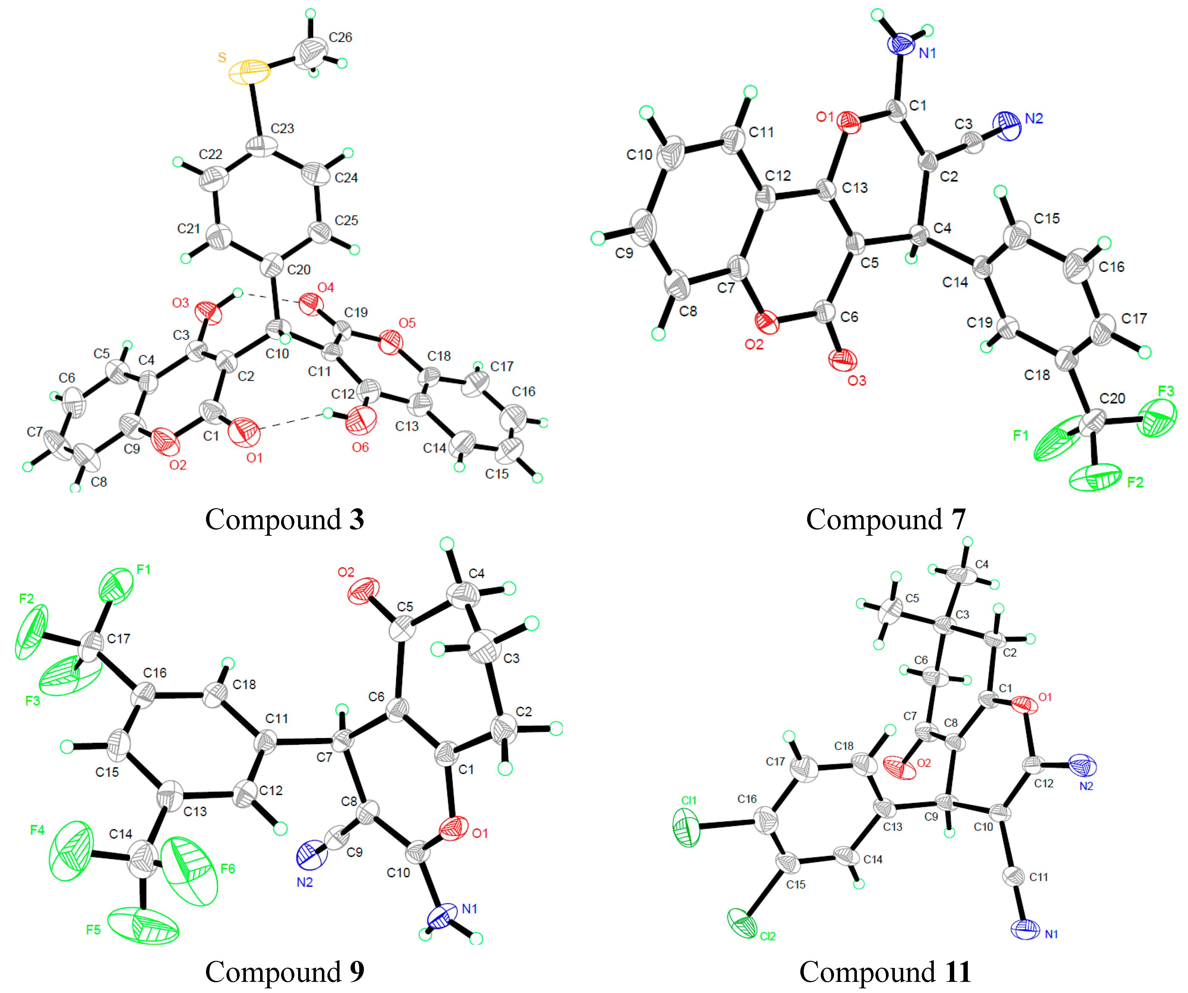
3.3. X-ray Crystallography

For X-ray diffraction experiments, single crystals of compounds 3, 7, 9 and 11 were grown from methanol. The X-ray diffraction data were collected on a Bruker SMART APEX II CCD diffractometer (Bruker Optics, Ettlingen, Germany) equipped with a graphite monochromated Mo Kα radiation (λ = 0.71073 Å) by using the ω-2θ scan technique at room temperature. The structure was solved by direct methods using SHELXS-97 (Sheldrick 1997, University of Gottingen, Germany) and refined using the full-matrix least squares method on F2 with anisotropic thermal parameters for all non-hydrogen atoms by using SHELXL-97 [15]. Hydrogen atoms were generated geometrically. The crystal data and details concerning data collection and structure refinement are given in Table 4. Molecular illustrations were prepared using the XP package. Parameters in CIF format are available as Electronic Supplementary Publication from Cambridge Crystallographic Data Centre.
CCDC 1048441-1048444 for compounds 3, 7, 9 and 11 contains the supplementary crystallographic data for this paper. These data can be obtained free of charge via http://www.ccdc.cam.ac.uk/conts/retrieving.html (or from the CCDC, 12 Union Road, Cambridge CB2 1EZ, UK; Fax: +44-1223-336033; E-mail: [email protected])
Table 4. Crystal data, data collection and structure refinement of compounds 3, 7, 9 and 11.
Compound 3Compound 7Compound 9Compound 11
FormulaC26H18O6SC20H11F3N2O3C18H12F6N2O2C18H16Cl2N2O2
Mr458.08384.31402.3362.06
Crystal systemTriclinicTriclinicTriclinicTriclinic
Space groupPīPīPīPī
a/Å10.947 (3)7.9416 (6)8.5762 (6)8.2045 (5)
b/Å20.402 (7)10.9917 (9)8.8891 (8)14.1658 (8)
c/Å20.694 (7)11.4755 (6)12.1026 (8)23.3891 (15)
α/°109.44 (3)112.429 (7)109.583 (7)94.540 (5)
β/°90.09 (3)105.094 (6)92.163 (6)97.474 (5)
γ/°93.29 (3)97.364 (6)97.257 (7)98.928 (5)
V/Å34350 (3)864.45 (11)859.13 (11)2648.9 (3)
Z2222
Dcalc/g·cm−31.4011.4471.5551.357
μ(Mo Kα)/mm−10.1910.1220.1460.38
θ range/°2.43 to 25.002.74 to 27.682.43 to 27.352.76 to 27.51
Reflections collected183555790581918090
No. unique data [R(int)]13442 [0.0377]3045 [0.0233]3030 [0.0213]9317 [0.0442]
No. data with I ≥ 2σ(I)5431217023186552
R10.07830.05950.07450.0824
ωR2(all data)0.24690.17740.22760.261

3.4. Quantum Chemical Calculations

All calculations were carried out using the Gaussian 09 package [16]. Density functional theory (DFT), Becke’s three-parameter hybrid function (B3LYP), and LYP correlation function were used to fully optimize all the geometries on the energy surface without constraints. To obtain precise results that are in conjunction with experimental results, three basis sets, namely 6-31G*, 6-31 + G**, and 6-311G*, were tested. Frequency calculations at the B3LYP (with basis sets 6-31G*) level of theory were carried out to confirm stationary points as minima and to obtain the zero-point energies and the thermal correlation data at 1 atm and 298 K.

3.5. Minimal Inhibitory Concentration (MIC) Assay

Based on the CLSI broth microdilution method [17], the determination of minimum inhibitory concentrations (MICs) via microdilution assay was performed in sterilized 96-well polypropylene microtiter plates (Sigma-Aldrich) in a final volume of 200 μL. Bacteria were grown overnight in nutrient broth. Mueller–Hinton (MH) broth (100 μL) containing bacteria (5 × 10⁵ CFU/mL) was added to 100 μL of the culture medium containing the test compound (0.12 μg/mL–256 μg/mL in serial 2-fold dilutions). The plates were incubated at 37 °C for 20 h in an incubator. About 50 µL of 0.2% triphenyl tetrazolium chloride (TTC), a colorimetric indicator, was added to each well of microtiter plates and incubated at 35 °C for 1.5 h. The TTC-based MIC was determined as the lowest concentration of oxacillin that showed no red color change indicating complete growth inhibition.

3.6. Cell Viability Assay

Viability of intestinal epithelial adenocarcinoma cell line (HuTu80), mammary adenocarcinoma cell line (4T1) and pancreatic cancer cell line (PANC1) was determined by using the MTT assay as described by Mosmann [18]. Cells reaching 70%–80% confluency were treated with various concentrations of the synthesized compounds with 1% dimethyl sulfoxide (DMSO) as a negative control. After 48 h incubation, 20 μL of MTT solution (5 mg/mL in PBS) was added and incubated for an additional 4 h. Subsequently, the medium was aspirated carefully, and 150 μL of DMSO was added. After incubation for 15 min, the optical density was measured at 490 nm using FlexStation 3 benchtop multi-mode microplate reader (Molecular Devices). Data were recorded and analyzed for the assessment of the effects of the test substances on cell viability and growth inhibition. The IC50 and IC90 values were calculated using regression equation as explained before. The results are presented as the average percentage viability to the negative control (1% DMSO). The percentage of cell viability was calculated using the following formula: % cell viability = (absorbance of treated/absorbance of untreated) × 100. The percentage of inhibition was plotted against the concentration in Microsoft excel and the IC50 was calculated using the regression equation.

4. Conclusions

In this work, we synthesized two new series of biscoumarin and dihydropyran derivatives; evaluated their antibacterial activities in vitro against one-drug-sensitive S. aureus (S. aureus ATCC 29213) strain and three MRSA strains (MRSA XJ 75302, Mu50, and USA 300 LAC); and then measured their antitumor activities on intestinal epithelial adenocarcinoma cell line (HuTu80), mammary adenocarcinoma cell line (4T1) and pancreatic cancer cell line (PANC1) in vitro.
In addition, X-ray structural analysis showed that biscoumarins 14 had two classical intramolecular O–H···O hydrogen bonds in their structures. Their corresponding intramolecular hydrogen bonds energy was calculated to be −116.20463, −115.7031595, −118.0346035 and −121.970228 kJ/mol, respectively.
Among the synthesized compounds, compounds 14 had more potent antitumor activity against the tested three cancer cell lines with the IC50 and IC90 values of 18.78–32.63 μg/mL and 36.05–64.55 μg/mL, respectively, which is much lower than that of the positive control drug carboplatin; compounds 1 and 2, with lower intramolecular hydrogen bonds energy, also showed the most potent antibacterial effect on four S. aureus bacterial strains with the MIC values of 2–16 μg/mL. The reason may be that intramolecular HB strength is related to the stability of chemical structure, which further affects the binding affinity between molecules and target protein.

Acknowledgments

This work was supported by grants from the National Natural Science Foundation of China (81274112; 81373986; 81473372; 81573468), Beijing Municipal Natural Science Foundation (7152106), Inheritance Program of China Academy of Chinese Medical Sciences (CM2014GD3002), Voluntary Program of China Academy of Chinese Medical Sciences (ZZ2014056) and the Innovation Plan of Science and Technology of Shaanxi Province (2014KTCL03-03).

Author Contributions

Feng Sui, Ming-Kai Li, Yun-Peng Sui, Hai-Ru Huo and Jia-Jia Xin conceived and designed the experiments; Jing Li, Yun-Peng Sui, Xiao-Jun Li, Xin-Liang Du, Hai Ma, Hai-Yu Zhou, Hong-Dan Zhan, Zhu-Ju Wang and Chun Li performed the experiments; Feng Sui, Jing Li, Yun-Peng Sui, Hai-Yu Zhou and Hong-Dan Zhan analyzed the data; Yun-Peng Sui and Jing Li wrote the paper; All authors read and approved the final manuscript.

Conflicts of Interest

The authors declare no conflict of interest.

References

  1. Newman, D.J.; Cragg, G.M. Natural products as sources of new drugs over the 30 years from 1981 to 2010. J. Nat. Prod. 2012, 75, 311–335. [Google Scholar] [CrossRef] [PubMed]
  2. Mishra, B.B.; Tiwari, V.K. Natural products: An evolving role in future drug discovery. Eur. J. Med. Chem. 2011, 46, 4769–4807. [Google Scholar] [CrossRef] [PubMed]
  3. Fylaktakidou, K.C.; Hadjipavlou-Litina, D.J.; Litinas, K.E.; Nicolaides, D.N. Natural and synthetic coumarin derivatives with anti-inflammatory/antioxidant activities. Curr. Pharm. Des. 2004, 10, 3813–3833. [Google Scholar] [CrossRef] [PubMed]
  4. Ostrov, D.A.; Hernandez Prada, J.A.; Corsino, P.E.; Finton, K.A.; Le, N.; Rowe, T.C. Discovery of novel DNA gyrase inhibitors by high-throughput virtual screening. Antimicrob. Agents Chemother. 2007, 51, 3688–3698. [Google Scholar] [CrossRef] [PubMed]
  5. Elinos-Baez, C.M.; Leon, F.; Santos, E. Effects of coumarin and 7OH-coumarin on BCL-2 and BAX expression in two human lung cancer cell lines in vitro. Cell Biol. Int. 2005, 29, 703–708. [Google Scholar] [CrossRef] [PubMed]
  6. Kawaii, S.; Tomono, Y.; Ogawa, K.; Sugiura, M.; Yano, M.; Yoshizawa, Y. The antiproliferative effect of coumarins on several cancer cell lines. Anticancer Res. 2001, 21, 917–923. [Google Scholar] [PubMed]
  7. Lopez-Gonzalez, J.S.; Prado-Garcia, H.; Aguilar-Cazares, D.; Molina-Guarneros, J.A.; Morales-Fuentes, J.; Mandoki, J.J. Apoptosis and cell cycle disturbances induced by coumarin and 7-hydroxycoumarin on human lung carcinoma cell lines. Lung Cancer 2004, 43, 275–283. [Google Scholar] [CrossRef] [PubMed]
  8. MacDonagh, L.; Gray, S.G.; Finn, S.P.; Cuffe, S.; O’Byrne, K.J.; Barr, M.P. The emerging role of microRNAs in resistance to lung cancer treatments. Cancer Treat. Rev. 2015, 41, 160–169. [Google Scholar] [CrossRef] [PubMed]
  9. Kaur, M.; Kohli, S.; Sandhu, S.; Bansal, Y.; Bansal, G. Coumarin: A Promising Scaffold for Anticancer Agents. Anticancer Agents Med. Chem. 2015, 15, 1032–1048. [Google Scholar] [CrossRef] [PubMed]
  10. Qu, D.; Li, J.; Yang, X.H.; Zhang, Z.D.; Luo, X.X.; Li, M.K.; Li, X. New Biscoumarin derivatives: synthesis, crystal Structure, theoretical study and antibacterial activity against Staphylococcus aureus. Molecules 2014, 19, 19868–19879. [Google Scholar] [CrossRef] [PubMed]
  11. Trendafilova, N.; Bauer, G.; Mihaylov, T. DFT and AIM studies of intramolecular hydrogen bonds in dicoumarols. Chem. Phys. 2004, 302, 95–104. [Google Scholar] [CrossRef]
  12. Mihaylov, T.; Georgieva, I.; Bauer, G.; Kostova, I.; Manolov, I.; Trendafilova, N. Theoretical study of the substituent effect on the intramolecular hydrogen bonds in di(4-hydroxycoumarin) derivatives. Int. J. Quantum Chem. 2006, 106, 1304–1315. [Google Scholar] [CrossRef]
  13. Li, J.; Hou, Z.; Chen, G.H.; Li, F.; Zhou, Y.; Xue, X.Y.; Li, Z.P.; Jia, M.; Zhang, Z.D.; Li, M.K.; et al. Synthesis, antibacterial activities, and theoretical studies of dicoumarols. Org. Biomol. Chem. 2014, 12, 5528–5535. [Google Scholar] [CrossRef] [PubMed]
  14. Khan, A.T.; Lal, M.; Ali, S.; Khan, M.M. One-pot three-component reaction for the synthesis of pyran annulated heterocyclic compounds using DMAP as a catalyst. Tetrahedron Lett. 2011, 52, 5327–5332. [Google Scholar] [CrossRef]
  15. Sheldrick, G.M. SHELXL-97, Program for Solution Crystal Structure and Refinement; University of Göttingen: Göttingen, Germany, 1997. [Google Scholar]
  16. Frisch, M.J.; Trucks, G.W.; Schlegel, H.B.; Scuseria, G.E.; Robb, M.A.; Cheeseman, J.R.; Scalmani, G.; Barone, V.; Mennucci, B.; Petersson, G.A.; et al. Gaussian 09, Revision D.01, Gaussian, Inc.: Wallingford, CT, USA, 2009.
  17. Prüller, S.; Frömke, C.; Kaspar, H.; Klein, G.; Kreienbrock, L.; Kehrenberg, C. Recommendation for a standardized method of broth microdilution susceptibility testing for porcine bordetella bronchiseptica. PLoS ONE 2015, 10, e0123883. [Google Scholar] [CrossRef] [PubMed]
  18. Mosmann, T. Rapid colorimetric assay for cellular growth and survival: Application to proliferation and cytotoxicity assays. J. Immunol. Methods 1983, 65, 55–63. [Google Scholar] [CrossRef]
  • Sample Availability: Samples of the compounds 113 are available from the authors.

