
Synthesis, Spectral Analysis and Preliminary in Vitro Evaluation of Some Tetrapyrrolic Complexes with 3d Metal Ions


 ,
,
Abstract
:
1. Introduction

2. Results and Discussion
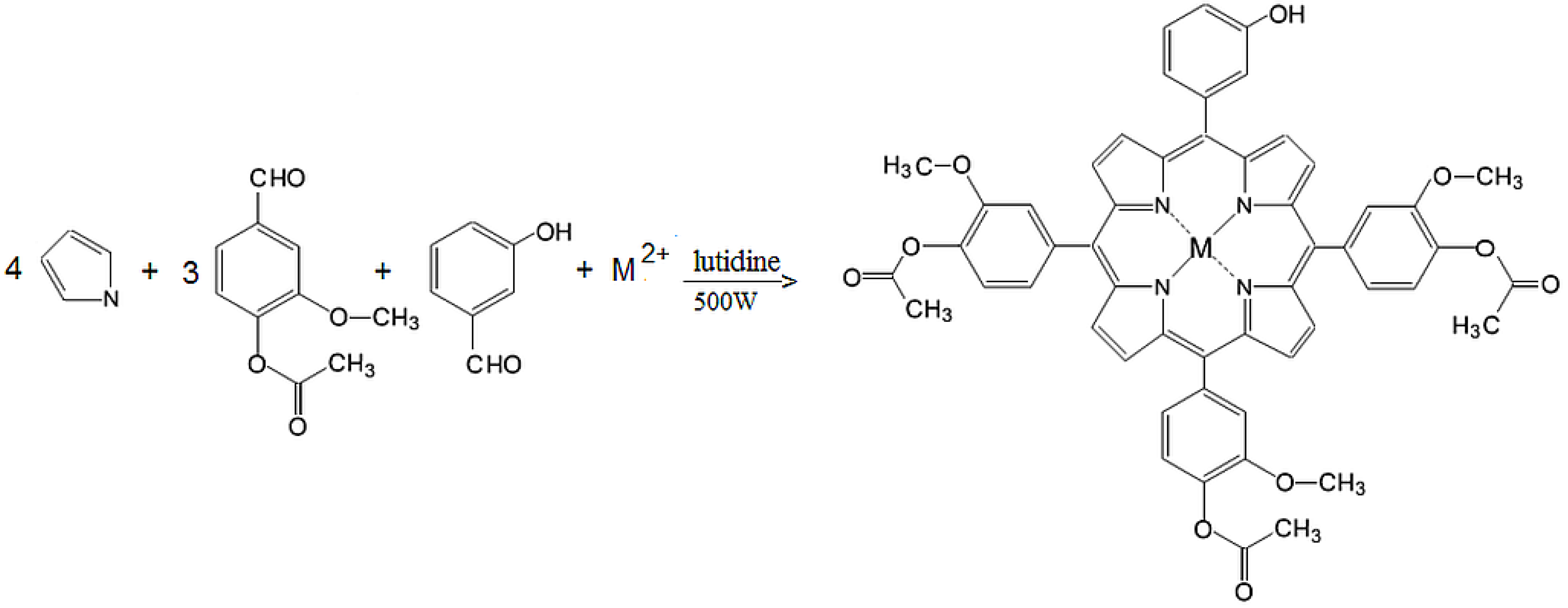
2.1. Chemistry
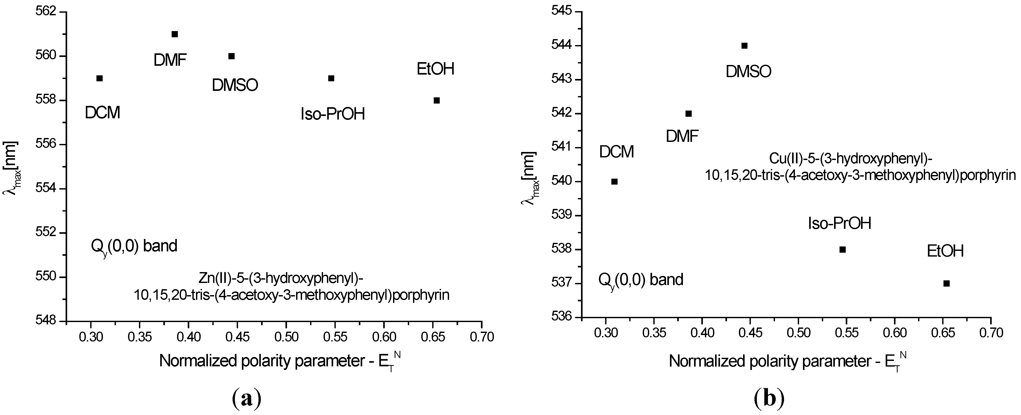
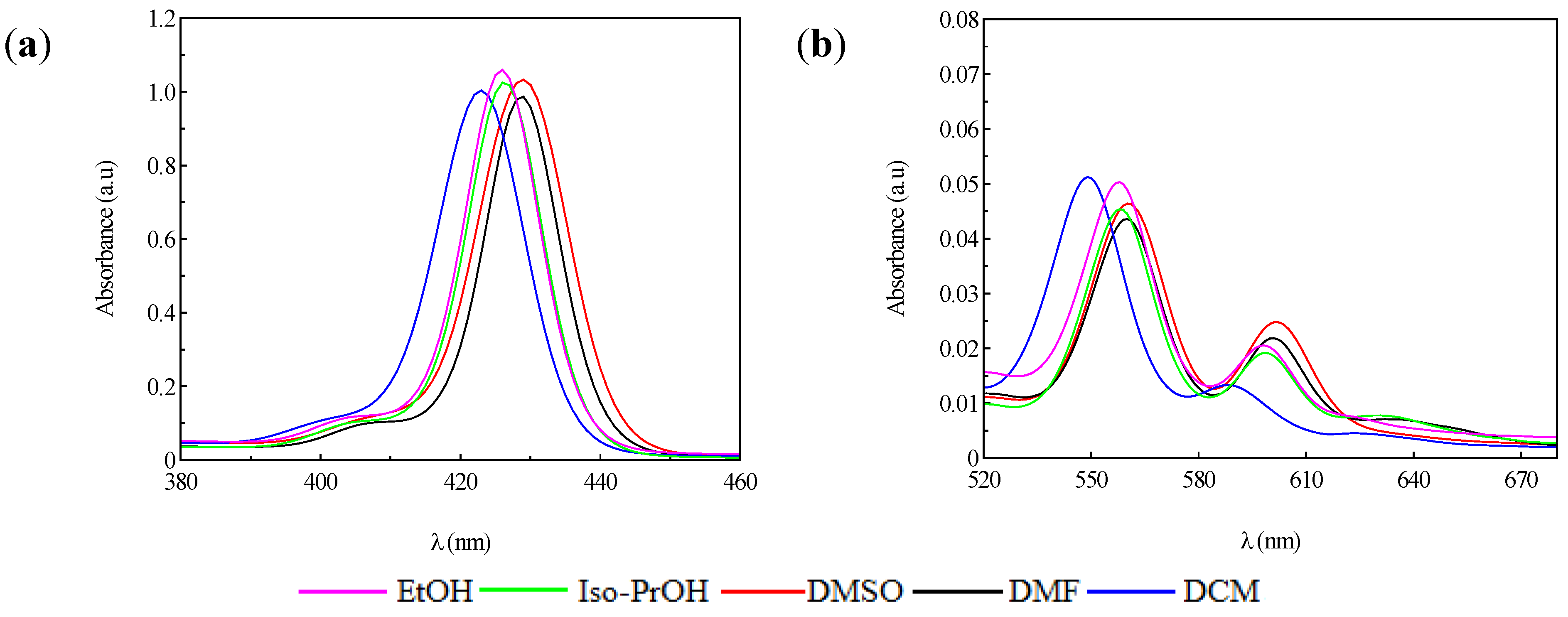
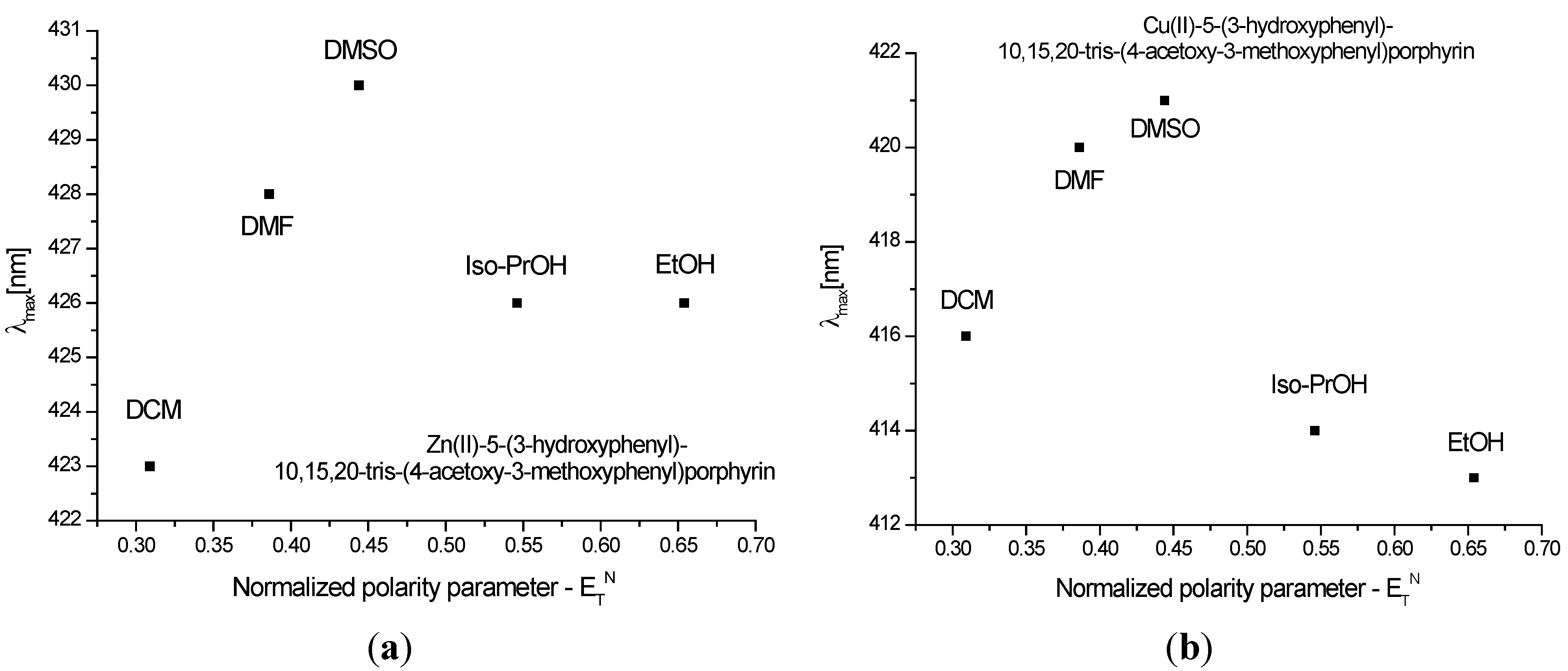
Absorption and Emission Spectra

| Solvent | Absorption λmax (nm) [lgε (L·mol−1·cm−1)] | Emission λmax (nm) |
|---|---|---|
| Soret Band B Qy (0,0) Qx(1,0) | Q(0,0) Q(0,1) | |
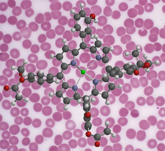
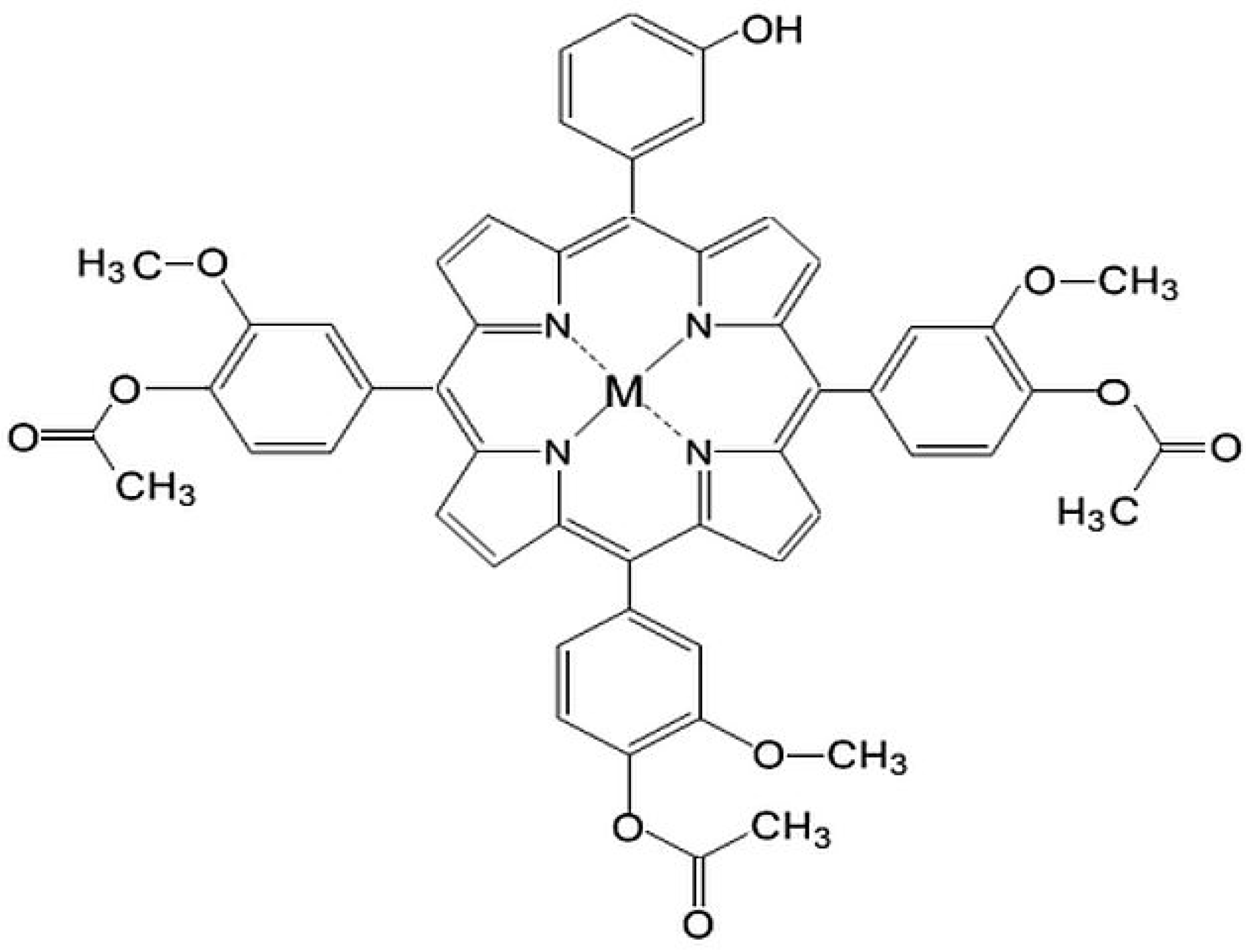
| Zn(II)-5-(3-hydroxyphenyl)-10,15,20-tris-(4-acetoxy-3-methoxyphenyl)porphyrin | ||
| EtOH | 426 [5.63] 558 [4.30] 598 [3.92] | 605 653 |
| Iso-PrOH | 426 [5.61] 559 [4.21] 599 [3.90] | 605 652 |
| DCM | 423 [5.60] 549 [4.30] 588 [3.71] | 603 650 |
| DMF | 428 [5.58] 561 [4.24] 600 [3.94] | 608 656 |
| DMSO | 430 [5.61] 560 [4.26] 602 [4.00] | 609 656 |
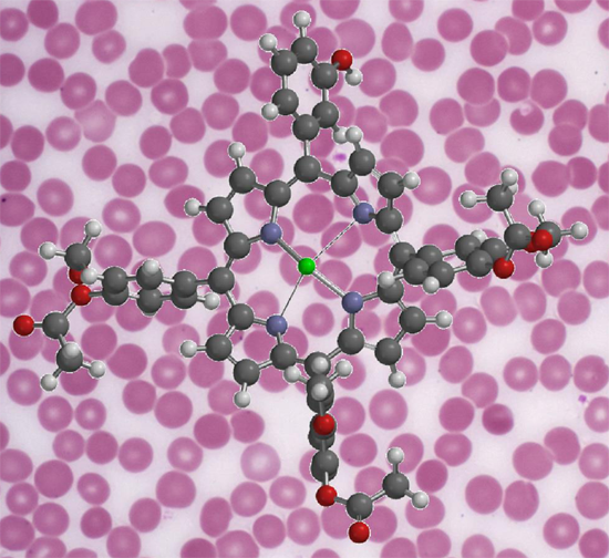
| Cu(II)-5-(3-hydroxyphenyl)-10,15,20-tris-(4-acetoxy-3-methoxyphenyl)porphyrin | ||
| EtOH | 413 [5.52] 537 [4.28] | - - |
| Iso-PrOH | 414 [5.58] 538 [4.36] | - - |
| DCM | 416 [5.50] 540 [4.14] | - - |
| DMF | 420 [5.52] 542 [4.20] | - - |
| DMSO | 421 [5.51] 544 [4.30] | - - |

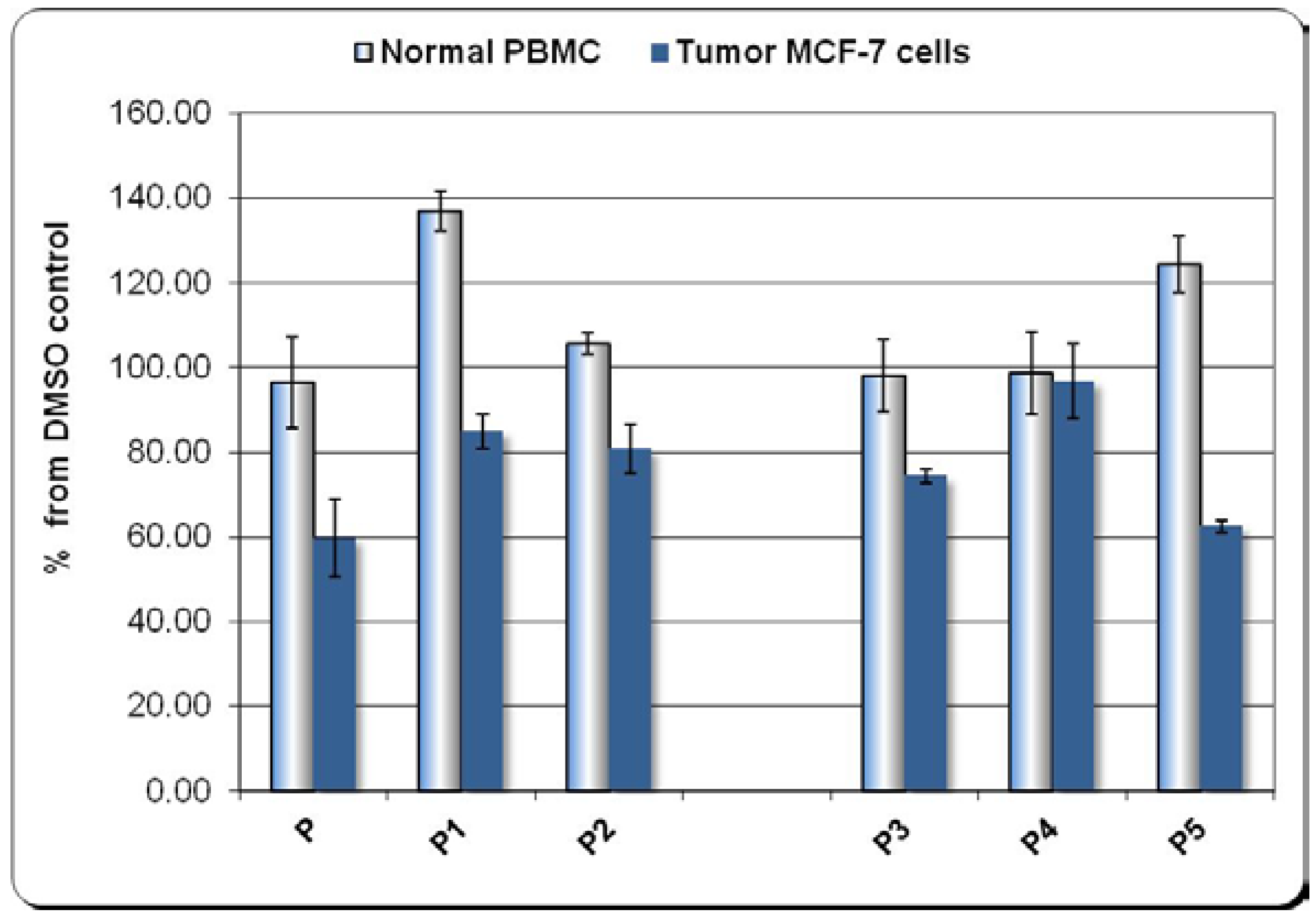
2.2. In Vitro Dark Cytotoxicity Study
The Effect of Porphyrinic Compounds on Human Tumor MCF-7 Cells

3. Experimental Section
3.1. General Information
3.2. Synthesis of Tetrapyrrolic Complexes
3.2.1. Synthesis of Zn(II)-5-(3-hydroxyphenyl)-10,15,20-tris-(4-acetoxy-3-methoxyphenyl)porphyrin (P1)

3.2.2. Synthesis of Cu(II)-5-(3-hydroxyphenyl)-10,15,20-tris-(4-acetoxy-3-methoxyphenyl)porphyrin (P2)
3.3. In Vitro Dark Cytotoxicity Tests
3.3.1. Preparation of Tetrapyrrolic Compounds for the in Vitro Study
3.3.2. Cells
3.3.3. Cell Cultures
3.3.4. The MTS Reduction Test
4. Conclusions
Author Contributions
Conflicts of Interest
References
- Ethirajan, M.; Chen, Y.; Josi, P.; Pandey, R.K. The role of porphyrin chemistry in tumor imaging and photodynamic therapy. Chem. Soc. Rev. 2011, 40, 340–362. [Google Scholar] [CrossRef] [PubMed]
- Celli, J.P.; Spring, B.Q.; Rizvi, I.; Evans, C.L.; Samkoe, K.S.; Verma, S.; Pogue, B.W.; Hasan, T. Imaging and Photodynamic Therapy: Mechanisms, Monitoring, and Optimization. Chem. Rev. 2010, 110, 2795–2838. [Google Scholar] [CrossRef] [PubMed]
- Lovell, J.F.; Liu, T.W.B.; Chen, J.; Zheng, G. Activatable Photosensitizers for Imaging and Therapy. Chem. Rev. 2010, 110, 2839–2857. [Google Scholar] [CrossRef] [PubMed]
- Detty, M.R.; Gibson, S.L.; Wagner, S.J. Current Clinical and Preclinical Photosensitizers for Use in Photodynamic Therapy. J. Med. Chem. 2004, 47, 3897–3915. [Google Scholar] [CrossRef] [PubMed]
- Hilderbrand, S.; Weissleder, R. Near-infrared fluorescence: Application to in vivo molecular imaging. Curr. Opin. Chem. Biol. 2010, 14, 71–79. [Google Scholar] [CrossRef] [PubMed]
- Chatterjee, D.K.; Fong, L.S.; Zhang, Y. Nanoparticles in photodynamic therapy: An emerging paradigm. Adv. Drug Deliv. Rev. 2008, 60, 1627–1637. [Google Scholar] [CrossRef] [PubMed]
- Bonneau, S.; Bizet, C.V.; Mojzisova, H.; Brault, D. Tetrapyrrole-photosensitizers vectorization and plasma LDL: A physic-chemical approach. Int. J. Pharm. 2007, 344, 78–87. [Google Scholar] [CrossRef] [PubMed]
- Moylan, C.; Sweed, A.M.K.; Shaker, Y.M.; Scanlan, E.M.; Senge, M.O. Lead structures for applications in photodynamic therapy. Efficient synthesis of amphiphilic glycosylated lipid porphyrin derivatives: Refining linker conjugation for potential PDT applications. Tetrahedron 2015, 71, 4145–4153. [Google Scholar] [CrossRef]
- O’Connor, A.E.; Gallagher, W.M.; Byrne, A.T. Porphyrin and nonporphyrin photosensitizers in oncology: Preclinical and clinical advances in photodynamic therapy. Photochem. Photobiol. 2009, 85, 1053–1074. [Google Scholar] [CrossRef] [PubMed]
- Stockert, J.C.; Cañete, M.; Juarranz, A.; Villanueva, A.; Horobin, R.W.; Borrell, J.I.; Teixidó, J.; Nonell, S. Porphycenes: Facts and prospects in photodynamic therapy of cancer. Curr. Med. Chem. 2007, 14, 997–1026. [Google Scholar] [CrossRef] [PubMed]
- Grosseweiner, L.I. The Science of Phototherapy; CRC Press: London, UK, 1994; Chapter 8; pp. 139–155. [Google Scholar]
- Schweiter, C.; Schmidt, R. Physical mechanisms of generation and deactivation of singlet oxygen. Chem. Rev. 2003, 103, 1685–1758. [Google Scholar] [CrossRef] [PubMed]
- Kessel, D. Correlation between subcellular localization and photodynamic efficacy. J. Porph. Phthal. 2004, 8, 1009–1014. [Google Scholar] [CrossRef]
- Shakiba, M.; Chen, J.; Zheng, G. Porphyrin nanoparticles in photomedicine, in Applicat. Nanosci. Photomed. 2015, 24, 511–526. [Google Scholar]
- Tovmasyan, A.; Babayan, N.; Poghosyan, D.; Margaryan, K.; Harutyunyan, B.; Grigoryan, R.; Sarkisyan, N.; Spasojevic, I.; Mamyan, S.; Sahakyan, L.; et al. Novel amphiphilic cationic porphyrin and its Ag(II) complex as potential anticancer agents. J. Inorg. Biochem. 2014, 140, 94–103. [Google Scholar] [CrossRef] [PubMed]
- Liu, T.W.; Huynh, E.; MacDonald, T.D.; Zheng, G. Porphyrins for Imaging, Photodynamic Therapy and Photothermal Therapy. In Cancer Theranostics; Chen, X., Wong, S., Eds.; Academic Press: Waltham, MA, USA, 2014; pp. 229–254. [Google Scholar]
- Goncalves, P.J.; Correa, D.S.; Franzen, P.L.; De Boni, L.; Almeida, L.M.; Mendonca, C.R.; Borissevitch, I.E.; Zilio, S.C. Effect of interaction with micelles on the excited-state optical properties of zinc porphyrins and J-aggregates formation. Spectrochim. Acta A Mol. Biomol. Spectrosc. 2013, 112, 309–317. [Google Scholar] [CrossRef] [PubMed]
- Phukan, S.; Mishra, B.; Chandra Shekar, K.P.; Kumar, A.; Kumar, D.; Mitra, S. Fluorescence spectroscopic studies on substituted porphyrins in homogeneous solvents and cationic micellar medium. J. Lumin. 2013, 134, 232–239. [Google Scholar] [CrossRef]
- Kee, H.L.; Bhaumik, J.; Diers, J.R.; Mroz, P.; Hamblin, M.R.; Bocian, D.F.; Lindsey, J.S.; Holten, D. Photophysical characterization of imidazolium-substituted Pd(II), In(III), and Zn(II) porphyrins as photosensitizers for photodynamic therapy. J. Photochem. Photobiol. A Chem. 2008, 200, 346–355. [Google Scholar] [CrossRef] [PubMed]
- McDonagh, F. Bilirubin Copper-Porphyrins and the Bronze-Baby Syndrome. J. Pediatr. 2011, 1, 160–164. [Google Scholar] [CrossRef] [PubMed]
- Rosenkranz, A.A.; Jans, D.A.; Sobolev, A.S. Targeted intracellular delivery of photosensitizers to enhance photodynamic efficiency. Immunol. Cell Biol. 2002, 78, 452–464. [Google Scholar] [CrossRef] [PubMed]
- Osterloh, J.; Vicente, M.G.H. Mechanisms of porphyrinoid localization in tumors. J. Porph. Phthal. 2002, 5, 305–325. [Google Scholar] [CrossRef]
- Boscencu, R.; Socoteanu, R.; Oliveira, A.S.; Vieira Ferreira, L.F.; Nacea, V.; Patrinoiu, G. Synthesis and characterization of some unsymmetrically-substituted mesoporphyrinic mono-hydroxyphenyl complexes of Copper(II). Pol. J. Chem. 2008, 82, 509–522. [Google Scholar]
- Boscencu, R.; Socoteanu, R.; Oliveira, A.S.; Ferreira, L.F.V. Studies on Zn(II) monohydroxyphenyl mesoporphyrinic complexes. Synthesis and characterization. J. Serb. Chem. Soc. 2008, 73, 713–726. [Google Scholar] [CrossRef]
- Boscencu, R.; Ilie, M.; Socoteanu, R.; Oliveira, A.S.; Constantin, C.; Neagu, M.; Manda, G.; Ferreira, L.F.V. Microwave synthesis, basic spectral and biological evaluation of some Copper(II) mesoporphyrinic complexes. Molecules 2010, 15, 3731–3743. [Google Scholar] [CrossRef] [PubMed]
- Boscencu, R. Unsymmetrical mesoporphyrinic complexes of Copper(II) and Zinc(II). Microwave-assisted synthesis, spectral characterization and cytotoxicity evaluation. Molecules 2011, 16, 5604–5617. [Google Scholar] [CrossRef]
- Boscencu, R.; Oliveira, A.S.; Ferreira, D.P.; Ferreira, L.F.V. Synthesis and spectral evaluation of some unsymmetrical mesoporphyrinic complexes. Int. J. Mol. Sci. 2012, 13, 8112–8125. [Google Scholar] [CrossRef] [PubMed]
- Boscencu, R.; Socoteanu, R.; Vasiliu, G.; Nacea, V. Synthesis under solvent free conditions of some unsymmetrically substituted porphyrinic compounds. Rev. Chim. 2014, 65, 888–891. [Google Scholar]
- Boscencu, R. Microwave Synthesis under Solvent-Free Conditions and Spectral Studies of Some Mesoporphyrinic Complexes. Molecules 2012, 17, 5592–5603. [Google Scholar] [CrossRef] [PubMed]
- Ferreira, L.F.V.; Ferreira, D.P.; Oliveira, A.S.; Boscencu, R.; Socoteanu, R.; Ilie, M.; Constantin, C.; Neagu, M. Synthesis, Photophysical and Cytotoxicity Evaluation of A3B Type Mesoporphyrinic Compounds. Dyes Pigment. 2012, 95, 296–303. [Google Scholar] [CrossRef]
- Gouterman, M. Optical Spectra and Electronic Structure of Porphyrins and Related Rings. In The Porphyrins; Dolphin, D., Ed.; Academic Press: New York, NY, USA, 1978; Volume 3, pp. 11–87. [Google Scholar]
- Gouterman, M.; Wagniere, G.H.; Snyder, L.C. Spectra of porphyrins: Part II. Four orbital model. J. Mol. Spectrosc. 1963, 11, 108–127. [Google Scholar]
- Rosa, A.; Ricciardi, G. Biophysical and Physiochemical Studies of Tetrapyrroles. In Handbook of Porphyrin Science; Kadish, K.M., Smith, K.M., Guilard, R., Eds.; World Scientific Publishing Co.: Singapore, 2012; Volume 22, pp. 170–236. [Google Scholar]
- Sample Availability: Not available.
© 2015 by the authors. Licensee MDPI, Basel, Switzerland. This article is an open access article distributed under the terms and conditions of the Creative Commons Attribution license ( http://creativecommons.org/licenses/by/4.0/).
Share and Cite
Socoteanu, R.; Manda, G.; Boscencu, R.; Vasiliu, G.; Oliveira, A.S. Synthesis, Spectral Analysis and Preliminary in Vitro Evaluation of Some Tetrapyrrolic Complexes with 3d Metal Ions. Molecules 2015, 20, 15488-15499. https://doi.org/10.3390/molecules200915488
Socoteanu R, Manda G, Boscencu R, Vasiliu G, Oliveira AS. Synthesis, Spectral Analysis and Preliminary in Vitro Evaluation of Some Tetrapyrrolic Complexes with 3d Metal Ions. Molecules. 2015; 20(9):15488-15499. https://doi.org/10.3390/molecules200915488
Chicago/Turabian StyleSocoteanu, Radu, Gina Manda, Rica Boscencu, Georgiana Vasiliu, and Anabela Sousa Oliveira. 2015. "Synthesis, Spectral Analysis and Preliminary in Vitro Evaluation of Some Tetrapyrrolic Complexes with 3d Metal Ions" Molecules 20, no. 9: 15488-15499. https://doi.org/10.3390/molecules200915488
APA StyleSocoteanu, R., Manda, G., Boscencu, R., Vasiliu, G., & Oliveira, A. S. (2015). Synthesis, Spectral Analysis and Preliminary in Vitro Evaluation of Some Tetrapyrrolic Complexes with 3d Metal Ions. Molecules, 20(9), 15488-15499. https://doi.org/10.3390/molecules200915488