Article

Synthesis, Molecular Structure and Spectroscopic Investigations of Novel Fluorinated Spiro Heterocycles

by
Mohammad Shahidul Islam
1,*,†,
Abdullah Mohammed Al-Majid
1,†,
Assem Barakat
1,2,*,†,
Saied M. Soliman
2,3,†,
Hazem A. Ghabbour
4,†,
Ching Kheng Quah
5,† and
Hoong-Kun Fun
4,5,†
1
Department of Chemistry, College of Science, King Saud University, P.O. Box 2455, Riyadh 11451, Saudi Arabia
2
Department of Chemistry, Faculty of Science, Alexandria University, P.O. Box 426, Ibrahimia, Alexandria 21321, Egypt
3
Department of Chemistry, Rabigh College of Science and Art, King Abdulaziz University, P.O. Box 344, Rabigh 21911, Saudi Arabia
4
Department of Pharmaceutical Chemistry, College of Pharmacy, King Saud University, P.O. Box 2457, Riyadh 11451, Saudi Arabia
5
X-ray Crystallography Unit, School of Physics, Universiti Sains Malaysia, Penang 11800, Malaysia
*
Authors to whom correspondence should be addressed.
These authors contributed equally to this work.
Molecules 2015, 20(5), 8223-8241; https://doi.org/10.3390/molecules20058223
Submission received: 7 April 2015 / Revised: 29 April 2015 / Accepted: 29 April 2015 / Published: 7 May 2015
(This article belongs to the Collection Heterocyclic Compounds)

Abstract

:
This paper describes an efficient and regioselective method for the synthesis of novel fluorinated spiro-heterocycles in excellent yield by cascade [5+1] double Michael addition reactions. The compounds 7,11-bis(4-fluorophenyl)-2,4-dimethyl- 2,4-diazaspiro[5.5] undecane-1,3,5,9-tetraone (3a) and 2,4-dimethyl-7,11-bis (4-(trifluoromethyl)phenyl)-2,4-diazaspiro[5.5]undecane-1,3,5,9-tetraone (3b) were characterized by single-crystal X-ray diffraction, FT-IR and NMR techniques. The optimized geometrical parameters, infrared vibrational frequencies and NMR chemical shifts of the studied compounds have also been calculated using the density functional theory (DFT) method, using Becke-3-Lee-Yang-Parr functional and the 6-311G(d,p) basis set. There is good agreement between the experimentally determined structural parameters, vibrational frequencies and NMR chemical shifts of the studied compounds and those predicted theoretically. The calculated natural atomic charges using NBO method showed higher polarity of 3a compared to 3b.The calculated electronic spectra are also discussed based on the TD-DFT calculations.

Graphical Abstract

1. Introduction

Spiro-compounds, especially heterocycles, are present in a large number of plants and animals. A wide range of natural alkaloids possessing a spiro-carbon in their molecules have proven their biological activity in agriculture and medicine. They also act as nicotinic receptor antagonists and have anticancer, antibiotic and antimicrobial properties. Additionally, a review of the literature has shown that the synthesis of fused or spiro-heterocyclic compounds derived from cycloalkanes has not been explored; however, in many cases, it has been observed that enlargement of the cycloalkane ring size influences the biological effect—cyclododecane derivatives appear to have an advantage in this regard [1,2,3,4,5,6,7,8,9].
As an example, we can highlight the fact that some diazaspiro[5.5]-undecane-1,3,5,9-tetraone motifs have a large range of therapeutic and biological properties such as anticonvulsant [10], potent sedative-hypnotic [11], CNS depressant properties [12], anti-fungicidal [13], and antibacterial activity [14].
On the other hand, the importance of fluorine in medicinal, agricultural, and material chemistry is well recognized and its role in drug design has been reviewed regularly over the years and several fluorinated compounds are currently being widely used to treat diverse diseases. It has also been highlighted that fluorine substitution can alter the chemical properties, disposition, and biological activity of many drugs and moreover, due to the special nature of fluorine atom, many properties of certain medicines, including enhanced binding interactions, metabolic stability, changes in physical properties, and selective reactivity, can be modified, and flourine inclusion can influence both the disposition of the drug and the interaction with the respective pharmacological target [15,16,17,18,19,20,21,22,23,24].
The Michael addition reaction is one of the most remarkable tools for C-C bond construction in organic synthesis [25,26,27,28,29,30,31,32]; in particular intermolecular double-Michael reactions are the most powerful tool for assembling spirocyclic products from the non-cyclic starting materials.
As a part of our ongoing program to develop efficient and robust methods for the preparation of biologically relevant compounds from readily available building blocks [25,26,27,28,29,30,31,32], we have extended our work to synthesize some new fluorine compounds derived from barbituric acid spiro-heterocycles. Herein, we report the regioselective synthesis in excellent yield of 7,11-bis(4-fluorophenyl)-2,4-dimethyl-2,4-diazaspiro[5.5]undecane-1,3,5,9-tetraone (3a) 2,4-dimethyl-7,11-bis(4-(trifluoromethyl)-phenyl)-2,4-diazaspiro[5.5]undecane-1,3,5,9-tetraone (3b). The studied compounds have been characterized using single crystal X-ray diffraction (XRD), FTIR and NMR spectroscopy. The two compounds differ in the F-substitution present on the phenyl rings. Since the substituent has peculiar characteristics influencing the electronic and spectroscopic aspects of compounds we employed density functional theory (DFT) in conjunction with the B3LYP/6-311G(d,p) to predict the electronic and spectroscopic properties (FTIR, UV-Vis and NMR) of the studied compounds.

2. Results and Discussion

2.1. Synthesis of 3a,b
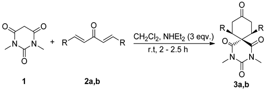
The synthetic pathway to the title compounds is summarized in Table 1. The starting compound, N,N-dimethylbarbituric acid (1) is commercially available, and (1E,4E)-1,5-bis(4-fluorophenyl)penta-1,4-dien-3-one (2a) and (1E,4E)-1,5-bis(4-(trifluoromethyl)phenyl)penta-1,4-dien-3-one (2b) were obtained by the condensation of p-fluorobenzaldehyde and p-trifluoromethylbenzaldehyde with acetone, respectively [28,29]. The reaction of 1 with equimolar amounts of the dienones 2a,b in DCM using NHEt2 as a base afforded the target compounds 7,11-bis(4-fluorophenyl)-2,4-dimethyl-2,4-diazaspiro[5.5]undecane-1,3,5,9-tetraone (3a) and 2,4-dimethyl-7,11-bis(4-(trifluoro- methyl)phenyl)-2,4-diazaspiro[5.5]undecane-1,3,5,9-tetraone (3b) regioselectively in excellent yield. The reaction between the nucleophilic center in 1 and the bielectrophilic centers in 2a,b, in principle, can yield two regioisomers, however, in the present study due to different reactivity of nucleophilic and electrophilic sites, only a single isomer was isolated. The compounds were characterized by 1H-, 13C-NMR and GCMS studies.
Table 1. Cascade [5+1] double Michael addition reaction as key step to form spirocyclic products 3a,b. Molecules 20 08223 i001
Entries aDiarylidene 2a,bR3Yield (%) b
12ap-FPh3a98
22bp-CF3Ph3b94
a Reaction conditions: N,N-dimethylbarbituric acid (1, 2 mmol), diarylideneacetone derivatives 2a,b (2 mmol); b isolated yield.
In the 1H-NMR spectra, the two N-Me protons at positions 8 and 10 of compounds 3a,b are strongly deshielded because these protons are flanked by two adjacent carbonyl groups and both N-Me groups are chemically non-identical because of the anisotropy effect caused by the carbonyl groups present at the C8 and C11 positions (Figure 1).
The 1H-NMR spectrum of the diazaspiro compound 3a shows a doublet of doublets at 2.55 and 2.59 with coupling constants Jgem = 15.40 Hz and Jea = 4.4 Hz, for the equatorial protons at the C2 and C6 positions, while another doublet of doublets appeared at 3.95 and 3.99 with coupling constants Jaa = 13.96 Hz and Jea = 4.40 Hz attributed to the axial protons at the C3 and C5 positions. On the other hand one triplet appeared at 3.64 with coupling constant Jaa/gem = 14.68 Hz for the axial protons present at the C2 and C6 positions (Figure 1).
Figure 1. Structure representation for compound 3a.
Molecules 20 08223 g001
Similarly the 1H-NMR spectrum of the diazaspiro compound 3b shows a doublet of doublets at 2.70 and 2.74 with coupling constants Jgem = 15.40 Hz and Jea = 4.4 Hz and one triplet at 3.56 with coupling constant Jaa/gem = 14.68 Hz, representing for the equatorial and axial protons at the C2 and C6 positions, respectively, while another doublet of doublets appeared at 4.25 and 4.28 with coupling constants Jaa = 13.96 Hz and Jea = 4.40 Hz due to the axial protons at the C3 and C5 positions. The 19F-NMR spectrum shows single signals at −112.36 and −62.65 for the specific F-atom and CF3-group respectively.
Here, the axial protons at positions C2, C3, C4 and C6 are less shielded as compared to the equatorial protons at the C2 and C6 position, because of the proximity of the phenyl ring to the C3 and C4positions, therefore the equatorial protons at positions C2 and C6 appeared at higher field (δ 2.55–2.74 ppm) than the axial protons. The anisotropic effect of C=O on Heq at C2 and C6 is also responsible for the observed diamagnetic shift of these protons.

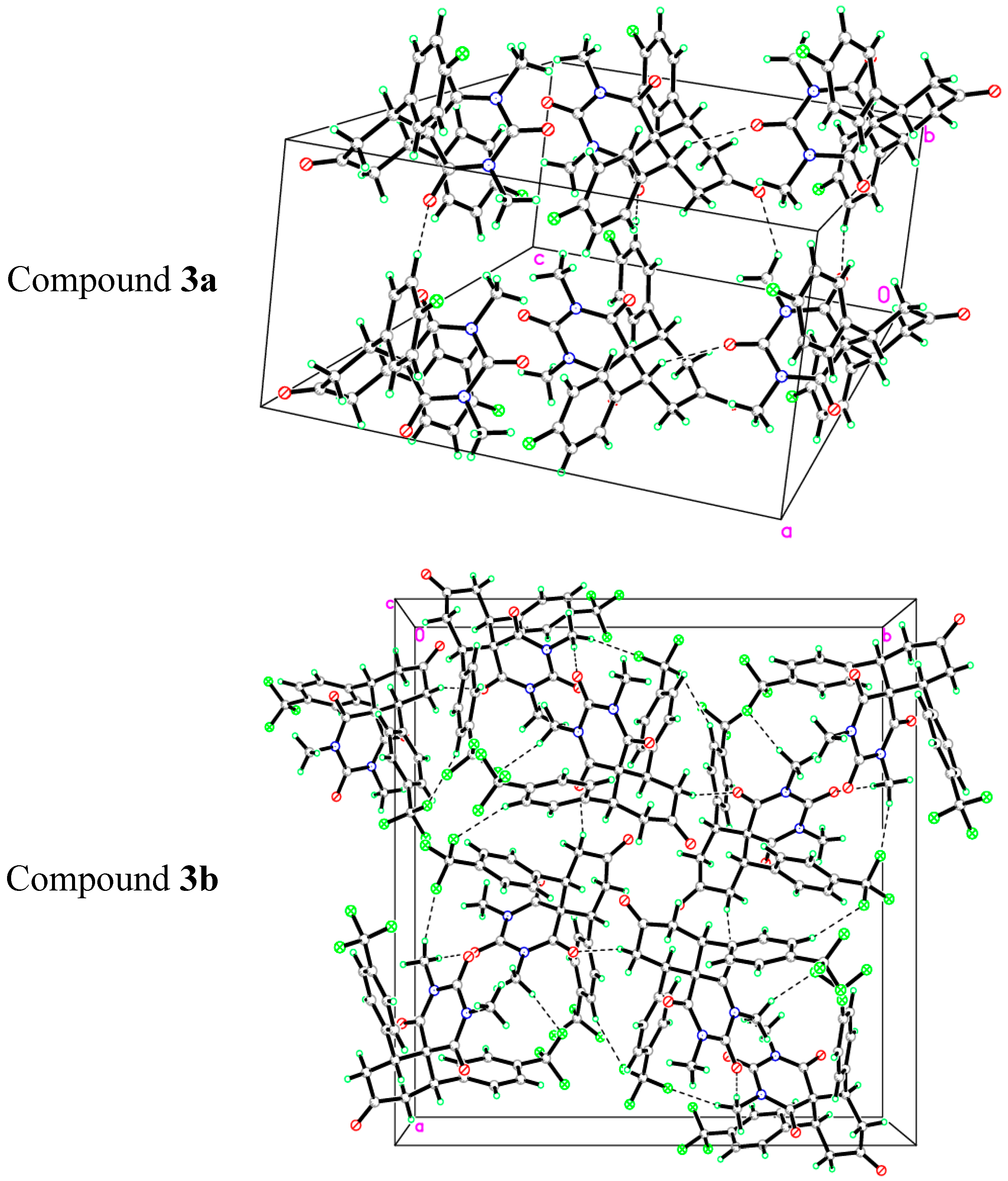
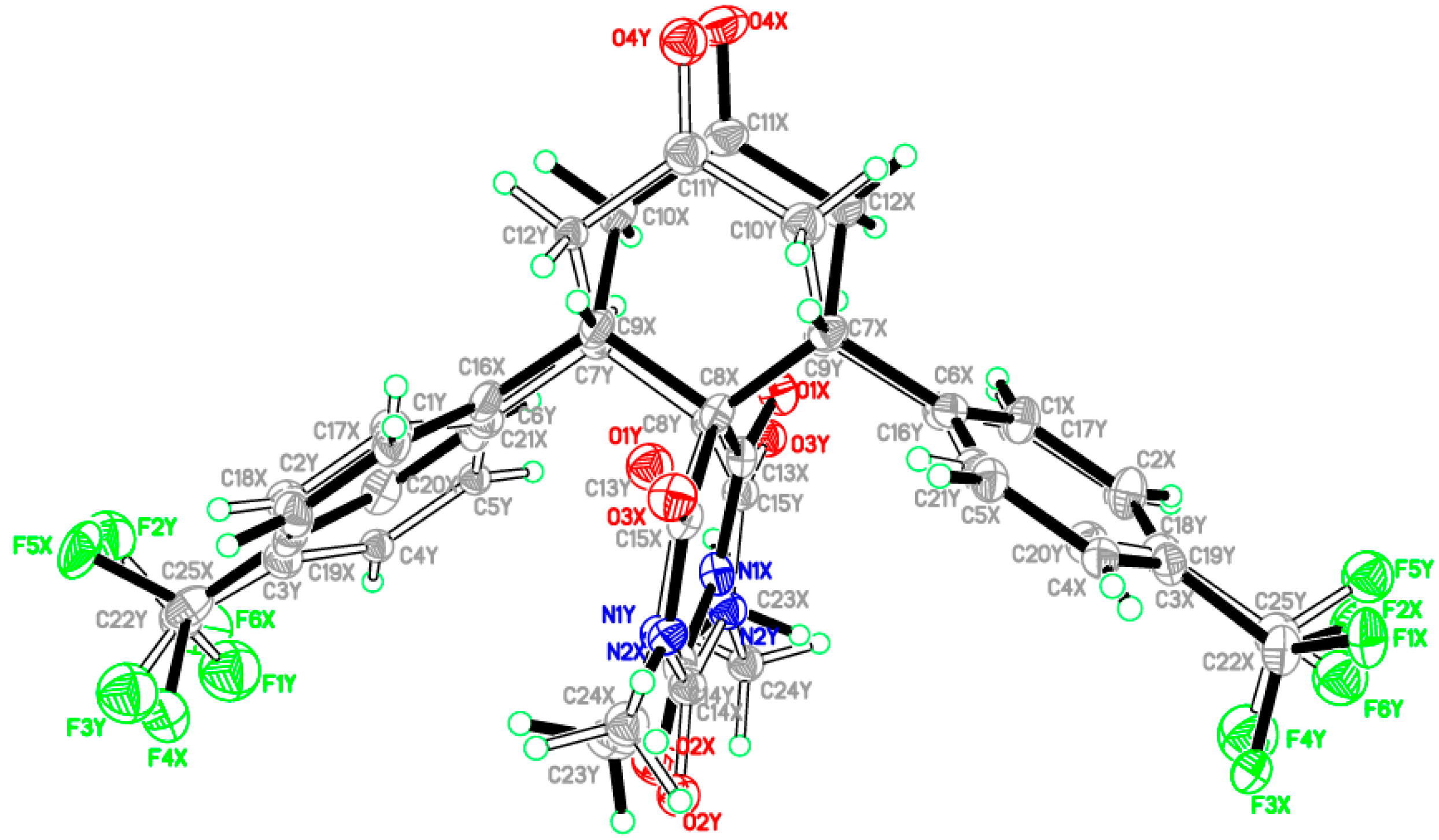
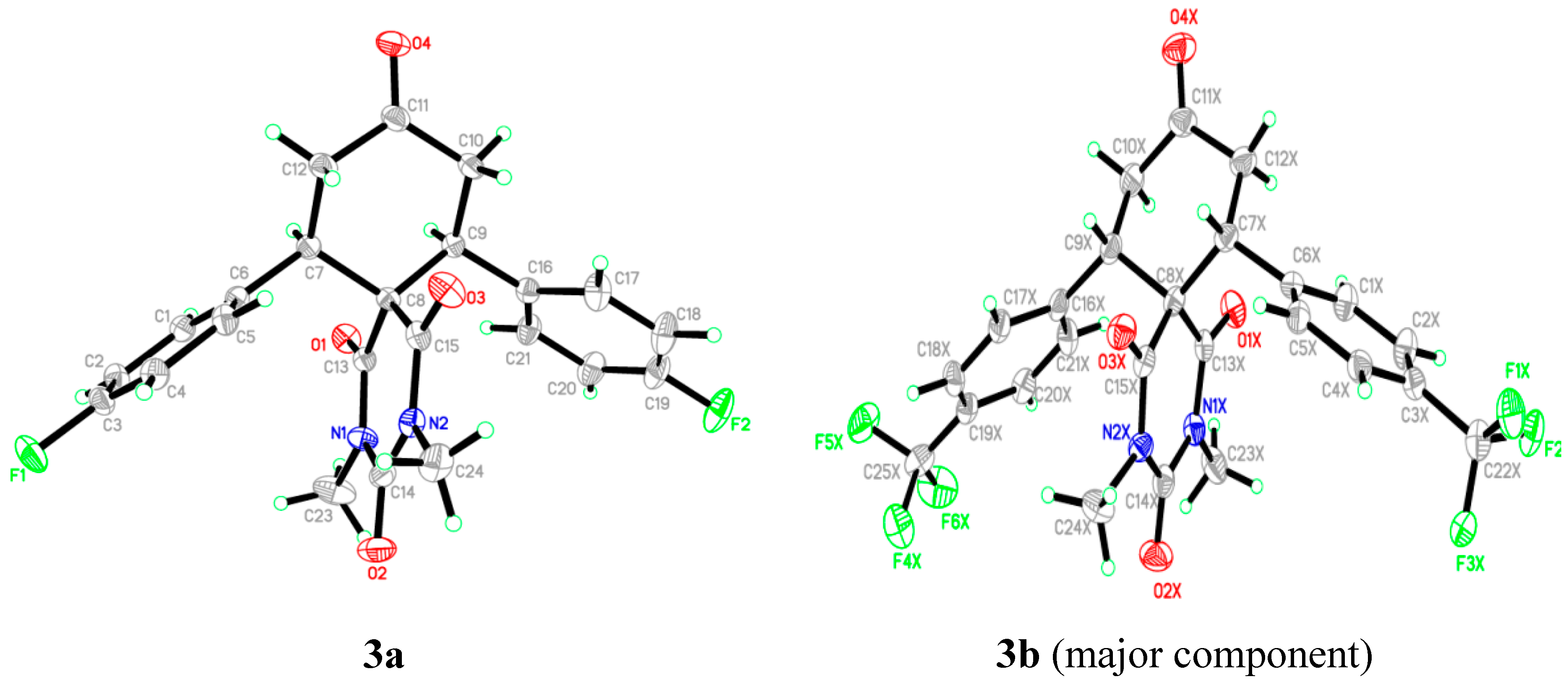
2.2. X-ray Crystal Structures of 3a,b

The compounds 3a,b were obtained as single crystals by slow diffusion at room temperature for 2 days with diethyl ether of pure compounds 3a,b in dichloromethane. Data was collected on a Bruker APEX-II D8 Venture area diffractometer (Bruker AXS GmbH, Karlsruhe, Germany), equipped with graphite monochromatic Mo Kα radiation at 100 (2) K. Cell refinement and data reduction was carried out by Bruker SAINT (Bruker AXS GmbH, Karlsruhe, Germany). SHELXS-97 [33,34] was used to solve these structures. The final refinement was carried out by full-matrix least-squares techniques with anisotropic thermal data for non-hydrogen atoms on 𝐹2. All the hydrogen atoms were placed in calculated positions (Table 2). Similarity and simulation restraints were applied for the disordered atoms. The crystal structures of 3a,b are shown in Figure 2. CCDC-1034099; and CCDC-1034833 contain the supplementary crystallographic data for these compounds. The data can be obtained free of charge from the Cambridge Crystallographic Data Centre via http://www.ccdc.cam.ac.uk/data_request/cif (or from the CCDC, 12 Union Road, Cambridge CB2 1EZ, UK; Fax: +44 1223 336033; E-mail: [email protected]).
Table 2. The crystal and experimental data of compounds 3a and 3b.
PropertiesCompound 3aCompound 3b
Empirical formulaC23H20F2N2O4C25H20F6N2O4
Formula weight426.41526.43
Crystal systemMonoclinicTetragonal
Space groupC2/cP-421c
Unit cell dimensionsa = 33.1745 (16)Å, α = 90.00°
b = 7.7860 (3)Å β = 100.68 (3)°
c = 15.866 (7)Å, γ = 90.00°
a = 22.1813 (10)Å, α = 90.00°
b = 22.1813 (10) Å, β = 90.00°
c = 9.2692 (4)Å, γ = 90.00°
Volume4027.2 (3)Å34560.5 (5)Å3
Density (calculated)1.407Mg/m31.533 Mg/m3
Absorption coefficient0.11 mm−10.14 mm−1
F(000)17762160
Crystal size0.65 × 0.27 × 0.15 mm0.59 × 0.33 × 0.21mm
Theta range for data collection2.5°–30.5°.2.4°–30.5°.
Index ranges−47 ≤ h ≤ 47, −11 ≤ k ≤ 11,
−22 ≤ l ≤ 22
−26 ≤ h ≤ 26, −26 ≤ k ≤ 26,
−10 ≤ l ≤ 11
Reflections collected/ unique112794/ 4917 [R(int) = 0.038]35961 / 4754 [R(int) = 0.024] 82456/4010[R(int) = 0.053]
Completeness to theta = 30.57° 99.7%99.9%
Goodness-of-fit on F21.031.09
Largest diff. peak and hole0.54 and −0.27 e·Å−30.27 and −0.26 e·Å−3
CCDC number10340991034833
The asymmetric unit of compound 3a contains one molecule (Figure 2). In the molecule, the pyrimidine rings are in chair form. Bond lengths and angles are within normal ranges. The cyclohexanone ring (C7–C12) is almost perpendicular to the mean plane of pyrimidine ring (C8/C13/N1/C14/N2/C15) with the dihedral angle of 86.76 (2)°. Also the phenyl rings (C1–C6) and (C16–C21) have dihedral angles of 72.74 (2)° and 58.59 (1)°, respectively, with the pyrimidine ring (C8/C13/N1/C14/N2/C15). In the crystal structure, Figure 3 (copound 3a), the molecules are linked via intermolecular C2-H2A•••O3, C9-H9A•••O2, C24-H24B•••F1 and C2-H24C•••O4 hydrogen bonds (see Supporting Information).
The asymmetric unit of compound 3b contains one molecule (Figure 2). In this, the whole molecule ring system is disordered over two sets of sites with refined site occupancies of 0.799 (1): 0.201 (1), suggesting 180° rotational disorder for the diazaspiro[5.5]undecane-1,3,5,9-tetraone ring system. The pyrimidine rings are in chair form conformation. Bond lengths and angles are within normal ranges. In the molecule (Figure 4), the cyclohexanone ring (C7–C12) is almost perpendicular to the mean plane of pyrimidine ring (C8/C13/N1/C14/N2/C15) with the dihedral angles of 85.04 (2) and 83.21 (4) for major and minor components, respectively. In the crystal structure, Figure 3, the molecules are linked into a 3-dimensional network via intermolecular C4X-H4XA•••F5X, C10X-H10B•••O3X, C23X-H23A•••F4X, C23X-H23B•••O2X and C24X-H24A•••F3X hydrogen bonds (see Supporting Information).
Figure 2. The ORTEP diagram of the final X-ray model of compounds 3a,b with displacement ellipsoids drawn at 30% probability level. The minor disorder component of compound 3b is omitted for clarity.
Molecules 20 08223 g002
Figure 3. View of the molecular packing in compounds 3a,b.
Molecules 20 08223 g003
Figure 4. The ORTEP diagram of the final X-ray model of compound 3b with full molecule disorder.
Molecules 20 08223 g004

2.3. Computational Details

All the quantum chemical calculations of the studied compounds were performed by applying DFT method with the B3LYP functional and 6-311G(d,p) basis set using Gaussian 03 software [35]. The input files were taken from the CIF obtained from our reported X‒ray single crystal measurements. The geometries were optimized by minimizing the energies with respect to all the geometrical parameters without imposing any molecular symmetry constraints. GaussView 4.1 [36] and Chemcraft [37] programs have been used to draw the structure of the optimized geometries and to visualize the HOMO and LUMO pictures. Frequency calculations at the optimized geometries were done to confirm the optimized structures to be an energy minimum. The true energy minimum at the optimized geometry of the studied compounds was confirmed by absence of any imaginary frequency modes. The electronic spectra of the studied compounds were calculated by the TD‒DFT method. The gauge including atomic orbital (GIAO) method was used for NMR calculations. The 1H and the 13C isotropic shielding tensors referenced to the TMS calculations were carried out at the same level of theory. The natural atomic charges were calculated using NBO method as implemented in the Gaussian 03 package [38] at the DFT/B3LYP level. The results of the computed geometrical parameters of compounds 3a,b are reported and discussed with respect to the X-ray data. This is followed by discussion of the experimental and theoretical electronic and spectroscopic properties (IR, UV-Vis and NMR) of the studied compounds. The electronic spectra are also discussed based on the TD-DFT results.

2.3.1. Optimized Molecular Geometry
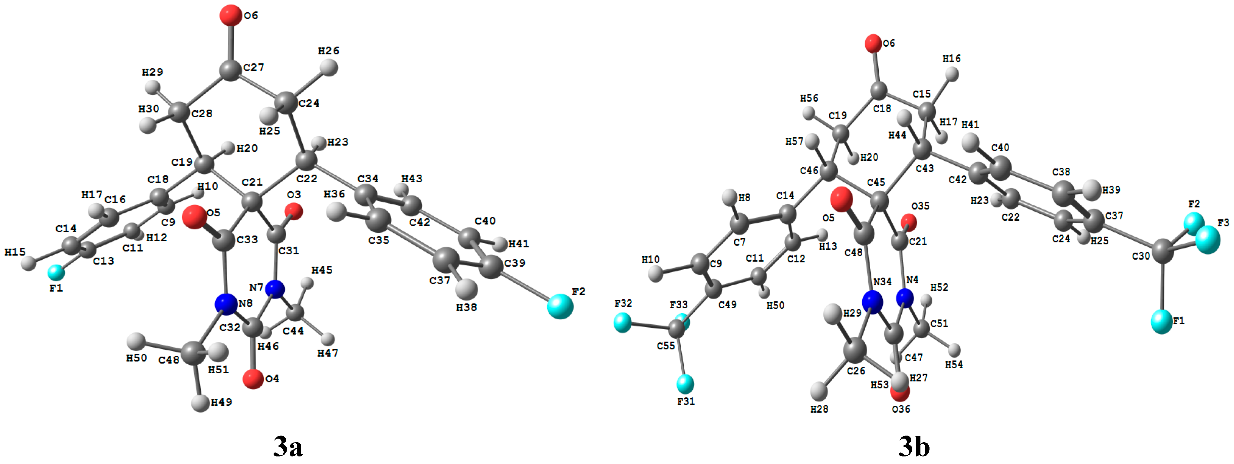
To clarify the electronic and spectroscopic properties of the studied compounds correctly, it is essential to examine the geometry of the studied compounds. A small change in the geometry can potentially cause substantial variations in their electronic and spectroscopic aspects. The optimized geometric structures of 3a,b are shown in Figure 5. The experimental and theoretical geometrical parameters (bond lengths and bond angles) are listed in Table S7 (Supplementary Information). The maximum deviations of the calculated bond length and bond angle values from the experimental data are 0.019Å (C21‒C22) and 1.915° (C18‒C19‒C21), The root mean square deviation (RMSD), the mean absolute deviation (MAD) and the correlation coefficient (R2) values between the experimental and calculated bond lengths are found to be 0.009 Å–0.078 Å–0.9979 and 0.017 Å–0.077 Å–0.9891 for 3a and 3b, respectively. Also, the RMSD, MAD and R2 values between the experimental and calculated bond angles are found to be 0.806°–3.552°–0.9840 and 1.674°–4.741°–0.9562 for for 3a and 3b, respectively. The deviations of the calculated geometric parameters from the experimental results because the latter were obtained at a very low temperature and the molecular rotations were restricted, while the theoretical results were obtained for a single molecule in the gas phase without any intermolecular interactions. The geometric parameters are predicted very well.
Figure 5. The optimized molecular structure of the studied compounds 3a,b.
Molecules 20 08223 g005
Charge distribution plays a vital role in the application of quantum chemical calculations to molecular systems because atomic charges affect many properties such as dipole moment, molecular polarizability and electronic structure of compound [39]. In this regard, the natural atomic charges (NAC) were calculated using NBO method at the DFT B3LYP/6-311G(d,p) level of theory and the results are given in Table 3. The natural charge calculations indicate the electronegative nature of the F, O and N-atoms. The maximum natural charges occur at the O-atoms are in the range −0.5416 to −0.6260 and −0.5377 to −0.6309 for 3a and 3b, respectively. The dipole moments of 3a and 3b are calculated to be 2.3021D and 0.9163D, respectively. Interestingly, 3a, which has F-substituent directly attached to the phenyl rings, is more polar than 3b, which has a CF3 substituent bonded to the phenyl rings. Since the skeleton of the spiro structure which has the electron rich oxygen atoms is common in both compounds so we could predict the polarity difference between the two molecules due to difference of the substituent at phenyl rings. In 3a, the F-atoms present at the para-position of the phenyl groups result in an electron withdrawing effect on these phenyl rings and may thus lead to a molecular system with electron withdrawing units at both ends of the molecule. It appears that this effect is smal in the 3b molecule and so a low dipole moment has been predicted.
Table 3. The natural atomic charges calculated at the B3LYP/6-311G (d,p) method.
3a3b
F1−0.3468H290.2283F1−0.3625H290.2228
F2−0.3468H300.2368F2−0.3629C301.1118
O3−0.6191C310.7398F3−0.3634F31−0.3620
O4−0.5981C320.8567N4−0.4888F32−0.3634
O5−0.6260C330.7240O5−0.6182F33−0.3637
O6−0.5416C34−0.0596O6−0.5377N34−0.4900
N7−0.4909C35−0.1934C7−0.1889O35−0.6309
N8−0.4889H360.2154H80.2176O36−0.5886
C9−0.1784C37−0.2606C9−0.1661C37−0.1514
H100.2162H380.2212H100.2209C38−0.1676
C11−0.2624C390.4327C11−0.1643H390.2222
H120.2221C40−0.2624C12−0.2038C40−0.1892
C130.4327H410.2221H130.2178H410.2177
C14−0.2606C42−0.1784C14−0.0235C42−0.0237
H150.2212H430.2162C15−0.4814C43−0.1975
C16−0.1934C44−0.3530H160.2295H440.2294
H170.2154H450.2226H170.2381C45−0.1819
C18−0.0596H460.2051C180.6236C46−0.1975
C19−0.1937H470.2052C19−0.4813C470.8539
H200.2275C48−0.3535H200.2382C480.7362
C21−0.1845H490.2266C210.7232C49−0.1515
C22−0.1937H500.2021C22−0.2037H500.2192
H230.2275H510.2021H230.2179C51−0.3552
C24−0.4814 C24−0.1625H520.2223
H250.2368 H250.2174H530.2068
H260.2283 C26−0.3536H540.2069
C270.6238 H270.2061C551.1118
C28−0.4814 H280.2068H560.2294

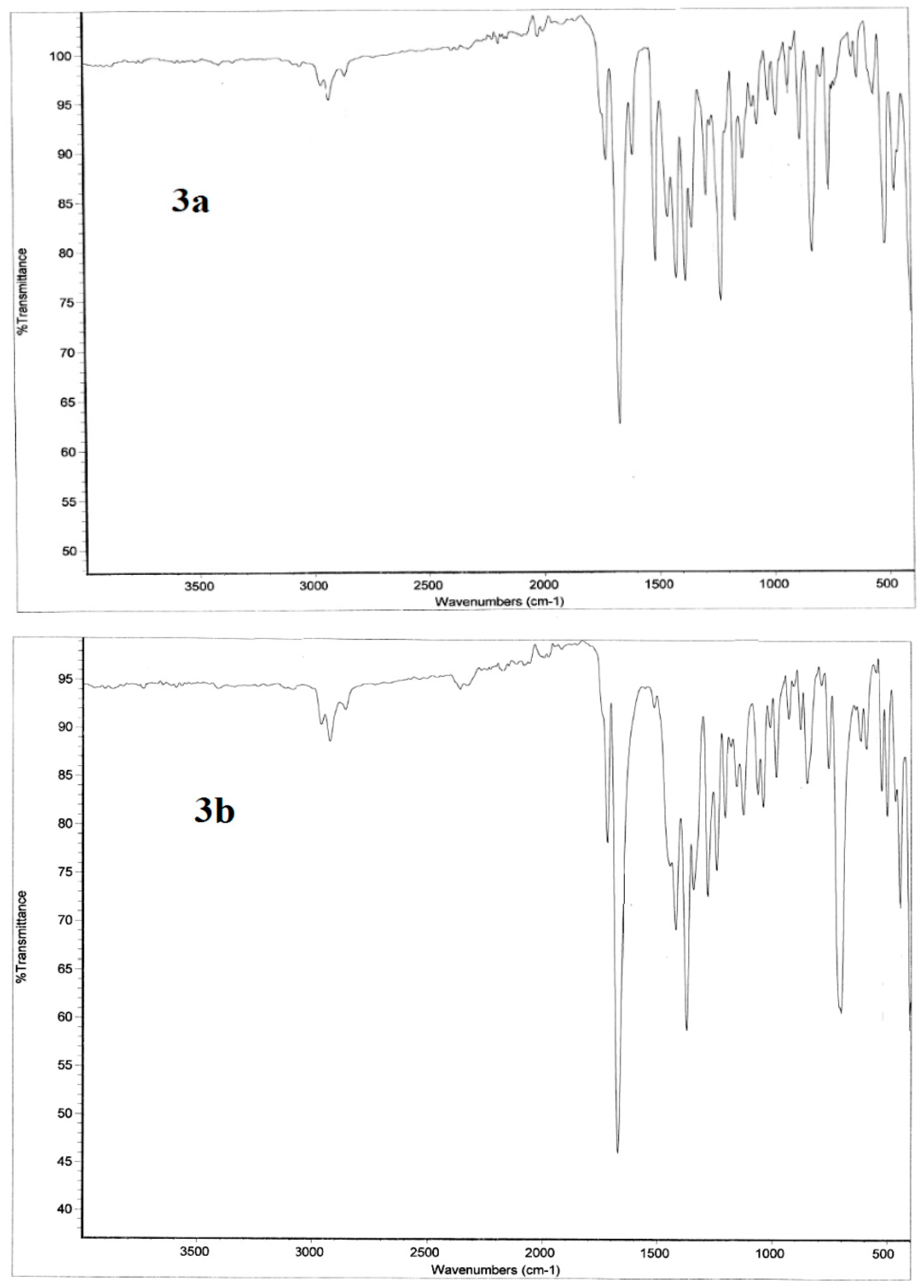
2.3.2. Infrared Vibrational Studies

The studied compounds belong to C1 point group. The compounds 3a,b consist of 51 and 57 atoms, having 147 and 165 normal vibrational modes, respectively. The assignments of the vibrational fundamental modes for the investigated compounds are given in Table 4. The experimental and simulated vibrational spectra are depicted in Figure 6 and Figure 7, respectively. The correlation coefficients between the calculated and experimental IR vibrational frequencies are 0.9994 and 0.9997 for 3a and 3b respectively. We noted that, the IR spectral bands in the region above 1650 cm−1 are almost the same for the two studied compounds. The region below 1650 cm−1 showed significant changes due to the variation of the F-substitution at the phenyl rings. The following are some of the important vibrational motions observed.
The C-H stretching vibrations of aromatic structures generally occur in the region 3100–3000 cm−1 [40] while the saturated hydrocarbon shows C-H stretching absorption bands in the region 3000–2840 cm−1 [41]. Both molecules have two types of C-H bonds: eight aromatic and 12 aliphatic C-H bonds. The aromatic stretching bands of the 3a and 3b molecules are calculated at 3096, 3095, 3080 and 3073 cm−1 (exp. 2956 cm−1) and 3101–3093, 3082–3074 cm−1 (exp. 2956 cm−1), respectively. For the aliphatic C-H bonds, the asymmetric C-H stretching vibrations of 3a and 3b are calculated at 3087, 3084, 3033–3007 cm−1 (exp. 2921 cm−1) and 3085, 3033–3008 cm−1 (exp. 2919 cm−1). The symmetric C-H modes usually appear at lower frequencies and higher intensity of 2970–2952 cm−1 (exp. 2850 cm−1) and 2971–2954 cm−1 (exp. 2851 cm−1) for the 3a and 3b, respectively. Moreover, the four carbonyl vibrations predicted at 1740–1668 cm−1 (exp. 1718–1688 cm−1) and 1743–1661 cm−1 (1715–1669 cm−1) for the 3a and 3b, respectively.
Figure 6. The experimental infrared vibrational spectra of the studied compounds 3a,b.
Molecules 20 08223 g006

Region below 1650 cm−1
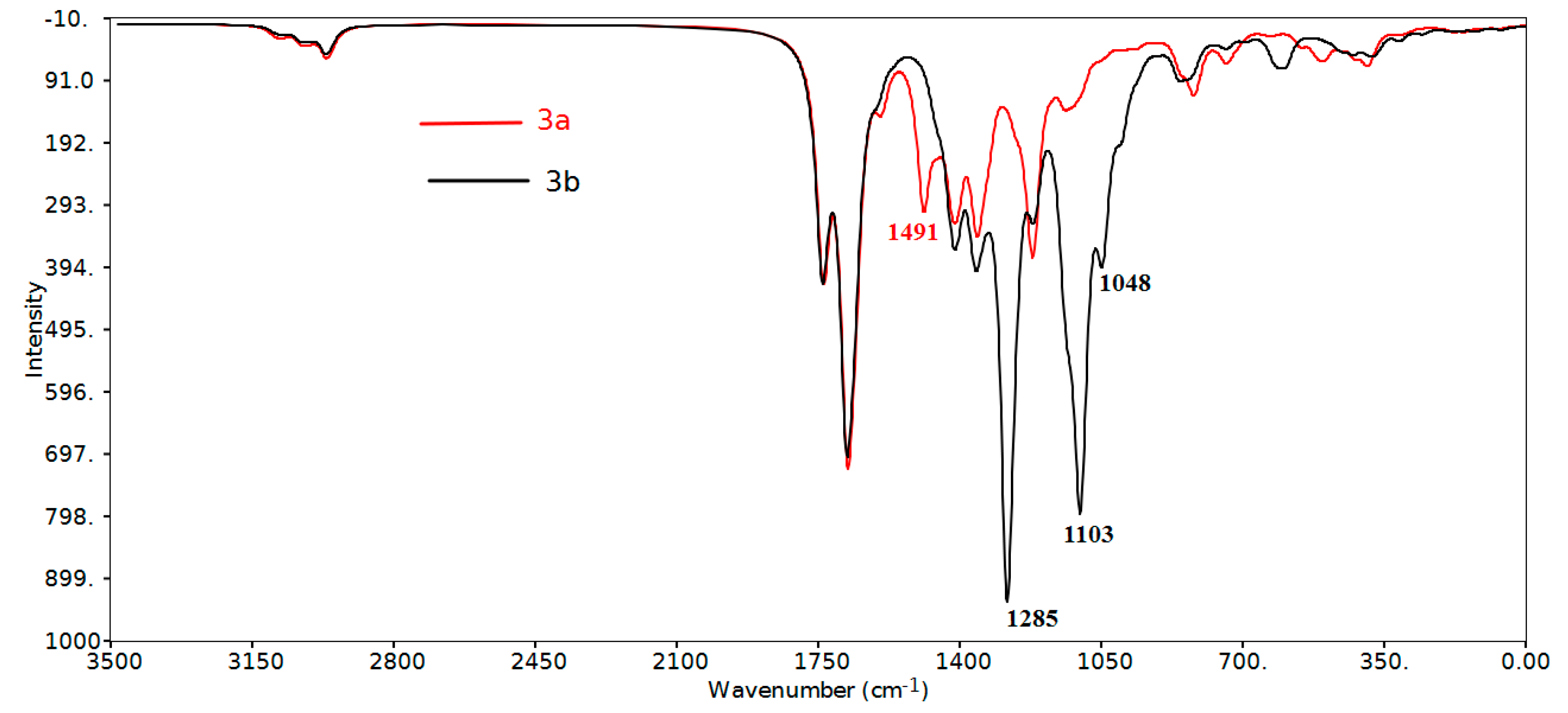
The asymmetric and symmetric bending vibrations of methyl groups normally appear in the 1470–1440 cm−1 and 1390–1370 cm−1 region, respectively [42,43]. These bending modes are predicted theoretically and observed experimentally in their characteristic regions (Table 4). The CH3 umbrella vibrations are calculated at 1422 and 1403–1355 cm−1 (exp. 1378, 1349 cm−1) and 1423 and 1400–1355 cm−1 (exp. 1373, 1343 cm−1) while the CH2 rocking vibrations are calculated at 1416–1404 cm−1 (exp. 1418 cm−1) and 1416–1405 cm−1 (exp. 1419 cm−1) for 3a and 3b, respectively.
Figure 7. The calculated scaled [44] infrared vibrational spectra of the studied compounds.
Molecules 20 08223 g007
Table 4. The calculated and experimental wavenumbers of the studied compounds 3a,b.
Assignment3a3b
CalculatedExperimentalCalculatedExperimental
υ(CH, aromatic)3096, 3095, 3080, 307329563101–3093, 3082–30742956
υ(CHasym, aliphatic)3087, 3084, 3033–300729213085, 3033–30082919
υ(CHsym, aliphatic)2970–295228502971–29542851
υ(C=O)1740–16681718-16881743–16611715–1669
υC=C (aromatic)1595–14911604, 15081604–14971512
δCH asym methyl1460–144914541463–14481448
δCH sym methyl1422, 1403–13551378, 13491423, 1400–13551373, 1343
δCH rocking CH21416–140414181416-14051419
δCH aromatic in planea1492, 1491, 1281–9981508, 1286–9801497, 1300–10201512, 1281–1008
υ(C-CF3) 1285, 12841281
υ(C-F)1223–121912241103, 1048 (asym) 1000 (sym)1038 (asym) 1008 (sym)
δCH aromatic out-of-plane948–806928–784965–823980–847
υ: streching δ: bending; a: mixed with other vibrations.

Region above 1650 cm−1

The aromatic ring C-H in-plane bending vibrations are usually weak and observed in the 1300–998 cm−1 region (exp. 1286–980 cm−1), while the C-H out-of-plane bending vibrations lie in the 965–806 cm−1 region (exp. 980–784 cm−1). We noted a high frequency C-H in-plane bending vibrations at 1497–1492 cm−1 (exp. 1512–1508 cm−1) mixed with the aromatic C=C stretching modes. Theoretically, this band was predicted to have higher intensity in 3a compared to 3b. Usually the aromatic C-F stretching vibrations appeared in the overlapping region with the aromatic C-H in plane vibrations. In the 3a molecule, the υC-F modes are predicted at 1223–1219 cm−1 (exp. 1224 cm−1). We noted the C-F in-plane and out-of-plane bending modes at 423 and 308 cm−1, respectively. Like the methyl group, the CF3 groups of the 3b molecule have two asymmetric and one symmetric stretching modes. The former is calculated at 1103 and 1048 cm−1 (exp. 1038 cm−1) while the latter at 1000 cm−1 (exp. 1008 cm−1). Moreover a strong intense C-C(CF3) stretching vibration at ~1285 cm−1 (exp. 1281 cm−1) has been predicted for compound 3b.

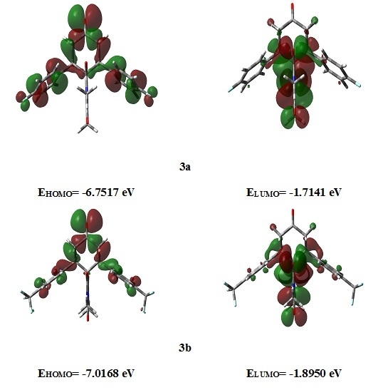
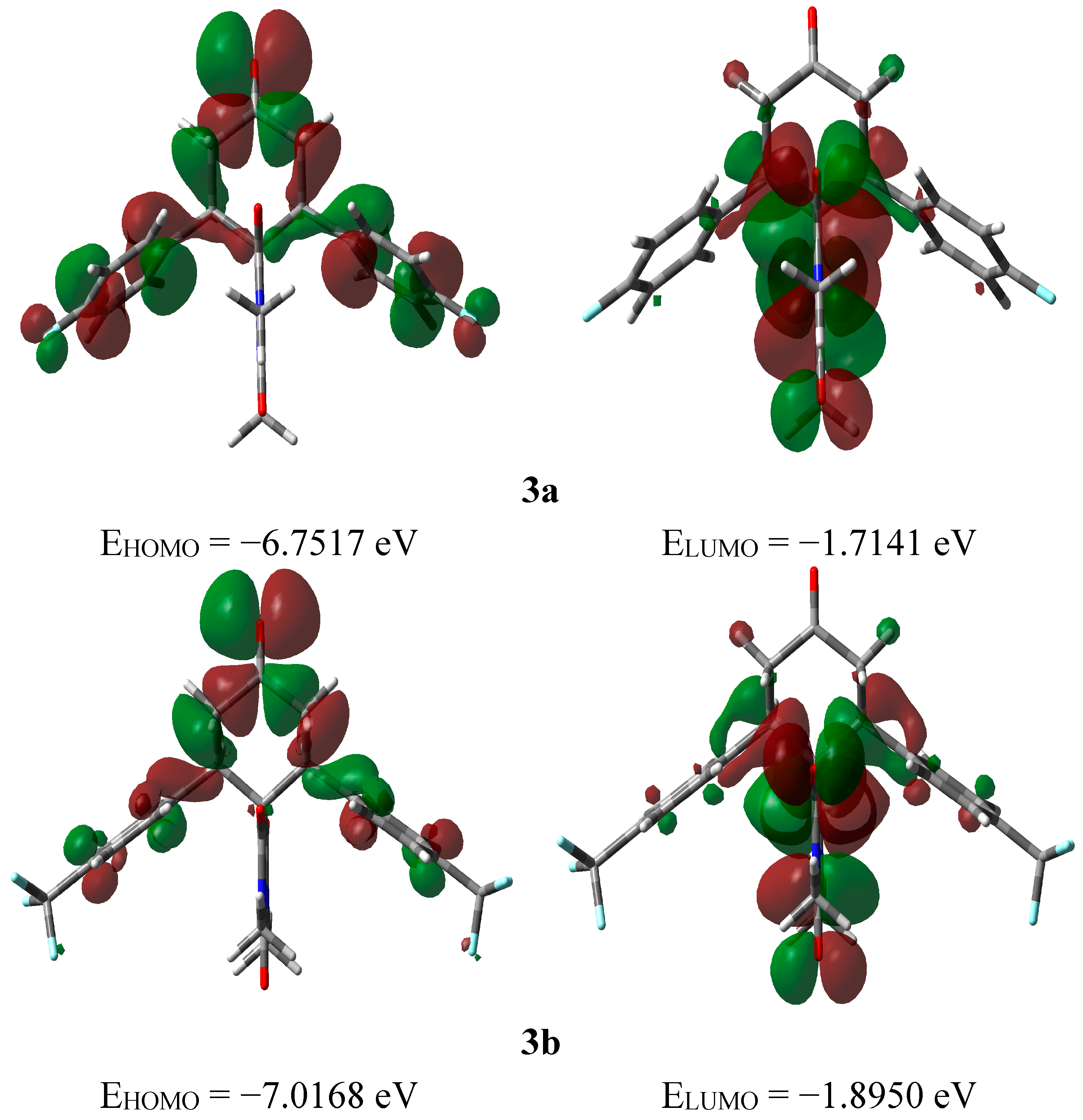
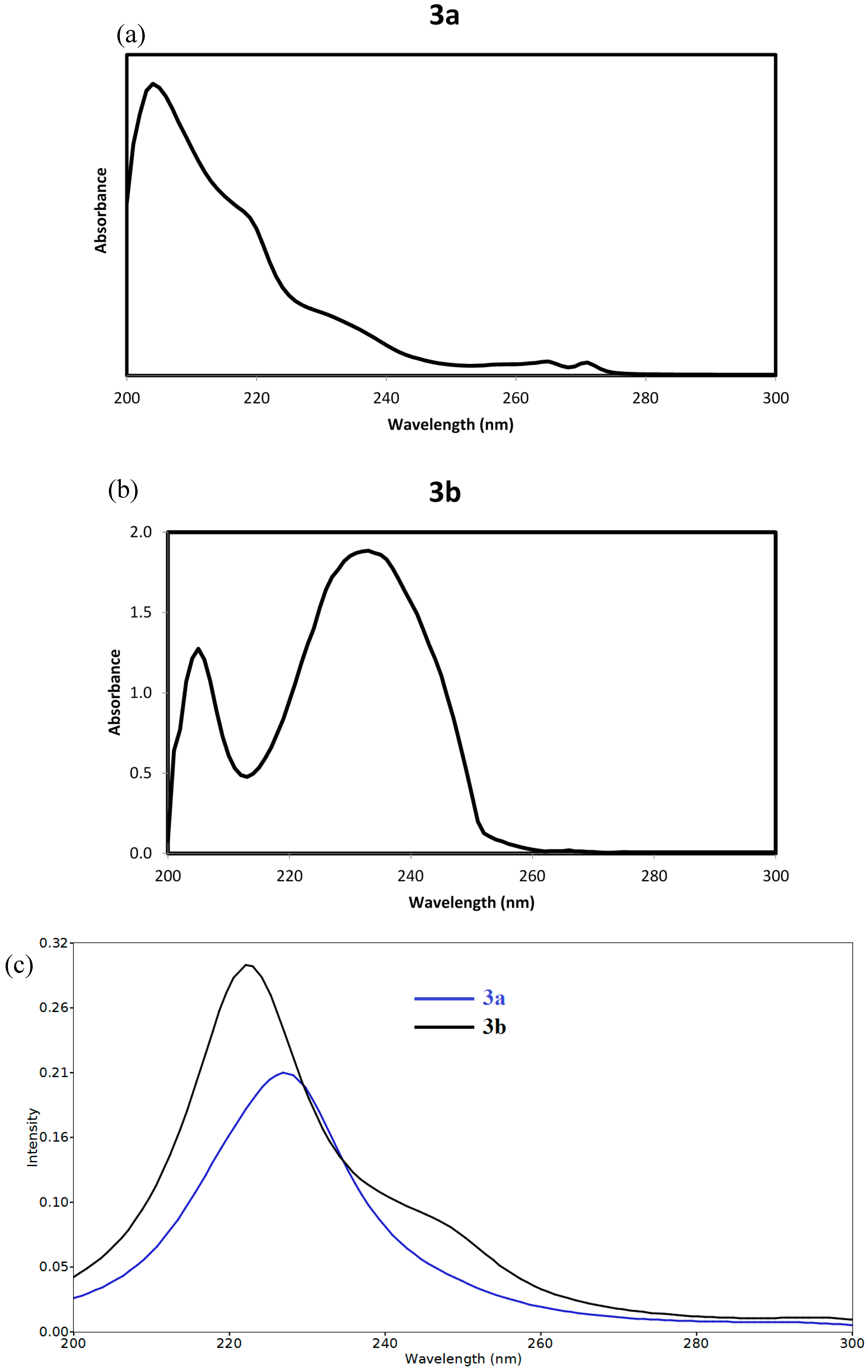
2.3.3. Frontier Molecular Orbitals
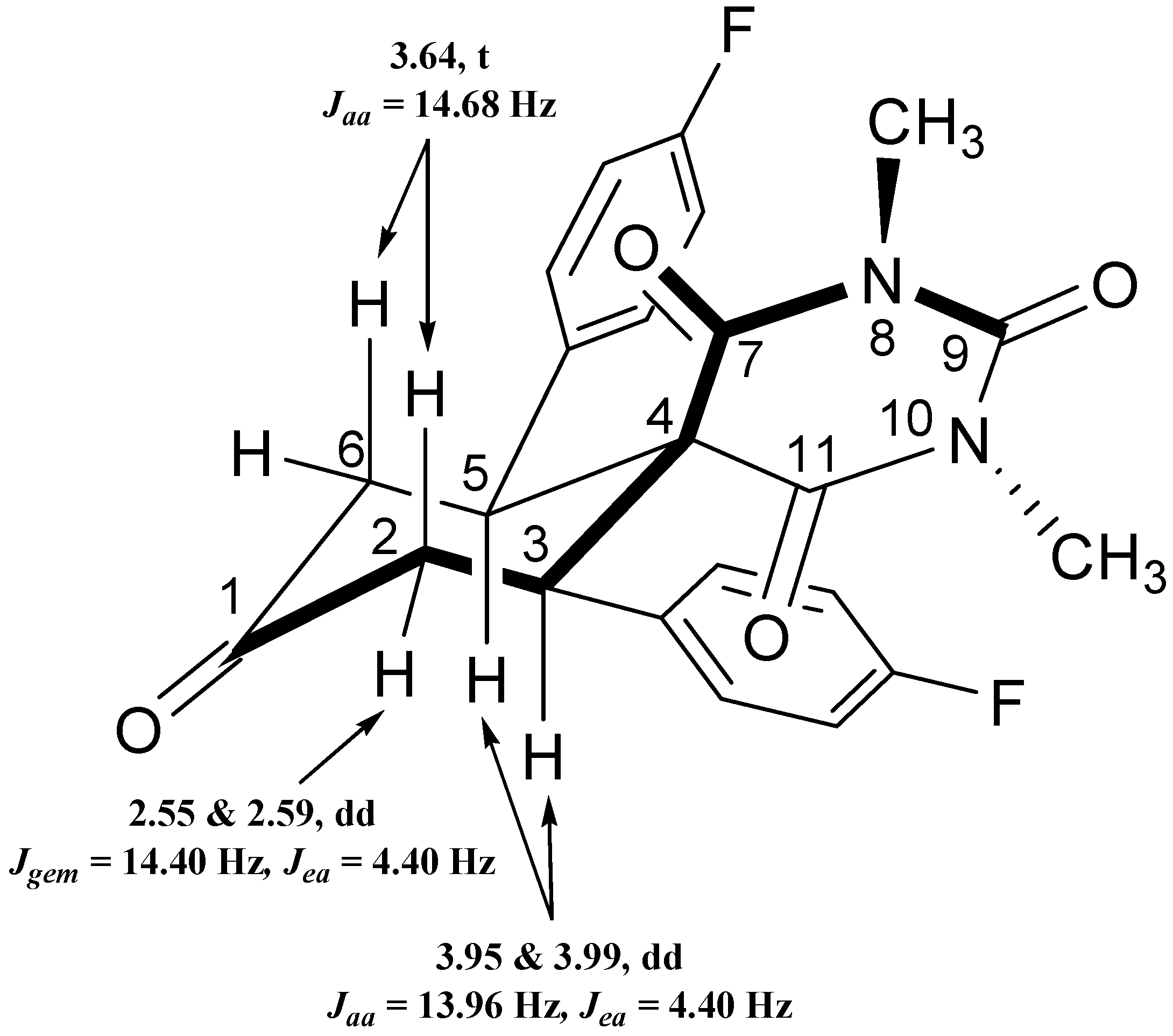
The electron densities of the frontier molecular orbitals (FMOs) were used for predicting the most reactive position in π-electron systems and also explained several types of reactions in conjugated system [45]. The energy values of the lowest unoccupied molecular orbital (ELUMO) and the highest occupied molecular orbital (EHOMO) and their energy gap (ΔE) reflect the chemical reactivity of the molecule. Recently, the energy gap between HOMO and LUMO has been used to prove the bioactivity from intramolecular charge transfer (ICT) [46,47]. The EHOMO, ELUMO and ΔE values of the studied compound were calculated by B3LYP/6‒311G(d,p) method and the HOMO and LUMO pictures are shown in Figure 8. The electron densities of the HOMO and LUMO are mainly localized on the π-system of the studied molecules. It is found that the EHOMO of 3a and 3b are −6.7517 and −7.0168 eV, respectively while the ELUMO values are −1.7141 and −1.8950 eV, respectively. The 3a molecule has lower energy gap (5.0377 eV) than the 3b one (5.1218 eV). The presence of F-atom directly attached to the phenyl ring enhances the ICT interactions. The time-dependant density functional theory (TD-DFT) enabled us to accurately describe the possible electronic transitions of the molecular systems. In this regard, the twenty spin allowed singlet-singlet electronic transitions calculated using TD-DFT results were collected in Table S8 (Supplementary Information). The experimental and calculated electronic spectra of 3a and 3b are shown in Figure 9.
Figure 8. The ground state isodensity surface plots for the frontier molecular orbitals.
Molecules 20 08223 g008
Figure 9. (a) The experimental electronic spectra of the studied compounds 3a; (b) The experimental electronic spectra of the studied compounds 3b; (c) The calculated electronic spectra of the studied compounds using TD-DFT method.
Molecules 20 08223 g009
Compound 3a showed an intense absorption band calculated at 228.3 nm (f = 0.1530) assigned to the H − 1/H→L + 2 and H→L + 3/L + 5 transitions. This band observed experimentally as a shoulder at 233 nm in dichloromethane. Compound 3b showed an intense transition band at 222.1 nm (f = 0.2486) and a shoulder at 248.5 nm (f = 0.0363) due to the H − 1→L + 1 and H→L + 1/L + 5 transitions, respectively. Experimentally, the longest wavelength band was observed at 232nm.

2.3.4. NMR Spectra

The isotropic magnetic shielding (IMS) values calculated using the GIAO approach at the 6-311G(d,p) level were used to predict the 13C and 1H chemical shifts (δcalc) for the studied compounds and the results are correlated with the experimental NMR data (δexp) in CDCl3 solvent. The experimental and theoretical 1H- and 13C-NMR chemical shift values of the studied compounds are given in Table S3 (Supplementary Information). According to these results, the calculated chemical shifts comply with the experimental findings. The correlations between experimental and the calculated chemical shifts are very good (Figures S1 and S2, Supplementary Materials). The correlation coefficients (R2) are calculated to be in the range 0.949–0.989. Generally, the aromatic carbons give signals in overlapped areas of the spectrum with chemical shift values from 100 to 150 ppm [48,49]. Based on the 13C-NMR spectra, the presence of a heteroatom such as fluorine attached to the carbon will shift the peak downfield (higher chemical shift). Since C13 and C39 of 3a are attached to the F-atoms, a high chemical shift is expected for these C-atoms (δcalc = 172.4 ppm). On other hand, the aliphatic C-atoms usually give signals below 50 ppm. The C30 and C55 have high chemical shift values (~134.24 ppm) because each of these C-atoms is attached to three F-atoms, which have high electronegativity.

3. Experimental Section

3.1. General

All glassware was oven-dried before use and the reactions were conducted under an inert atmosphere. The progress of the reaction was monitored by TLC (Silica Gel 60 F–254 thin layer plates, Merck, Schwalbach; Hessen, Germany). The chemicals were purchased from Aldrich (Gillingham, Dorset, UK) and Fluka Chemie GmbH (Buchs, Switzerland), etc., and were used without further purification, unless otherwise stated. Petroleum ether (PE), hexane and ethyl acetate were distilled prior to use especially for column chromatography. All the major solvents were dried by using standard drying techniques mentioned in the literature. Melting points were measured on a Gallenkamp melting point apparatus in open glass capillaries and are uncorrected. IR Spectra were measured as KBr pellets on a 6700 FT-IR Nicolet spectrophotometer (Thermo Fisher Scientific, Madison, WI, USA). The NMR spectra were recorded on a Jeol-400 NMR spectrometer (Peabody, MA, USA). 1H-NMR (400 MHz), and 13C-NMR (100 MHz) were recorded in deuterated chloroform (CDCl3). Chemical shifts (δ) are referred in terms of ppm and J-coupling constants are given in Hz. Mass spectrometric analysis was conducted by using ESI mode on an Agilent Technologies 6410–triple quad LC/MS instrument (Santa Clara, CA, USA). Elemental analysis was carried out on a Perkin Elmer 2400 Elemental Analyzer, CHN mode (Waltham, MA, USA). The electronic spectra of the studied compounds 3a and 3b were measured using a Perkin Elmer Lambda 35, UV/Vis spectrophotometer (Waltham, MA, USA).

3.2. General Procedure of Double Michael Addition Reaction for the Synthesis of Spiro-Compounds 3a,b (GP1)

A solution of N,N-dimethylbarbituric acid (1, 2 mmol) and diarylidene acetone derivatives 2a,b (2 mmol) in dry CH2Cl2 (10 mL) was charged into a 50 mL round bottom flask under inert atmosphere. Et2NH (2.5 mmol) was then added to the reaction mixture which was stirred at room temperature for up to 1.5–2 h, until TLC showed complete consumption of both the reactants. After the completion of reaction, the crude product was directly subjected to column chromatography using 100–200 mesh silica gel and ethyl acetate/n-hexane (2:8, v/v) as an eluent to afford the pure products 3a,b. The solid products were further crystallized from a mixture of CHCl3/n-heptane.

3.2.1. 7,11-bis(4-Fluorophenyl)-2,4-dimethyl-2,4-diazaspiro[5.5]undecane-1,3,5,9-tetraone (3a)

Diarylideneacetone 2a (540 mg, 2 mmol) was reacted with compound 1 (312.1 mg, 2 mmol) according to GP1 to yield a white solid of spiro-product 3a (835 mg, 1.96 mmol, 98%); m.p. 175–177 °C; UV-Vis: 204 nm, 218 nm (sh) and 233 nm (sh) (in Dichloromethane); 1H-NMR (CDCl3) δ: 2.55 and 2.59 (dd, 2H, Jgem = 15.40 Hz, Jea = 4.40 Hz, CH2(e)), 2.88 (s, 3H, -NCH3), 3.02 (s, 3H, –NCH3), 3.64 (t, 2H, Jaa = 14.68 Hz, CH2(a)), 3.95 & 3.99 (dd, 2H, Jaa = 13.96 Hz, Jae = 4.40 Hz, CH), 6.89–6.94 (m, 4H, Ar-H), 7.01–7.04 (m, 4H, Ar-H): 13C-NMR (CDCl3) δ: 27.92, 28.34, 42.93, 49.56, 60.82, 115.73 and 115.95 (d, J2 = 21.4 Hz), 115.10 and 129.18 (d, J3 = 7.65 Hz), 132.78 and 132.81 (d, J4 = 3.06 Hz), 149.33, 161.20 & 163.67 (d, J1 = 147 Hz), [ArC1, C2, C3& C4 are split into doublets due to 19F], 168.74, 170.48, 207.27; IR (KBr, cm−1) νmax = 2920, 1718, 1618, 1507, 1418, 1378, 1223, 1159, 828, 753, 510, 466; [Anal. Calcd. for C23H20F2N2O4: C, 50.39; H, 3.68; N, 5.11; Found: C, 50.51; H, 3.73; N, 5.17]; LC/MS (ESI, m/z): [M+], found 426.21, C23H20F2N2O4 requires 426.14.

3.2.2. 2,4-Dimethyl-7,11-bis (4-(trifluoromethyl)phenyl)-2,4-diazaspiro[5.5]undecane-1,3,5,9-tetraone (3b)

Diarylideneacetone 2b (684 mg, 2 mmol) was reacted with compound 1 (312.1 mg, 2 mmol) according to GP1 to yield the white solid spiro-product 3b (936 mg, 1.88 mmol, 94%); m p. 180–182 °C; UV-Vis: (in ethanol): 204nm and 232nm; 1H-NMR (CDCl3) δ: 2.70 and 2.74 (dd, 2H, Jgem = 15.40 Hz, Jea = 4.40 Hz, CH2(e)), 3.02 (s, 3H, -NCH3), 3.04 (s, 3H, –NCH3), 3.56 (t, 2H, Jaa = 14.68 Hz, CH2(e)), 4.25 and 4.28 (dd, 2H, Jaa = 13.96 Hz, Jae = 4.40 Hz, CH), 6.75–7.14 (m, 4H, Ar-H); 13C-NMR (CDCl3) δ: 28.21, 28.63, 44.17, 45.44, 61.51, 125.48, 125.87, 126.96, 139.84, 149.93, 168.60, 170.91, 205.78; IR (KBr, cm−1) νmax = 2956, 2919, 1715, 1669, 1419, 1373, 1280, 702, 500, 444; [Anal. Calcd. for C23H16F6N2O4: C, 55.43; H, 3.24; N, 5.62; Found: C, 55.29; H, 3.17; N, 5.75]; LC/MS (ESI, m/z): [M+], found 498.19, C23H16F6N2O4 requires 498.10.

4. Conclusions

In conclusion, we have synthesized novel spiro-heterocyclic molecules by room temperature cascade [5+1] double Michael addition reactions using diethylamine as a strong base. The molecular structures of the studied compounds have been optimized using the DFT/B3LYP method and 6-311G(d,p) basis set. The calculated bond distances and bond angles showed good agreement with our reported X-ray crystal structures. The IR vibrational frequencies are calculated and the fundamental bands were assigned and compared with the experimental data. The regions above 1650 cm−1 have common spectral patterns in both compounds but this is not the case for the region below this value. The aromatic υC-F modes are predicted at 1223–1219 cm−1 (exp. 1224 cm−1) while the asymmetric and symmetric C-F stretching modes are calculated at 1,103 and 1048 cm−1 (exp. 1038 cm−1) and 1000 cm−1 (exp. 1008 cm−1), respectively. The natural atomic charges at the different atomic sites calculated using NBO method were used to describe the higher polarity of 3a compared to 3b.The calculated electronic spectra using the TD-DFT showed the most intense absorption band at 228.3 nm (f = 0.1530) for 3a and 222.1 nm (f = 0.2486) for 3b. The GIAO 1H- and 13C-NMR chemical shift values correlated well with the experimental data (R2 = 0.949–0.989). The F-atoms has significant effect on the chemical shifts of the C-atoms attached to them.

Supplementary Materials

Supplementary materials can be accessed at: https://www.mdpi.com/1420-3049/20/05/8223/s1.

Acknowledgments

The authors extend their appreciation to the Deanship of Scientific Research at King Saud University for funding the work through the research group project Number RGP- VPP- 044.

Author Contributions

Mohammad Shahidul Islam: Mainly Carried out the synthesis and characterization part. Abdullah Mohammed Al-Majid: Provided the lab infrastructure. Assem Barakat: Designed the study, grew the X-ray crystals and manuscript preparation. Saied M. Soliman: carried out computional studies. Hazem A. Ghabbour, Ching Kheng Quah and Hoong-Kun Fun: X-ray crystallographic analysis and report writing.

Conflicts of Interest

The authors declare no conflict of interest.

References

  1. Pradhan, R.; Patra, M.; Behera, A.K.; Mishra, B.K.; Behera, R.K. A synthon approach to spiro compounds. Tetrahedron 2006, 62, 779–828. [Google Scholar] [CrossRef]
  2. Karatholuvhu, M.S.; Sinclair, A.; Newton, A.F.; Alcaraz, M.-L.; Stockman, R.A.; Fuchs, P.L. A concise total synthesis of DL-histrionicotoxin. J. Am. Chem. Soc. 2006, 128, 12656–12657. [Google Scholar] [CrossRef]
  3. Taylor, E.C.; Berrier, J.V.; Cocuzza, A.J.; Kobylecki, R.; McCormack, J.J. Pteridines. 41. Synthesis and dihydrofolate reductase inhibitory activity of some cycloalka[g]pteridines. J. Med. Chem. 1977, 20, 1215–1218. [Google Scholar] [CrossRef] [PubMed]
  4. Naydenova, E.; Pencheva, N.; Popova, J.; Stoyanov, N.; Lazarova, M.; Aleksiev, B. Aminoderivatives of cycloalkanespirohydantoins: Synthesis and biological activity. Il Farmaco 2002, 57, 189–194. [Google Scholar] [CrossRef] [PubMed]
  5. Takahashi, K.; Witkop, B.; Brossi, A.; Maleque, M.A.; Albuquerque, E.X. Total synthesis and electrophysiological properties of natural (–)-perhydrohistrionicotoxin, its unnatural (+) antipode and their 2-depentyl analogs. Helv. Chim. Acta 1982, 65, 252–261. [Google Scholar] [CrossRef]
  6. Kim, H.S.; Nagai, Y.; Ono, K.; Begum, K.; Wataya, Y.; Hamada, Y.; Tsuchiya, K.; Masuyama, A.; Nojima, M.; McCullough, K.J. Synthesis and antimalarial activity of novel medium-sized 1,2,4,5-tetraoxacycloalkanes. J. Med. Chem. 2001, 44, 2357–2361. [Google Scholar] [CrossRef] [PubMed]
  7. Reddy, D.M.; Qazi, N.A.; Sawant, S.D.; Bandey, A.H.; Srinivas, J.; Shankar, M.; Singh, S.K.; Verma, M.; Chashoo, G.; Saxena, A.; et al. Design and synthesis of spiro derivatives of parthenin as novel anti-cancer agents. Eur. J. Med. Chem. 2011, 46, 3210–3217. [Google Scholar] [CrossRef] [PubMed]
  8. Srivastav, N.; Mittal, A.; Kumar, A. A novel chiral auxiliary from chiral spiranes. cis, cis-(+)-and (–)-spiro [4.4] nonane-1, 6-diol as chiral modifier in lithium aluminium hydride reduction of phenyl alkyl ketones. J. Chem. Soc. Chem. Commun. 1992, 493–494. [Google Scholar]
  9. Longeon, A.; Guyot, M.; Vacelet, J. Araplysillins-I and -II: Biologically active dibromotyrosine derivatives from the sponge Psammaplysilla Arabica. Experentia 1990, 46, 548–550. [Google Scholar] [CrossRef]
  10. Osman, A.N.; Kandeel, M.M.; Said, M.M.; Ahmed, E.M. Synthesis and anticonvulsant activity of some spiro compound derived from barbituric and thiobarbituric acids. Indian. J. Chem. Sect. B Org. Chem. Incl. Med. Chem. 1996, 35, 1073–1078. [Google Scholar]
  11. Kesharwani, S.; Sahu, N.K.; Kohli, D.V. Synthesis and biological evaluation of some new spiro derivatives of barbituric acid. Pharm. Chem. J. 2009, 43, 315–319. [Google Scholar] [CrossRef]
  12. Behera, R.K.; Behera, A.K.; Pradhan, R.; Pati, A.; Patra, M. Studies on spiroheterocycles Part-II: Heterocyclisation of the spiro compounds containing cyclohexanone and thiobarbituric acid with different bidentate nucleophilic reagents. Synth. Commun. 2006, 36, 3729–3742. [Google Scholar] [CrossRef]
  13. Behera, R.K.; Behera, A.K.; Pradhan, R.; Pati, A.; Patra, M. Phosphorus, Studies on spiroheterocycles part-III: Synthesis of diazaspiroundecanetetraone derivatives containing biological active heterocycles. Sulfur Silicon Relat. Elem. 2009, 184, 753–765. [Google Scholar] [CrossRef]
  14. Goel, B.; Sharma, S.; Bajaj, K.; Bansal, D.; Singh, T.; Malik, N.; Lata, S.; Tyagi, C.; Panwar, H.; Agarwal, A.; Kumar, A. Synthesis and CNS depressant of newer spirobarbiturates. Indian J. Pharm. Sci. 2005, 67, 194–199. [Google Scholar]
  15. Tamejiro, H.; Yamamoto, H. Organofluorine Compounds: Chemistry and Applications, 1st ed.; Springer: Heidelberg, Germany, 2000; pp. 1–19. [Google Scholar]
  16. Park, B.K.; Kitteringham, N.L.; O’Neill, P.M. Metabolism of fluorine-containing drugs. Annu. Rev. Pharmacol. Toxicol. 2001, 41, 443–470. [Google Scholar] [CrossRef] [PubMed]
  17. Kirsch, P. Modern Fluoroorganic Chemistry; Wiley-VHC: Weinheim, Germany, 2004; pp. 1–19. [Google Scholar]
  18. Nyffeler, P.T.; Duron, S.G.; Burkart, M.D.; Vincent, S.P.; Wong, C.-H. Selectfluor: Mechanistic Insight and Applications. Angew. Chem. Int. Ed. 2004, 44, 192–212. [Google Scholar] [CrossRef]
  19. Kirk, J.L. Fluorine in medicinal chemistry: Recent therapeutic application of fluorinated small molecules. J. Fluor. Chem. 2006, 127, 1013–1029. [Google Scholar] [CrossRef]
  20. Dinoiu, V. Chemical fluorination of organic compounds. Rev. Roum. Chim. 2007, 52, 219–234. [Google Scholar]
  21. Hagmann, W.K. The many roles for fluorine in medicinal chemistry. J. Med. Chem. 2008, 51, 4359–4369. [Google Scholar] [CrossRef] [PubMed]
  22. Filler, R.; Saha, R. Fluorine in medicinal chemistry: A century of progress and a 60 year retrospective of selected highlights. Future Med. Chem. 2009, 5, 777–791. [Google Scholar] [CrossRef]
  23. Janin, Y.L. Preparation and Chemistry of 3/5-Halogenopyrazoles. Chem. Rev. 2012, 112, 3924–3958. [Google Scholar] [CrossRef] [PubMed]
  24. Liang, T.; Neumann, C.N.; Ritter, T. Introduction of Fluorine and Fluorine-Containing Functional Groups. Angew. Chem. Int. Ed. 2013, 52, 8214–8264. [Google Scholar] [CrossRef]
  25. Michael, A.J. Ueber die Addition von Natriumacetessig-und Natriummalonsäureäthern zu den Aethern ungesättigter Säuren. Prakt. Chem. 1887, 35, 349–356. [Google Scholar] [CrossRef]
  26. Jung, M.E. Comprehensive Organic Synthesis; Trost, B.M., Fleming, I., Semmelhack, M.F., Eds.; Pergamon: Oxford, NY, USA, 1991; Volume 4, pp. 1–68. [Google Scholar]
  27. Perlmutter, P. Conjugate Addition Reactions in Organic Synthesis; Elsevier Science: New York, NY, USA, 1992. [Google Scholar]
  28. Barakat, A.; Islam, M.S.; Al Majid, A.M.A.; Al-Othman, Z.A. Highly Enantioselective Friedel-Crafts Alkylation of Indoles with α,β-Unsaturated Ketones with simple Cu(II)-Oxazoline-Imidazoline Catalysts. Tetrahedron 2013, 69, 5185–5192. [Google Scholar] [CrossRef]
  29. Islam, M.S.; Al Majid, A.M.A.; Al-Othman, Z.A.; Barakat, A. Highly enantioselective Friedel-Crafts alkylation of indole with electron deficient trans-b-nitroalkenes using Zn(II)-oxazoline-imidazoline catalysts. Tetrahedron-Asymmetry 2014, 25, 245–251. [Google Scholar] [CrossRef]
  30. Barakat, A.; Al-Najjar, H.J.; Al-Majid, A.M.; Soliman, S.M.; Mabkhot, Y.N.; Al-Agamy, M.H.M.; Ghabbour, H.A.; Fun, H-K. Synthesis, molecular structure investigations and antimicrobial activity of 2-thioxothiazolidin-4-one derivatives. J. Mol. Struct. 2015, 1081, 519–529. [Google Scholar] [CrossRef]
  31. Barakat, A.; Al Majid, A.M.A.; Al-Najjar, H.J.; Mabkhot, Y.N.; Sumaira, J.; Sammer, Y.; Choudhary, M.I. Zwitterionic pyrimidinium adducts as antioxidants with therapeutic potential as nitric oxide scavenger. Euro. J. Med. Chem. 2014, 84, 146–154. [Google Scholar] [CrossRef]
  32. Islam, M.S.; Barakat, A.; Al Majid, A.M.A.; Ghabbour, H.A.; Fun, H.-K.; Siddiqui, M.R. Stereoselective synthesis of diazaspiro[5.5]undecane derivatives via base promoted [5+1] double Michael addition of N,N-dimethylbarbituric acid to diaryliedeneacetones. Arab. J. Chem 2015, in press. [Google Scholar] [CrossRef]
  33. Sheldrick, G.M. A short history of SHELX. Acta Crystallogr. 2008, 112, 112–122. [Google Scholar] [CrossRef]
  34. Spek, A.L. Structure validation in chemical crystallography. Acta Crystallogr. 2009, 148, 148–155. [Google Scholar]
  35. Frisch, M.J.; Trucks, G.W.; Schlegel, H.B.; Scuseria, G.E.; Robb, M.A.; Cheeseman, J.R. Gaussian-03; Revision C.01; Gaussian, Inc.: Wallingford, CT, USA, 2004. [Google Scholar]
  36. Dennington, R., II; Keith, T.; Millam, J. Gauss View; Version 4.1.2; Semichem Inc.: Shawnee Mission, KS, USA, 2007. [Google Scholar]
  37. Zhurko, G.A.; Zhurko, D.A. ChemCraft: Tool for Treatment of Chemical Data, Late version build 08 (freeware). 2005. Available online: http://www.chemcraftprog.com (accessed on 5 May 2015).
  38. Glendening, E.D.; Reed, A.E.; Carpenter, J.E.; Weinhold, F. NBO; Version 3.1; TCI, University of Wisconsin: Madison, WI, USA, 1998. [Google Scholar]
  39. Sidir, I.; Sidir, Y.G.; Kumalar, M.; Tasal, E. Ab initio Hartree-Fock and density functional theory investigations on the conformational stability, molecular structure and vibrational spectra of 7-acetoxy-6-(2,3-dibromopropyl)-4,8-dimethyl -coumarin molecule. J. Mol. Struct. 2010, 964, 134–151. [Google Scholar] [CrossRef]
  40. Bellamy, L.J. The Infrared Spectra of Complex Molecules; John Wiley and Sons Inc.: New York, NY, USA, 1975. [Google Scholar]
  41. Silverstein, R.M.; Basseler, G.C.; Morill, C. Spectroscopic Identification of Organic Compounds; Wiley: New York, NY, USA, 1981. [Google Scholar]
  42. Smith, B. Infrared Spectral Interpretation, A Systematic Approach; CRC Press: Washington, DC, USA, 1999. [Google Scholar]
  43. Dollish, F.R.; Fateley, W.G.; Bentley, F.F. Characteristic Raman Frequencies of Organic Compounds; John Wiley & Sons: New York, NY, USA, 1997. [Google Scholar]
  44. Chamundeeswari, S.P.V.; Samuel, E.R.J.J.; Sundaraganesan, N. Theoretical and experimental studies on 2-(2-methyl-5-nitro-1-imidazolyl)ethanol. Eur. J. Chem. 2011, 2, 136–145. [Google Scholar]
  45. Fukui, K.; Yonezawa, T.; Shingu, H.J. A molecular-orbital theory of reactivity in aromatic hydrocarbons. J. Chem. Phys. 1952, 20, 722–725. [Google Scholar] [CrossRef]
  46. Padmaja, L.; Ravikumar, C.; Sajan, D.; Joe, I.H.; Jayakumar, V.S.; Pettit, G.R.; Neilsen, F.O. Density functional study on the structural conformations and intramolecular charge transfer from the vibrational spectra of the anticancer drug combretastatin–A2. J. Raman Spectrosc. 2009, 40, 419–428. [Google Scholar] [CrossRef]
  47. Ravikumar, C.; Joe, I.H.; Jayakumar, V.S. Charge transfer interactions and nonlinear optical properties of push-pull chromophore benzaldehyde phenylhydrazone: A vibrational approach. Chem. Phys. Lett. 2008, 460, 552–558. [Google Scholar] [CrossRef]
  48. Göğer, N.G.; Parlatan, H.K.; Basan, H.; Berkkan, A.; Özden, T. Quantitative determination of azathioprine in tablets by H NMR spectroscopy. J. Pharm. Biomed. Anal. 1999, 21, 685–689. [Google Scholar] [CrossRef] [PubMed]
  49. Barakat, A.; Al-Najjar, H.J.; Al-Majid, A.M.; Soliman, S.M.; Mabkhot, Y.N.; Rafi Shaik, M.; Ghabbour, H.A.; Fun, H.-K. Synthesis, NMR, FT-IR, X-ray structural characterization, DFT analysis and Isomerism aspects of 5-(2,6-dichlorobenzylidene)pyrimidine-2,4,6(1H,3H,5H)-trione. Spectrochim. Acta Part A 2015, 147, 107–116. [Google Scholar] [CrossRef]
  • Sample Availability: Samples 3a,b are available from the authors.

