Review

Antioxidants of Edible Mushrooms

1
Department for Chemistry and Biochemistry, Faculty of Agriculture, University of Belgrade, Nemanjina 6, Belgrade 11080, Serbia
2
Department for Industrial Microbiology, Faculty of Agriculture, University of Belgrade, Nemanjina 6, Belgrade 11080, Serbia
3
Institute of Chemistry, Technology and Metallurgy, University of Belgrade, Njegoseva 12, Belgrade 11001, Serbia
4
Institute of Chemical Engineering, Faculty of Technology and Metallurgy, University of Belgrade, Karnegijeva 4, Belgrade 11060, Serbia
5
Faculty of Chemistry, University of Belgrade, Studentski trg 12–16, Belgrade 11000, Serbia
6
Plant Research International, Wageningen University and Research Centre, Droevendaalsesteeg 1, Wageningen 6700 AA, The Netherlands
*
Author to whom correspondence should be addressed.
Molecules 2015, 20(10), 19489-19525; https://doi.org/10.3390/molecules201019489
Submission received: 4 September 2015 / Revised: 19 October 2015 / Accepted: 21 October 2015 / Published: 27 October 2015
(This article belongs to the Special Issue Antioxidants—A Risk-Benefit Analysis for Health)

Abstract

:
Oxidative stress caused by an imbalanced metabolism and an excess of reactive oxygen species (ROS) lead to a range of health disorders in humans. Our endogenous antioxidant defense mechanisms and our dietary intake of antioxidants potentially regulate our oxidative homeostasis. Numerous synthetic antioxidants can effectively improve defense mechanisms, but because of their adverse toxic effects under certain conditions, preference is given to natural compounds. Consequently, the requirements for natural, alternative sources of antioxidant foods identified in edible mushrooms, as well as the mechanistic action involved in their antioxidant properties, have increased rapidly. Chemical composition and antioxidant potential of mushrooms have been intensively studied. Edible mushrooms might be used directly in enhancement of antioxidant defenses through dietary supplementation to reduce the level of oxidative stress. Wild or cultivated, they have been related to significant antioxidant properties due to their bioactive compounds, such as polyphenols, polysaccharides, vitamins, carotenoids and minerals. Antioxidant and health benefits, observed in edible mushrooms, seem an additional reason for their traditional use as a popular delicacy food. This review discusses the consumption of edible mushrooms as a powerful instrument in maintaining health, longevity and life quality.

1. Introduction

Inadequate nutrition due to modern lifestyle and the increase of average longevity are the two key reasons for the growing incidence of disease all over the world. Oxidative stress caused by an imbalanced metabolism and an excess of reactive oxygen species (ROS) end into a range of disorders i.e., metabolic disease, heart disease, severe neural disorders such as Alzheimer’s and Parkinson’s, premature aging and some cancers. ROS are not only generated internally, in the organism, but also through various external sources like ultraviolet light, ionizing radiation, chemotherapeutics, inflammatory cytokines, and environmental toxins. Inhaling toxic chemicals from the environment has become unavoidable in modern civilization.
Apart of the endogenous antioxidant defense mechanisms of an organism, its dietary intake is another very important source of antioxidants and may contribute to oxidative homeostasis. Antioxidant supplements or antioxidant-containing foods may be used to help the organism to reduce oxidative damage as well to protect food quality by preventing oxidative deterioration. Just as in food production and packing, antioxidants are extensively used in health care, anti-aging and cosmetics. The growing preference for healthy food, cosmetics, and health and wellness products is influencing the growth of the antioxidants market. In addition, increasing demand for nutritional products and cosmetics obtained from natural sources is also driving the natural antioxidants market [1]. Population growth and the increasing healthcare spending levels have led to a consistent increase in the demand for antioxidant products. The global market for antioxidants is growing fast and is expected to more than double from $103.6 million in 2011 to reach $246.1 million in 2018 [2,3].
The antioxidants market by product type is segmented into natural antioxidants and synthetic antioxidants. Natural antioxidants are categorized into plant and fungal extracts, spices (rosemary, thyme, marjoram, oregano, sage, basil, pepper, clove, cinnamon, and nutmeg), flavonoids, ubiquinol (fully reduced form of coenzyme Q10), glutathione, zink (Zn), selenium (Se), vitamin A (including carotenoids), vitamin C and vitamin E (including tocopherols and tocotrienols) [4].
Synthetic phenolic antioxidants include butylated hydroxyanisole (BHA), butylated hydroxytoluene (BHT) and others e.g., propyl gallate, tert-butylhydroquinone (TBHQ), ethoxyquin (EQ), that all effectively inhibit oxidation [5]. However, some synthetic antioxidants may cause adverse toxic effects under certain conditions [6,7]. BHA, which is very often used as an additive in food industry, can have negative effects on the regulation of the activity of mitogen-activated protein kinase (MAPK) depending on the dosage [7,8]. Several synthetic antioxidants are authorized for use as feed additives in the European Union [9].
In recent years, the restriction on the use of synthetic antioxidants, such as BHA and BHT, has caused a rapidly increased interest towards natural antioxidant substances [6,7]. Requirements for natural alternative sources of antioxidant foods and ingredients derive primarily from consumers.
In recent years edible mushrooms have attracted attention as a commercial source of antioxidants [6,7,10]. They might be used directly in enhancement of antioxidant defenses through dietary supplementation to reduce the level of oxidative stress. There is a wealth of evidence to support the effectiveness of such a strategy in vitro.
Edible mushrooms include many fungal species that are either harvested wild or cultivated. Cultivated mushrooms as well as the more common wild mushrooms are often available in markets; porcini (Boletus edulis) or other ectomycorhyzal mushrooms may be collected on a smaller scale by private gatherers [11,12].
The true nutritive value of mushrooms has rapidly become known and recognized not only by mushroom researchers and farmers but also by the general consumers [13]. In addition to their good flavor, mushrooms possess favorable chemical composition with high amounts of functional proteins, low total fat level, and the high proportion of polyunsaturated fatty acids (PUFA), making them well suited for low calorie diets. Edible mushrooms provide a nutritionally significant content of vitamins (B1, B2, B12, C, D, and E) [14,15,16]. Moreover, mushrooms have a low glycemic index, and high mannitol, which is especially beneficial for diabetics. Mushrooms have very low sodium (Na) concentration, which is beneficial for hypertensive patients and a high content of potassium (K) and phosphorus (P), which is an important orthomolecular aspect [13]. In Asia mushrooms are used as important source of home remedies against various diseases elicited by oxidative stress [10].
There is no easy distinction between edible and medicinal mushrooms because many of the common edible species have therapeutic properties [12,17]. Besides antioxidant properties, mushrooms have received considerable attention for their biological activities, such as antitumor, antiviral, anticomplementary, anticoagulant, antidiabetic, hypolipidemic, hepatoprotective, immunostimulant and immunological activities, which made them suited for use in food, cosmetics, biomedicine, agriculture, environmental protection and wastewater management [7,18,19].
To date, a numerous edible wild mushroom species, growing in various ecological conditions, are known [20]. The common mushroom species produced in suitable ecological conditions are: Agaricus spp., Lentinula edodes (shiitake), Pleurotus spp. (oyster), Volvariella volvacea (straw), Hericium erinaceus (Lion’s head or pom pom), Auricularia auricula-judae (ear), Grifola frondosa (maitake), Ganoderma lucidum (lingzhi), Flammulina velutipes, Tremella fuciformis, Pholiota nameko, Lepista nuda (blewit) and Coprinus comatus (shaggy mane). Those of highest economic value are usually produced under artificial conditions, i.e., on a well defined substrate and under full climatization. These are mostly Agaricus bisporus (button mushroom), Lentinula edodes, Pleurotus spp., and Flammulina velutipes [20]. Mushrooms production is continuously increasing, with China being the biggest producer around the world [12] (Figure 1).
The Netherlands can be distinguished as the country at the forefront of European mushroom cultivation. Over the past 40 years a unique industry was established, resolute research carried on and exemplary education in mushroom growing was organized. The most important European country for the import of fresh and canned mushrooms is Germany. Mushroom production in Poland has increased intensely over the last 20 years and is now the largest in Europe [21].
In this work we review the antioxidant compounds identified in edible mushrooms, as well as the mechanistic aspects behind their antioxidant properties. The present review supplies a critical overview and is meant to promote further research and development of mushrooms.
Figure 1. Map of edible mushroom species that are commonly grown commercially all over the world.
Molecules 20 19489 g001

2. ROS and Antioxidants in Cell Metabolism and Their Consequences in Human Cells and Health

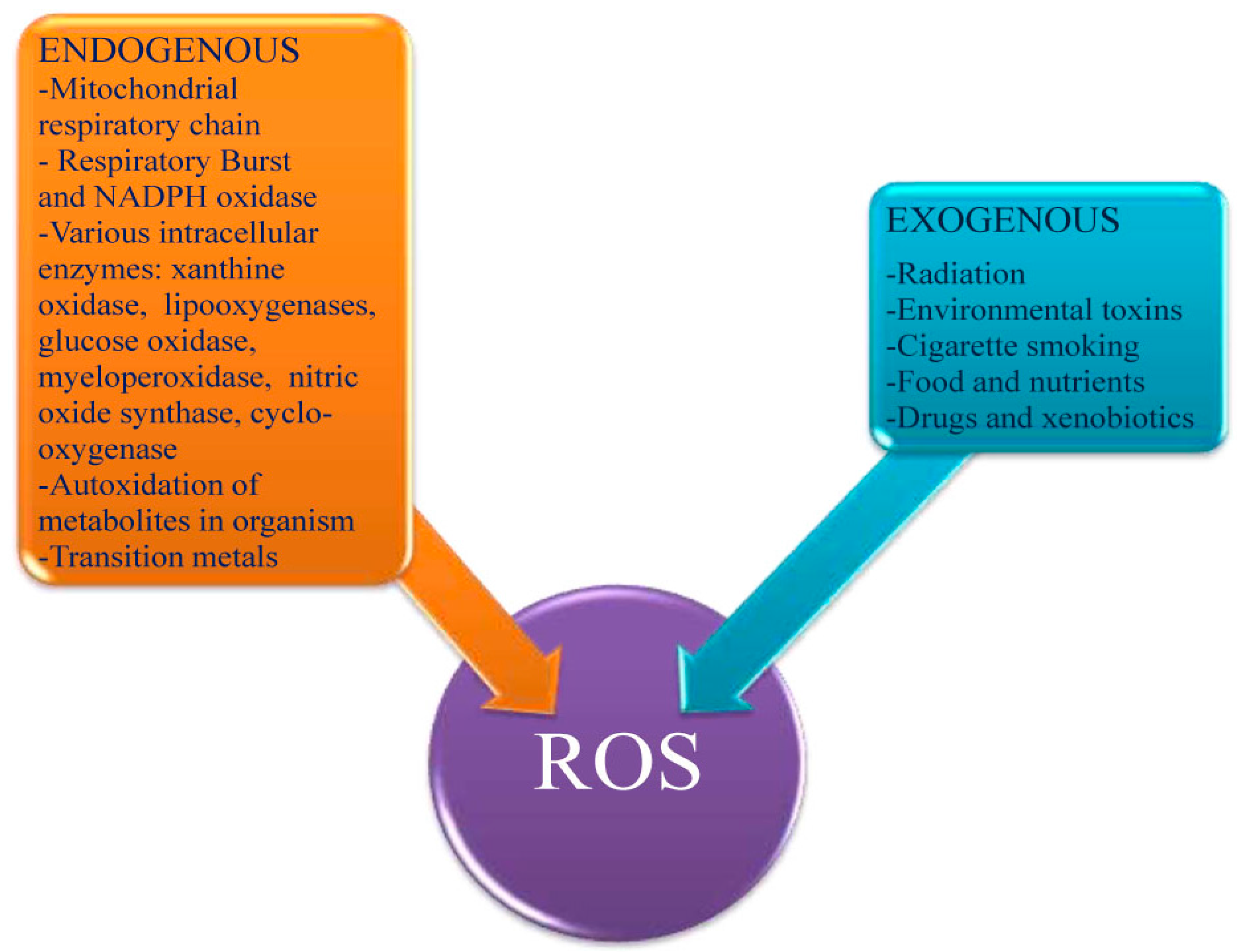
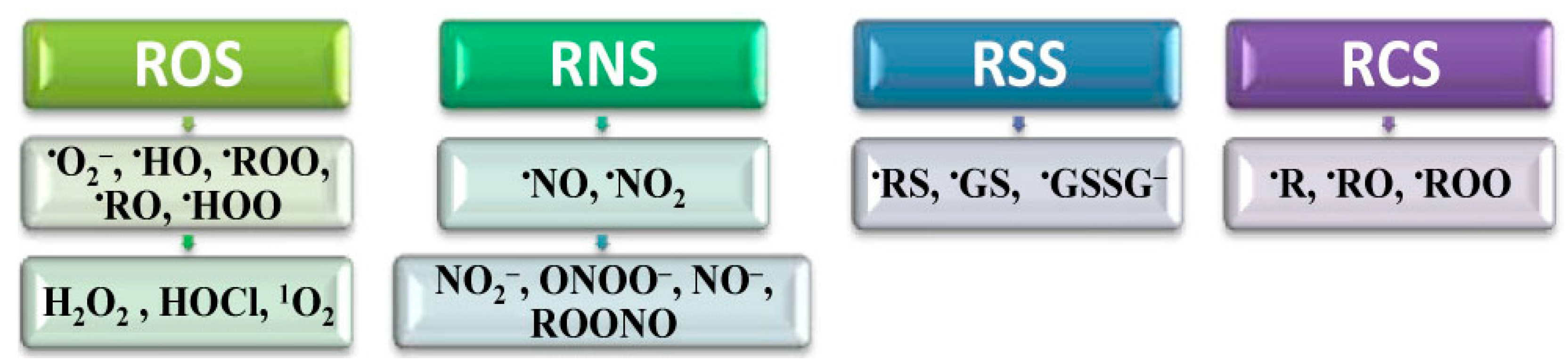
2.1. Introduction to ROS

In the mid-1950s, Harman published a “Free-Radical Theory of Ageing”, speculating that endogenous oxygen radicals were generated in cells and resulted in a pattern of cumulative damage. When the supply of antioxidants was insufficient, Harman speculated, the resulting cell damage triggers a cascade of events that leads to disease development and death [14,22].
Since that hypothesis, our knowledge on involvement of free radicals and antioxidants in living processes has grown enormously. The field of free radicals or more common reactive species (RS) research in biological systems has become one of the most dynamic.
Homeostasis is strongly influenced by many RS [23], such as ROS, reactive nitrogen (RNS), reactive carbon (RCS) and reactive sulfur species (RSS) (Figure 2). There are also many other RS consisting of halogens and related compounds [23].
ROS represent the most important class of reactive species generated in living systems [6,23]. In eukaryotic cells over 90% of ROS are produced by mitochondria via escape of electrons from the mitochondrial electron transport system (ETS), mainly from coenzyme Q to molecular oxygen (O2), resulting in the generation of superoxide anion radical (O2), named “primary” ROS. Further, O2 spontaneously or enzymatically, can be dismutated into hydrogen peroxide (H2O2) and O2. H2O2 is not a free radical, but is chemically more active than O2 due to which it is included in the ROS group. It possesses the ability to form the more damaging OH, through a combination of the Fenton and Haber-Weiss reactions [23,24]. Finally, ·OH interacts with one more electron and proton resulting in the formation of a water molecule (H2O). In biological systems, this reaction is mainly realized through abstraction of a hydrogen atom which originates from different compounds such as proteins and lipids, resulting frequently in initiation of radical chain processes. Besides of mitochondrial ETS, minor ROS are generated by ETS located in/at endoplasmic reticulum (ER), plasmatic, and nuclear membranes [23].
Figure 2. Classification of reactive species (RS) in living systems. Depending on the active center (ac) they are classified as: reactive oxygen species (ROS), ac-oxygen; reactive nitrogen species (RNS), ac-nitrogen; reactive carbon species (RCS), ac-carbon and reactive sulfur species (RSS), ac-sulfur.
Molecules 20 19489 g002
Diverse oxidases are also rather powerful ROS producers [23,25] (Figure 3). The physiological role of xanthine oxidase (XO) in total ROS balance, has received substantial attention. It is believed that under hypoxic conditions this enzyme may be the main ROS producer [26]. Additionally, autoxidation in the organism of different small molecules, such as adrenalin and norepinephrine, is also coupled with ROS production [27]. Even growth factors generate high levels of ROS that can perturb the normal redox balance and shift cells into a state of oxidative stress [14].
Figure 3. Endogenous and exogenous factors inducing ROS generation.
Molecules 20 19489 g003
There are multiple external factors that induce ROS generation (Figure 3). Environmental pollution, radiation, cigarette smoking, certain foods, and drugs are the major exogenous sources of ROS. Many xenobiotics, especially different homo- and heterocyclic compounds, are clearly related as ROS generators in autooxidation reactions [6,23]. Inadequate nutrition, due to modern lifestyle, may also indirectly result in oxidative stress which impairs the cellular defense mechanisms.

2.2. Positive Effects of ROS in Homeostasis

ROS are a double-edge sword [28]. Despite their harmful effects the beneficial physiological cellular use of ROS is now being demonstrated in different fields [29]. Low physiological levels of ROS function as secondary messengers in intracellular signaling and are required for normal cell functions (Table 1). It is well documented that low levels of ROS can modulate cell proliferation, apoptosis, and gene expression through activation of transcription factors, like Nuclear Factor Kappa B (NF-κB) and hypoxia-inducible-factor-1α (HIF) [28,30,31,32,33,34].
Table 1. ROS signaling is integrated into many cellular pathways [28,29].
No.Cellular Pathways
1proliferation and survival pathways through mitogen-activated protein kinase (MAPK), phosphoinositide 3-kinase (PI3), phosphatase and tensin homolog (PTEN), and protein tyrosine phosphatases
2ROS homeostasis and antioxidant gene regulation through redox effector factor-1 (Ref-1), NF-E2-related factor (Nrf-2), thioredoxin
3Aging through p66Shc, a member of the Src homologous-collagen homologue (ShcA) adaptor protein family
4DNA damage response through ataxia-telangiectasia mutated kinase (ATM); this may lead to inhibition of the mammalian target of the rapamycin complex 1 (mTORC1) resulting in suppression of protein synthesis and activation of autophagy
5Iron homeostasis through iron-regulatory proteins (IRP) and iron-responsive elements (IRE)
A moderate increase of ROS may protect from infections caused by a broad range of microorganisms [35]. The production of O2 and H2O2 by activated phagocytes is the classic example of the deliberate metabolic generation of ROS for useful purposes [28,36]. Moderate amounts of mitochondrial O2 and H2O2 have important roles in a range of cellular signaling processes and can activate signaling pathways that promote cell survival and disease resistance due to hormesis [28,37]. Generation of O2, HOCl, and H2O2 by phagocytes is important for defense against various bacterial and fungal strains [38]. For example individuals with chronic granulomatous disease who have deficiencies in generating ROS, are highly susceptible to infection by a broad range of microbes including Salmonella enterica, Staphylococcus aureus, Serratia marcescens, and Aspergillus spp. [39,40]. ·O2 is produced also by several cell types other than phagocytes, including lymphocytes and fibroblasts [28].
Detoxification reactions, ensured by the cytochrome P450 family, are dependent on the integrity of the microsomal ROS generating system. Nicotinamide adenosine dinucleotide phosphate (NADPH) supplies electrons, required for the reduction of O2 and the formation of ROS by microsomal monooxygenases, which have cytochrome P450 as a central link. They oxidize hydrophobic toxic substances, steroids and drugs, transforming them into hydrophilic compounds, which are removed from the body [41,42].
Further, a moderate increase of ROS is involved in apoptosis which eliminates cancerous and other life-threatening cells [35]. Apoptosis sometimes called “a guardian angel” or “cell policeman” [42], is carried out by a multistage chain of reactions, arises in cancer cells, in which ROS act as triggers and essential mediators [42].
Mitochondria play a critical role in apoptosis [43]. Apoptotic signals promote accumulation of the p53 protein that triggers the release of ROS, cytochrome C and a few other regulators from mitochondria [42]. ROS can act as signaling intermediates for cytokines, including interleukin 1 (IL-1) and the tumor necrosis factor (TNF) family [28,44,45,46]. Members of the TNF cytokine family, such as TNFα and Fas ligand (FasL), play important roles in inflammation and immunity [47,48]. Although TNF-related apoptosis-inducing ligand (TRAIL) is a member of this family, this molecule can induce (most, but not all) cancer cell death by its binding to the death receptors (DR), while causing almost no cytotoxicity to normal cells [47,49,50]. Normal cells are reported to show TRAIL-resistance with their preferential expression of decoy receptors (DcRs), which inhibit apoptotic signaling [51].
TRAIL-induced activation is a cascade of proteolytic enzymes, called caspases, which digest a number of cancer cell proteins and promote a caspase-activated deoxyribonuclease. Cleavage of the critical proteins and deoxyribonucleic acid (DNA) results in apoptotic cell death [42]. Also some reports suggest that TRAIL can induce necroptosis in cancer cells via ROS signaling [47,52,53,54].
In modern medicine generation of ROS by prooxidant compounds increasingly attracts attention as a therapeutic strategy for cancer [35]. For example, the compound 4-benzyl-2-methyl-1,2,4-thia-diazolidine-3,5-dione (TDZD-8) can induce depletion of thiols and rapid accumulation of ROS and selectively kill leukemic cells that express stem-cell markers, with minimal toxicity to normal hematopoietic stem cells [55]. Because tumor stem cells are thought to be the subpopulation of cells that are highly resistant to chemotherapy and play a critical role in disease relapse after treatment, the potency of the prooxidative compound in removing these cells underscores a key role of the redox system in regulating survival of stem cells and highlights the promising therapeutic potential of using a redox-based strategy in cancer treatment [35].
A transient increase of levels of oxidative stress by physical exercise reflects a potentially health promoting process at least in regards to prevention of insulin resistance and type 2 diabetes mellitus [56]. Physical training has been shown to improve glucose metabolism [57] by inducing molecular regulators of insulin sensitivity and endogenous antioxidant defense [56]. Exercise, as well as weight loss, has been linked to activation of mitochondrial metabolism, and reduced mitochondrial metabolism has been functionally connected with type 2 diabetes [58]. Muscle tissue is also known to generate ROS, especially during contraction and physical exercise [59].

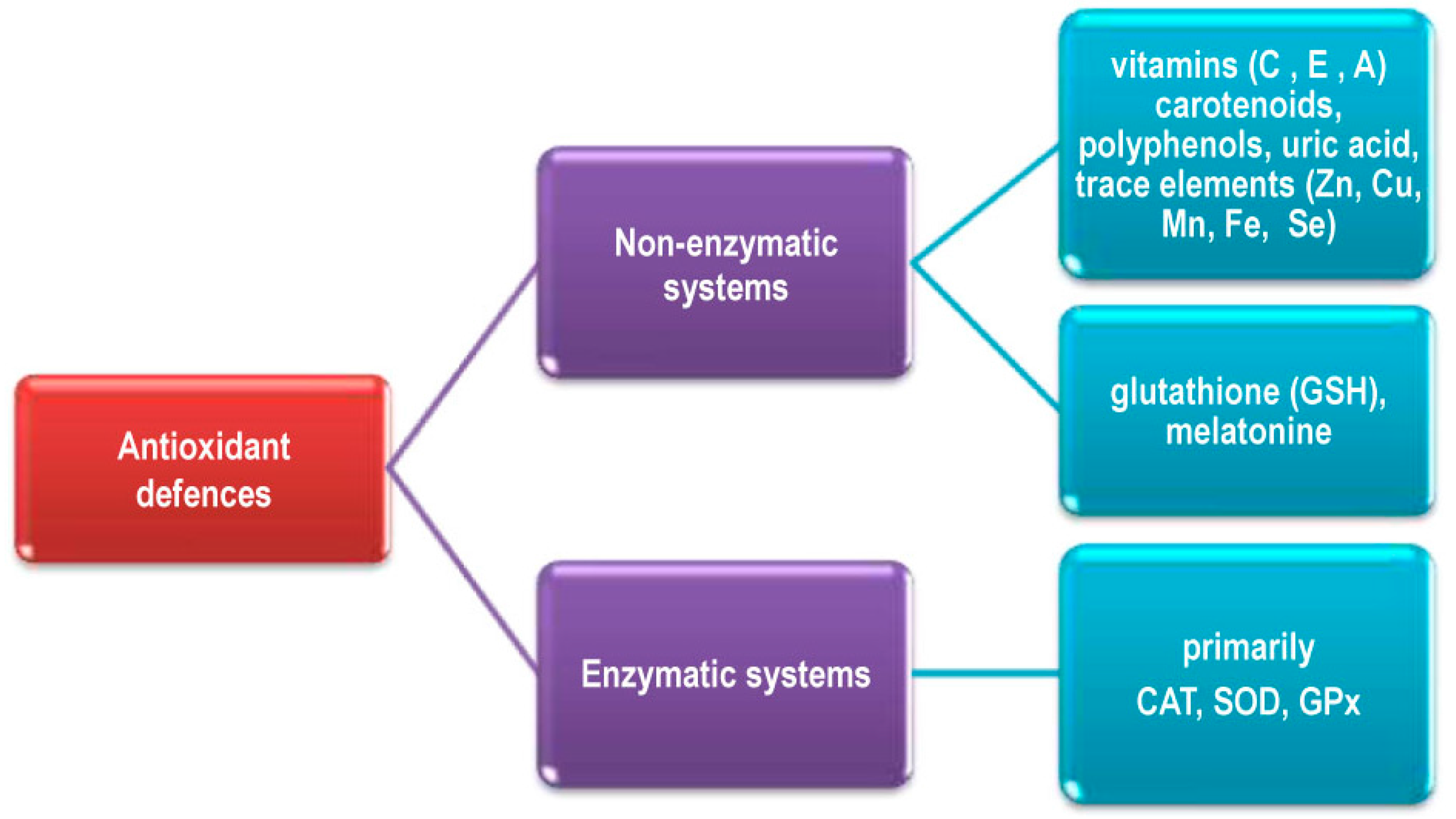
2.3. Elimination of ROS in Living Systems

Living organisms possess a multilevel and complicated antioxidant system operating either to eliminate ROS, or to minimize their negative effects. Major ROS defense mechanisms include enzymatic and nonenzymatic systems (Figure 4). The ROS which are not neutralized can target biological molecules such as DNA, lipids and proteins, which can result in cell death or dysfunction and accelerated ageing and age-related diseases [14,23].
Figure 4. Major ROS defense mechanisms in the organism.
Molecules 20 19489 g004
There are many different endogenous enzymatic antioxidant defense systems in the organism, either in intracellular or extracellular medium. They primarily include superoxide dismutase (SOD), catalase (CAT), glutathione peroxidases (GPx), and glutathione redutase (Gred) (Figure 4). Red blood cells are particularly sensitive to oxidative environments throughout the body and, as a consequence of their iron (Fe) content, are capable of producing their own ROS. However, the presence of antioxidant enzymes such as SOD and CAT, and methemoglobin reductase which catalyzes the reduction of methemoglobin to hemoglobin, minimized these processes [24,60]. The red blood cells and hepatocytes possess the highest level of CAT activity in the human body [60,61].
Nonenzymatic ROS defense mechanisms include low molecular mass antioxidants such as vitamins C and E, carotenoids including vitamin A, polyphenols, uric acid, (Figure 4) and large molecules i.e., albumin, ceruloplasmin, transferrin, ferritin [62]. In addition, Zn, copper (Cu), manganese (Mn), Fe, and Se are key components of enzymes with antioxidant functions and are designated as antioxidant micronutrients. Most of them are available to the human organism as food or supplement components [23,25,62,63,64].
Very important nonenzymatic antioxidants are glutathione (GSH), synthesized by most living organisms, and melatonin. As melatonin can directly cross the mitochondrial membranes, it plays a very significant role in the protecting mitochondria from oxidative damage. Probably due to antioxidant activity melatonin can improve glucose metabolism via correction of insulin production protecting pancreatic β-cells against ROS induced damage [65]. Other hormones. i.e., estrogen and angiotensin, also express antioxidant activity [63,66,67].
Alterations of the balance between ROS production and the capacity to rapidly detoxify reactive intermediates lead to oxidative stress. It has been implicated in a wide variety of states, processes and disease, e.g., aging, ischemia-reperfusion (I/R) injury, muscle damage, hypertension, atherosclerosis, diabetes, renal diseases, liver diseases, neurological diseases including Parkinson’s disease, Alzheimer’s disease and other forms of dementia, as well as diverse cancers. Oxidative stress also contributes to various gastrointestinal (GI) diseases including gastroduodenal ulcers, inflammatory bowel disease, and GI malignancies such as gastric and colorectal cancer [23,25,68].

2.4. Regulation of Antioxidant Systems

Under oxidative stress, an organism develops responses to prevent or neutralize negative ROS effects. These responses are mainly based on up-regulation of antioxidant and related enzymes. Mechanisms include ROS sensing, transduction of signals through specific pathways and up-regulation of target genes to enhance level of their products [23].
The human organism possesses a complicated system of adaptive responses to ROS exposure. Usually, it has several components (Figure 5). Under low intensity oxidative stress, the Kelch-like ECH-associated protein 1/NF-E2-related factor 2 (Keap1/Nrf2) system up-regulates genes encoding antioxidant enzymes. It is known to be activated by minute amounts of ROS. Intermediate intensity oxidative stress up-regulates antioxidant enzymes and induces inflammation proteins and heat shock proteins via NF-κB, activator protein-1 (AP1), MAPKs and heat shock factor (HSF). Under high intensity oxidative stress perturbations of mitochondrial permeability transition pore, activation of apoptosis cascade, destruction of electron transporters take place, which may culminate in apoptosis and/or necrosis [23,69].
Figure 5. Human and animal organism system of adaptive response to ROS exposure.
Molecules 20 19489 g005
Up-regulation of antioxidant systems increases their capability to eliminate ROS creating in this way autoregulated negative feedback control loop.

3. Antioxidant Compounds in Mushrooms

A whole range of edible mushrooms were reported to possess antioxidant activity (Table 2). It is generally accepted that extracts of fungi contain many components, each of which has its own specific biological effects [70,71]. Antioxidant compounds found in fruit bodies, mycelium and broth confirmed to be phenolics, flavonoids, glycosides, polysaccharides, tocopherols, ergothioneine, carotenoids, and ascorbic acid [16,17,72,73,74,75,76,77,78,79,80,81,82,83,84,85,86,87,88,89,90,91,92,93,94,95,96,97,98,99,100,101,102,103,104,105,106,107,108,109,110,111,112,113,114,115,116,117,118,119,120,121,122,123,124,125,126,127,128,129,130,131,132,133,134,135,136,137,138,139,140,141,142,143,144,145,146,147,148].
Table 2. Some studies of antioxidative properties of wild and cultivated mushrooms.
Mushroom SpeciesReferences
Agaricus bisporus, Agaricus brasiliensis (=Agaricus blazei ss. Heinem.), Agrocybe aegerita, Auricularia auricular, Auricularia cornea, Auricularia polytricha, Auricularia mesenterica, Auricularia fuscosuccinea, Agrocybe cylindracea, Amanita rubescens, Agaricus arvensis, Armillariella mellea, Agaricus silvicola, Agaricus silvaticus, Agaricus romagnesii, Antrodia camphorate[75,77,81,82,83,90,91,93,97,103,104,112,115,117,121,123,125,129,130,131,132,133,135,139,140,142,144]
Boletus edulis, Boletus badius [91,123,144]
Cantharellus lutescens, Cantharellus clavatus, Cantharellus cibarius, Cordyceps sinensis, Calvatia gigantea, Cerrena unicolor, Coprinus comatus[16,91,93,97,104,115,134,140,142,144,147]
Dictophora indusiata [114]
Flammulina velutipes (white), Flammulina velutipes (yellow) [95,103,105,136,137,138]
Inonotus obliquus [78,79,80]
Ganoderma lucidum, Ganoderma tsugae, Grifola frondosa, Ganoderma applanatum, Geastrum arenarius, Geastrum saccatum, Ganoderma atrum [74,75,76,86,87,88,89,91,93,94,96,98,99,107,109,110,111,114,118,119,127,142,145,146]
Hericium erinaceus, Hericium coralloides, Hydnum repandum, Hygrophorus agathosmus, Hypsizigus marmoreus, Hypholoma fasciculare, Helvella crispa[91,97,103,113,114,115,123,140,147]
Lepista nuda, Lentinus edodes, Lactarius sanguifluus, Lentinus squarrosulus, Lactarius deliciosus, Lentius sajor-caju, Leucopaxillus giganteus, Lactarius piperatus, Laetiporus sulphureus, Lycoperdon molle, Lycoperdon perlatum, Lactarius piperatus [73,74,81,84,91,95,97,99,103,104,106,115,120,122,125,126,128,133,140,144,145,147]
Morchella esculenta, Morchella conica, Macrolepiota procera, Morchella angusticeps, Macrolepiota procera[85,91,118,140]
Pleurotus ostreatus, Pleurotus eryngii, Pleurotus citrinopileatus, Pleurotus djamor, Pleurotus sajor-caju, Pleurotus cystidiosus, Pleurotus australis, Pleurotus tuber-regium, Phellinus linteus, Phellinus rimosus, Phellinus merrillii, Polyporus squamosus, Picoa juniperi, Pleurotus florida, Pleurotus pulmonarius, Paecilomyces japonica, Piptoporus betulinus[50,75,81,88,91,92,93,95,97,102,103,104,108,115,116,124,133,141,142,144,148]
Russula brevipes, Russula cyanoxantha, Russula delica, Ramaria botrytis, Russula vinosa [91,101,104,123,140]
Sparassis crispa, Suillus bellini, Suillus luteus, Suillus granulatus, Sarcodon imbricatus, Schizophyllum commune [72,91,123,125,140,147]
Tricholoma acerbum, Tricholoma equestre, Tricholoma giganteum, Tricholomopsis rutilans, Termitomyces microcarpus, Termitomyces schimperi, Termitomyces mummiformis, Termitomyces tylerance, Termitomyces heimii, Termitomyces albuminosus, Termitomyces robustus, Terfezia claveryi, Tremella fuciformis, Trametes (Coriolus) versicolor, Trametes orientalis [74,90,91,93,99,100,114,115,118,123,133,140,145,147]
Verpa conica, Volvariella volvacea [103,104,106,120]
The various methods used in order to measure the antioxidative properties of mushroom compounds or extracts are appropriate for various levels of antioxidative activity, such as methods based on the transfer of electrons and hydrogen atoms, the ability to chelate ferrous (Fe2+) and cupric (Cu2+) ions, the electron spin resonance (ESR) method, erythrocyte hemolysis, and the monitoring of the activity of SOD, CAT and GPx [16,17,72,73,74,75,76,77,78,79,80,81,82,83,84,85,86,87,88,89,90,91,92,93,94,95,96,97,98,99,100,101,102,103,104,105,106,107,108,109,110,111,112,113,114,115,116,117,118,119,120,121,122,123,124,125,126,127,128,129,130,131,132,133,134,135,136,137,138,139,140,141,142,145,146,147].
It has been established that mushroom antioxidants can demonstrate their protective properties at different stages of the oxidation process and by different mechanisms [4,6,7]. There are two main types of mushroom antioxidants, namely, primary (chain breaking, free radical scavengers) and secondary or preventive [4,6,7,16,72,73,74,75,76,77,78,79,80,81,82,83,84,85,86,87,88,89,90,91,92,93,94,95,96,97,98,99,100,101,102,103,104,105,106,107,108,109,110,111,112,113,114,115,116,117,118,119,120,121,122,123,124,125,126,127,128,129,130,131,132,133,134,135,136,137,138,139,140,141,142,145,146,147]. Secondary antioxidants are the consequence of deactivation of metals, inhibition or breakdown of lipid hydroperoxides, regeneration of primary antioxidants, singlet oxygen (1O2) quenching, etc. Some mushroom substances that exhibit antioxidant activity function as inducers and/or cell signals, leading to changes in gene expression, which result in the activation of enzymes that eliminate ROS [6,108,109,110,111,149].
Different analytical methods have been applied for its identification and quantification: high performance liquid chromatography (HPLC) and gas chromatography (GC) coupled to distinct detection devices, nuclear magnetic resonance (NMR), Fourier transform infrared (FT-IR), UV-VIS spectroscopy and various spectrophotometric assays [16,17,72,73,74,75,76,77,78,79,80,81,82,83,84,85,86,87,88,89,90,91,92,93,94,95,96,97,98,99,100,101,102,103,104,105,106,107,108,109,110,111,112,113,114,115,116,117,118,119,120,121,122,123,124,125,126,127,128,129,130,131,132,133,134,135,136,137,138,139,140,141,142,143,144,145,146,147,148].

3.1. Polyphenols Including Flavonoids

Polyphenols are the most abundant antioxidants in the diet [150]. Research on the effects of dietary polyphenols on human health has developed considerably in the past 20 years. One of the major difficulties of elucidating the health effects of polyphenols is the large number of phenolic compounds found in food [150,151], yielding differing biological activities [152].
These compounds may be classified into different groups as a function of the number of phenol rings and the structural elements which bind these rings to each other. Thus a distinction is made between the phenolic acids, flavonoids, stilbenes, and lignans. In addition to this diversity, most polyphenols are present in food in the form of esters, glycosides, or polymers. These substances cannot be absorbed in their native form and must be hydrolyzed by intestinal enzymes or by the colonic microflora before absorption. In the course of absorption, polyphenols are conjugated and this process mainly includes methylation, sulfation, and glucuronidation. Circulating polyphenols are conjugated derivatives that are extensively bound to albumin [153]. Nonconjugated metabolites are generally either absent from the blood or present in low concentrations [152]. Major differences in bioavailability of polyphenols are now well established facts and the influence of structural factors is better understood [152,153,154].
The main phenolic compounds found in mushrooms are phenolic acids [6]. Phenolic acids can be divided into two major groups, hydroxybenzoic acids and hydroxycinnamic acids, which are derived from the non-phenolic molecules benzoic and cinnamic acid, respectively [6,153,154].
Hydroxybenzoic acid derivatives commonly occur in the bound form and are typically a component of a complex structure like lignins and hydrolyzable tannins. They can also be found linked to sugars or organic acids. Hydroxycinnamic acid derivatives are mainly present in the bound form, linked to cell-wall structural components, such as cellulose, lignin, and proteins, as well as associated to organic acids, such as tartaric or quinic acids (i.e., chlorogenic acids), through ester bonds [6,153].
The hydroxycinnamic acids are more common than the hydroxybenzoic acids and consist chiefly of p-coumaric, caffeic, ferulic, and sinapic acids. These acids are rarely found in the free form, except in processed food that has undergone freezing, sterilization, or fermentation. The bound forms are glycosylated derivatives or esters of quinic acid, shikimic acid, and tartaric acid [153,154].
The most common (prevalent) benzoic acid derivatives found in mushrooms are reported to be p-hydroxybenzoic, protocatechuic, gallic, gentisic, homogentisic, vanillic, 5-sulphosalicylic, syringic, veratric, vanillin [6]. The majority of identified cinnamic acid derivatives in mushrooms are: p-coumaric, o-coumaric, caffeic, ferulic, sinapic, 3-o-caffeoylquinic, 4-o-caffeoylquinic, 5-o-caffeoylquinic [6]. Besides, presence of ellagic and tannic acids is observed [6].
Natural polyphenolic compounds exert their antioxidant effect by quenching free radical species and/or promoting endogenous antioxidant capacity. Furthermore, some of them stimulate synthesis of endogenous antioxidant molecules in cells via activating the Nrf/ARE pathway [149,150]. Cells respond to polyphenols mainly through direct interactions with receptors or enzymes involved in signal transduction, which may result in modification of the redox status of the cell and may trigger a series of redox-dependent reactions [150,155,156,157].
Apart from the antioxidant capacity, most of these compounds appear to have a number of different molecular targets, impinging on several signaling pathways, and showing pleiotropic activity on cells. For instance, polyphenolics can modulate activity of NF-κB or Sirutin1 (SIRT1) exerting neuroprotective effects [154,158]. SIRT1 acts as a “rescue gene”, capable to repair damages caused by the action of free radicals and to prevent premature death of cells. The gene also affects the mitochondria to produce greater amounts of energy what is typical for the metabolism of younger cells. As a result, SIRT1 is believed to be a principal regulator of lifespan [158,159].
Besides antioxidant properties, polyphenols possess pro-oxidative capacity based upon the structure of the particular polyphenol and the cellular redox context that may include increased levels of oxidant scavenging proteins or decreased levels of oxidized proteins and lipids [159]. For example, epigallocatechin-3-gallate (EGCG) induces the activation of protein caspases-3 and c-Jun N-terminal kinases (JNKs), which belong to the group of MAPKs that have a role in the process of programmed cell death, i.e., apoptosis. These various effects are dependent on cell type, stress conditions, concentrations and the time of exposure of EGCG. It is possible that low concentrations of EGCG activate MAPK, leading to antioxidant responsive element (ARE)-mediated gene expression, whereas higher concentrations and sustained activation of MAPKs lead to apoptosis [7,160].
Polyphenols such as tannic acid, gallic acid, and catechin compounds are also known to interact with steroid receptors. This may lead to a change of the mitochondrial transmembrane potential and ultimately to a decrease or increase in ROS activation, depending on cell systems [142]. As antioxidants, polyphenols may improve cell survival; as prooxidants, they may induce apoptosis and prevent tumor growth, and combat against bacterial and viral infections by direct oxidative damage or by a variety of innate and adaptive mechanisms [150,161].
Recently, Lagunes and Trigos [162] showed that polyphenols, e.g., curcumin, resveratrol and quercetin, have pro-oxidant activity; they act as photosensitizers in the generation of 1O2. Resveratrol and curcumin have been reported as efficient quenchers of 1O2 [163,164]. On the other hand, they have also been reported as molecules capable of absorbing ultraviolet-visible (UV-Vis) light and able to transfer it to O2 to generate 1O2 [162,165]. Recent studies have shown that curcumin is able to inhibit melanoma cells proliferation, under UVA-Vis light, suggesting that it could be used as an adjunctive therapy in the treatment of cancerous diseases [166]. Resveratrol and curcumin are lipophilic molecules and mix very well between membrane lipids and lipoproteins; their levels in tissues may be higher than those detected in blood [167]. High consumption of dietary supplementation with resveratrol and curcumin could lead to a high bioavailability in tissues and when these are exposed to light, it would allow resveratrol and curcumin to act as photosensitizers, showing a pro-oxidant activity by generation of 1O2. 1O2 is a powerful oxidizing agent initiator of oxidation processes in biological systems [168], by which resveratrol and curcumin would favour a pro-oxidant effect against normal cells [162]. Analogous processes are likely to occur with lipophilic polyphenols of mushrooms, which requires attention should be given to possible harmful effects.

Flavonoids

Flavonoid research has intensified since the discovery of the “French paradox” [169,170]. This phenomenon was observed in the French population as a significantly lower rate of heart diseases despite high consumption of saturated fat and smoking habits and was related to moderate and regular consumption of red wine rich in flavonoid compounds [170].
Basic structure of flavonoid has a flavan nucleus consisting of two benzene rings (A and B) combined by an oxygen-containing pyran ring (C). The various classes of flavonoids differ in their level of oxidation of the C ring of the basic 4-oxoflavonoid (2-phenyl-benzo-γ-pyrone) nucleus. The common six subclasses of flavonoids are flavonols, flavones, isoflavones, flavanones, anthocyanidins, and flavanols [153,169,171]. Flavonoids are most frequently found in nature in the form of glycosylate or esterificate conjugates but can also occur as aglycones in food, especially as a result of the food processing. Flavonols are the most abundant flavonoids in foods [170,171].
In general, it can be assumed that only plants possess the biosynthetic ability to produce flavonoids, while animals and fungi are not capable of it [6]. However, the presence of flavonoids is reported in different edible mushrooms, e.g., myricetin, chrysin, catechin, hesperetin, naringenin, naringin, formometin, biochanin, pyrogallol, resveratrol, quercetin, rutin, kaempferol [6]. More recently, the analysis of the methanolic extract of Cantharellus cibarius showed that phenols were its major antioxidant components but followed by flavonoids, whose content was approximately 86% of the total phenol content [16].
Mechanisms of the antioxidant action of flavonoids can include direct scavenging of RS, chelating of trace metal ions involved in RS formation, inhibition of enzymes, e.g., XO and lipoxygenases (LOXs), involved in RS production, and regeneration of membrane-bound antioxidants such as α tocopherol [169]. It is generally considered that the primary mechanism of the radical scavenging activity of flavonoids is hydrogen atom donation. [170,171].
The capacity of flavonoids to inhibit ROS is governed by the presence and position of the multiple hydroxyl groups in their structure. A double bond and carbonyl function in the heterocycle or polymerization of the nuclear structure increases activity by affording a more stable flavonoid radical through conjugation and electron delocalization [170,171]. Mechanisms of flavonoid action, such as modulation of signalling pathways and gene expression, could also contribute to protective properties of flavonoids [153].
Further, flavonoids may act as pro-oxidants based upon on a different number of OH groups in the B ring and presence of transient metal ions [170]. The flavonoid phenoxyl radical could interact with O2, generating quinones and superoxide anion (O2), rather than terminating the chain reaction. This reaction may take place in the presence of high levels of transient metal ions and may be responsible for the undesired prooxidant effect of flavonoids [170].
El Amrani et al. [172] reported oxidative DNA cleavage induced by binding of a Fe3+-flavonoid complex to DNA. Likewise, reacting with Cu2+ quercetin induces extensive DNA damage, but kaempferol and luteolin induce only slight DNA damage, even in the presence of Cu2+ [170]. These complexes may play an important role in their potency for biological action such as angiogenesis and immune-endothelial cell adhesion, which, respectively, are important processes in the development of cancer and atherosclerosis [173].
Also, it has been shown that quercetin could aggravate the severity of renal cell carcinoma through the tumorigenic action mechanism due to pro-oxidant effect [174]. It act as an antioxidant in low doses, or as pro-oxidant at higher doses [175].Concerning the absorption and bioavailability of quercetin, it has been demonstrated that in an aglycosylated form it becomes a lipophilic molecule and can be easily absorbed through epithelia in cells of the colon [162,176]. In a similar manner to resveratrol, quercetin would be able to be incorporated into cellular membranes, and under specific conditions, show a pro-oxidant activity by generation of 1O2 [162].

3.2. Polysaccharides

Polysaccharides, including polysaccharide-protein complexes, have been recognized as a class of major bioactive constituents of edible and medicinal mushrooms [70,71]. They act primarily as adaptogens and immunostimulators. The immunostimulatory effect of mushroom polysaccharides is prophylactic primarily and it belongs to non-invasive treatments, playing a role in the prevention of infectious diseases and of tumor metastases [7,70,71,177,178].
Another widely reported activity of mushroom polysaccharides is antioxidative [7,72,73,74,75,76,77,99,145]. Many mushroom polysaccharides have been reported to have significant antioxidant activities based on various in vitro and in vivo assays [7,179]. Their antioxidative activity is attributed to the ability of RS scavenging, their reduction property and ability to chelate Fe2+, lipid peroxidation inhibition, erythrocyte hemolysis and the increase of enzymes activities in eukaryotic as well as in prokaryotic cells as well as in taking part in antioxidative processes, such as SOD, CAT and GPx [7,108,109,110,111].
The extraction procedure and the method used for the purification of the obtained fractions of polysaccharides depend in many aspects on the type of a mushrooms and physical properties of polysaccharides. Usually these polysaccharides were isolated from the hot-water extract simply by ethanol precipitation without further purification [7,72,73,74,75,76,77,99]. Purification of the crude polysaccharides is usually accomplished by chromatographic methods such as size-exclusion (SEC) and ion-exchange chromatography (IEC) [99,178], as well as by enzymatic purification using cellulases, amylases and proteases [7,77]. Besides, new extraction procedures were reported, as ultrasonic-assisted extraction [145,180] and more recently, extraction procedure with a freeze-thawing process [181]. Klaus et al. [76] have managed to encapsulate polysaccharide extracts from G. frondosa in alginate gel beads to protect them from external influences and this could contribute to a more widespread use.
Some studies have found that the purified mushroom polysaccharides had lower antioxidant activities than the original crude extracts [99,146]. Polysaccharides in the fungal cell wall may be bound by covalent (ester) linkages with proteins via remains of tyrosine and/or with ferulic acid as a result of lignin degradation processes [7,74]. However, other studies reported higher antioxidant activity in the pure polysaccharide fraction, e.g., the A. brasiliensis polysaccharides obtained by pronase deproteinization, demonstrated a high antioxidative activity against OH and O2 radicals measured by electron paramagnetic resonance (EPR) spin-trapping spectroscopy. These polysaccharides consisted mainly of (1→6)-β-d-glucans [77].
The major antioxidant effects of mushrooms are attributed to β-glycans. Except reported antioxidant properties, α-glycans are eukaryotic nutrient components and are easily degraded by mammalian enzymes. β-Glycans from various mushrooms can be taken up by the M cells of Peyer’s patches, and/or interact with dendritic cells (DCs) in the small intestine to activate systemic organism responses [182,183].
The ability of the polysaccharide molecules to scavenge free radicals may be conditioned by the presence of hydrogen from specific, certain monosaccharide units, and the type of their binding in side branches of the main chain [84,184]. The enhanced antioxidant activity of the polymers over the monomeric form may be due to the greater ease of abstraction of the anomeric hydrogen from one of the internal monosaccharide units rather than from the reducing end [184].
Recently, Kishk and Al-Sayed [185] reported that the OH scavenging mechanism of polysaccharides was perhaps similar to that of phenol compounds by hydrogen atom transfer (HAT) reactions. However, the HAT reaction is more likely to occur in the neutral polysaccharides, while the electron transfer (ET) mechanism is usually occur in the acidic polysaccharides.
Mushroom polysaccharides and glycoconjugates may be useful in creating new natural-based medications or dietary supplements and helpful in the prevention and treatment of oxidative stress-mediated disorders. ROS are produced within the GI tract. Despite the protective barrier provided by the mucosa, ingested materials and microbial pathogens can induce oxidative injury and GI inflammatory responses involving the epithelium and immune/inflammatory cells. High antioxidative capacities of polysaccharides from edible mushrooms can prevent lipid peroxidation [72,73,74,75,76,77] and the pathogenesis of various GI diseases including peptic ulcers [186], GI cancers [182], and inflammatory bowel disease which is in part due to oxidative stress [25].
Cytotoxic effects of polysaccharide extracts of higher fungi on normal cells have not been reported up-to-date [7,73,77]. Furthermore, it was confirmed that polysaccharide extract of G. lucidum, in vitro, stimulates proliferation of HTR-8/SVneo trophoblast cells which are essential for normal placentation, establishment of pregnancy and maintenance of fetal growth in humans [73,187].
The polysaccharide extracts proved to be heat stable and retained high antioxidant potential, despite all the treatments applied in their preparation [72,73,74,75,76,77].

3.3. Vitamins

3.3.1. Vitamin C

Vitamin C, also known as l-ascorbic acid, is the primary antioxidant in plasma and cells [25]. It is an essential nutrient for a limited species of animals, including humans, and therefore must be ingested to avoid a potentially lethal condition [6,25,188]. Likewise, ascorbic acid is a normal skin constituent found at high levels in both the dermis and epidermis [189,190]. Ageing, however, causes a decline in vitamin C content in both layers. Excessive exposures to UV light or pollutants (e.g., cigarette smoke and ozone) may also lower its content, primarily in the epidermis [189,190].
Vitamin C was detected in various edible mushrooms [6,16]. It was quantified by HPLC or spectrophotometric assay, based on the reaction with 2,6-dichlorophenolindophenol [6,16]. Kozarski et al. [16], found 100 mg/100 g dry weight (DW) of ascorbic acid in the methanolic extract of the wild edible mushroom Cantharellus cibarius. It was higher than its content in some fruits and vegetables which are usually recommended as a good source of vitamin C [191]. Reported amount of ascorbic acid found in strawberries is 60 mg/100 g, citrus fruits 30–50 mg/100 g, while apples, pears and plums represent only a very modest source of ascorbic acid (3–5 mg/100 g) [191].
Grangeia et al. [192] found high concentrations of ascorbic acid in the methanolic extracts of different saprotrophic and mycorrhizal wild edible mushrooms (81.32–400.36 mg/100 g DW).
Ascorbic acid has been shown as an effective RS scavenger [188,191]. In studies with human plasma lipids it appeared that ascorbate was far more effective in inhibiting lipid peroxidation initiated by a lipid peroxyl radical (LOO) initiator than other plasma components, such as protein thiols, urate, bilirubin, and vitamin E. In the aqueous phase, ascorbic acid can protect biomembranes against peroxidative damage by the efficiently trapping LOO before they can initiate lipid peroxidation [188,191]. It may also recycle vitamin E, the main lipid-soluble antioxidant. Ascorbic acid reacts rapidly with the tocopherol radical by reducing the ascorbate radical (semidehydroascorbate) to ascorbate by NADH-dependent semidehydroascorbate reductase [6,25]:
Ascorbic acid + Tocopherol → Ascorbate + Tocopherol Ascorbate + NADH → Ascorbate + NAD
Although the main function of ascorbic acid is antioxidant, under certain circumstances, it can also have a pro-oxidant effect, in particular by maintaining the transition metal ions, Fe3+ and Cu2+ in their reduced forms. These metal ions react then with H2O2 to form the highly reactive ·OH in the Fenton reaction [25,191]. But there is currently no clear evidence that these reactions are of significance in vivo [25,191]:
Fe3+ + Ascorbic acid → Fe2+ + Ascorbate Fe2+ + H2O2 OH + OH + Fe3+

3.3.2. Vitamin E

The term “vitamin E” does not refer to a single molecule but rather is frequently used to designate a family of chemically related compounds, namely tocopherols and tocotrienols, which share a common structure with a chromanol ring and isoprenic side chain [188]. α, β, γ, and δ Tocopherols were identified and quantified in edible mushrooms. However till now, tocotrienols were not detected in any of the available studies [6].
Biologically the most active form of vitamin E is α tocopherol whose main role is to protect cell membranes from lipid peroxidation (LPO) [25,188]. α Tocopherol terminates the activity of LPO by scavenging ·LOO, but during this reaction is itself converted into a reactive radical [6,25]:
LH + Oxidant initiator → L L + O2LOO LOO + Tocopherol → LOOH + Tocopherol
α-Tocopherol can also act as a prooxidant and reduces Fe3+ or Cu2+ [193]. The ability of α tocopherol to act as a pro- or antioxidant depends mainly on its amount which is available for scavenging of ROS [193]. Epidemiological studies indicate that diets rich in fruits and vegetables lower GI cancer rates, but supplementation of exogenous vitamin E and other antioxidants (β-carotene, vitamins A, C and Se) have not been shown to prevent GI cancers [194]. In some cases, vitamin E increases cancer risks [195]. Likewise, supplementing vitamin E has been shown to significantly increase lung cancer progression and reduce survival rate in mouse models [196]. As the expression of endogenous antioxidant genes is reduced by the introduction of antioxidant supplements, the tumor transcriptome profile is altered with a downregulation of p53 (tumor suppressor protein gene) expression level accordingly [197]. These changes provide an explanation on why antioxidants may promote cancer development [195].
The amounts of tocopherols detected in edible mushrooms were much lower that those measured in some groceries which are usually recommended as a good source of vitamin E [6,139,147,192]. Compared with foods high in vitamin E content, e.g., almonds (26.2 mg/100 g), roasted sunflower seeds (36.3 mg/100 g), avocados (2.1 mg/100 g), tofu (5.3 mg/100 g), shrimp (2.2 mg/100 g) etc., the tocopherols content in edible mushrooms is measured to be between 0.02 and 200 μg/100 g DW [139,147,192,198].

3.3.3. Vitamin A Including Carotenoids

Carotenoids are natural pigments. There are several dozen carotenoids in foods. The predominant carotenoids in the diet and human body are represented by β-carotene, α-carotene, lycopene, lutein and β-cryptoxanthin, of which β-carotene, α-carotene, and β-cryptoxanthin are able to function as provitamin A and play important roles as vitamin A dietary sources [199]. Recent interest in carotenoids has focused on the role of lycopene in human health. It does not have pro-vitamin A properties. Because of the unsaturated nature of lycopene it is considered to be a potent antioxidant and a 1O2 quencher. Lycopene was shown to cross the blood brain barrier and be present in the central nervous system in low concentrations. Significant reduction in the levels of lycopene was reported in Parkinson’s disease and vascular dementia patients [199,200].
Particularly, β-carotene and lutein were found in several mushroom species [6,16,201]. Carotenoids found in the pink-red Cantharellus cinnabarinus and the orange Cantharellus friesii are composed almost entirely of canthaxanthin, a pigment also found in the salmon [201]. It might explain the use of chanterelles by Chinese herbalists in treating night blindness [201]. Canthaxanthin is reported to protect human tissues from oxidative damage and is sold as an antioxidant [201].
Carotenoids can react with ROS and become radicals themselves. They function as a chain-breaking antioxidant in a lipid environment, especially under low oxygen partial pressure. The extensive systems of double bonds make carotenoids susceptible to attack LOO, resulting in the formation of inactive products [202,203].
Carotenoids reactivity depends on the length of conjugated double bonds chain and the characteristics of the end groups [202]. Carotenoid radicals are stable by virtue of the delocalization of the unpaired electron over the conjugated polyene chain of the molecules. This delocalization also allows additional reactions which occur at many sites on the radical. The carotenoid radicals are very short-lived species [202].
The antioxidant properties of carotenoids can be reversed to pro-oxidant behavior depending on oxygen pressure or carotenoid concentration [14,25,203]. It is established that β-carotene loses its antioxidant activity when the normal ambient oxygen pressure increases, and has an autocatalytic pro-oxidant effect which increases with its concentration [7,203]. Studies of food supplementation with large doses of β-carotene in smokers have shown an increase in cancer risk, possibly because β-carotene under intense oxidative stress (e.g., induced by heavy smoking) gives breakdown products that reduce plasma vitamin A and worsen the lung cell proliferation induced by smoke [149,203]. Frequent use of vitamin C and vitamin E in the period after breast cancer diagnosis was associated with a decreased likelihood of recurrence, whereas frequent use of combination carotenoids was associated with increased mortality [204].

3.3.4. Vitamin D

Vitamin D refers to a group of fat-soluble secosteroids responsible for enhancing intestinal absorption of calcium (Ca), Fe, magnesium (Mg), P and Zn [205,206,207]. The importance of vitamin D in bone (Ca2+ ion homeostasis) is well established, and vitamin D has been the subject of increased attention in recent years for its role in muscle function, immunology, heart and cardiovascular disease, cancer, and insulin secretion [208].
In humans, the most important compounds in this group are vitamin D3, also known as cholecalciferol and vitamin D2, or ergocalciferol. Both cholecalciferol and ergocalciferol are inactive themselves, but are metabolized in the liver to 25-hydroxyvitamin D and further in the kidney to the biologically active form, 1,2,5-dihydroxyvitamin D. The serum concentration of 25-hydroxyvitamin D is considered to be a good indicator of vitamin D status in humans [209].
Vitamin D3 originates from animal sources, and vitamin D2 is derived predominantly from mushrooms and yeast [208]. Vitamin D2 contents in mushrooms, cultivated or harvested wild, is available but varies significantly with and within different species and by developmental stage [210,211]. Differences of climate, habitat, and degree of latitude may cause variation in ergocalciferol contents of mushrooms [210]. For example, content of only 0–3.75 μg/100 g of fresh weight has been found in A. bisporus [210,212,213,214] and 0.04 [215] to 21.8–109.6 μg/100 g DW [216] in L. edodes.
The vitamin D2 content of mushrooms can be increased dramatically by UV irradiation, whereby it is formed from ergosterol that is present in large amounts [208,217,218,219,220]. Outila et al. [209] showed for the first time that ergocalciferol was well absorbed in humans from lyophilized and homogenized wild edible mushrooms.
Vitamin D3 and D2, its active metabolite 1,25-dihydroxycholecalciferol and also 7-dehydrocholesterol (pro-Vitamin D3) are a membrane antioxidants and inhibit iron-dependent liposomal lipid peroxidation [207]. The structural basis for the antioxidant ability of these vitamin D compounds is considered in terms of their molecular relationship to cholesterol and ergosterol [207].
Biologically active, 1,25-dihydroxyvitamin D, binds to the vitamin D receptor on cells and plays a role not only in Ca2+ uptake but also in differentiation [50,208,211]. Furthermore, by decreasing membrane fluidity by the membrane interaction that is thought to lead to the observed inhibition of iron-dependent liposomal lipid peroxidation [207], Vitamin D could help inhibit the growth of cancer cells (especially metastatic cells), which often have increased membrane fluidity compared to normal cells [221].
Van Griensven and Verhoeven reported [50] that crude polysaccharides extract of Phellinus linteus increased the mitochondrial membrane potential (MMP) and causes apoptotic death of THP-1 monocytes. They concluded that there are two major factors determining differentiation and/or death of polysaccharide extract treated THP-1 cells, i.e., a death receptor (TRAIL-like) binding activity on the one hand, and oxidative stress and Ca2+ homeostasis on the other. It was confirmed that P. linteus polysaccharides extract increased mitochondrial Ca2+ concentration, resulting in breakdown of the outer mitochondrial membrane and induction of apoptosis [222]. Apoptosis through the mitochondrial pathway merely seems to be caused by disturbance of the mitochondrial Ca2+ homeostasis [223]. Changes in the MMP and Ca2+ homeostasis play a prominent role in the pathogenesis of age related loss of neuronal function, such as that occurring in Alzheimer’s disease [224] and Parkinson's disease [225]. Hyperpolarizing compounds e.g., vitamine D2, in P. linteus might restore Ca2+ homeostasis and thereby prevent the loss of neuronal function [50].

3.3.5. Ergothioneine

Ergothioneine (ET) is an unusual sulfur-containing derivative of the amino acid, histidine, which is obtained exclusively through the diet. Recently, a highly specific transporter for ET (ETT) was identified (integral membrane protein, OCTN1) in mammalian tissues, which explains abundant tissue levels of ET and implies its physiological role [226,227]. Cells lacking ETT are more susceptible to oxidative stress, resulting in increased mitochondrial DNA damage, protein oxidation and lipid peroxidation. ET is concentrated in mitochondria, suggesting a specific role in protecting mitochondrial components, such as DNA, from oxidative damage associated with mitochondrial generation of O2. Because of its dietary origin and the toxicity associated with its depletion, ET may represent a new vitamin whose physiological roles include antioxidant protection [226,227].
Mushrooms are a primary source of ET containing from 400 to 2500 mg/g DW [17,143]. Weigand-Heller et al. [135] reported that ET from A. bisporus is bioavailable as assessed by red blood cell uptake postprandially. Consumption is associated with an attenuated postprandial triglyceride (TG) response.
By analyzing the fruiting bodies and mycelia of 29 edible and medicinal mushrooms species Chen et al. [17] confirmed presence of ET in all samples. The highest amount was observed among fruiting bodies of edible species, P. citrinopileatus, P. ostreatus (Korea), P. ostreatus (Taiwan) and P. salmoneostramineus (2850.7, 1829.4, 1458.4 and 1245.0 mg/kg, respectively) whereas among mycelia, P. eryngii contained the highest amount (1514.6 mg/kg). Dubost et al. [143] also found the ET content of P. ostreatus to be the highest (2590 mg/kg DW). It seems that mushrooms are an abundant source of ET [17,136,137,138,143].

3.4. Minerals

Mushrooms are generally capable of accumulating trace elements and then become their source in the food chain [228,229]. Trace elements Zn, Cu, Mn and Fe are cofactors of enzymes with antioxidant functions and are designated as antioxidant micronutrients [25]. Se is a major antioxidant in the form of selenoproteins that reduce the cytotoxic effects of ROS. GPx and selenoprotein-P are dominant Se compounds secreted in the blood [229]. In mushrooms, several Se compounds have been identified including selenomethionine, selenocysteine, Se-methylselenocysteine, selenite and seleno-polysaccharides [230,231,232].
Factors influencing the bioaccumulation of trace elements in mushrooms and the biological importance of the accumulation process itself are poorly understood. However, the following fundamentals have been recognized: natural factors (bedrock geochemistry), metalliferous areas and environmental pollution, fungal environment, bioaccumulation of trace elements in fruit bodies is highly specific: some macrofungi are able to accumulate elements much more effectively than others, the accumulation process can be highly element specific- macrofungi may even discriminate elements with similar properties and chemical behavior (homologues) [229,233]. Moreover, wild-grown edible mushrooms can accumulate minerals essential to humans in a much greater extent than cultivated edible mushroom [229].
However, it should also be kept in mind that these essential metals can also produce toxic effects when their intake is excessively elevated [228,229]. For example, in vitro and in vivo experiments have indicated that Se prevents carcinogenesis, but a high concentration of Se can be cytotoxic [234]. Se conjugates with two GSH to form the metabolite selenodiglutathione (GSSeSG), a potent compound that enhances ROS production, DNA damage and apoptosis [235]. Besides exogenous antioxidant treatments, the modulation of the endogenous antioxidant system can also be cytotoxic. Over expressed glutamate cysteine ligase (GCL), GCL catalytic subunit (GCLC) or GCL modifier subunit (GCLM) can lead to an imbalanced GSH/GSSG ratio. Excess GSH shifts the cells to reductive stress, which can cause mitochondrial oxidation and cytotoxicity [195,236].
Many edible mushroom species are known to accumulate high levels of heavy metals, mainly cadmium (Cd) and lead (Pb) [237,238,239,240]. Divalent metal cations belonging to the transition elements may pass into the mitochondria through membrane channels for essential group elements [241]. For example Pb2+ ions seem to be transported into human and animal mitochondria through the Ca2+ transporters [241,242]. Also, after reaching the cytoplasm, Pb2+ continues its destructive mimicking action by occupying the Ca2+ binding sites on numerous Ca-dependent proteins. Pb2+ has high affinity for Ca-binding sites in the proteins; a pM concentration of Pb2+ can replace Ca2+ in μM concentrations [242].
Cd2+ ions can replace Fe2+ and Cu2+ from a number of cytoplasmic and membrane proteins like ferritin, thereby causing rise in the concentration of Fe2+ and Cu2+ ions which may be associated with the production of oxidative stress via the Fenton reaction [243]. Another mechanism of Cd2+ toxicity may be carried out in the body by Zn2+ binding proteins. Cd2+ can bind up to ten times more strongly than Zn2+ in certain biological systems and is notoriously difficult to remove [242].
Another target for Pb2+ and Cd2+ attack could be a thiol group (-SH) containing enzymes involved in antioxidant mechanisms, i.e., Cd2+ forms Cd-Se complexes in the active centre of GPx and inhibits the enzyme activity [242]. Likewise, Cd2+ and Pb2+ inhibit complex III of the mitochondrial electronic transport chain, divert the electron flow and increase production of ROS [244] which may damage the mitochondrial membrane and trigger onset of apoptosis. In addition, changes in mitochondrial oxidative metabolism may lead to energy deficit thereby affecting the essential cellular functions. Thus, Cd and Pb are capable of eliciting of ROS, which could be the main mechanism of cellular toxicity induced by these heavy metals [241,242,245].

4. Conclusions

Oxidative stress plays a significant role in ageing processes and increases the risk of chronic diseases. The ability to resist or prevent oxidative stress is a key determinant of longevity. Enhancement of antioxidant defenses through dietary supplementation would seem to provide a reasonable approach to reduce the level of oxidative stress. There is a wealth of evidence to support the effectiveness of such a strategy in vitro [14].
Edible mushrooms have been related to significant antioxidant properties due to their bioactive compounds, such as polyphenols, polysaccharides, vitamins, carotenoids and minerals. Wild or cultivated, they are a primary source of ergothioneine which has specific role in protecting mitochondrial components from oxidative damage associated with generation of ·O2 via the escape of electrons from the mitochondrial ETS. Likewise they are becoming increasingly important in our nutrition due to their high amounts of valuable proteins and low total fat levels, making them well suited for the prevention and treatment of obesity. Mushrooms are also very appreciated for their texture, flavor, and versatility in culinary activities. They can be easily incorporated into any kind of dish, improving the dietary diversity without adding many calories. Antioxidant and health benefits, observed in edible mushrooms, seem an additional reason for their use as a popular delicacy food. Besides, mushroom antioxidants are particularly of interest to the present generation because they have the potential to substantially reduce the expensive, high-tech, disease treatment approaches presently being employed in healthcare [2,148].
Nonetheless, successful implementation of mushroom antioxidants still remains insufficiently explored. To act in vivo, antioxidants would need to be incorporated into the tissues in the correct location and at a suitable concentration relative to the oxidizing agent and the molecule to be protected. In addition, some of mushroom ROS scavengers act in oxidation-reduction reactions that are reversible, and some can act both as antioxidants and pro-oxidants, depending on the conditions. Besides, the human population is heterogeneous regarding ROS levels which in moderate concentrations are necessary for a number of protective reactions. Intake of mushroom antioxidants could protect against cancer and other degenerative diseases in people with innate or acquired high levels of ROS. Abundant antioxidants might suppress these protective functions, particularly in people with a low innate baseline level of ROS [42,56,195,204,246].
Likewise, standardization of antioxidant dietary supplements from mushrooms is still in progress [13]. There are insufficient data to determine which antioxidant components are more effective and have a higher safety profile, purified mushroom extracts and fractions thereof, or the original crude. One of the main targets of mushroom antioxidants researches should be to postulate these standards.

Acknowledgments

The study was financed by EU Commission project AREA, no. 316004 and the Serbian Ministry of Science and Technological Development, project numbers III 46001, III 46010 and III 43004.

Conflicts of Interest

The authors declare no conflict of interest.

References

  1. Antioxidants Market—Global Industry Analysis, Size, Share, Growth, Trends and Forecast, 2014–2020. Available online: http://www.prnewswire.com/news-releases/antioxidants-market-global-industry-analysis-size-share-growth-trends-and-forecast-2014-2020-300098101.html (accessed on 26 June 2015).
  2. Frost & Sullivan. Report, 2010. Available online: http://www.frost.com/prod/servlet/cio/236145272 (accessed on 30 June 2015).
  3. Knott, M. Natural Demand. Available online: http://www.foodmanufacture.co.uk/Ingredients/Natural-demand (accessed on 28 June 2015).
  4. Brewer, M.S. Natural antioxidants: Sources, compounds, mechanism of action, and potential applications. Compr. Rev. Food Sci. Food Saf. 2011, 10, 221–247. [Google Scholar] [CrossRef]
  5. Venkatesh, R.; Sood, D. Review of the Physiological Implications of Antioxidants in Food Interactive Qualifying; Project Report; Faculty of the Worcester Polytechnic Institute: Worcester, MA, USA, 2011; pp. 1–72. [Google Scholar]
  6. Ferreira, I.C.F.R.; Barros, L.; Abreu, R.M.V. Antioxidants in wild mushrooms. Curr. Med. Chem. 2009, 16, 1543–1560. [Google Scholar] [CrossRef] [PubMed]
  7. Kozarski, M.S.; Klaus, A.S.; Niksic, M.P.; van Griensven, L.J.L.D.; Vrvic, M.M.; Jakovljevic, D.M. Polysaccharides of higher fungi: Biological role, structure and antioxidative activity. Chem. Ind. 2014, 68, 305–320. [Google Scholar] [CrossRef]
  8. Yu, R.; Tan, T.H.; Kong, A.N. Butylated hydroxyanisole and its metabolite tert-butylhydroquinone differentially regulate mitogen-activated protein kinases. The role of oxidative stress in the activation of mitogen-activated protein kinases by phenolic antioxidants. J. Biol. Chem. 1997, 272, 28962–28970. [Google Scholar] [CrossRef] [PubMed]
  9. Lundebye, A.K.; Hove, H.; Mage, A.; Bohne, V.J.; Hamre, K. Levels of synthetic antioxidants (ethoxyquin, butylated hydroxytoluene and butylated hydroxyanisole) in fish feed and commercially farmed fish. Food Addit. Contam. Part A Chem. Anal. Control Expo Risk Assess. 2010, 27, 1652–1657. [Google Scholar] [CrossRef] [PubMed]
  10. Khatua, S.; Paul, S.; Acharya, K. Mushroom as the potential source of new generation of antioxidant: A review. Res. J. Pharm. Technol. 2013, 6, 496–505. [Google Scholar]
  11. Chang, S.T.; Miles, P.G. Mushrooms: Cultivation, Nutritional Value, Medicinal Effect, and Environmental Impact, 2nd ed.; CRC Press: Boca Raton, FL, USA, 2008. [Google Scholar]
  12. Valverde, M.E.; Hernandez-Perez, T.; Paredes-Lopez, O. Edible mushrooms: improving human health and promoting quality life. Int. J. Microbiol. 2015, 2015, 376387:1–376387:14. [Google Scholar] [CrossRef] [PubMed]
  13. Chang, S.T.; Wasser, S.P. The role of culinary-medicinal mushrooms on human welfare with a pyramid model for human health. Int. J. Med. Mushrooms 2012, 14, 95–134. [Google Scholar] [CrossRef] [PubMed]
  14. Finkel, T.; Holbrook, N.K. Oxidants, oxidative stress and the biology of ageing. Nature 2000, 408, 239–247. [Google Scholar] [CrossRef] [PubMed]
  15. Mattila, P.; Konko, K.; Eurola, M.; Pihlava, J.M.; Astola, J.; Vahteristo, L.; Hietaniemi, V.; Kumpulainen, J.; Valtonen, M.; Piironen, V. Contents of vitamins, mineral elements, and some phenolic compounds in cultivated mushrooms. J. Agric. Food Chem. 2001, 49, 2343–2348. [Google Scholar] [CrossRef] [PubMed]
  16. Kozarski, M.; Klaus, A.; Vunduk, J.; Zizak, Z.; Niksic, M.; Jakovljevic, D.; Vrvic, M.M.; van Griensven, L.J.L.D. Nutraceutical properties of the methanolic extract of edible mushroom Cantharellus cibarius (Fries): Primary mechanisms. Food Funct. 2015, 6, 1875–1886. [Google Scholar] [CrossRef] [PubMed]
  17. Chen, S.Y.; Ho, K.J.; Hsieh, Y.J.; Wang, L.T.; Mau, J.L. Contents of lovastatin, γ-aminobutyric acid and ergothioneine in mushroom fruiting bodies and mycelia. LWT Food Sci. Technol. 2012, 47, 274–278. [Google Scholar] [CrossRef]
  18. Van Griensven, L.J.L.D. Culinary-medicinal mushrooms: Must action be taken? Int. J. Med. Mushrooms 2009, 11, 281–286. [Google Scholar] [CrossRef]
  19. Loria-Kohen, V.; Lourenco-Nogueira, T.; Espinosa-Salinas, I.; Marin, F.R.; Soler-Rivas, C.; Ramirez de Molina, A. Nutritional and functional properties of edible mushrooms: A food with promising health claims. J. Pharm. Nutr. Sci. 2014, 4, 187–198. [Google Scholar] [CrossRef]
  20. Caglarirmak, N. Edible Mushrooms: An Alternative Food Item. In Economical and Societal Features, Proceedings of the 7th International Conference on Mushroom Biology and Mushroom Products (ICMBMP7), Convention Centre, Arcachon, France, 4–7 October 2011; p. 548.
  21. The Situation in Hungarian Mushroom Production and Possibilities of Development. Available online: http://phd.lib.uni-corvinus.hu/369/2/gyorfi_julia_ten.pdf (accessed on 29 June 2015).
  22. Harman, D. Aging: A theory based on free radical and radiation chemistry. J. Gerontol. 1957, 2, 298–300. [Google Scholar] [CrossRef]
  23. Lushchak, V.I. Free radicals, reactive oxygen species, oxidative stress and its classification. Chem. Biol. Interact. 2014, 224, 164–175. [Google Scholar] [CrossRef] [PubMed]
  24. Chirico, E.N.; Pialoux, V. Role of oxidative stress in the pathogenesis of sickle cell disease. IUBMB Life 2012, 64, 72–80. [Google Scholar] [CrossRef] [PubMed]
  25. Bhattacharyya, A.; Chattopadhyay, R.; Mitra, S.; Crowe, S.E. Oxidative stress: An essential factor in the pathogenesis of gastrointestinal mucosal diseases. Physiol. Rev. 2014, 94, 329–354. [Google Scholar] [CrossRef] [PubMed]
  26. Nanduri, J.; Vaddi, D.R.; Khan, S.A.; Wang, N.; Makerenko, V.; Prabhakar, N.R. Xanthine oxidase mediates hypoxia-inducible factor-2a degradation by intermittent hypoxia. PLoS ONE 2013, 8, e75838:1–e75838:9. [Google Scholar] [CrossRef] [PubMed]
  27. Saller, S.; Merz-Lange, J.; Raffael, S.; Hecht, S.; Pavlik, R.; Thaler, C.; Berg, D.; Berg, U.; Kunz, L.; Mayerhofer, A. Norepinephrine, active norepinephrine transporter, and norepinephrine-metabolism are involved in the generation of reactive oxygen species in human ovarian granulosa cells. Endocrinology 2012, 153, 1472–1483. [Google Scholar] [CrossRef] [PubMed]
  28. Poljsak, B.; Suput, D.; Milisav, I. Achieving the balance between ROS and antioxidants: When to use the synthetic antioxidants. Oxid. Med. Cell. Longev. 2013, 2013, 956792:1–956792:11. [Google Scholar] [CrossRef] [PubMed]
  29. Ray, P.D.; Huang, B.-W.; Tsuji, Y. Reactive oxigen species (ROS) homeostasis and redox regulation in cellular signalling. Cell. Signal. 2012, 24, 981–990. [Google Scholar] [CrossRef] [PubMed]
  30. Murrell, G.A.C.; Francis, M.J.O.; Bromley, L. Modulation of fibroblast proliferation by oxygen free radicals. Biochem. J. 1990, 265, 659–665. [Google Scholar] [CrossRef] [PubMed]
  31. Kim, K.M.; Kim, P.K.; Kwon, Y.G.; Bai, S.K.; Nam, W.D.; Kim, Y.M. Regulation of apoptosis by nitrosative stress. J. Biochem. Mol. Biol. 2002, 35, 127–133. [Google Scholar] [CrossRef] [PubMed]
  32. Felty, Q.; Xiong, W.C.; Sun, D.; Sarkar, S.; Singh, K.P.; Parkash, J.; Roy, D. Estrogen-induced mitochondrial reactive oxygen species as signal-transducing messengers. Biochemistry 2005, 44, 6900–6909. [Google Scholar] [CrossRef] [PubMed]
  33. Schreck, R.; Rieber, P.; Baeuerle, P.A. Reactive oxygen intermediates as apparently widely used messengers in the activation of the NF-κB transcription factor and HIV-1. EMBO J. 1991, 10, 2247–2258. [Google Scholar] [PubMed]
  34. Kroncke, K.D. Nitrosative stress and transcription. J. Biol. Chem. 2003, 384, 1365–1377. [Google Scholar] [CrossRef] [PubMed]
  35. Trachootham, D.; Lu, W.; Ogasawara, M.A.; Rivera-Del Valle, N.; Huang, P. Redox regulation of cell survival. Antioxid. Redox Signal. 2008, 10, 1343–1374. [Google Scholar] [CrossRef] [PubMed]
  36. Halliwell, B.; Cross, C.E. Oxygen-derived species: Their relation to human disease and environmental stress. Environ. Health Perspect. 1994, 102, 5–12. [Google Scholar] [CrossRef] [PubMed]
  37. Rattan, S.I.S.; Demirovic, D. Hormesis can and does work in humans. Dose-Response 2010, 8, 58–63. [Google Scholar] [CrossRef] [PubMed]
  38. Babior, B.M.; Woodman, R.C. Chronic granulomatous disease. Semin. Hematol. 1990, 27, 247–259. [Google Scholar] [PubMed]
  39. Fang, F.C. Antimicrobial reactive oxygen and nitrogen species: Concepts and controversies. Nat. Rev. Microbiol. 2004, 2, 820–832. [Google Scholar] [CrossRef] [PubMed]
  40. Fang, F.C. Antimicrobial actions of reactive oxygen species. MBIO 2011. [Google Scholar] [CrossRef] [PubMed]
  41. Ghosh, M.K.; Mukhopadhyay, M.; Chatterjee, I.B. NADPH-initiated P450-dependent free-iron-dependent microsomal lipid peroxidation: Specific prevention by ascorbic acid. Mol. Cell Biochem. 1997, 166, 35–44. [Google Scholar] [CrossRef] [PubMed]
  42. Salganik, R.I. The benefits and hazards of antioxidants: Controlling apoptosis and other protective mechanisms in cancer patients and the human population. J. Am. Coll. Nutr. 2001, 20, 464S–472S. [Google Scholar] [CrossRef] [PubMed]
  43. Kroemer, G.; Zamzami, N.; Susin, S.A. Mitochondrial control of apoptosis. Immunol. Today 1997, 18, 44–51. [Google Scholar] [CrossRef]
  44. Meier, B.; Radeke, H.H.; Selle, S.; Younes, M.; Sies, H.; Resch, K.; Habermehl, G.G. Human fibroblasts release reactive oxygen species in response to interleukin-1 or tumour necrosis factor-α. Biochem. J. 1989, 263, 539–545. [Google Scholar] [CrossRef] [PubMed]
  45. Tiku, M.L.; Liesch, J.B.; Robertson, F.M. Production of hydrogen peroxide by rabbit articular chondrocytes. Enhancement by cytokines. J. Immunol. 1990, 145, 690–696. [Google Scholar] [PubMed]
  46. Lo, Y.Y.C.; Cruz, T.F. Involvement of reactive oxygen species in cytokine and growth factor induction of c-fos expression in chondrocytes. J. Biol. Chem. 1995, 270, 11727–11730. [Google Scholar] [CrossRef] [PubMed]
  47. Zhang, M.; Harashima, N.; Moritani, T.; Huang, W.; Harada, M. The roles of ROS and caspases in TRAIL-induced apoptosis and necroptosis in human pancreatic cancer cells. PLoS ONE 2015, 10, e0127386:1–e0127386:21. [Google Scholar] [CrossRef] [PubMed]
  48. Ashkenazi, A.; Dixit, V.M. Apoptosis control by death and decoy receptors. Curr. Opin. Cell Biol. 1999, 11, 255–260. [Google Scholar] [CrossRef]
  49. Almasan, A.; Ashkenazi, A. Apo2L/TRAIL: Apoptosis signaling, biology, and potential for cancer therapy. Cytokine Growth Factor Rev. 2003, 14, 337–348. [Google Scholar] [CrossRef]
  50. Van Griensven, L.J.L.D.; Verhoeven, H.A. Phellinus linteus polysaccharide extracts increase the mitochondrial membrane potential and cause apoptotic death of THP-1 monocytes. Chin. Med. 2013, 8, 25:1–25:13. [Google Scholar] [CrossRef] [PubMed]
  51. Pan, G.; Ni, J.; Wei, Y.F.; Yu, G.; Gentz, R.; Dixit, V.M. An antagonist decoy receptor and a death domain-containing receptor for TRAIL. Science 1997, 277, 815–818. [Google Scholar] [CrossRef] [PubMed]
  52. Jouan-Lanhouet, S.; Arshad, M.I.; Piquiet-Pellorce, C.; Martin-Chouly, C.; Moigne-Muller, G.L.; van Herreweghe, F.; Takahashi, N.; Sergent, O.; Lagadic-Gossmann, D.; Vandenabeele, P.; et al. TRAIL induces necroptosis involving RIPK1/RIPK3-dependent PARP-1 activation. Cell. Death Differ. 2012, 19, 2003–2014. [Google Scholar] [CrossRef] [PubMed]
  53. Meurette, O.; Rebillard, A.; Huc, L.; Moigne, G.L.; Merino, D.; Micheau, O.; Lagadic-Gossmann, D.; Dimanche-Boitrel, M.T. TRAIL induces receptor-interacting protein 1-dependent and caspase-dependent necrosis-like cell dearth under acidic extracellular conditions. Cancer Res. 2007, 67, 218–226. [Google Scholar] [CrossRef] [PubMed]
  54. Karl, I.; Jossberger-Werner, M.; Schmidt, N.; Horn, S.; Goebeler, M.; Leverkus, M.; Wajant, H.; Giner, T. TRAF2 inhibits TRAIL- and CD95L-induced apoptosis and necroptosis. Cell. Death Dis. 2014, 5, e1444:1–e1444:12. [Google Scholar] [CrossRef]
  55. Guzman, M.L.; Li, X.; Corbett, C.A.; Rossi, R.M.; Bushnell, T.; Liesveld, J.L.; Hebert, J.; Young, F.; Jordan, C.T. Rapid and selective death of leukemia stem and progenitor cells induced by the compound 4-benzyl, 2-methyl, 1,2,4-thiadiazolidine, 3,5 dione (TDZD-8). Blood 2007, 110, 4436–4444. [Google Scholar] [CrossRef] [PubMed]
  56. Ristow, M.; Zarse, K.; Oberbach, A.; Kloting, N.; Birringer, M.; Kiehntopf, M.; Stumvoll, M.; Kahn, R.C.; Bluher, M. Antioxidants prevent health-promoting effects of physical exercise in humans. PNAS 2009, 106, 8665–8670. [Google Scholar] [CrossRef] [PubMed]
  57. James, D.E.; Kraegen, E.W.; Chisholm, D.J. Effect of exercise training on whole-body insulin sensitivity and responsiveness. J. Appl. Physiol. 1984, 56, 1217–1222. [Google Scholar] [PubMed]
  58. Simoneau, J.A.; Kelley, D.E. Altered glycolytic and oxidative capacities of skeletal muscle contribute to insulin resistance in NIDDM. J. Appl. Physiol. 1997, 83, 166–171. [Google Scholar] [PubMed]
  59. Powers, S.K.; Jackson, M.J. Exercise-induced oxidative stress: cellular mechanisms and impact on muscle force production. Physiol. Rev. 2008, 88, 1243–1276. [Google Scholar] [CrossRef] [PubMed]
  60. Cristiana, F.; Zamosteanu, N.; Albu, E. Homocysteine in red blood cells metabolism—Pharmacological approaches. In Blood Cell—An Overview of Studies in Hematology; Moschandreou, T.E., Ed.; In Tech: Iasi, Romania, 2012; Chapter 3. [Google Scholar]
  61. Pandey, K.B.; Rizvi, S.I. Biomarkers of oxidative stress in red blood cells. Biomed. Pap. Med. Fac. Univ. Palacky Olomouc Czechoslov. 2011, 155, 131–136. [Google Scholar] [CrossRef]
  62. Lindeman, J.H.; Lentjes, E.G.; Houdkamp, E.; van Zoeren-Grobben, D.; Schrijver, J.; Berger, H.M. Effect of an exchange transfusion on plasma antioxidants in the newborn. Pediatrics 1992, 90, 200–203. [Google Scholar] [PubMed]
  63. Díaz-Reinoso, B.; Moure, A.; Domínguez, H.; Parajo, J.C. Antioxidant extraction by supercritical fluids. In Supercritical Fluid Extraction of Nutraceuticals and Bioactive Compounds; Martinez, J.L., Ed.; CRC Press: Boca Raton, FL, USA, 2007; Chapter 9; pp. 275–305. [Google Scholar]
  64. Durackova, Z. Oxidants, antioxidants and oxidative stress. In Mitochondrial Medicine Mitochondrial Metabolism, Diseases, Diagnosis and Therapy; Gvozdjakova, A., Ed.; Springer: New York, NY, USA, 2008; Chapter 2; pp. 19–54. [Google Scholar]
  65. Lardone, P.; Alvarez-Sanchez, N.; Guerrero, J.; Carrillo-Vico, A. Melatonin and glucose metabolism: Clinical relevance. Curr. Pharm. Des. 2014, 20, 4841–4853. [Google Scholar] [PubMed]
  66. Chakrabarti, S.; Lekontseva, O.; Davidge, S.T. Estrogen is a modulator of vascular inflammation. IUBMB Life 2008, 60, 376–382. [Google Scholar] [CrossRef] [PubMed]
  67. Strehlow, K.; Rotter, S.; Wassmann, S.; Adam, O.; Grohe, C.; Laufs, K.; Bohm, M.; Nickenig, G. Modulation of antioxidant enzyme expression and function by estrogen. Circ. Res. 2003, 93, 170–177. [Google Scholar] [CrossRef] [PubMed]
  68. Sasaki, M.; Joh, T. Oxidative stress and ischemia-reperfusion injury in gastrointestinal tract and antioxidant, protective agents. J. Clin. Biochem. Nutr. 2007, 40, 1–12. [Google Scholar] [CrossRef] [PubMed]
  69. Lushchak, V.I. Glutathione homeostasis and functions: Potential targets for medical interventions. J. Amino Acids 2012, 2012, 736837:1–736837:26. [Google Scholar] [CrossRef] [PubMed]
  70. Wasser, S.P.; Weis, A.L. Therapeutic effects of substances occurring in higher Basidiomycetes mushrooms: A modern perspective. Crit. Rev. Immunol. 1999, 19, 65–96. [Google Scholar] [PubMed]
  71. Wasser, S.P. Medicinal mushroom science: History, current status, future trends, and unsolved problems. Int. J. Med. Mushrooms 2010, 12, 1–16. [Google Scholar] [CrossRef]
  72. Klaus, A.; Kozarski, M.; Niksic, M.; Jakovljevic, D.; Todorovic, N.; van Griensven, L.J.L.D. Antioxidative activities and chemical characterization of polysaccharides extracted from the basidiomycete Schizophyllum commune. LWT Food Sci. Technol. 2011, 44, 2005–2011. [Google Scholar] [CrossRef]
  73. Klaus, A.; Kozarski, M.; Niksic, M.; Jakovljevic, D.; Todorovic, N.; van Griensven, L.J.L.D.; Stefanoska, I. The edible mushroom Laetiporus sulphureus as potential source of natural antioxidants. Int. J. Food Sci. Nutr. 2013, 64, 599–610. [Google Scholar] [CrossRef] [PubMed]
  74. Kozarski, M.; Klaus, A.; Niksic, M.; Vrvic, M.M.; Todorovic, N.; van Griensven, L.J.L.D.; Jakovljevic, D. Antioxidative activities and chemical characterization of polysaccharide extracts from the widely used mushrooms Ganoderma applanatum, Ganoderma lucidum, Lentinus edodes and Trametes versicolor. J. Food Compos. Anal. 2012, 26, 144–153. [Google Scholar] [CrossRef]
  75. Kozarski, M.; Klaus, A.; Niksic, M.; Jakovljevic, D.; Helsper, J.P.F.G.; van Griensven, L.J.L.D. Antioxidative and immunomodulating activities of polysaccharide extracts of the medicinal mushrooms Agaricus bisporus, Agaricus brasiliensis, Ganoderma lucidum and Phellinus linteus. Food Chem. 2011, 129, 1667–1675. [Google Scholar] [CrossRef]
  76. Klaus, A.; Kozarski, M.; Vunduk, J.; Todorovic, N.; Jakovljevic, D.; Zizak, Z.; Pavlovic, V.; Levic, S.; Niksic, M; van Griensven, L.J.L.D. Biological potential of extracts of the wild edible Basidiomycete mushroom Grifola frondosa. Food Res. Int. 2015, 67, 272–283. [Google Scholar] [CrossRef]
  77. Kozarski, M.; Klaus, A.; Jakovljevic, D.; Todorovic, N.; Niksic, M.; van Griensven, L.J.L.D.; Vrvic, M.M. Dietary polysaccharide extracts of Agaricus brasiliensis fruiting bodies: Chemical characterization and bioactivities at different levels of purification. Food Res. Int. 2014, 64, 53–64. [Google Scholar] [CrossRef]
  78. Glamoclija, J.; Ciric, A.; Nikolic, M.; Fernandes, A.; Barros, L.; Calhelha, R.C.; Ferreira, I.C.F.R.; Sokovic, M.; van Griensven, L.J.L.D. Chemical characterization and biological activity of Chaga (Inonotus obliquus), a medicinal “mushroom”. J. Ethnopharmacol. 2015, 162, 323–332. [Google Scholar] [CrossRef] [PubMed]
  79. Debnath, T.; Park, D.K.; Lee, B.R.; Jin, H.L.; Lee, S.Y.; Samad, N.B.; Lim, B.O. Antioxidant activity of Inonotus obliquus grown on germinated brown rice extracts. J. Biochem. 2013, 37, 456–464. [Google Scholar]
  80. Nakajima, Y.; Sato, Y.; Konishi, T. Antioxidant small phenolic ingredients in Inonotus obliquus (Persoon) Pilat (Chaga). Chem. Pharm. Bull. 2007, 55, 1222–1226. [Google Scholar] [CrossRef] [PubMed]
  81. Reis, F.S.; Martins, A.; Barros, L.; Ferreira, I.C.F.R. Antioxidant properties and phenolics profile of the most widely appreciated cultivated mushrooms: A comparative study between in vivo and in vitro samples. Food Chem. Toxicol. 2012, 50, 1201–1207. [Google Scholar] [CrossRef] [PubMed]
  82. Stojkovic, D.; Reis, F.S.; Glamoclija, J.; Ciric, A.; Barros, L.; van Griensven, L.J.L.D.; Sokovic, M.; Ferreira, I.C.F.R. Cultivated strains of Agaricus bisporus and A. brasiliensis: Chemical characterization and evaluation of antioxidant and antimicrobial properties for final healthy product—Natural preservatives in yoghurt. Food Funct. 2014, 5, 1602–1612. [Google Scholar] [CrossRef] [PubMed]
  83. Ker, Y.B.; Chen, K.C.; Chyau, C.C.; Chen, C.C.; Guo, J.H.; Hsien, C.L.; Wang, H.E.; Peng, C.C.; Chang, C.H.; Peng, R.Y. Antioxidant capability of polysaccharides fractionated from submerge-cultured Agaricus blazei Mycelia. J. Agric. Food Chem. 2005, 53, 7052–7058. [Google Scholar] [CrossRef] [PubMed]
  84. Lo, T.C.T.; Chang, C.A.; Chiuc, K.H.; Tsayd, P.K.; Jena, J.F. Correlation evaluation of antioxidant properties on the monosaccharide components and glycosyl linkages of polysaccharide with different measuring methods. Carbohydr. Polym. 2011, 86, 320–327. [Google Scholar] [CrossRef]
  85. Heleno, S.A.; Stojkovic, D.; Barros, L.; Glamoclija, J.; Sokovic, M.; Martins, A.; Queiroz, M.J.R.P.; Ferreira, I.C.F.R. A comparative study of chemical composition, antioxidant and antimicrobial properties of Morchella esculenta (L.) Pers. from Portugal and Serbia. Food Res. Int. 2013, 51, 236–243. [Google Scholar] [CrossRef]
  86. Li, W.J.; Nie, S.P.; Liu, X.Z.; Zhang, H.; Yang, Y.; Yu, Q.; Xie, M.Y. Antimicrobial properties, antioxidant activity and cytotoxicity of ethanol-soluble acidic components from Ganoderma atrum. Food Chem. Toxicol. 2012, 50, 689–694. [Google Scholar] [CrossRef] [PubMed]
  87. Yeh, J.Y.; Hsieh, L.H.; Wu, K.T.; Tsai, C.F. Antioxidant properties and antioxidant compounds of various extracts from the edible Basidiomycete Grifola frondosa (Maitake). Molecules 2011, 16, 3197–3321. [Google Scholar] [CrossRef] [PubMed]
  88. Ajith, T.A.; Janardhanan, K.K. Indian medicinal mushrooms as a source of antioxidant and antitumor agents. J. Clin. Biochem. Nutr. 2007, 40, 157–162. [Google Scholar] [CrossRef] [PubMed]
  89. Liu, W.; Wang, H.; Pang, X.; Yao, W.; Gao, X. Characterization and antioxidant activity of two low-molecular-weight polysaccharides purified from the fruiting bodies of Ganoderma lucidum. Int. J. Biol. Macromol. 2010, 46, 451–457. [Google Scholar] [CrossRef] [PubMed]
  90. Mau, J.L.; Chao, G.R.; Wu, K.T. Antioxidant properties of methanolic extracts from several ear mushrooms. J. Agric. Food Chem. 2001, 49, 5461–5467. [Google Scholar] [CrossRef] [PubMed]
  91. Puttaraju, N.G.; Venkateshaiah, S.U.; Dharmesh, S.M.; Urs, S.M.N.; Somasundaram, R. Antioxidant activity of indigenous edible mushrooms. J. Agric. Food Chem. 2006, 54, 9764–9772. [Google Scholar] [CrossRef] [PubMed]
  92. Shin, K.H.; Lim, S.S.; Lee, S.H.; Lee, Y.S.; Cho, S.Y. Antioxidant and immunostimulating activities of the fruiting bodies of Paecilomyces japonica, a new type of Cordyceps sp. Ann. N. Y. Acad. Sci. 2001, 928, 261–273. [Google Scholar] [CrossRef] [PubMed]
  93. Song, W.; van Griensven, L.J.L.D. Pro- and antioxidative properties of medicinal mushroom extracts. Int. J. Med. Mushrooms 2008, 10, 315–324. [Google Scholar]
  94. Tseng, Y.H.; Yang, J.H.; Mau, J.L. Antioxidant properties of polysaccharides from Ganoderma tsugae. Food Chem. 2008, 107, 732–738. [Google Scholar] [CrossRef]
  95. Yang, J.H.; Lin, H.C.; Mau, J.L. Antioxidant properties of several commercial mushrooms. Food Chem. 2002, 77, 229–235. [Google Scholar] [CrossRef]
  96. Ferreira, I.C.; Heleno, S.A.; Reis, F.S.; Stojkovic, D.; Queiroz, M.J.; Vasconcelos, M.H.; Sokovic, M. Chemical features of Ganoderma polysaccharides with antioxidant, antitumor and antimicrobial activities. Phytochemistry 2015, 114, 38–55. [Google Scholar] [CrossRef] [PubMed] [Green Version]
  97. Ren, L.; Hemar, Y.; Perera, C.O.; Lewis, G.; Krissansen, G.W.; Buchanan, P.K. Antibacterial and antioxidant activities of aqueous extracts of eight edible mushrooms. Bioact. Carbohydr. Diet. Fibre 2014, 3, 41–51. [Google Scholar] [CrossRef]
  98. Yu, Y.; Guzha, N.; Ying, T. Extraction of Polysaccharide from Ganoderma lucidum assisted ultrafiltration and optimization of free radical scavenging capacity. J. Chin. Inst. Food Sci. Technol. 2014, 34, 40–46. [Google Scholar]
  99. Siu, K.C.; Chen, X.; Wu, J.Y. Constituents actually responsible for the antioxidant activities of crude polysaccharides isolated from mushrooms. J. Funct. Foods 2014, 11, 548–556. [Google Scholar] [CrossRef]
  100. Zheng, Y.; Li, Y.; Wang, W.D. Optimization of ultrasonic-assisted extraction and in vitro antioxidant activities of polysaccharides from Trametes orientalis. Carbohydr. Polym. 2014, 111, 3215–3323. [Google Scholar] [CrossRef] [PubMed]
  101. Liu, Q.; Tian, G.; Yan, H.; Geng, X.; Cao, Q.; Wang, H.; Ng, T.B. Characterization of polysaccharides with antioxidant and hepatoprotective activities from the wild edible mushroom Russula vinosa lindblad. J. Agric. Food Chem. 2014, 62, 8858–8866. [Google Scholar] [CrossRef] [PubMed]
  102. Wang, Z.B.; Pei, J.J.; Ma, H.L.; Cai, P.F.; Yan, J.K. Effect of extraction media on preliminary characterizations and antioxidant activities of Phellinus linteus polysaccharides. Carbohydr. Polym. 2014, 109, 49–55. [Google Scholar] [CrossRef] [PubMed]
  103. Fu, H.; Shieh, D.; Ho, C. Antioxidant and free radical scavenging activities of edible mushrooms. J. Food Lipids 2002, 9, 35–46. [Google Scholar] [CrossRef]
  104. Elmastas, M.; Isildak, O.; Turkekul, I.; Temur, N. Determination of antioxidant activity and antioxidant compounds in wild edible mushrooms. J. Food Comp. Anal. 2007, 20, 337–345. [Google Scholar] [CrossRef]
  105. Bao, H.N.D.; Osako, K.; Ohshima, T. Value-added use of mushroom ergothioneine as a colour stabilizer in processed fish meats. J. Sci. Food Agric. 2010, 90, 1634–1641. [Google Scholar] [CrossRef] [PubMed]
  106. Cheung, L.M.; Cheung, P.C.K.; Ooi, V.E.C. Antioxidant activity and total phenolics of edible mushroom extracts. Food Chem. 2003, 81, 249–255. [Google Scholar] [CrossRef]
  107. Heleno, S.A.; Barros, L.; Martins, A.; Queiroz, M.J.R.P.; Santos-Buelga, C.; Ferreira, I.C.F.R. Fruiting body, spores and in vitro produced mycelium of Ganoderma lucidum from Northeast Portugal: A comparative study of the antioxidant potential of phenolic and polysaccharidic extracts. Food Res. Int. 2012, 46, 135–140. [Google Scholar] [CrossRef] [Green Version]
  108. Chang, H.Y.; Ho, Y.L.; Sheu, M.J.; Lin, Y.H.; Tseng, M.C.; Wu, S.H.; Huang, G.J.; Chang, Y.S. Antioxidant and free radical scavenging activities of Phellinus merrillii extracts. Bot. Stud. 2007, 48, 407–417. [Google Scholar]
  109. Guo, C.Y.; Ji, S.Z.; Ping, C.X. Modulatory effect of Ganoderma lucidum polysaccharides on serum antioxidant enzymes activities in ovarian cancer rats. Carbohydr. Polym. 2009, 78, 258–262. [Google Scholar]
  110. Ping, C.X.; Yan, C.; Bing, L.S.; Guo, C.Y.; Yun, L.J.; Ping, L.L. Free radical scavenging of Ganoderma lucidum polysaccharides and its effect on antioxidant enzymes and immunity activities in cervical carcinoma rats. Carbohydr. Polym. 2009, 77, 389–393. [Google Scholar]
  111. Jia, J.; Zhang, X.; Hu, Y.S.; Wu, Y.; Wang, Q.Z.; Li, N.N.; Guo, Q.C.; Dong, X.C. Evaluation of in vivo antioxidant activities of Ganoderma lucidum polysaccharides in STZ diabetic rats. Food Chem. 2009, 115, 32–36. [Google Scholar] [CrossRef]
  112. Fan, L.; Zhang, S.; Yu, L.; Ma, L. Evaluation of antioxidant property and quality of breads containing Auricularia auricula polysaccharide flour. Food Chem. 2007, 101, 1158–1163. [Google Scholar] [CrossRef]
  113. Lee, Y.L.; Jian, S.Y.; Lian, P.Y.; Mau, J.L. Antioxidant properties of extracts from a white mutant of the mushroom Hypsizigus marmoreus. J. Food Comp. Anal. 2008, 21, 116–124. [Google Scholar] [CrossRef]
  114. Mau, J.L.; Lin, H.C.; Song, S.F. Antioxidant properties of several specialty mushrooms. Food Res. Int. 2002, 35, 519–526. [Google Scholar] [CrossRef]
  115. Murcia, M.A.; Martinez-Tome, M.; Jimenez, A.M.; Vera, A.M.; Honrubia, M.; Parras, P.J. Antioxidant activity of edible fungi (truffles and mushrooms): Losses during industrial processing. Food Prot. 2002, 65, 1614–1622. [Google Scholar]
  116. Song, Y.S.; Kim, S.H.; Sa, J.H.; Jin, C.; Lim, C.J.; Park, E.H. Antiangiogenic, antioxidant and xanthine oxidase inhibition activities of the mushroom Phellinus linteus. J. Ethnopharmacol. 2003, 88, 113–116. [Google Scholar] [CrossRef]
  117. Acharya, K.; Samui, K.; Rai, M.; Dutta, B.B.; Acharya, R. Antioxidant and nitric oxide synthase activation properties of Auricularia auricula. Indian J. Exp. Biol. 2004, 42, 538–540. [Google Scholar] [PubMed]
  118. Mau, J.L.; Chang, C.N.; Huang, S.J.; Chen, C.C. Antioxidant properties of methanolic extracts from Grifola frondosa, Morchella esculenta and Termitomyces albuminosus mycelia. Food Chem. 2004, 87, 111–118. [Google Scholar] [CrossRef]
  119. Acharya, K.; Yonzone, P.; Rai, M.; Rupa, A. Antioxidant and nitric oxide synthase activation properties of Ganoderma applanatum. Indian J. Exp. Biol. 2005, 43, 926–929. [Google Scholar] [PubMed]
  120. Cheung, L.M.; Cheung, P.C.K. Mushroom extracts with antioxidant activity against lipid peroxidation. Food Chem. 2005, 89, 403–409. [Google Scholar] [CrossRef]
  121. Lo, K.M.; Cheung, P.C.K. Antioxidant activity of extracts from the fruiting bodies of Agrocybe aegerita var. alba. Food Chem. 2005, 89, 533–539. [Google Scholar] [CrossRef]
  122. Choi, Y.; Lee, S.M.; Chun, J.; Lee, H.B.; Lee, J. Influence of heat treatment on the antioxidant activities and polyphenolic compounds of Shiitake (Lentinus edodes) mushroom. Food Chem. 2006, 99, 381–387. [Google Scholar] [CrossRef]
  123. Ribeiro, B.; Rangel, J.; Valentao, P.; Baptista, P.; Seabra, R.M.; Andrade, P.B. Contents of carboxylic acids and two phenolics and antioxidant activity of dried portuguese wild edible mushrooms. J. Agric. Food Chem. 2006, 54, 8530–8537. [Google Scholar] [CrossRef] [PubMed]
  124. Hu, S.H.; Liang, Z.C.; Chia, Y.C.; Lien, J.L.; Chen, K.S.; Lee, M.Y.; Wang, J.C. Antihyperlipidemic and antioxidant effects of extracts from Pleurotus citrinopileatus. J. Agric. Food Chem. 2006, 54, 2103–2110. [Google Scholar] [CrossRef] [PubMed]
  125. Barros, L.; Ferreira, M.-J.; Queirós, B.; Ferreira, I.C.F.R.; Baptista, P. Total phenols, ascorbic acid, β-carotene and lycopene in Portuguese wild edible mushrooms and their antioxidant activities. Food Chem. 2007, 103, 413–419. [Google Scholar] [CrossRef]
  126. Barros, L.; Baptista, P.; Ferreira, I.C.F.R. Effect of Lactarius piperatus fruiting body maturity stage on antioxidant activity measured by several biochemical assays. Food Chem. Toxicol. 2007, 45, 1731–1737. [Google Scholar] [CrossRef] [PubMed]
  127. Dore, C.M.P.G.; Azevedo, T.C.G.; de Souza, M.C.R.; Rego, L.A.; de Dantas, J.C.M.; Silva, F.R.F.; Rocha, H.A.O.; Basela, I.G.; Leite, E.L. Antiinflammatory, antioxidant and cytotoxic actions of beta-glucan-rich extract from Geastrum saecatum mushroom. Int. Immunopharmacol. 2007, 7, 1160–1169. [Google Scholar] [CrossRef] [PubMed]
  128. Kitzberger, C.S.G.; Smania, A.; Pedrosa, R.C.; Ferreira, S.R.S. Antioxidant and antimicrobial activities of shiitake (Lentinula edodes) extracts obtained by organic solvents and supercritical fluids. J. Food Eng. 2007, 80, 631–638. [Google Scholar] [CrossRef]
  129. Ng, L.T.; Wu, S.J.; Tsai, J.Y.; Lai, M.N. Antioxidant activities of cultured Armillariella mellea. Prikl. Biokhim. Mikrobiol. 2007, 43, 495–500. [Google Scholar] [CrossRef] [PubMed]
  130. Oliveira, O.M.; Vellosa, J.C.; Fernandes, A.S.; Buffa-Filho, W.; Hakime-Silva, R.A.; Furlan, M.; Brunetti, I.L. Antioxidant activity of Agaricus blazei. Fitoterapia 2007, 78, 263–264. [Google Scholar] [CrossRef] [PubMed]
  131. Barros, L.; Falcao, S.; Baptista, P.; Freire, C.; Vilas-Boas, M.; Ferreira, I.C.F.R. Antioxidant activity of Agaricus sp. Mushrooms by chemical, biochemical and electrochemical assays. Food Chem. 2008, 111, 61–66. [Google Scholar] [CrossRef]
  132. Soares, A.A.; de Souza, C.G.M.; Daniel, F.M.; Ferrari, G.P.; da Costa, S.M.G.; Peralta, R.M. Antioxidant activity and total phenolic content of Agaricus brasiliensis (Agaricus blazei Murril) in two stages of maturity. Food Chem. 2009, 112, 775–781. [Google Scholar] [CrossRef]
  133. Obodai, M.; Ferreira, I.C.F.R.; Fernandes, A.; Barros, L.; Narh Mensah, D.L.; Dzomeku, M.; Urben, A.F.; Prempeh, J.; Takli, R.K. Evaluation of the chemical and antioxidant properties of wild and cultivated mushrooms of Ghana. Molecules 2014, 19, 19532–19548. [Google Scholar] [CrossRef] [PubMed]
  134. Jaszek, M.; Osinska-Jaroszuk, M.; Janusz, G.; Matuszewska, A.; Stefaniuk, D.; Sulej, J.; Polak, J.; Ruminowicz, M.; Grzywnowicz, K.; Jarosz-Wilkolazka, A. New bioactive fungal molecules with high antioxidant and antimicrobial capacity isolated from Cerrena unicolor idiophasic cultures. Biomed. Res. Int. 2013, 2013, 497492:1–497492:11. [Google Scholar] [CrossRef] [PubMed]
  135. Weigand-Heller, J.; Kris-Etherton, P.M.; Beelman, R.B. The bioavailability of ergothioneine from mushrooms (Agaricus bisporus) and the acute effects on antioxidant capacity and biomarkers of inflammation. Prev. Med. 2012, 54, S75–S78. [Google Scholar] [CrossRef] [PubMed]
  136. Encarnacion, A.B.; Fagutao, F.; Jintasataporn, O.; Worawattanamateekul, W.; Hirono, I.; Ohshima, T. Application of ergothioneine-rich extract from an edible mushroom Flammulina velutipes for melanosis prevention in shrimp, Penaeus monodon and Litopenaeus vannamei. Food Res. Int. 2012, 45, 232–237. [Google Scholar] [CrossRef]
  137. Encarnacion, A.B.; Fagutao, F.; Hirono, I.; Ushio, H.; Ohshima, T. Effects of ergothioneine from mushrooms (Flammulina velutipes) on melanosis and lipid oxidation of kuruma shrimp (Marsupenaeus japonicus). J. Agric. Food Chem. 2010, 58, 2577–2585. [Google Scholar] [CrossRef] [PubMed]
  138. Bao, H.N.D.; Ushio, H.; Ohshima, T. Antioxidative activity and antidiscoloration efficacy of ergothioneine in mushroom (Flammulina velutipes) extract added to beef and fish meats. J. Agric. Food Chem. 2008, 56, 10032–10040. [Google Scholar] [CrossRef] [PubMed]
  139. Barros, L.; Correia, D.M.; Ferreira, I.C.F.R.; Baptista, P.; Santos-Buelga, C. Optimization of the determination of tocopherols in Agaricus sp. edible mushrooms by a normal phase liquid chromatographic method. Food Chem. 2008, 110, 1046–1050. [Google Scholar] [CrossRef] [PubMed]
  140. Barros, L.; Duenas, M.; Ferreira, I.C.F.R.; Baptista, P.; Santos-Buelga, C. Phenolic acids determination by HPLC-DAD-ESI/MS in sixteen different Portuguese wild mushrooms species. Food Chem. Toxicol. 2008, 47, 1076–1079. [Google Scholar] [CrossRef] [PubMed]
  141. Vunduk, J.; Klaus, A.; Kozarski, M.; Petrovic, P.; Zizak, Z.; Niksic, M.; van Griensven, L.J.L.D. Did the “Iceman” know better: Screening of the medicinal properties of Piptoporus betulinus. Int. J. Med. Mushrooms 2015, in press. [Google Scholar]
  142. Wei, S.; Helsper, J.P.F.G.; van Griensven, L.J.L.D. Phenolic compounds present in medicinal mushroom extracts generate reactive oxygen species in human cells in vitro. Int. J. Med. Mushrooms 2008, 10, 1–13. [Google Scholar] [CrossRef]
  143. Dubost, N.J.; Beelman, R.B.; Peterson, D.; Royse, D.J. Identification and quantification of ergothioneine in cultivated mushrooms by liquid chromatography-mass spectroscopy. Int. J. Med. Mushrooms 2006, 8, 215–222. [Google Scholar] [CrossRef]
  144. Muszynska, B.; Sulkowska-Ziaja, K.; Ekiert, H. Phenolic acids in selected edible basidiomycota species: Armillaria mellea, Boletus badius, Boletus edulis, Cantharellus cibarius, Lactarius deliciosus and Pleurotus ostreatus. Acta Sci. Pol. Hortorum Cultus 2013, 12, 107–116. [Google Scholar]
  145. Cheung, Y.C.; Siu, K.C.; Liu, Y.S.; Wu, J.Y. Molecular properties and antioxidant activities of polysaccharideprotein complexes from selected mushrooms by ultrasound assisted extraction. Process. Biochem. 2012, 47, 892–895. [Google Scholar] [CrossRef]
  146. Chen, Y.; Xie, M.Y.; Nie, S.P.; Li, C.; Wang, Y.X. Purification, composition analysis and antioxidant activity of a polysaccharide from the fruiting bodies of Ganoderma atrum. Food Chem. 2008, 107, 231–241. [Google Scholar] [CrossRef]
  147. Barros, L.; Venturini, B.A.; Baptista, P.; Estevinho, L.M.; Ferreira, I.C. Chemical composition and biological properties of Portuguese wild mushrooms: a comprehensive study. J. Agric. Food Chem. 2008, 56, 3856–3862. [Google Scholar] [CrossRef] [PubMed]
  148. Suabjakyong, P.; Saiki, R.; van Griensven, L.; Higashi, K.; Nishimura, K.; Igarashi, K.; Toida, T. Polyphenol extract from Phellinus igniarius protects against acrolein toxicity in vitro and provides protection in a mouse stroke model. PLoS ONE 2015, 10, e0122733:1–e0122733:14. [Google Scholar] [CrossRef] [PubMed]
  149. Finley, J.W.; Kong, A.N.; Hintze, K.J.; Jeffery, E.H.; Ji, L.L.; Lei, X.G. Antioxidants in foods: State of the science important to the food industry. J. Agric. Food Chem. 2011, 59, 6837–6846. [Google Scholar] [CrossRef] [PubMed]
  150. Scalbert, A.; Johnson, I.T.; Saltmarsh, M. Polyphenols: antioxidants and beyond. Am. J. Clin. Nutr. 2005, 81, 215S–217S. [Google Scholar] [PubMed]
  151. Vujovic, D.; Pejin, B.; Popovic-Djordjevic, J.; Velickovic, M.; Tesevic, V. Phenolic natural products of the wines obtained from three new Merlot clone candidates. Nat. Prod. Res. 2015. [Google Scholar] [CrossRef] [PubMed]
  152. Kuntz, S.; Wenzel, U.; Daniel, H. Comparative analysis of the effects of flavonoids on proliferation, cytotoxicity, and apoptosis in human colon cancer cell lines. Eur. J. Nutr. 1999, 38, 133–142. [Google Scholar] [CrossRef] [PubMed]
  153. Manach, C.; Scalbert, A.; Morand, C.; Remesy, C.; Jimenez, L. Polyphenols: Food sources and bioavailability. Am. J. Clin. Nutr. 2004, 79, 727–747. [Google Scholar] [PubMed]
  154. Choi, D.Y.; Lee, Y.J.; Hong, J.T.; Lee, H.J. Antioxidant properties of natural polyphenols and their therapeutic potentials for Alzheimer’s disease. Brain Res. Bull. 2012, 87, 144–153. [Google Scholar] [CrossRef] [PubMed]
  155. Halliwell, B.; Rafter, J.; Jenner, A. Health promotion by flavonoids, tocopherols, tocotrienols, and other phenols: Direct or indirect effects? Antioxidant or not? Am. J. Clin. Nutr. 2005, 81, 268S–276S. [Google Scholar] [PubMed]
  156. Moskaug, J.O.; Carlsen, H.; Myhrstad, M.C.W.; Blomhoff, R. Polyphenols and glutathione synthesis regulation. Am. J. Clin. Nutr. 2005, 81, 277S–283S. [Google Scholar] [PubMed]
  157. Forman, H.J.; Torres, M.; Fukuto, J. Redox signaling. Mol. Cell Biochem. 2002, 234–235, 49–62. [Google Scholar] [CrossRef] [PubMed]
  158. Guarente, L. Sirtuins in aging and disease. Cold Spring Harbor Symp. Quant. Biol. 2007, 72, 483–488. [Google Scholar] [CrossRef] [PubMed]
  159. Davenport, A.M.; Huber, F.M.; Hoelz, A. Structural and functional analysis of human SIRT1. J. Mol. Biol. 2014, 426, 526–541. [Google Scholar] [CrossRef] [PubMed]
  160. Kim, H.S.; Quon, M.J.; Kim, J.A. New insights into the mechanisms of polyphenols beyond antioxidant properties; lessons from the green tea polyphenol, epigallocatechin 3-gallate. Redox Biol. 2014, 2, 187–195. [Google Scholar] [CrossRef] [PubMed]
  161. Paiva, C.N.; Bozza, M.T. Are Reactive Oxygen Species Always Detrimental to Pathogens? Antioxid. Redox. Signal. 2014, 20, 1000–1037. [Google Scholar] [CrossRef] [PubMed]
  162. Lagunes, I.; Trigos, A. Photo-oxidation of ergosterol: Indirect detection of antioxidants photosensitizers or quenchers of singlet oxygen. J. Photochem. Photobiol. B 2015, 145, 30–34. [Google Scholar] [CrossRef] [PubMed]
  163. Das, K.C.; Das, C.K. Curcumin (diferuloylmethane), a singlet oxygen (1O2) quencher. Biochem. Biophys. Res. Commun. 2002, 295, 62–66. [Google Scholar] [CrossRef]
  164. Celaje, J.A.; Zhang, D.; Guerrero, A.M.; Selke, M. Chemistry of trans-resveratrol with singlet oxygen: [2 + 2] addition, [4 + 2] addition, and formation of the phytoalexin moracin M. Org. Lett. 2011, 13, 4846–4849. [Google Scholar] [CrossRef] [PubMed]
  165. Fotiou, S.; Fotiou, D.; Alamanou, A.; Deliconstantinos, G. Resveratrol activation of nitric oxide synthase in rabbit brain synaptosomes: singlet oxygen (1O2) formation as a causative factor of neurotoxicity. In Vivo 2010, 24, 49–53. [Google Scholar] [PubMed]
  166. Buss, S.; Dobra, J.; Goerg, K.; Hoffmann, S.; Kippenberger, S.; Kaufmann, R.; Hofmann, M.; Bernd, A. Visible light is a better co-inducer of apoptosis for curcumin-treated human melanoma cells than UVA. PLoS ONE 2013, 8, e79748:1–e79748:8. [Google Scholar]
  167. Timmers, S.; Auwerx, J.; Schrauwen, P. The journey of resveratrol from yeast to human. Aging 2012, 4, 146–158. [Google Scholar] [PubMed]
  168. Bilski, P.; Li, M.Y.; Ehrenshaft, M.; Daub, M.E.; Chignell, C.F. Vitamin B6 (pyridoxine) and its derivatives are efficient singlet oxygen quenchers and potential fungal antioxidants. Photochem. Photobiol. 2000, 71, 129–134. [Google Scholar] [CrossRef]
  169. Renaud, S.C.; de Lorgeril, M. Wine, alcohol, platelets, and the French paradox for coronary heart disease. Lancet 1992, 339, 1523–1526. [Google Scholar] [CrossRef]
  170. Amic, D.; Davidovic-Amic, D.; Beslo, D.; Rastija, V.; Lucic, B.; Trinajstic, N. SAR and QSAR of the antioxidant activity of flavonoids. Curr. Med. Chem. 2007, 14, 827–845. [Google Scholar] [CrossRef] [PubMed]
  171. Farkas, O.; Jakus, J.; Heberger, K. Quantitative structure–antioxidant activity relationships of flavonoid compounds. Molecules 2004, 9, 1079–1088. [Google Scholar] [CrossRef] [PubMed]
  172. El Amrani, F.B.A.; Perello, L.; Real, J.A.; Gonzalez-Alvarez, M.; Alzuet, G.; Garcia-Granda, S.; Borras, J.; Montejo-Bernardo, J. Oxidative DNA cleavage induced by an iron(III) flavonoid complex: Synthesis, crystal structure and characterization of chlorobis(flavonolato)(methanol) iron(III) complex. J. Inorg. Biochem. 2006, 100, 1208–1218. [Google Scholar] [CrossRef] [PubMed]
  173. Kim, J.D.; Liu, L.; Guo, W.; Meydani, M. Chemical structure of flavonols in relation to modulation of angiogenesis and immune-endothelial cell adhesion. J. Nutr. Biochem. 2006, 17, 165–176. [Google Scholar] [CrossRef] [PubMed]
  174. Hsieh, C.L.; Peng, C.C.; Cheng, Y.M.; Lin, L.Y.; Ker, Y.B.; Chang, C.H.; Chen, K.C.; Peng, R.Y. Quercetin and ferulic acid aggravate renal carcinoma in long-term diabetic victims. J. Agric. Food Chem. 2010, 58, 9273–9280. [Google Scholar] [CrossRef] [PubMed]
  175. Vargas, A.J.; Burd, R. Hormesis and synergy: Pathways and mechanisms of quercetin in cancer prevention and management. Nutr. Rev. 2010, 68, 418–428. [Google Scholar] [CrossRef] [PubMed]
  176. Murota, K.; Terao, J. Antioxidative flavonoid quercetin: implication of its intestinal absorption and metabolism. Arch. Biochem. Biophys. 2003, 417, 12–17. [Google Scholar] [CrossRef]
  177. Ooi, V.E.C.; Liu, F. Immunomodulation and anti-cancer activity of polysaccharide-protein complexes. Curr. Med. Chem. 2000, 7, 715–729. [Google Scholar] [CrossRef] [PubMed]
  178. Zhang, M.; Cui, S.W.; Cheung, P.C.K.; Wang, Q. Antitumor polysaccharides from mushrooms: A review on their isolation process, structural characteristics and antitumor activity. Trends Food Sci. Technol. 2007, 18, 4–19. [Google Scholar] [CrossRef]
  179. Lindequist, U.; Niedermayer, T.H.J.; Julich, W.D. The pharmacological potential of mushrooms. Evid. Based Complement. Alternat. Med. 2005, 2, 285–299. [Google Scholar] [CrossRef] [PubMed]
  180. Alzorqi, I.; Manickam, S. Effects of axial circulation and dispersion geometry on the scale-up of ultrasonic extraction of polysaccharides. AlChE J. 2015, 61, 1483–1491. [Google Scholar] [CrossRef]
  181. Smiderle, F.R.; Baggio, C.H.; Borato, D.G.; Santana-Filho, A.P.; Sassaki, G.L.; Iacomini, M.; van Griensven, L.J.L.D. Anti-inflammatory properties of the medicinal mushroom Cordyceps militaris might be related to its linear (1–3)-β-d-Glucan. PLoS ONE 2014, 9, e110266:1–e110266:26. [Google Scholar]
  182. Batbayar, S.; Lee, D.H.; Kim, H.W. Immunomodulation of fungal β-glucan in host defense signaling by dectin-1. Biomol. Ther. 2012, 20, 433–445. [Google Scholar] [CrossRef] [PubMed]
  183. Vos, A.P.; M’Rabet, L.; Stahl, B.; Boehm, G.; Garssen, J. Immune-modulatory effects and potential working mechanisms of orally applied nondigestible carbohydrates. Crit. Rev. Immunol. 2007, 27, 97–140. [Google Scholar] [CrossRef] [PubMed]
  184. Tsiapali, E.; Whaley, S.; Kalbfleisch, J.; Ensley, H.E.; Browder, I.W.; Williams, D.L. Glucans exhibit weak antioxidant activity, but stimulate macrophage free radical activity. Free Radic. Biol. Med. 2001, 30, 393–402. [Google Scholar] [CrossRef]
  185. Kishk, Y.F.M.; Al-Sayed, H.M.A. Free-radical scavenging and antioxidative activities of some polysaccharides in emulsions. LWT Food Sci. Technol. 2007, 40, 270–277. [Google Scholar] [CrossRef]
  186. Cipriani, T.R.; Mellinger, C.G.; de Souza, L.M.; Baggio, C.H.; Freitas, C.S.; Marques, M.C.; Gorin, P.A.; Sassaki, G.L.; Iacomini, M. A polysaccharide from a tea (infusion) of Maytenus ilicifolia leaves with anti-ulcer protective effects. J. Nat. Prod. 2006, 69, 1018–1021. [Google Scholar] [CrossRef] [PubMed]
  187. Jia, R.Z.; Ding, G.C.; Gu, C.M.; Huang, T.; Rui, C.; Wang, Y.X.; Lu, Q. CDX2 enhances HTR-8/SVneo trophoblast cell invasion by altering the expression of matrix metalloproteinases. Cell Physiol. Biochem. 2014, 34, 628–636. [Google Scholar]
  188. Sies, H.; Stahl, W.; Sundquist, A.R. Antioxidant functions of vitamins—vitamin-E and vitamin-C, beta-carotene, and other carotenoids. Ann. N. Y. Acad. Sci. 1992, 669, 7–20. [Google Scholar] [CrossRef] [PubMed]
  189. Shindo, Y.; Witt, E.; Han, D.; Epstein, W.; Packer, L. Enzymic and non-enzymic antioxidants in epidermis and dermis of human skin. J. Investig. Dermatol. 1994, 102, 122–124. [Google Scholar] [CrossRef] [PubMed]
  190. Rhie, G.; Shin, M.H.; Seo, J.Y.; Choi, W.W.; Cho, K.H.; Kim, K.H.; Park, K.C.; Eun, H.C.; Chung, J.H. Aging- and photoaging-dependent changes of enzymic and nonenzymic antioxidants in the epidermis and dermis of human skin in vivo. J. Investig. Dermatol. 2001, 117, 1212–1217. [Google Scholar] [CrossRef] [PubMed]
  191. Davey, M.W.; van Montagu, M.; Inze, D.; Sanmartin, M.; Kanellis, A.; Smirnoff, N.; Benzie, I.J.J.; Strain, J.J.; Favell, D.; Fletcher, J. Plant l-ascorbic acid: Chemistry, function, metabolism, bioavailability and effects of processing. J. Sci. Food Agric. 2000, 80, 825–860. [Google Scholar] [CrossRef]
  192. Grangeia, C.; Heleno, S.A.; Barros, L.; Martins, A.; Ferreira, I.C.F.R. Effects of trophism on nutritional and nutraceutical potential of wild edible mushrooms. Food Res. Int. 2011, 44, 1029–1035. [Google Scholar] [CrossRef]
  193. Yamamoto, K.; Niki, E. Interaction of alpha-tocopherol with iron: antioxidant and prooxidant effects of alpha-tocopherol in the oxidation of lipids in aqueous dispersions in the presence of iron. Biochim. Biophys. Acta 1998, 958, 19–23. [Google Scholar] [CrossRef]
  194. Bjelakovic, G.; Nikolova, D.; Simonetti, R.G.; Gluud, C. Antioxidant supplements for prevention of gastrointestinal cancers: a systematic review and meta-analysis. Lancet 2004, 364, 1219–1228. [Google Scholar] [CrossRef]
  195. Tong, L.; Chuang, C.C.; Wu, S.; Zuo, L. Reactive oxygen species in redox cancer therapy. Cancer Lett. 2015, 367, 18–25. [Google Scholar] [CrossRef] [PubMed]
  196. Sayin, V.I.; Ibrahim, M.X.; Larsson, E.; Nilsson, J.A.; Lindahl, P.; Bergo, M.O. Antioxidants accelerate lung cancer progression in mice. Sci. Transl. Med. 2014, 6, 221ra15. [Google Scholar] [CrossRef] [PubMed]
  197. Savas, E.; Aksoy, N.; Pehlivan, Y.; Sayiner, Z.A.; Ozturk, Z.A.; Tabur, S.; Orkmez, M.; Onat, A.M. Evaluation of oxidant and antioxidant status and relation with prolidase in systemic sclerosis. Wien. Klin. Wochenschr. 2014, 126, 341–346. [Google Scholar] [CrossRef] [PubMed]
  198. Top 10 Foods Highest in Vitamin E You Can’t Miss. Available online: http://healthaliciousness.com/artcles/vitamin-E.php (accessed on 1 July 2015).
  199. Rao, A.V.; Rao, L.G. Carotenoids and human health. Pharmacol. Res. 2007, 55, 207–216. [Google Scholar] [CrossRef] [PubMed]
  200. Foy, C.J.; Passmore, A.P.; Vahidassr, M.D.; Young, I.S.; Lawson, J.T. Plasma chain-breaking antioxidants in Alzheimer’s disease, vascular dementia and Parkinson’s disease. QJM 1999, 92, 39–45. [Google Scholar] [CrossRef] [PubMed]
  201. Pilz, D.; Norvell, L.; Danell, E.; Molina, R. General Technical Report PNW-GTR-576; United States Department of Agriculture (USDA): Portland, OR, USA, 2003. [Google Scholar]
  202. Britton, G. Structure and properties of carotenoids in relation to function. FASEB J. 1995, 9, 1551–1558. [Google Scholar] [PubMed]
  203. Paiva, S.A.; Russell, R.M. Beta-carotene and other carotenoids as antioxidants. J. Am. Coll. Nutr. 1999, 18, 426–433. [Google Scholar] [CrossRef] [PubMed]
  204. Greenlee, H.; Kwan, M.L.; Kushi, L.H.; Song, J.; Castillo, A.; Weltzien, E.; Quesenberry, C.P., Jr.; Caan, B.J. Antioxidant supplement use after breast cancer diagnosis and mortality in the life after cancer epidemiology (LACE) cohort. Cancer 2012, 118, 2048–2058. [Google Scholar] [CrossRef] [PubMed]
  205. Watkins, D.W.; Khalafi, R.; Cassidy, M.M.; Vahouny, G.V. Alterations in calcium, magnesium, iron, and zinc metabolism by dietary cholestyramine. Dig. Dis. Sci. 1985, 30, 477–482. [Google Scholar] [CrossRef] [PubMed]
  206. Pointillart, A.; Denis, I.; Colin, C. Effects of dietary vitamin D on magnesium absorption and bone mineral contents in pigs on normal magnesium intakes. Magnes Res. 1995, 8, 19–26. [Google Scholar] [PubMed]
  207. Wiseman, H. Vitamin D is a membrane antioxidant Ability to inhibit iron-dependent lipid peroxidation in liposomes compared to cholesterol, ergosterol and tamoxifen and relevance to anticancer action. FEBS Lett. 1993, 326, 285–288. [Google Scholar] [CrossRef]
  208. Phillips, K.M.; Horst, R.L.; Koszewski, N.J.; Simon, R.R. Vitamin D4 in mushrooms. PLoS ONE 2012, 7, e40702:1–e40702:20. [Google Scholar] [CrossRef] [PubMed]
  209. Outila, T.A.; Mattila, P.H.; Piironen, V.I.; Lamberg-Allardt, C.J.E. Bioavailability of vitamin D from wild edible mushrooms (Cantharellus tubaeformis) as measured with a human. Am. J. Clin. Nutr. 1999, 69, 95–98. [Google Scholar] [PubMed]
  210. Mattila, P.H.; Piironen, V.I.; Uusi-Rauva, E.J.; Koivistoinent, P.E. Vitamin D contents in edible mushrooms. J. Agric. Food Chem. 1994, 42, 2449–2453. [Google Scholar] [CrossRef]
  211. Shao, S.; Hernandez, M.; Kramer, J.K.G.; Rinker, D.L.; Tsao, R. Ergosterol profiles, fatty acid composition, and antioxidant activities of button mushrooms as affected by tissue and developmental stage. J Agric. Food Chem. 2010, 58, 11616–11625. [Google Scholar] [CrossRef] [PubMed]
  212. Souci, S.W.; Fachman, W.; Kraut, H. Food Composition and Nutrition Tables, 3rd ed.; Wissenschaftliche Verlagsgesellschaft mbH: Stuttgart, Germany, 1986. [Google Scholar]
  213. Holland, B.; Welch, A.A.; Unwin, I.D.; Buss, D.H.; Paul, A.A.; Southgate, D.A.T. McCance and Widdowson’s The Composition of Foods; The Royal Society of Chemistry and Ministry of Agriculture, Fisheries and Food, Richard Clay Ltd.: Bungay, Suffolk, England, UK, 1991. [Google Scholar]
  214. Moller, A. Levnedsmiddeltabeller; Statens Levnedmiddelinstitute: Frederikshavn, Germany, 1985. [Google Scholar]
  215. Takeuchi, A.; Okano, T.; Teraoka, S.; Murakami, Y.; Kobayashi, T. High-performance liquid chromatographic determination of vitamin D in foods, feeds and pharmaceuticals by successive use of reversed-phase and straight-phasecolumns. J. Nutr. Sei. Vitaminol. 1984, 30, 11–25. [Google Scholar] [CrossRef]
  216. Takamura, K.; Hoshino, H.; Sugahara, T.; Amano, H. Determination of vitamin D2 in shiitake mushroom by highperformance liquid chromatography. J. Chromatogr. 1991, 545, 201–204. [Google Scholar] [CrossRef]
  217. Koyyalamudi, S.R.; Jeong, S.C.; Song, C.H.; Cho, K.Y.; Pang, G. Vitamin D2 formation and bioavailability from Agaricus bisporus button mushrooms treated with ultraviolet irradiation. J. Agric. Food Chem. 2009, 57, 3351–3355. [Google Scholar] [CrossRef] [PubMed]
  218. Koyyalamudi, S.R.; Jeong, S.C.; Pang, G.; Teal, A.; Biggs, T. Concentration of vitamin D2 in white button mushrooms (Agaricus bisporus) exposed to pulsed UV light. J. Food Comp. Anal. 2011, 24, 976–979. [Google Scholar] [CrossRef]
  219. Roberts, J.S.; Teichert, A.; McHugh, T.H. Vitamin D2 formation from postharvest UV-B treatment of mushrooms (Agaricus bisporus) and retention during storage. J. Agric. Food Chem. 2008, 56, 4541–4544. [Google Scholar] [CrossRef] [PubMed]
  220. Teichmann, A.; Dutta, P.C.; Staffas, A.; Jagerstad, M. Sterol and vitamin D2 concentrations in cultivated and wild grown mushrooms: effect of UV radiation. LWT Food Sci Technol. 2007, 40, 815–822. [Google Scholar] [CrossRef]
  221. Taraboletti, G.; Perin, L.; Bottazzi, B.; Mantovani, A.; Giavazzi, R.; Salmona, M. Membrane fluidity affects tumor-cell motility, invasion and lung-colonizing potential. Int. J. Cancer 1989, 44, 707–713. [Google Scholar] [CrossRef] [PubMed]
  222. Orrenius, S.; Zhivotovsky, B.; Nicotera, P. Regulation of cell death: The calcium-apoptosis link. Nat. Rev. Mol. Cell Biol. 2003, 4, 552–565. [Google Scholar] [CrossRef] [PubMed]
  223. Giorgi, G.; Baldassari, F.; Bononi, A.; Bonora, M.; de Marchi, E.; Marchi, S.; Missiroli, S.; Patergnani, S.; Rimessi, A.; Suski, J.M.; et al. Mitochondrial Ca2+ and apoptosis. Cell Calcium 2012, 52, 36–43. [Google Scholar] [CrossRef] [PubMed]
  224. Blanchard, B.J.; Stockwell, B.R.; Ingram, V.M. Eliminating membrane depolarization caused by the Alzheimer peptide Aβ(1–42, aggr.). Biochem. Biophys. Res. Commun. 2002, 293, 1204–1208. [Google Scholar] [CrossRef]
  225. Ho, P.W.L.; Ho, J.W.M.; Liu, H.F.; So, D.H.F.; Tse, Z.H.M.; Chan, K.H.; Ramsden, D.B.; Ho, S.L. Mitochondrial neuronal uncoupling proteins: A target for potential disease-modification in Parkinson’s disease. Transl. Neurodegener. 2012, 1, 3:1–3:9. [Google Scholar] [CrossRef] [PubMed] [Green Version]
  226. Grundemann, D.; Harlfinger, S.; Golz, S.; Geerts, A.; Lazar, A.; Berkels, R.; Jung, N.; Rubbert, A.; Schomig, E. Discovery of the ergothioneine transporter. Proc. Natl. Acad. Sci. USA 2005, 102, 5256–5261. [Google Scholar] [CrossRef] [PubMed]
  227. Grundemann, D. The ergothioneine transporter controls and indicates ergothioneine activity. Prev. Med. 2012, 54, S71–S74. [Google Scholar] [CrossRef] [PubMed]
  228. Radulescu, C.; Stihi, C.; Busuioc, G.; Gheboianu, A.I.; Popescu, I.V. Studies concerning heavy metals bioaccumulation of wild edible mushrooms from industrial area by using spectrometric techniques. Bull. Environ. Contam. Toxicol. 2010, 84, 641–646. [Google Scholar] [CrossRef] [PubMed]
  229. Falandysz, J.; Borovicka, B. Macro and trace mineral constituents and radionuclides in mushrooms: Health benefits and risks. Appl. Microbiol. Biotechnol. 2013, 97, 477–501. [Google Scholar] [CrossRef] [PubMed]
  230. Turlo, J.; Gutkowska, B.; Klimaszewska, M.; Kapusta, C.; Schneider, K.; Sikora, M.; Cieslak, M.; Kazmierczak-Baranska, J.; Gorski, A.; Purchla, S.; et al. Selenium-Enriched Polysaccharide Fraction Isolated from Mycelial Culture of Lentinula edodes (Berk.)—Preliminary Analysis of the Structure and Biological Activity. In Mycosourced Molecules and Nutritional Quality, Proceedings of the 7th International Conference on Mushroom Biology and Mushroom Products (ICMBMP7), Convention Centre, Arcachon, France, 4–7 October 2011; pp. 242–246.
  231. Milovanovic, I.; Brceski, I.; Stajic, M.; Knezevic, A.; Vukojevic, J. Potential enrichment of medicinal mushrooms with selenium to obtain new dietary supplements. Int. J. Med. Mushrooms 2013, 15, 449–455. [Google Scholar] [CrossRef] [PubMed]
  232. Milovanovic, I.; Stajic, M.; Stanojkovic, T.; Knezevic, A.; Vukojevic, J. Effects of selenium presence in mycelia of Ganoderma species (higher basidiomycetes) on their medicinal properties. Int. J. Med. Mushrooms 2015, 17, 11–20. [Google Scholar] [CrossRef] [PubMed]
  233. Vunduk, J.; Klaus, A.; Kozarski, M.; Dordevic, R.; Jovanovic, L.; Niksic, M. Zeolites as possible biofortifiers in Maitake cultivation. Arch. Biol. Sci. 2014, 66, 123–129. [Google Scholar] [CrossRef]
  234. Lipinski, B. Rationale for the treatment of cancer with sodium selenite. Med. Hypotheses 2005, 64, 806–810. [Google Scholar] [CrossRef]
  235. Tobe, T.; Ueda, K.; Ando, M.; Okamoto, Y.; Kojima, N. Thiol-mediated multiple mechanisms centered on selenodiglutathione determine selenium cytotoxicity against MCF-7 cancer cells. J. Biol. Inorg. Chem. 2015, 20, 687–694. [Google Scholar] [CrossRef] [PubMed]
  236. Zhang, H.; Limphong, P.; Pieper, J.; Liu, Q.; Rodesch, C.K.; Christians, E.; Benjamin, I.J. Glutathione-dependent reductive stress triggers mitochondrial oxidation and cytotoxicity. FASEB J. 2012, 26, 1442–1451. [Google Scholar] [CrossRef] [PubMed]
  237. Stihi, C.; Radulescu, C.; Busuioc, G.; Popescu, I.V.; Gheboianu, A.; Ene, A. Studies on accumulation of heavy metals from substrate to edible wild mushrooms. Rom. J. Phys. 2011, 56, 257–264. [Google Scholar]
  238. Das, N. Heavy metals biosorption by mushrooms. Nat. Prod. Radiance 2005, 4, 454–459. [Google Scholar]
  239. Kalac, P.; Ninanska, M.; Bevilaqua, D.; Staskova, I. Concentartion of mercury, copper, cadmium and lead in fruiting bodies of edible mushrooms in the vicinity of a mercury smelter and a copper smelter. Sci. Total Environ. 1996, 177, 251–258. [Google Scholar] [CrossRef]
  240. Mironczuk-Chodakowska, I.; Socha, K.; Witkowska, A.M.; Zujko, M.E.; Borawska, M.H. Cadmium and lead in wild edible mushrooms from the eastern region of Poland s “Green Lungs”. Pol. J. Environ. Stud. 2013, 22, 1759–1765. [Google Scholar]
  241. Hagemeyer, J. Heavy Metal Stress in Plants: From Molecules to Ecosystems; Prasad, M.N.V., Ed.; Springer: New York, NY, USA, 1999; p. 401. [Google Scholar]
  242. Sharma, B.; Singh, S.; Siddiqi, N.J. Biomedical implications of heavy metals induced imbalances in redox systems. Biomed. Res. Int. 2014, 2014, 640754:1–640754:26. [Google Scholar]
  243. Casalino, E.; Sblano, C.; Landriscina, C. Enzyme activity alteration by cadmium administration to rats: The possibility of iron involvement in lipid peroxidation. Arch. Biochem. Biophys. 1997, 346, 171–179. [Google Scholar] [CrossRef] [PubMed]
  244. Wang, Y.; Fang, J.; Leonard, S.S.; Rao, K.M.K. Cadmium inhibits the electron transfer chain and induces reactive oxygen species. Free Radic. Biol. Med. 2004, 36, 1434–1443. [Google Scholar] [CrossRef] [PubMed]
  245. Lopez, E.; Arce, C.; Oset-Gasque, M.J.; Canadas, S.; Gonzalez, M.P. Cadmium induces reactive oxygen species generation and lipid peroxidation in cortical neurons in culture. Free Radic. Biol. Med. 2006, 40, 940–951. [Google Scholar] [CrossRef] [PubMed]
  246. Fukumura, H.; Sato, M.; Kezuka, K.; Sato, I.; Feng, X.; Okumura, S.; Fujita, T.; Yokoyama, U.; Eguchi, H.; Ishikawa, Y.; et al. Effect of ascorbic acid on reactive oxygen species production in chemotherapy and hyperthermia in prostate cancer cells. J. Physiol. Sci. 2012, 62, 251–257. [Google Scholar] [CrossRef] [PubMed]

