Article

Three New Triterpene Esters from Pumpkin (Cucurbita maxima) Seeds

Osaka University of Pharmaceutical Sciences, 4-20-1 Nasahara, Takatsuki, Osaka, 569-1094, Japan
*
Author to whom correspondence should be addressed.
Molecules 2014, 19(4), 4802-4813; https://doi.org/10.3390/molecules19044802
Submission received: 7 February 2014 / Revised: 27 March 2014 / Accepted: 8 April 2014 / Published: 16 April 2014
(This article belongs to the Section Natural Products Chemistry)

Abstract

:
Three new multiflorane-type triterpene esters, i.e. 7α-hydroxymultiflor-8-ene-3α,29-diol 3-acetate-29-benzoate (1), 7α-methoxymultiflor-8-ene-3α,29-diol 3,29-dibenzoate (2), and 7β-methoxymultiflor-8-ene-3α,29-diol 3,29-dibenzoate (3), were isolated from seeds of Cucurbita maxima, along with the known compound, multiflora-7,9(11)-diene-3α,29-diol 3,29-dibenzoate (4). Compound 1 exhibited melanogenesis inhibitory activities comparable with those of arbutin. In cytotoxicity assays, compounds 1 and 3 exhibited weak cytotoxicity, with IC50 values of 34.5–93.7 μM against HL-60 and P388 cells.

1. Introduction

Pumpkins, including Cucurbita moschata, C. pepo, and C. maxima, are gourd squashes of the genus Cucurbita and the family Cucurbitaceae. Cucurbita moschata seeds have been used as an anthelmintic [1], and Cucurbita pepo seeds, as an anthelmintic and a diuretic [2]. The isolation of 3-p-aminobenzoyl multiflorane-type triterpenes, namely 3-O-p-aminobenzoyl-29-O-benzoylmultifrora-8-ene-3α,7β,29-triol and 3-O-p-aminobenzoyl-29-O-benzoylmultifrora-7,9(11)-diene-3α,29-diol, and 7-epi zucchini factor A, and debenzoyl zucchini factor B from C. pepo seeds has been reported [3,4].
Cucurbita maxima (English name: squash, pumpkin, Japanese name: Kabocha) is indigenous to the plateaus of central and south America, but is nowadays cultivated throughout the world. Its fruits, flowers, and seeds have been eaten as vegetables containing vitamins A, C, and E. Several triterpenes, such as cucurbita-5,24-dienol [5] and α- and β-amyrin [6], are present in the seeds of Cucurbita maxima. Additionally, it was demonstrated that the seeds and flowers of Cucurbita maxima contain sterols [6,7,8]. Recently we have reported the isolation of six multiflorane-type triterpenes including three new triterpenes: 7α-methoxymultiflor-8-ene-3α,29-diol 3-acetate-29-benzoate, 7-oxomultiflor-8-ene-3α,29-diol 3-acetate-29-benzoate, and multiflora-7,9(11)-diene-3α,29-diol 3-p-hydroxybenzoate-29-benzoate, from seeds of C. maxima produced in Japan, and the melanogenesis inhibitory and cytotoxic activities of these compounds [9]. In a continuing study to explore new compounds possessing potent biological activities from C. maxima seeds, we have isolated four multiflorane-type triterpenes from seeds of C. maxima produced in India, and determined the structures of three new compounds: 7α-hydroxymultiflor-8-ene-3α,29-diol 3-acetate-29-benzoate (1) 7α-methoxymultiflor-8-ene-3α,29-diol 3,29-dibenzoate (2), and 7β-methoxymultiflor-8-ene-3α,29-diol 3,29-dibenzoate (3). In addition, 13, were evaluated for inhibitory effects on α-MSH-induced melanogenesis in B16 melanomas, and cytotoxic activities against the HL-60 and P388 leukemia cell lines.

2. Results and Discussion

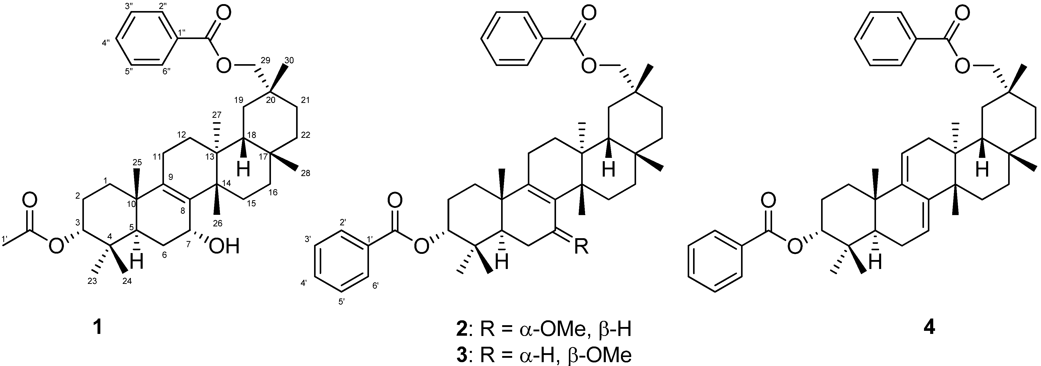
Four multiflorane-type triterpenes, including three new compounds, i.e. 7α-hydroxymultiflor-8-ene-3α,29-diol 3-acetate-29-benzoate (1), 7α-methoxymultiflor-8-ene-3α,29-diol 3,29-dibenzoate (2), and 7β-methoxymultiflor-8-ene-3α,29-diol 3,29-dibenzoate (3), were isolated from the MeOH extract of C. maxima seeds (Figure 1). The known compound, i.e. multiflora-7,9(11)-diene-3α,29-diol 3,29-dibenzoate (4), was identified by comparing its MS and 1H-NMR data with published values [10].
Figure 1. Chemical structures of isolated compounds 14.
Molecules 19 04802 g001
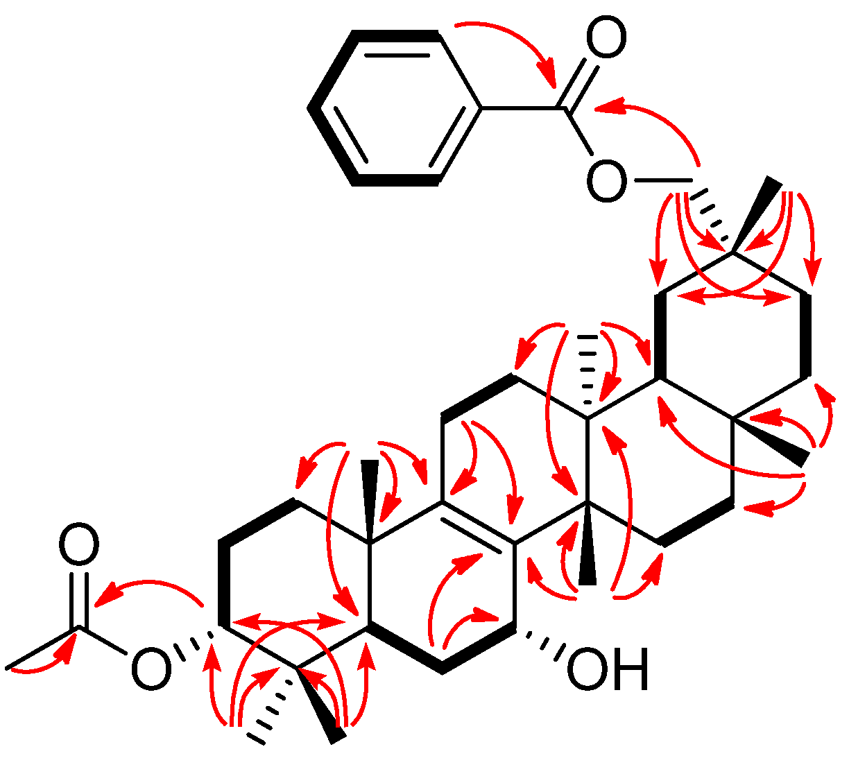
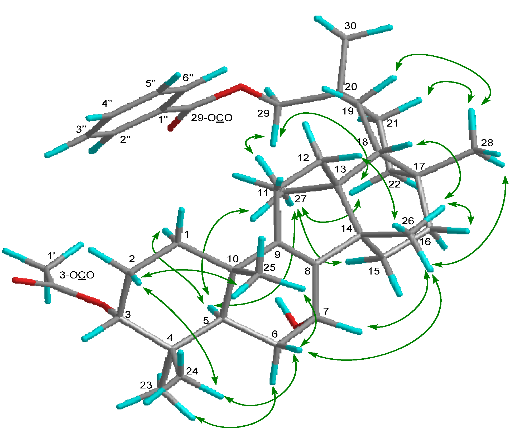
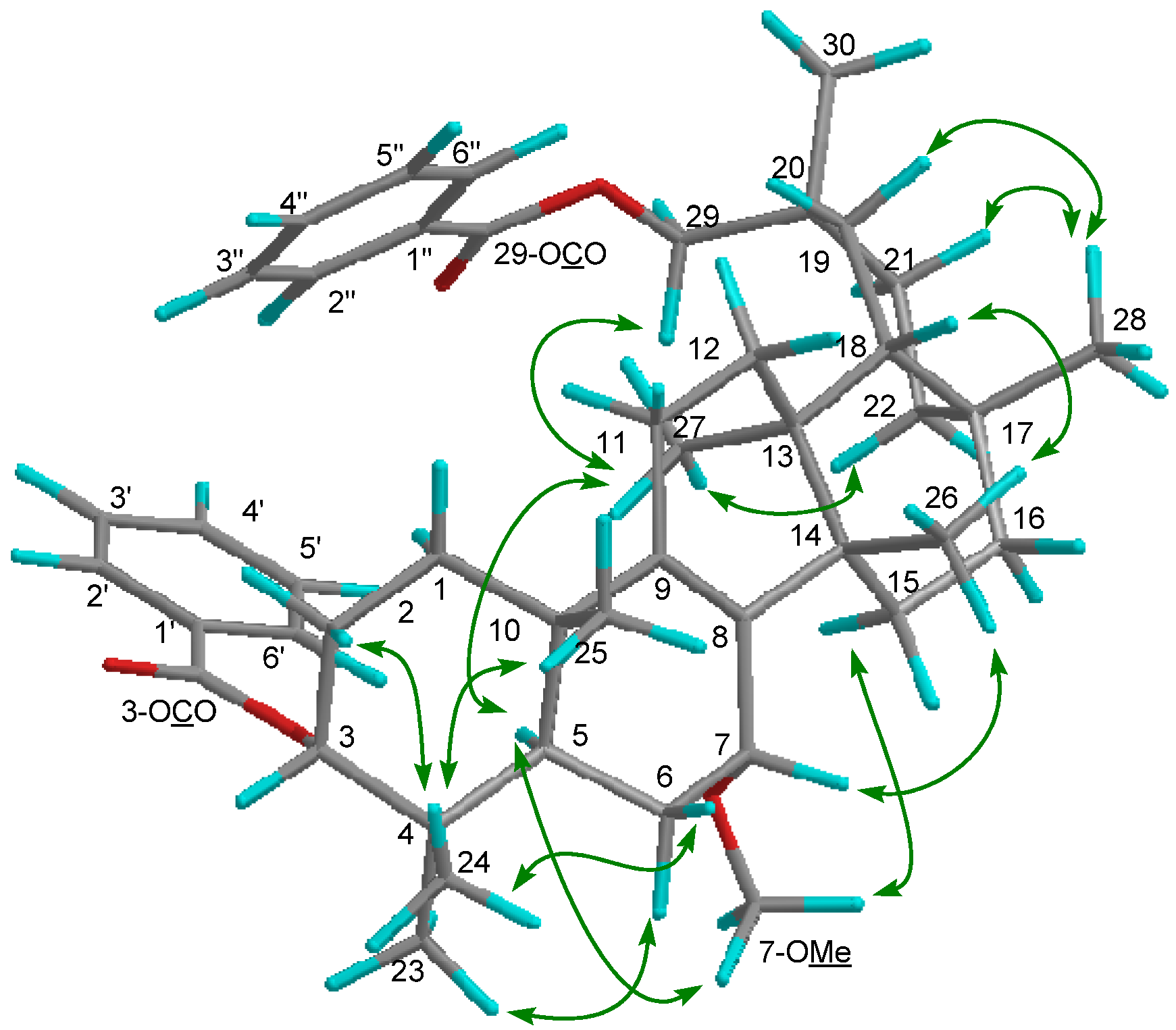
Compound 1 exhibited a [M–H2O]+ ion in the HREIMS data at m/z 586.4019 compatible with the molecular formula C39H54O4 (calcd. 586.4023), therefore it was suggested that the molecular formula of 1 is C39H56O5. The IR spectrum showed the presence of a hydroxy group (νmax 3437 cm−1) and ester carbonyl groups (νmax 1718, 1272, 1244 cm−1). The 1H and 13C-NMR spectra (Table 1) displayed signals for seven tertiary methyl groups [δH 0.89, 0.91, 0.92, 1.03, 1.06, 1.10, 1.12 (each s)], an oxymethylene [δH 4.14 (2H, brs); δC 72.5 (t)], two oxymethines [δH 4.16 (brs), 4.69 (t); δC 64.3 (d), 77.4 (d)], a tetrasubstituted olefin [δC 136.4 (s), 140.1 (s)], an acetoxy group [δH 2.07 (s); δC 21.4 (q), 170.9 (s)], and a benzoyl group [δH 7.47 (tt), 7.58 (tt), 8.07 (dd); δC 128.4 (d), 129.4 (d), 130.6 (s), 132.9 (d), 166.7 (s)]. The 1H- and 13C-NMR spectra are similar to those of 3-O-p-aminobenzoyl-29-O-benzoylmultiflor-8-ene-3α,7α,29-triol [3] except for the absence of a p-aminobenzoyl group at C-3 and existence of an acetyl group in 1. In the HMBC experiment, the following correlations were observed: Me-23 [δH 0.89 (s)] to C-3 [δc 77.4 (d)], C-4, C-5, and C-24; Me-24 [δH 0.91 (s)] to C-3, C-4, C-5, and Me-23; Me-25 [δH 0.92 (s)] to C-1, C-5, C-9 [δC 140.1 (s)], and C-10; Me-26 [δH 1.03 (s)] to C-8 [δC 136.4 (s)], C-13, C-14, and C-15; Me-27 [δH 1.06 (s)] to C-12, C-13, C-14, and C-18; Me-28 [δH 1.12 (s)] to C-16, C-17, C-18, and C-22; H2-29 [δH 4.14 (2H, brs)] to C-19, C-20, C-21, C-30, and 29-OCO [δC 166.7 (s)]; Me-30 [δH 1.10 (s)] to C-19, C-20, C-21, and C-29; H-3 [δH 4.69 (t)] to 3-OCO [δC 170.9 (s)]; H-5 and H-6α to C-7 [δC 64.3 (d)]; H-6α and H2-11 to C-8 [δC 136.4 (s)]; H-11 and H-12δ to C-9 [δC 140.1 (s)]. In the 1H-1H COSY experiment, H-7 [δH 4.16 (brs)] correlated with H2-6 [δH 1.60, 1.74] (Figure 2). The following significant NOE interactions were observed in 1: H-5/H-1α, Me-27; Me-23/H-6α; Me-27/H-15α, H-22α, and H2-29; H-2β /Me-24, and Me-25; Me-25/H-6β; Me-26/H-6β, H-7β, H-12β, H-16β, and Me-28; Me-28/H-19β, H-21β (Figure 3). In addition, the NOE correlations between H-7 and Me-26 suggested that the hydroxy group at C-7 is in the α (axial)-orientation (Figure 3). The configuration of the acetoxy group at C-3 was established as the α (axial)-orientation due to the coupling constants of H-3 [δH 4.69 (t, J3β.2α;3β,2β = 3.0 Hz)] and NOEs between H-3 and Me-24. Therefore, the structure of 1 was determined to be as shown in Figure 1.
Figure 2. Key HMBC and 1H-1H COSY correlations of compound 1.
Molecules 19 04802 g002
Figure 3. Selected NOE correlations of compound 1.
Molecules 19 04802 g003
Table 1. 1H (600 MHz) and 13C (150 MHz) NMR spectroscopic data of compounds 13 (CDCl3) a.
123
PositionδC, typeδH (J in Hz)δC, typeδH (J in Hz)δC, typeδH (J in Hz)
129.6tα1.33m29.8tα1.48m30.6tα1.40m
β1.47mβ1.53mβ1.52m
223.3tα1.66m23.6tα1.80m23.3tα1.80m
β1.88mβ1.98mβ1.97m
377.4d4.69t (3.0)78.1d4.95t (2.9)78.2d4.91t (3.0)
436.3s36.7s37.0s
539.6d1.91m39.9d2.19dd (12.6, 1.2)44.1d1.70m
628.8tα1.74m22.5tα1.95m25.3tα2.21m
β1.60mβ1.34mβ1.51m
764.3d4.16brs73.8d3.54brs78.8d3.98brt (7.6)
8136.4s135.3s136.7s
9140.1s139.7s140.3s
1038.6s38.6s38.2s
1120.9t1.932H, m20.9t1.972H, m20.8tα2.03m
β1.91m
1231.19tα1.34m31.3tα1.35m30.7tα1.51m
β1.61mβ1.61mβ1.40m
1336.8s37.0s38.2s
1441.9s41.8s40.9s
1526.1tα2.05m25.4tα2.19m26.3tα1.78m
β1.50mβ1.26mβ1.83m
1636.9tα1.54m36.9tα1.56m36.5tα1.61m
β1.67mβ1.61mβ1.53m
1731.2s31.1s31.2s
1844.4d1.61m44.0d1.60m42.8d1.66m
1928.4tα1.90m28.8tα1.86m29.8tα1.40m
β1.56mβ1.49mβ1.50m
2031.7s31.9s32.2s
2130.1tα1.47m29.9tα1.48m29.1t1.522H, m
β1.59mβ1.53m
2235.0tα1.87m35.6tα1.84d (4.4)37.1tα1.68m
β0.97mβ0.96mβ1.01m
2327.4q0.89s27.5q0.97s27.6q0.96s
2422.1q0.91s22.4q1.00s21.7q1.02s
2518.1q0.92s18.2q0.98s20.2q1.10s
2625.1q1.03s26.0q1.05s27.8q1.29s
2719.0q1.06s19.0q1.082s18.0q0.95s
2831.17q1.12s31.3q1.13s30.7q1.17s
2972.5t4.142H, brs72.9tA4.08d (10.8)74.0tA4.05d (10.6)
B4.16d (10.8)B4.11d (10.6)
3030.6q1.10s29.8q1.084s28.1q1.12s
3-OCO170.9s166.3s165.9s
1'21.4q2.07s130.8bs130.6s
2', 6'129.6cd8.05 bdd (7.4, 1.4)129.4d7.99dd (7.4, 1.4)
3', 5'128.4dd7.45 ctt (7.4, 1.4)128.5d7.45tt (7.4, 1.4)
4'132.7ed7.55 dtt (7.4, 1.4)132.8d7.56tt (7.4, 1.4)
29-OCO166.7s166.6s166.8s
1''130.6s130.7bs130.9s
2'', 6''129.4d8.07dd (7.4, 1.2)129.4cd8.04 bdd (7.4, 1.4)129.5d8.04dd (7.3, 1.7)
3'', 5''128.4d7.47tt (7.4, 1.2)128.3dd7.43 ctt (7.4, 1.4)128.4d7.43tt (7.3, 1.7)
4''132.9d7.58tt (7.4, 1.2)132.6ed7.54 dtt (7.4, 1.4)132.7d7.55tt (7.3, 1.7)
7-OMe54.9q3.24s55.0q3.35s
a Assignments were based on 1H-1H COSY, HMQC, HMBC and NOESY supectroscopic data. b−e Interchengeable.
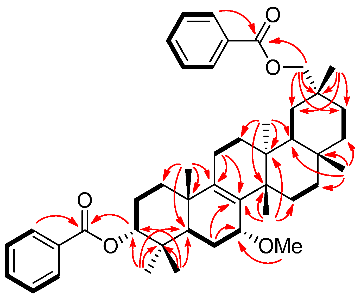
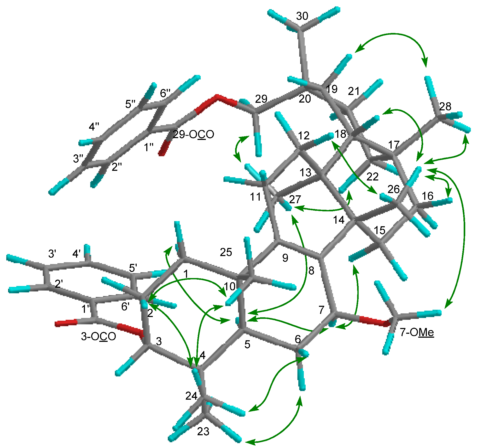
Compound 2 exhibited a [M]+ ion in the HREIMS data at m/z 680.4447 compatible with the molecular formula C45H60O5 (calcd. 680.4441). The IR spectrum showed absorption indicating ester carbonyl groups (νmax 1717, 1274 cm−1). The 1H- and 13C-NMR spectra (Table 1) displayed signals for seven tertiary methyl groups [δH 0.97, 0.98, 1.00, 1.05, 1.082, 1.084, 1.13 (each s)], an oxymethylene [δH 4.08, 4.16 (each d); δC 72.9 (t)], two oxymethines [δH 3.54 (brs), 4.95 (t); δC 73.8 (d), 78.1 (d)], a tetrasubstituted olefin [δC 135.3 (s), 139.7 (s)], and two benzoyl groups [δH 7.43 (tt), 7.45 (tt), 7.54 (tt), 7.55 (tt), 8.04 (dd), 8.05 (dd); δC 128.3 (d), 128.4 (d), 129.4 (d), 129.6 (d), 130.7 (s), 130.8 (s), 132.6 (d), 132.7 (d), 166.3 (s), 166.6 (s)]. The above data suggested that the structure of 2 is similar to that of 7α-methoxymultiflor-8-ene-3α,29-diol 3-acetate-29-benzoate [9], except for the lack of the 3-O-acetyl group and the existence of a 3-O-benzoyl group. In the HMBC experiment, the following correlations were observed: H2-29 [δH 4.08, 4.16 (each d)] to 29-OCO [δC 166.6 (s)]; H-3 [δH 4.95 (t)] to 3-OCO [δC 166.3 (s)]; H-5 and H-6α to C-7 [δC 73.8 (d)]; H-6α, H-7β, and Me-26 to C-8 [δC 135.3 (s)]; H-7β, H2-11 and Me-25 to C-9 [δC 139.7 (s)]. In the 1H-1H COSY experiment, H-7 [δH 3.54 (brs)] correlated with H2-6 [δH 1.34, 1.95] (Figure 4). Additionally, the NOEs were observed H-7/H-15β, Me-26; 7-OMe/H-5 and H-15α; suggested that the methoxy group at C-7 was in the α-orientation (Figure 5). The configuration of the acetoxy group at C-3 was established as the α-orientation due to the significant NOEs between H-3 and Me-24, and the coupling constants of H-3 [δH 4.95 (t, J = 2.9 Hz)]. Therefore, 2 was established as shown in Figure 1.
Figure 4. Key HMBC and 1H-1H COSY correlations of compound 2.
Molecules 19 04802 g004
Figure 5. Selected NOE correlations of compound 2.
Molecules 19 04802 g005
Compound 3 exhibited a [M]+ ion in the HREIMS data at m/z 680.4446 compatible with the molecular formula C45H60O5 (calcd. 680.4440). The IR spectrum showed absorption indicating ester carbonyl groups (νmax 1717, 1272 cm−1). The 1H- and 13C-NMR spectra (Table 1) displayed signals for seven tertiary methyl groups [δH 0.95, 0.96, 1.02, 1.10, 1.12, 1.17, 1.29 (each s)], an oxymethylene [δH 4.05, 4.11 (each d); δC 74.0 (t)], two oxymethines [δH 3.98 (brt), 4.91 (t); δC 78.2 (d), 78.8 (d)], a tetrasubstituted olefin [δC 136.7 (s), 140.3 (s)], and two benzoyl group [δH 7.43 (tt), 7.45 (tt), 7.55 (tt), 7.56 (tt), 7.99 (dd), 8.04 (dd); δC 128.4 (d), 128.5 (d), 129.4 (d), 129.5 (d), 130.6 (s), 130.9 (s), 132.7 (d), 132.8 (d), 165.9 (s), 166.8 (s)]. The 1H- and 13C-NMR spectra of 3 were very similar to those of 2, except for the H-7 signal [δH 3.98 (brt, J = 7.6 Hz): δC 78.8 (d) in 3; δH 3.54 (brs): δC 73.8 (d) in 2]. The coupling constants of H-7, and the NOE correlations of H-7/H-5α, and H-15α; 7-OMe/Me-26 suggested that the methoxy group at C-7 is in the β (equatorial)-orientation (Figure 6). The configuration of the benzoyl group at C-3 was established as the α-orientation due to the coupling constants of H-3 [δH 4.91 (t, J = 3.0 Hz)]. The above data established that 3 was a 7β-methoxy epimer of 2 (Figure 1).
Figure 6. Selected NOE correlations of compound 3.
Molecules 19 04802 g006
Multiflorane-type triterpenes are unusual, and most of them have been isolated from cucurbitaceae plants, such as Cucumis melo [11], Cucurbita pepo [3,4], Momordica cochinchinensis [12], and Trichosanthes kirilowii [13]. Only a few of their biological activities, such as anti-tumor promoting activities [14], anti-oxidant effects [15], cytotoxic activities [9,16], and melanogenesis inhibitory activities [9,16], have been reported. In this study, we evaluated them for melanogenesis inhibitory effects and cytotoxic activities against cancer cell lines. Melanogenesis plays an important role to protect the skin from UV irradiation. However, overproduction of melanin causes esthetic and dermatological problems [17], thus, several hypopigmenting products have been developed [17]. In this study, three new multiflorane triterpenes 13 from C. maxima were evaluated for inhibitory activities against α-MSH-induced melanogenesis in B16 melanomas (Table 2). To determine the safe concentration, cytotoxicities of compounds against B16 4A5 cells were examined by an MTT assay. Compound 2 did not exhibit cytotoxicity at 10–100 μM. Compounds 1 and 3 showed no toxicities at 10 μM, although they decreased cell viabilities at higher concentrations (1: 88.0% at 30 μM, 58.4% at 100 μM; 3: 86.3% at 30 μM, 67.2% at 100 μM). In the melanogenesis inhibitory assay, compound 1 reduced melanin content (88.5%) at a non-toxic concentration, 10 μM. The melanogenesis inhibitory activity of compound 1 was comparable with that of the positive control, arbutin (melanin content 88.9% at 10 μM), which has been recognized as a useful depigmentation compound for skin whitening in the cosmetic industry [18]. These results suggested that compound 2 may be valuable as a potential skin-whitening agent. Compounds 2 (10–100 μM) and 3 (10 μM) did not show any melanogenesis inhibitory activities.
Table 2. Melanogenesis inhibitory activity and cytotoxicity in B16 mouse melanoma cells of multiflorane-type triterpenes isolated from Cucurbita maxima seeds a.
CompoundConc. (μM)IC50 (μM)
1030100300
1melanin content88.5 ± 2.7 **65.8 ± 1.7 **35.7 ± 0.8 **46.5
cell viability105.4 ± 4.588.0 ± 1.058.4 ± 7.7 **>100
2melanin content96.3 ± 2.2105.5 ± 4.5108.3 ± 1.0>100
cell viability103.4 ± 3.6105.8 ± 5.0104.9 ± 7.7>100
3melanin content96.9 ± 9.279.4 ± 4.5 **60.1 ± 1.8 **>100
cell viability94.6 ± 0.486.3 ± 2.4 *67.2 ± 4.8**>100
4 bmelanin content98.4 ± 3.2102.2 ± 11.795.4 ± 8.4>100
cell viability110.8 ± 4.3103.0 ± 8.2101.1 ± 5.9>100
Arbutin cmelanin content88.9 ± 2.3 **72.3 ± 3.1 **55.3 ± 1.0 **33.8 ± 2.8 **124.6
cell viability100.0 ± 2.794.4 ± 1.289.9 ± 0.3 **81.9 ± 3.2 **>300
a Melanin content (%) and cell viability (%) were determined based on the absorbance at 450 nm, and 540 nm, respectively, by comparison with values for DMSO (100%). Each value represents the mean ± standard deviation (S.D.) of three determinations. Asterisks denote significant differences from control group, * p < 0.05, ** p < 0.01. The concentration of DMSO in the sample solution was 2 μL/mL. b Melanogenesis inhibitory and cytotoxicity data from [9]. c Reference compound.
Three triterpenes and a reference compound, 5-fluorouracil (5-FU), were also evaluated for cytotoxic activities against human leukemia (HL-60) and murine leukemia (P388) cell lines by means of the MTT assay. Compounds 1 and 3 exhibited weak cytotoxicities against HL-60 (IC50 1:89.2 μM; 3:64.6 μM) and P388 (IC50 1:93.7 μM; 3:34.5 μM). Compound 2 did not show activities against either cell line (IC50 each > 100 μM). In our previous study, several mutiflorane-type triterpenes were evaluated for their cytotoxic activities, and they showed no or weak activities, except 7-oxomultiflor-8-ene-3α,29-diol 3-acetate-29-benzoate, having a conjugated enone [9]. Results of this and previous studies suggest that a conjugated enone moiety strengthens the cytotoxic activities of multiflorane-type triterpenes.

3. Experimental

3.1. General Experimental Procedures

Chemicals and reagents were purchased as follows: fetal bovine serum (FBS) from Invitrogen (Carlsbad, CA, USA), 3-(4,5-dimethyl-2-thiazolyl)-2,5-diphenyl-2H-tetrazolium bromide (MTT) from Sigma-Aldrich Japan Co. (Tokyo, Japan), Roswell Park Memorial Institute (RPMI) 1640 medium, Dulbecco’s modified Eagle’s medium (D-MEM), and antibiotics from Nacalai tesque, Inc. (Kyoto, Japan). All other chemicals and reagents were of analytical grade. Melting points were determined on a Yanagimoto micro-melting point apparatus and are uncorrected. Optical rotations were measured with a JASCO DIP-1000 digital polarimeter. IR spectra were recorded on a Perkin-Elmer 1720X FTIR spectrophotometer. The 1H- (600 MHz) and 13C- (150 MHz) NMR spectra were recorded on an Agilent vnmrs600 instrument in CDCl3 with tetramethylsilane as the internal standard. The EIMS was recorded on a Hitachi 4000H double-focusing mass spectrometer (70 eV). Silica gel (70–230 mesh, Merck, Darmstadt, Germany) and silica gel 60 (230–400 mesh, Nacalai tesque, Inc.) were used for column chromatography and medium-pressure liquid chromatography, respectively. HPLC was carried out on an SiO2 column [Cosmosil 5SL-II column (Nacalai tesque, Inc.), 25 cm × 20 mm i.d.] at 25 °C with n-hexane/EtOAc [20:1 (HPLC system I), 10:1 (HPLC system II), and 5:1 (HPLC system III), flow rate 8.0 mL/min], and on ODS column [Cosmosil 5C18-MS-II column (Nacalai tesque, Inc.), 25 cm × 20 mm i.d.] at 25 °C with Me2CO:H2O [10:1 (HPLC system IV) and 9:1 (HPLC system V), flow rate 8.0 mL/min].

3.2. Plant Material

The seeds of Cucurbita maxima, produced in India, were purchased from Takada Seeds Co., Ltd. (Osaka, Japan) in 2011. A voucher specimen was deposited in the Herbarium of the Laboratory of Medicinal Chemistry, Osaka University of Pharmaceutical Sciences.

3.3. Extraction and Isolation

The seeds of Cucurbita maxima (10 kg) produced in India, were subjected to extraction with MeOH under reflux (30 L, one week, four times). The MeOH extract (310 g) was then partitioned between Et2O and H2O. The Et2O-soluble fraction (150 g) was subjected to SiO2 column chromatography (CC) [SiO2 (3.5 kg); CHCl3/MeOH 1:0, 10:1, 5:1, and 0:1 in increasing order of polarity] resulting in 9 fractions (Fr. A–I). Fr. B, eluted with CHCl3, was subjected to SiO2 CC to yield 18 fractions, B1–B18. Preparative HPLC of B7 (42.5 mg) (HPLC system II), eluted with hexane/EtOAc (10:1), gave 4 (28.0 mg; tR 23.2 min). Fr. C, eluted with CHCl3, was subjected to SiO2 CC to yield 24 fractions, C1–C24. Preparative HPLC of C5 (67.9 mg)(HPLC system II), eluted with hexane/EtOAc (10:1), gave Fr. C5-4 (44.4 mg; tR 11.2 min), and then re-preparative HPLC gave 2 (11.1 mg; tR 36.0 min)(HPLC system I). Preparative HPLC (HPLC system II) of C6 (64.0 mg), eluted with hexane/EtOAc (10:1), gave 15 fractions; C6-1–C6-15, and preparative HPLC (HPLC system II) of C7 (15.4 mg), eluted with hexane/EtOAc (10:1), gave 15 fractions; C7-1–C7-15. Preparative HPLC of C6-6 (2.9 mg; tR 13.0 min) and C7-6 (0.4 mg) combination gave 3 (1.0 mg; tR 59.0 min)(HPLC system IV). Fr. E, eluted with CHCl3, was fractionated with SiO2 CC to E1–E11. Preparative HPLC (HPLC system III) of E8 (119.8 mg), eluted with hexane/EtOAc (5:1), gave Fr. E8-10 (8.8 mg; tR 34.8 min), and then re-preparative HPLC (HPLC system V) gave 1 (1.6 mg; tR 13.6 min).

3.4. Product Characterization Data

7α-Hydroxymultiflor-8-ene-3α,29-diol 3-acetate-29-benzoate (1): Colorless crystal (MeOH); mp 66–68 °C; Molecules 19 04802 i001 −85.8 (c = 0.2, CHCl3); UV (EtOH) λmax (logε) 205.0 (3.80), 220.0 (3.94), 232.5 (3.98), 270.5 (3.41), 280.0 (3.32), 321.0 (2.87) nm; IR (KBr) νmax 3437, 2938, 2876, 1718, 1272, 1244, 1110, 750, 729, 710 cm−1; 1H and 13C-NMR data see Table 1; EIMS m/z 586 [M–H2O]+ (26), 540 (33), 527 (40), 511 (100), 389 (12), 387 (9), 253 (15), 225 (16); HREIMS m/z 586.4019 (calcd for C39H54O4: 586.4023).
7α-Methoxymultiflor-8-ene-3α,29-diol 3,29-dibenzoate (2): Colorless crystal (MeOH); mp 88–90 °C; Molecules 19 04802 i002 −20.7 (c = 0.7, CHCl3); UV (EtOH) λmax (logε) 238.5 (3.70), 271.5 (3.21), 278.5 (3.11) nm; IR (KBr) νmax:2948, 2883, 1717, 1456, 1367, 1314, 1274, 1113, 1069, 716 cm−1;1H and 13C-NMR data see Table 1; EIMS m/z 680 (6) [M]+, 648 [M–MeOH]+ (12), 526 (25), 511 (100), 389 (8), 355 (10), 324 (8); HREIMS m/z 680.4447 (calcd for C45H60O5: 680.4441).
-Methoxymultiflor-8-ene-3α,29-diol 3,29-dibenzoate (3).Colorless crystal (MeOH); mp 67–68 °C; Molecules 19 04802 i001 −43.7 (c = 0.2, CHCl3); UV (EtOH) λmax (logε) 206.5 (3.86), 220.0 (3.97), 235.0 (4.00), 271.0 (3.29), 280.0 (3.16) nm; IR (KBr) νmax:3437, 1717, 1272, 1110, 1028, 975, 711 cm−1; 1H and 13C-NMR data see Table 1; EIMS 680 (65) [M]+, 665 (38), 648 [M–MeOH]+ (14), 526 (29), 511 (100), 393 (18), 381 (18), 354 (28); HREIMS m/z 680.4446 [M]+ (calcd for C45H60O5: 680.4440).

3.5. Cell Cultures

The cell lines HL-60 (human leukemia) and P388 (murine leukemia) were grown in RPMI 1640 medium, while B16 4A5 cells were grown in D-MEM. The medium was supplemented with 10% FBS and antibiotics (100 units/mL penicillin and 100 μg/mL streptomycin). The cells were incubated at 37 °C in a 5% CO2 humidified incubator.

3.6. Determination of B16 4A5 Cells Proliferation

The assay of B16 4A5 cells proliferation was examined according to a method reported previously [9].

3.7. Assay of Melanin Content

The assay of melanin content was performed as described previously [9].

3.8. Cytotoxicity Assay against Cancer Cell Lines

The cytotoxicity assay was determined previously [19].

4. Conclusions

In this study, we isolated three new multiflorane-type triterpene esters, i.e. 7α-hydroxymultiflor-8-ene-3α,29-diol 3-acetate-29-benzoate (1), 7α-methoxymultiflor-8-ene-3α,29-diol 3,29-dibenzoate (2), and 7β-methoxymultiflor-8-ene-3α,29-diol 3,29-dibenzoate (3), from pumpkin seeds. Isolated compounds were evaluated for melanogenesis inhibitory and cytotoxic activities. In the melanogenesis inhibitory assay, we revealed that compound 1 possessed melanogenesis inhibitory activities comparable with arbutin at a non-toxic concentration. In a cytotoxicity assay against cancer cell lines, none of the compounds showed remarkable cytotoxic activities. We will continue to explore other biological activities of multiflorane-type triterpenes.

Supplementary Materials

Supplementary materials can be accessed at: https://www.mdpi.com/1420-3049/19/4/4802/s1.

Acknowledgments

We thank Katsuhiko Minoura and Mihoyo Fujitake (Osaka University of Pharmaceutical Sciences) for NMR and MS measurements.

Author Contributions

T. Kikuchi performed the isolation, structure elucidation, and evaluation of bioactivities, and prepared the manuscript. S. Ueda, J. Kanazawa, and H. Naoe contributed to the isolation and structure elucidation. T. Yamada and R. Tanaka supervised whole research project.

Conflicts of Interest

The authors declare no conflict of interest.

References

  1. Okada, M.; Mitsuhashi, H. Newly Revised Illustrated Medicinal Plants of World; Hokuryukan Pablishing Co., Ltd.: Tokyo, Japan, 2002; p. p. 514. [Google Scholar]
  2. Chevallier, A. The Encyclopedia of Medicinal Plants; Seibundo Shinkosha Pablishing Co., Ltd.: Tokyo, Japan, 2000; p. p. 194. [Google Scholar]
  3. Appendino, G.; Jakupovic, J.; Belloro, E.; Marchesini, A. Secondary metabolites from edible plants and spices. Part 4. Multiflorane triterpenoid esters from pumpkin. An unexpected extrafolic source of PABA. Phytochemistry 1999, 51, 1021–1026. [Google Scholar] [CrossRef]
  4. Appendino, G.; Jakupovic, J.; Belloro, E.; Marchesini, A. Triterpenoid p-aminobenzoates from the seeds of zucchini. Fitoterapia 2000, 71, 258–263. [Google Scholar] [CrossRef]
  5. Akihisa, T.; Ghosh, P.; Thakur, S.; Rosenstein, F.U.; Tamura, T.; Matsumoto, T. Widespread occurrence of cucurbita-5,24-dienol in Cucurbitaceae. Yukagaku 1986, 35, 1036–1040. [Google Scholar]
  6. Cattel, L.; Balliano, G.; Caputo, O. Sterols and triterpenes from Cucurbita maxima. Planta Med. 1979, 37, 264–267. [Google Scholar] [CrossRef]
  7. Fenner, G.P.; Patterson, G.W.; Koines, P.M. Sterol composition during the life cycle of the soybean and the squash. Lipids 1986, 21, 48–51. [Google Scholar] [CrossRef]
  8. Fenner, G.P.; Patterson, G.W.; Lusby, W.R. Developmental regulation of sterol biosynthesis in Cucurbita. maxima L. Lipids 1989, 24, 271–277. [Google Scholar] [CrossRef]
  9. Kikuchi, T.; Takebayashi, M.; Shinto, M.; Yamada, T.; Tanaka, R. Three new multiflorane-type triterpenes from pumpkin (Cucurbita maxima) seeds. Molecules 2013, 18, 5568–5579. [Google Scholar] [CrossRef]
  10. Ukiya, M.; Akihisa, T.; Tokuda, H.; Toriumi, M.; Mukainaka, T.; Banno, N.; Kimura, Y.; Hasegawa, J.; Nishino, H. Inhibitory effects of cucurbitane glycosides and other triterpenoids from the fruit of Momordica grosvenori on Epstein-Barr virus early antigen induced by tumor promoter 12-O-tetradecanoylphorbol-13-acetate. J. Agric. Food Chem. 2002, 50, 6710–6715. [Google Scholar] [CrossRef]
  11. De Marino, S.; Festa, C.; Zollo, F.; Iorizzi, M. Phenolic glycosides from Cucumis melo var. inodorus seeds. Phytochem. Lett. 2009, 2, 130–133. [Google Scholar] [CrossRef]
  12. De Shan, M.; Hu, L.H.; Chen, Z.L. A new multiflorane triterpenoid ester from Momordica cochinchinensis Spreng. Nat. Prod. Lett. 2001, 15, 139–145. [Google Scholar] [CrossRef]
  13. Ma, Y.P.; Li, N.; Gao, J.; Fu, K.L.; Qin, Y.; Li, G.Y.; Wang, J.H. A new peroxy-multiflorane triterpene ester from the processed seeds of Trichosanthes kirilowii. Helv. Chim. Acta 2011, 94, 1881–1887. [Google Scholar]
  14. Akihisa, T.; Tokuda, H.; Ichiishi, E.; Mukainaka, T.; Toriumi, M.; Ukiya, M.; Yasukawa, K.; Nishino, H. Anti-tumor promoting effects of multiflorane-type triterpenoids and cytotoxic activity of karounidiol against human cancer cell lines. Cancer Lett. 2001, 173, 9–14. [Google Scholar] [CrossRef]
  15. Liu, C.H.; Yen, M.H.; Tsang, S.F.; Gan, K.H.; Hsu, H.Y.; Lin, C.N. Antioxidant triterpenoids from the stems of Momordica charantia. Food Chem. 2009, 118, 751–756. [Google Scholar]
  16. Tanaka, R.; Kikuchi, T.; Nakasuji, S.; Ue, Y.; Shuto, D.; Igarashi, K.; Okada, R.; Yamada, T. A novel 3α-p-nitrobenzoylmultiflora-7:9(11)-diene-29-benzoate and two new triterpenoids from the seeds of zucchini (Cucurbita pepo L.). Molecules 2013, 18, 7448–7459. [Google Scholar] [CrossRef]
  17. Lin, J.W.; Chiang, H.M.; Lin, Y.C.; Wen, K.C. Natural products with skin-whitening effects. J. Food Drug Anal. 2008, 16, 1–10. [Google Scholar]
  18. Lim, Y.J.; Lee, E.H.; Kang, T.H.; Ha, S.K.; Oh, M.S.; Kim, S.M.; Yoon, T.J.; Kang, C.; Park, J.H.; Kim, S.Y. Inhibitory effects of arbutin on melanin biosynthesis of α-melanocyte stimulating hormone-induced hyperpigmentation in cultured Brownish guinea pig skin tissues. Arch. Pharm. Res. 2009, 32, 367–373. [Google Scholar] [CrossRef]
  19. Yamada, T.; Muroga, Y.; Jinno, M.; Kajimoto, T.; Usami, Y.; Numata, A.; Tanaka, R. New class azaphilone produced by a marine fish-derived Chaetomium globosum. The stereochemistry and biological activities. Bioorg. Med. Chem. 2011, 19, 4106–4113. [Google Scholar]
  • Sample Availability: Not available.