Phytochemical Analysis of Pfaffia glomerata Inflorescences by LC-ESI-MS/MS

Abstract
:1. Introduction
2. Results and Discussion
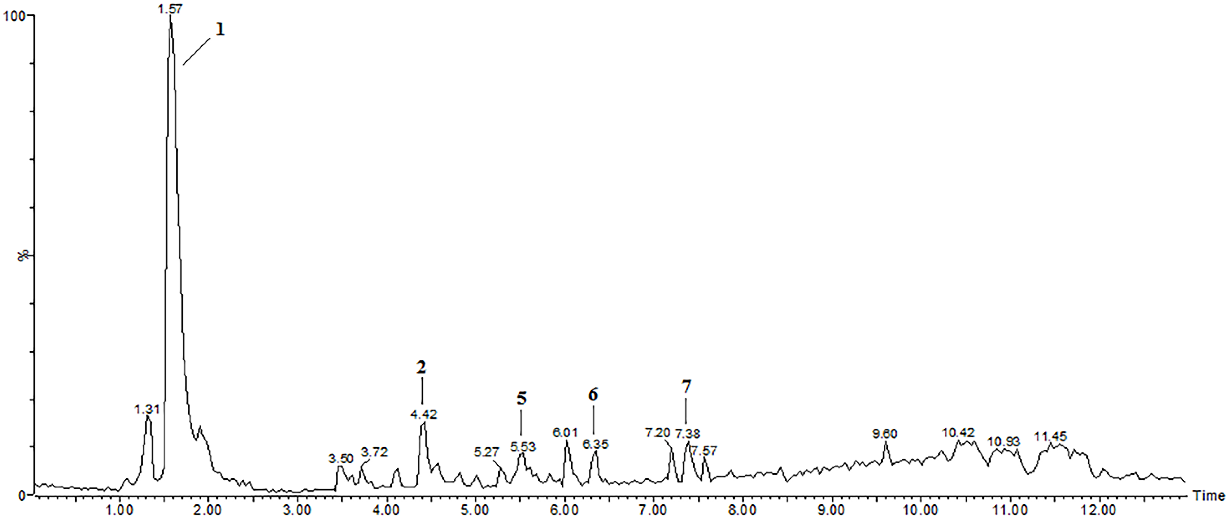
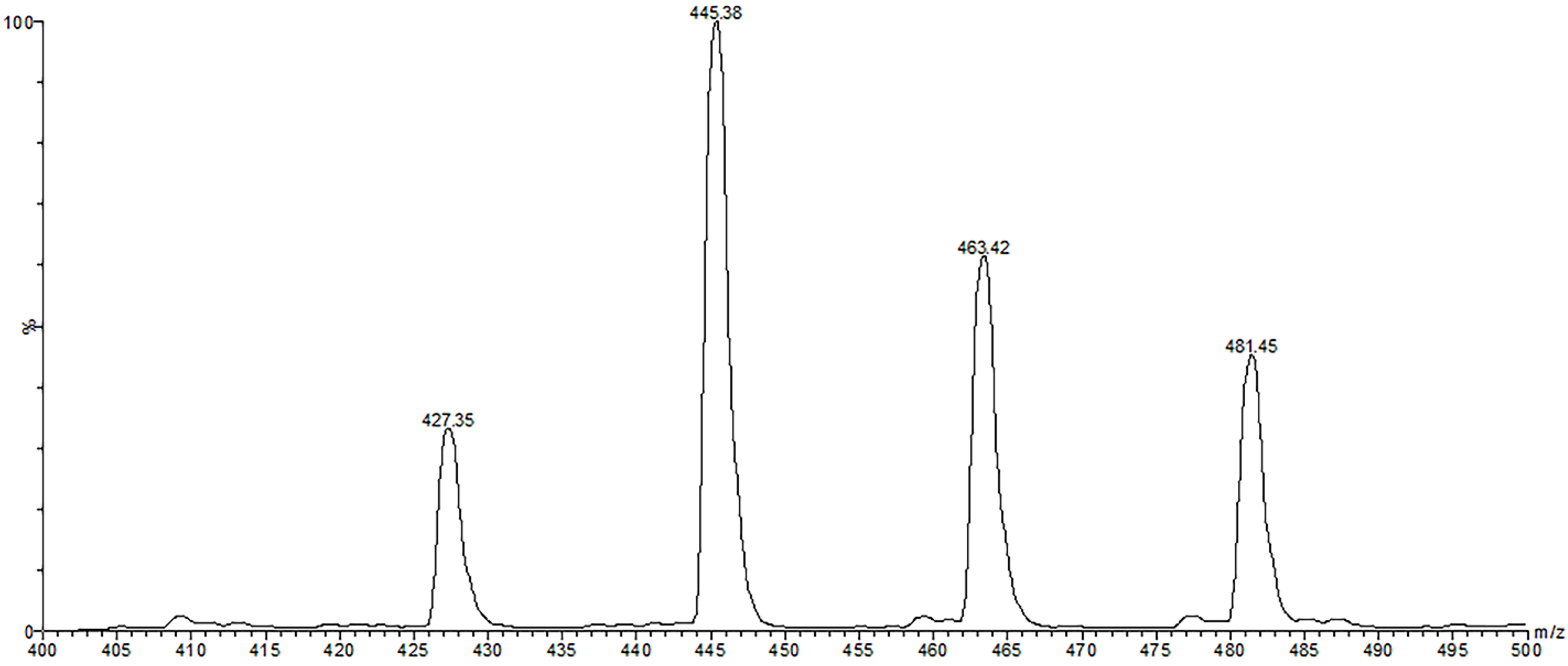
2.1. Identification of Compounds in P. glomerata Inflorescences by LC-ESI-MS/MS in Positive Ion Mode

| Peak a | tr (min) | Positive Ion Mode | Negative Ion Mode | Tentative Assignment | ||
|---|---|---|---|---|---|---|
| Precursor Ion (m/z) | Fragment Ions d (m/z) | Precursor Ion (m/z) | Fragment Ions d (m/z) | |||
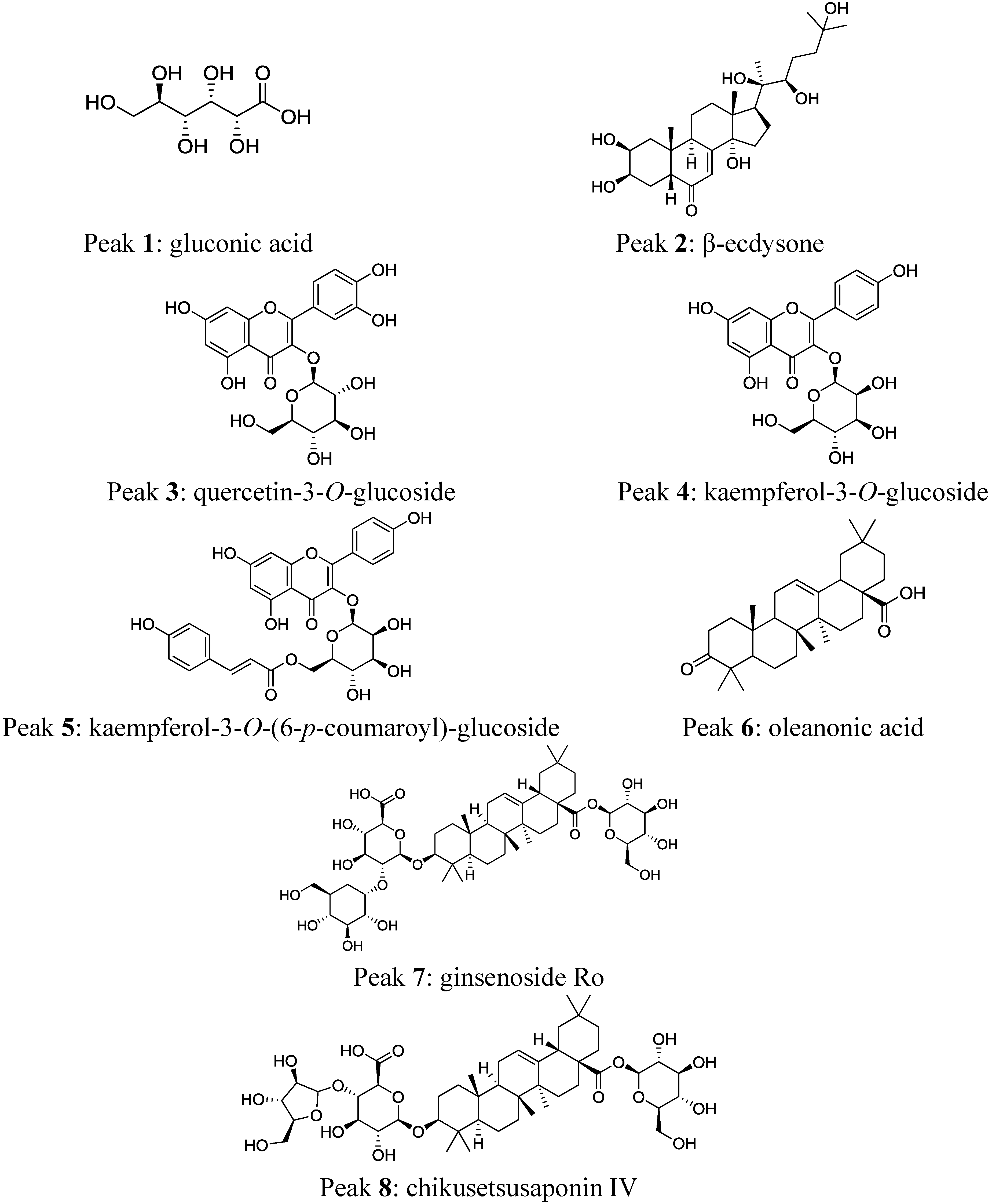
| 1 | 1.6 | 235 [M+K]+ c | 118, 59 | 195 [M-H]− | 177, 159, 129, 99, 75 | gluconic acid |
| 2 | 4.4 | 481 [M+H]+ b | 463, 445, 427, 409, 371, 165 | 525 [M+HCOO]− c | 479, 461, 319, 159 | β-ecdysone |
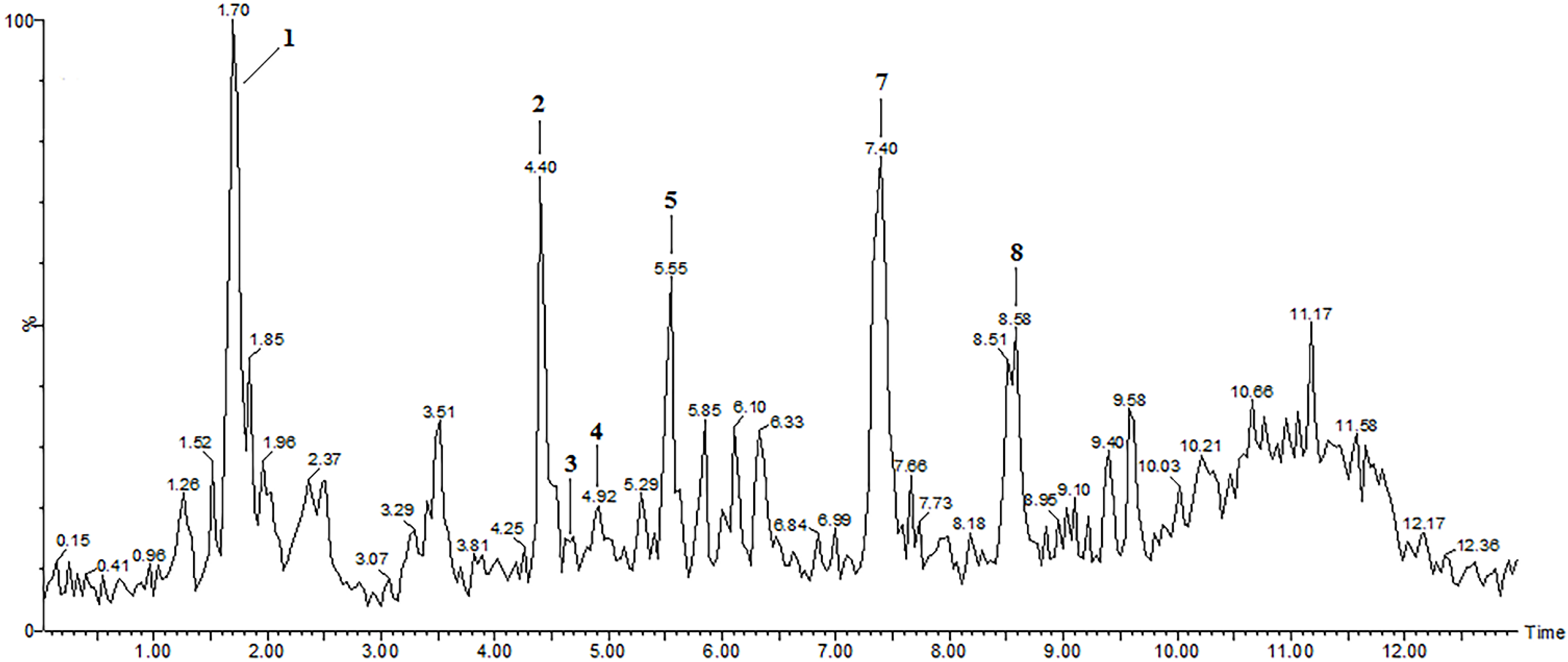
| 3 | 4.7 | ND | ND | 463 [M-H]− | 301/300 | quercetin-3-O-glucoside |
| 4 | 4.9 | ND | ND | 447 [M-H]− | 284 | kaempferol-3-O-glucoside |
| 5 | 5.5 | 595 [M+H]+ | 309, 287, 147 | 593 [M-H]− | 447, 307, 285 | kaempferol-3-O-(6-p-coumaroyl)-glucoside |
| 6 | 6.4 | 455 [M+H]+ | 438, 410, 147 | ND | ND | oleanonic acid |
| 7 | 7.4 | 439 [aglycone+H-H2O]+ | 393, 249, 203, 191 | 955 [M-H]− | 910, 793, 631 | ginsenoside Ro |
| 8 | 8.6 | ND | ND | 925 [M-H]− | 793,763 | chikusetsusaponin IV |
2.2. Identification of Compounds in P. glomerata Inflorescences by LC-ESI-MS/MS in Negative Ion Mode
3. Experimental Section
3.1. Chemicals and Materials
3.2. Plant Material and Sample Preparation
3.3. LC-ESI-MS and LC-ESI-MS/MS Analyses
4. Conclusions
Acknowledgments
Author Contributions
Conflicts of Interest
References
- Nascimento, E.X.; Mota, J.H.; Vieira, M.C.; Zárate, N.A.H. Produção de biomassa de Pfaffia glomerata (Spreng.) Pedersen e Plantago major L. em cultivo solteiro e consorciado. Cienc. Agrotec. 2007, 31, 724–730. [Google Scholar] [CrossRef]
- Festucci-Buselli, R.A.; Contim, L.A.S.; Barbosa, L.C.A.; Stuart, J.J.; Otoni, W.C. Biosynthesis and potencial functions of the ecdysteroid 20-hydroxyecdysone—A review. Botany 2008, 86, 978–987. [Google Scholar] [CrossRef]
- Flores, R.; Brondani, D., Jr.; Cezarotto, V., Jr.; Giacomelli, S.R.; Nicoloso, F.T. Micropropagation and β-ecdysone content of the Brazilian ginsengs Pfaffia glomerata and Pfaffia tuberosa. In Vitro Cell. Dev. Biol.-Plant 2010, 46, 210–217. [Google Scholar] [CrossRef]
- Carulo, M.F. Use of SFC in extraction of adaptogens from Brazilian plants. Am. J. Anal. Chem. 2012, 3, 977–982. [Google Scholar] [CrossRef]
- Freitas, C.S.; Baggio, C.H.; da Silva-Santos, J.E.; Rieck, L.; Santos, C.A.M.; Junior, C.C.; Ming, L.C.; Cortez, D.A.G.; Marques, M.C.A. Involvement of nitric oxide in the gastroprotective effects of an aqueous extract of Pfaffia glomerata (Spreng) Pedersen, Amaranthaceae, in rats. Life Sci. 2004, 74, 1167–1179. [Google Scholar] [CrossRef]
- Daniel, J.F.S.; Alves, K.Z.; Jacques, J.D.S.; Souza, P.V.S.; Carvalho, M.G.; Freire, R.B.; Ferreira, D.T.; Freire, M.F.I. Free radical scavenging activity of Pfaffia glomerata (Spreng.) Pedersen (Amaranthaceae). Indian J. Pharmacol. 2005, 37, 174–178. [Google Scholar] [CrossRef]
- Leal, P.F.; Kfouri, M.B.; Alexandre, F.C.; Fagundes, F.H.R.; Prado, J.M.; Toyama, M.H.; Meireles, M.A.A. Brazilian Ginseng extraction via LPSE and SFE: Global yields, extraction kinetics, chemical composition and antioxidant activity. J. Supercrit. Fluids 2010, 54, 38–45. [Google Scholar] [CrossRef]
- Neto, A.G.; da Silva Filho, A.A.; Costa, J.M.L.C.; Vinholis, A.H.C.; Souza, G.H.B.; Cunha, W.R.; Silva, M.L.A.; Albuquerque, S.; Bastos, J.K. Evaluation of the trypanocidal and antileishmanial in vitro activity of the crude hydroalcoholic extract of Pfaffia glomerata (Amaranthaceae) roots. Phytomedicine 2004, 11, 662–665. [Google Scholar] [CrossRef]
- Neto, A.G.; Costa, J.M.; Belati, C.C.; Vinholis, A.H.; Possebom, L.S.; da Silva Filho, A.A.; Cunha, W.R.; Carvalho, J.C.; Bastos, J.K.; Silva, M.L. Analgesic and anti-inflammatory activity of a crude root extract of Pfaffia glomerata (Spreng) Pedersen. J. Ethnopharmacol. 2005, 96, 87–91. [Google Scholar] [CrossRef]
- Shiobara, Y.; Inoue, S.S.; Kato, K.; Nishiguchi, Y.; Oishi, Y.; Nishimoto, N.; de Oliveira, F.; Akisue, G.; Akisue, M.K.; Hashimoto, G. A nortriterpenoid, triterpenoids and ecdystereoids from Pfaffia glomerata. Phytochemistry 1993, 32, 1527–1530. [Google Scholar] [CrossRef]
- Iarema, L.; da Cruz, A.C.F.; Saldanha, C.W.; Dias, L.L.C.; Vieira, R.F.; de Oliveira, E.J.; Otoni, W.C. Photoautotrophic propagation of Brazilian ginseng [Pfaffia glomerata (Spreng.) Pedersen]. Plant Cell Tissue Org. 2012, 110, 227–238. [Google Scholar] [CrossRef]
- Zimmer, A.R.; Bruxel, F.; Bassani, V.L.; Gosmann, G. HPLC method for the determination of ecdysterone in extractive solution from Pfaffia glomerata. J. Pharm. Biomed. 2006, 40, 450–453. [Google Scholar] [CrossRef]
- Serra, L.Z.; Felipe, D.F.; Cortez, D.A.G. Quantification of β-ecdysone in differents parts of Pfaffia glomerata by HPLC. Rev. Bras. Farmacogn. 2012, 22, 1349–1354. [Google Scholar]
- Lafont, R.; Dinan, L. Practical uses for ecdysteroids in mammals including humans: An update. J. Insect Sci. 2003, 3, 1–30. [Google Scholar] [CrossRef]
- Nsimba, R.Y.; Kikuzaki, H.; Konishi, Y. Ecdysteroids act as inhibitors of calf skin collagenase and oxidative stress. J. Biochem. Mol. Toxicol. 2008, 22, 240–250. [Google Scholar] [CrossRef]
- Dinan, L.; Lafont, R. Effects and applications of arthropod steroid hormones (ecdysteroids) in mammals. J. Endocrinol. 2006, 191, 1–8. [Google Scholar] [CrossRef]
- Kim, H.Y.; Park, H.M.; Lee, C.H. Mass spectrometry-based chemotaxonomic classification of Penicillium species (P. echinulatum, P. expansum, P. solitum, and P. oxalicum) and its correlation with antioxidant activity. J. Microbiol. Methods 2012, 90, 327–335. [Google Scholar] [CrossRef]
- Swatsitang, P.; Tucker, G.; Robards, K.; Jardine, D. Isolation and identification of phenolic compounds in Citrus sinensis. Anal. Chim. Acta 2000, 417, 231–240. [Google Scholar] [CrossRef]
- Ramachandran, S.; Fontanille, P.; Pandey, A.; Larroche, C. Fed-batch production of gluconic acid by terpene-treated Aspergillus niger spores. Appl. Biochem. Biotechnol. 2008, 151, 413–423. [Google Scholar] [CrossRef]
- Amin, M.A.; Abd El Rehim, S.S.; El-Lithy, A.S. Pitting and pitting control of Al in gluconic acid solutions—Polarization, chronoamperometry and morphological studies. Corros. Sci. 2010, 52, 3099–3108. [Google Scholar] [CrossRef]
- Duckstein, S.M.; Stintzing, F.C. Comprehensive study of the phenolics and saponins from Helleborus niger L. leaves and stems by liquid chromatography/tandem mass spectrometry. Chem. Biodivers. 2014, 11, 276–298. [Google Scholar] [CrossRef]
- Li, Y.; Warren, J.T.; Boysen, G.; Gilbert, L.I.; Gold, A.; Sangaiah, R.; Ball, L.M.; Swenberg, J.A. Profiling of ecdysteroids in complex biological samples using liquid chromatography/ion trap mass spectrometry. Rapid Commun. Mass Spectrom. 2006, 20, 185–192. [Google Scholar] [CrossRef]
- Stevens, J.F.; Reed, R.L.; Morré, J.T. Characterization of phytoecdysteroid glycosides in meadowfoam (Limnanthes alba) seed meal by positive and negative ion LC-MS/MS. J. Agric. Food Chem. 2008, 56, 3945–3952. [Google Scholar] [CrossRef]
- Wang, Y.H.; Avula, B.; Jadhav, A.N.; Smillie, T.J.; Khan, I.A. Structural characterization and identification of ecdysteroids from Sida rhombifolia L. in positive electrospray ionization by tandem mass spectrometry. Rapid Commun. Mass Spectrom. 2008, 22, 2413–2422. [Google Scholar] [CrossRef]
- Wainwright, G.; Prescott, M.C.; Lomas, L.O.; Webster, S.G.; Rees, H.H. Development of a new high-performance liquid chromatography-mass spectrometric method for the analysis of ecdysteroids in biological extracts. Arch. Insect Biochem. 1997, 35, 21–31. [Google Scholar] [CrossRef]
- Destrez, B.; Pinel, G.; Bichon, E.; Monteau, F.; Lafont, R.; Le Bizec, B. Detection of 20-hydroxyecdysone in calf urine by comparative liquid chromatography/high-resolution mass spectrometry and liquid chromatography/tandem mass spectrometry measurements: Application to the control of the potential misuse of ecdysteroids in cattle. Rapid Commun. Mass Spctrom. 2008, 22, 4073–4080. [Google Scholar] [CrossRef]
- Miyashita, M.; Matsushita, K.; Nakamura, S.; Akahane, S.; Nakagawa, Y.; Miyagawa, H. LC/MS/MS identification of 20-hydroxyecdysone in a scorpion (Liocheles australasiae) and its binding affinity to in vitro-translated molting hormone receptors. Insect Biochem. Mol. 2011, 41, 932–937. [Google Scholar] [CrossRef]
- Laghari, A.Q.; Memon, S.; Nelofar, A.; Laghari, A.H. Tecomella undulate G. Don: A rich source of flavonoids. Ind. Crop. Prod. 2013, 43, 213–217. [Google Scholar] [CrossRef]
- Atoui, A.K.; Mansouri, A.; Boskou, G.; Kefalas, P. Tea and herbal infusions: Their antioxidant activity and phenolic profile. Food Chem. 2005, 89, 27–36. [Google Scholar] [CrossRef]
- Zhao, Y.; Chen, P.; Lin, L.; Harnly, J.M.; Yu, L.L.; Li, Z. Tentative identification, quantitation, and principal component analysis of green pu-erh, green, and white teas using UPLC/DAD/MS. Food Chem. 2011, 126, 1269–1277. [Google Scholar] [CrossRef]
- Kajdžanoska, M.; Gjamovski, V.; Stefova, M. HPLC-DAD-ESI-MSn identification of phenolic compounds in cultivated strawberries from Macedonia. Maced. J. Chem. Chem. Eng. 2010, 29, 181–194. [Google Scholar]
- Karioti, A.; Bilia, A.R.; Skaltsa, H. Quercus ilex L.: A rich source of polyacylated flavonoid glucosides. Food Chem. 2010, 123, 131–142. [Google Scholar] [CrossRef]
- Seeram, P.N.; Lee, R.; Scheuller, S.H.; Heber, D. Identification of phenolic compounds in strawberries by liquid chromatography electrospray ionization mass spectroscopy. Food Chem. 2006, 97, 1–11. [Google Scholar] [CrossRef]
- Mellou, F.; Lazari, D.; Skaltsa, H.; Tselepis, A.D.; Kolisis, F.N.; Stamatis, H. Biocatalytic preparation of acylated derivatives of flavonoid glycosides enhances their antioxidant and antimicrobial activity. J. Biotechnol. 2005, 116, 295–304. [Google Scholar] [CrossRef]
- Van der Doelen, G.A.; van den Berg, K.J.; Boon, J.J.; Shibayama, N.; Renè De La Rie, E.; Genuit, W.J.L. Analysis of fresh triterpenoid resins and aged triterpenoid varnishes by high-performance liquid chromatography–atmospheric pressure chemical ionisation (tandem) mass spectrometry. J. Chromatogr. A 1998, 809, 21–37. [Google Scholar] [CrossRef]
- Chen, Q.; Zhang, Y.; Zhang, W.; Chen, Z. Identification and quantification of oleanolic acid and ursolic acid in Chinese herbs by liquid chromatography—Ion trap mass spectrometry. Biomed. Chromatogr. 2011, 25, 1381–1388. [Google Scholar] [CrossRef]
- Huang, L.; Chen, T.; Ye, Z.; Chen, G. Use of liquid chromatography–atmospheric pressure chemical ionization-ion trap mass spectrometry for identification of oleanolic acid and ursolic acid in Anoectochilus roxburghii (wall.) Lindl. J. Mass Spectrom. 2007, 42, 910–917. [Google Scholar] [CrossRef]
- Li, Y.J.; Wei, H.L.; Qi, L.W.; Chen, J.; Ren, M.T.; Li, P. Characterization and identification of saponins in Achyranthes bidentata by rapid-resolution liquid chromatography with electrospray ionization quadrupole time-of-flight tandem mass spectrometry. Rapid Commun. Mass Spectrom. 2010, 24, 2975–2985. [Google Scholar] [CrossRef]
- Liu, X.R.; Zheng, X.F.; Ji, S.Z.; Lv, Y.H.; Zheng, D.Y.; Xia, Z.F.; Zhang, W.D. Metabolomic analysis of thermally injured and/or septic rats. Burns 2010, 36, 992–998. [Google Scholar] [CrossRef]
- Yang, H.; Lin, W.; Zhang, J.; Lin, W.; Xu, P.; Li, J.; Ling, X. Metabonomic analysis of the toxic effects of TM208 in rat urine by HPLC-ESI-IT-TOF/MS. J. Chromatogr. B 2014, 959, 49–54. [Google Scholar] [CrossRef]
- Deng, J.; Yang, Y. Chemical fingerprint analysis for quality assessment and control of Bansha herbal tea using paper spray mass spectrometry. Anal. Chim. Acta 2013, 785, 82–90. [Google Scholar] [CrossRef]
- Li, J.; Qi, H.; Qi, L.W.; Yi, L.; Li, P. Simultaneous determination of main phytoecdysones and triterpenoids in radix achyranthis bidentatae by high-performance liquid chromatography with diode array-evaporative light scattering detectors and mass spectrometry. Anal. Chim. Acta 2007, 596, 264–272. [Google Scholar] [CrossRef]
- Destrez, B.; Pinel, G.; Monteau, F.; Lafont, R.; Le Bizec, B. Detection and identification of 20-hydroxyecdysone metabolites in calf urine by liquid chromatography-high resolution or tandem mass spectrometry measurements and establishment of their kinetics of elimination after 20-hydroxyecdysone administration. Anal. Chim. Acta 2009, 637, 178–184. [Google Scholar]
- Sánchez-Rabaneda, F.; Jáuregui, O.; Casals, I.; Andrés-Lacueva, C.; Izquierdo-Pulido, M.; Lamuela-Raventós, R.M. Liquid chromatographic/electrospray ionization tandem mass spectrometric study of the phenolic composition of cocoa (Theobroma cacao). J. Mass Spectrom. 2003, 38, 35–42. [Google Scholar] [CrossRef]
- Singh, A.P.; Wilson, T.; Luthria, D.; Freeman, M.R.; Scott, R.M.; Bilenker, D.; Shah, S.; Somasundaram, S.; Vorsa, N. LC-MS–MS characterisation of curry leaf flavonols and antioxidant activity. Food Chem. 2011, 127, 80–85. [Google Scholar] [CrossRef]
- Hvattum, E.; Ekeberg, D. Study of the collision-induced radical cleavage of flavonoid glycosides using negative electrospray ionization tandem quadrupole mass spectrometry. J. Mass Spectrom. 2003, 38, 43–49. [Google Scholar] [CrossRef]
- Gouveia, S.; Castilho, P.C. Characterisation of phenolic acid derivatives and flavonoids from different morphological parts of Helichrysum obconicum by a RP-HPLC–DAD-(−)–ESI-MSn method. Food Chem. 2011, 129, 333–344. [Google Scholar] [CrossRef]
- Castillo-Muñoz, N.; Gómez-Alonso, S.; García-Romero, E.; Gómez, M.V.; Velders, A.H.; Hermosín-Gutiérrez, I. Flavonol 3-O-glycosides series of Vitis vinifera cv. Petit Verdot red wine grapes. J. Agric. Food Chem. 2009, 57, 209–219. [Google Scholar] [CrossRef]
- Abu-Reidah, I.M.; Arráez-Román, D.; Quirantes-Piné, R.; Fernández-Arroyo, S.; Segura-Carretero, A.; Fernández-Gutiérrez, A. HPLC–ESI-Q-TOF-MS for a comprehensive characterization of bioactive phenolic compounds in cucumber whole fruit extract. Food Res. Int. 2002, 46, 108–117. [Google Scholar] [CrossRef]
- Falcão, S.I.; Vale, N.; Gomes, P.; Domingues, M.R.M.; Freire, C.; Cardoso, S.M.; Vilas-Boas, M. Phenolic profiling of Portuguese propolis by LC–MS spectrometry: Uncommon propolis rich in flavonoid glycosides. Phytochem. Anal. 2012, 24, 309–318. [Google Scholar] [CrossRef]
- Zehl, M.; Braunberger, C.; Conrad, J.; Crnogorac, M.; Krasteva, S.; Vogler, B.; Beifuss, U.; Krenn, L. Identification and quantification of flavonoids and ellagic acid derivatives in therapeutically important Drosera species by LC–DAD, LC–NMR, NMR, and LC–MS. Anal. Bioanal. Chem. 2011, 400, 2565–2576. [Google Scholar] [CrossRef]
- Ferreres, F.; Gil-Izquierdo, A.; Vinholes, J.; Silva, S.T.; Valentão, P.; Andrade, P.B. Bauhinia forficata link authenticity using flavonoids profile: Relation with their biological properties. Food Chem. 2012, 134, 894–904. [Google Scholar] [CrossRef]
- Ramirez, J.E.; Zambrano, R.; Sepúlveda, B.; Simirgiotis, M.J. Antioxidant properties and hyphenated HPLC-PDA-MS profiling of chilean Pica mango fruits (Mangifera indica L. Cv. piqueño). Molecules 2014, 19, 438–458. [Google Scholar] [CrossRef]
- Gouveia, S.C.; Castilho, P.C. Analysis of phenolic compounds from different morphological parts of Helichrysum devium by liquid chromatography with on-line UV and electrospray ionization mass spectrometric detection. Rapid Commun. Mass Spetrom. 2009, 23, 3939–3953. [Google Scholar] [CrossRef]
- Plazonić, A.; Bucar, F.; Maleš, Ž.; Mornar, A.; Nigović, B.; Kujundžić, N. Identification and quantification of flavonoids and phenolic acids in burr parsley (Caucalis platycarpos L.), using high-performance liquid chromatography with diode array detection and electrospray ionization mass spectrometry. Molecules 2009, 14, 2466–2490. [Google Scholar] [CrossRef]
- Kite, G.C.; Veitch, N.C. Identification of common glycosyl groups of flavonoid O-glycosides by serial mass spectrometry of sodiated species. Rapid Commun. Mass Spectrom. 2011, 25, 2579–2590. [Google Scholar]
- Yang, W.Z.; Ye, M.; Qiao, X.; Liu, C.F.; Miao, W.J.; Bo, T.; Tao, H.Y.; Guo, D.A. A strategy for efficient discovery of new natural compounds by integrating orthogonal column chromatography and liquid chromatography/mass spectrometry analysis: Its application in Panax ginseng, Panax quinquefolium and Panax notoginseng to characterize 437 potential new ginsenosides. Anal. Chim. Acta 2012, 739, 56–66. [Google Scholar] [CrossRef]
- Sun, X.; Chen, P.; Cook, S.L.; Jackson, G.P.; Harnly, J.M.; Harrington, P.B. Classification of cultivation locations of Panax quinquefolius L samples using high performance liquid chromatography–electrospray ionization mass spectrometry and chemometric analysis. Anal. Chem. 2012, 84, 3628–3634. [Google Scholar] [CrossRef]
- Yan, Z.; Lin, G.; Ye, Y.; Wang, Y.; Yan, R. Triterpenoid saponins profiling by adducts-targeted neutral loss triggered enhanced resolution and product ion scanning using triple quadrupole linear ion trap mass spectrometry. Anal. Chim. Acta 2014, 819, 56–64. [Google Scholar] [CrossRef]
- Sha, Y.; Yan, M.C.; Liu, J.; Liu, Y.; Cheng, M.S. Facile synthesis of oleanolic acid monoglycosides and diglycosides. Molecules 2008, 13, 1472–1486. [Google Scholar] [CrossRef]
- Waffo-Téguo, P.; Voutquenne, L.; Thoison, O.; Dumontet, V.; Nguyen, V.H.; Lavaud, C. Acetylated glucuronide triterpene bidesmosidic saponins from Symplocos glomerata. Phytochemistry 2004, 65, 741–750. [Google Scholar] [CrossRef]
- Sample Availability: Samples of β-ecdysone standard and extract of P. glomerata inflorescences are available from the authors.
© 2014 by the authors. Licensee MDPI, Basel, Switzerland. This article is an open access article distributed under the terms and conditions of the Creative Commons Attribution license ( http://creativecommons.org/licenses/by/4.0/).
Share and Cite
Felipe, D.F.; Brambilla, L.Z.S.; Porto, C.; Pilau, E.J.; Cortez, D.A.G. Phytochemical Analysis of Pfaffia glomerata Inflorescences by LC-ESI-MS/MS. Molecules 2014, 19, 15720-15734. https://doi.org/10.3390/molecules191015720
Felipe DF, Brambilla LZS, Porto C, Pilau EJ, Cortez DAG. Phytochemical Analysis of Pfaffia glomerata Inflorescences by LC-ESI-MS/MS. Molecules. 2014; 19(10):15720-15734. https://doi.org/10.3390/molecules191015720
Chicago/Turabian StyleFelipe, Daniele F., Lara Z. S. Brambilla, Carla Porto, Eduardo J. Pilau, and Diógenes A. G. Cortez. 2014. "Phytochemical Analysis of Pfaffia glomerata Inflorescences by LC-ESI-MS/MS" Molecules 19, no. 10: 15720-15734. https://doi.org/10.3390/molecules191015720
APA StyleFelipe, D. F., Brambilla, L. Z. S., Porto, C., Pilau, E. J., & Cortez, D. A. G. (2014). Phytochemical Analysis of Pfaffia glomerata Inflorescences by LC-ESI-MS/MS. Molecules, 19(10), 15720-15734. https://doi.org/10.3390/molecules191015720