Abstract
Rubus parvifolius L. (Rp) is a medicinal herb that possesses antibacterial activity. In this study, we extracted the volatile oil from the leaves of Rp to assess its antibacterial activity and analyze its chemical composition. A uniform distribution design was used to optimize the extraction procedure, which yielded 0.36% (w/w) of light yellowish oil from the water extract of Rp leaves. We found that the extracted oil effectively inhibited the growth of a wide range of Gram positive and negative bacteria, including Staphylococcus aureus, Staphylococcus epidermidis, Enterococcus faecalis, Escherichia coli, Pseudomonas aeruginosa, Acinetobacter baumanii, Bacillus cloacae, and Klebsiella pneumoniae. We further analyzed the components contained in the hydro-distillated Rp volatile oil by gas chromatography-mass spectroscopy. Twenty nine compounds were identified, including 4-hydroxy-3-methoxystyrene (66%), 3,7,11,15-tetramethyl-2-hexadecen-1-ol (10%) and 4-tert-butylbenzoic acid (2%). Our results suggest that one or multiple constituents contained in Rp volatile oil may account for its antibacterial activity.
1. Introduction
Rubus parvifolius L. (Rp) is a deciduous thorny shrub widely distributed in East and South Asia, and used in herbal medicines for the treatment of many inflammatory and infectious diseases [,]. Previous studies demonstrated that the extracts of Rp were effective in shortening bleeding and coagulation time, and increasing tolerance to hypoxia [,,]. The extracts also showed hepatoprotective effects and functioned as antioxidants [,,,,]. The leaves of Rp can inhibit the growth of E. coli and Pasteurella []. In addition, Rp volatile oil is effective against other bacteria, including Staphylococcus aureus, Staphylococcus epidermidis, Bacillus subtilis, Micrococcus luteus, White candidiasis and Enterococcus faecalis [,]. However, the detailed chemical composition responsible for the biological activities of the volatile oil that is extracted from the leaves of Rp remains elusive.
To investigate the antibacterial activity of the volatile oil extracted from the leaves of Rp, in this study we used a uniform distribution design to optimize the volatile oil extraction method. The light-yellow oil obtained from Rp leaves was tested for its antibacterial activity. We further applied gas chromatography-mass spectrometry (GC-MS) to determine the contents of Rp volatile oil. Our study suggests that the Rp volatile oil and its components could be utilized as drug-leads for the development of novel anti-inflammatory and antibacterial agents.
2. Results and Discussion
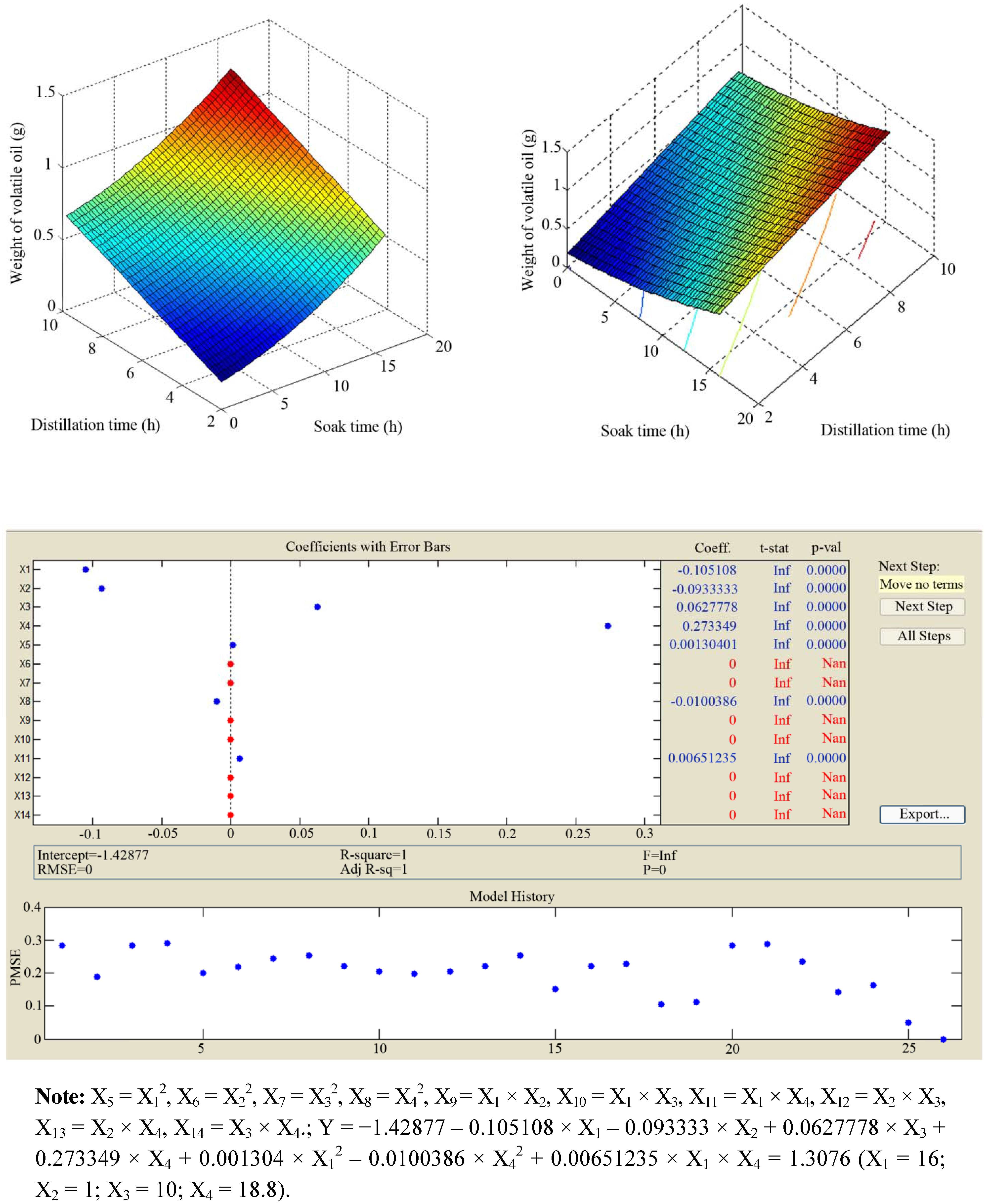
2.1. Uniform Distribution Experimental Design of Extraction
Previous studies found that the yield of volatile oil extracted from Rubus parvifolius L. was very low []. In our study, we applied a Clevenger apparatus to achieve a higher yield. NaCl (80 g/L) was also added during the extraction. A uniform distribution experimental design was used to optimize the extraction efficiency (Table 1 and Figure 1).
Table 1.
U9 (34) Uniform distribution of volatile oil extracted from Rubus parvifolius L. leaves.
| No. | Soak time X1 (h) | Grinding degree X2 (mesh) | Distillation time X3 (h) | Ratio of solvent to leaves X4 ( v/w) | Weight of volatile oil (g) |
|---|---|---|---|---|---|
| 1 | 0 | < 20 | 5 | 20 | 0.15 |
| 2 | 2 | 20–40 | 9 | 16 | 0.85 |
| 3 | 4 | > 40 | 4 | 12 | 0.29 |
| 4 | 6 | < 20 | 8 | 8 | 0.16 |
| 5 | 8 | 20–40 | 3 | 22 | 0.21 |
| 6 | 10 | > 40 | 7 | 18 | 0.65 |
| 7 | 12 | < 20 | 2 | 14 | 0.39 |
| 8 | 14 | 20–40 | 6 | 10 | 0.28 |
| 9 | 16 | > 40 | 10 | 24 | 0.85 |
Figure 1.
Results of the uniform distribution experimental design.
Regression analysis indicated that independent variable tests were statistically significant (p < 0.05). Combination of errors value was 0 with adjustment R2 of 1, indicating that all the experimental points were predicted by the regression equation. Thus, the equation was accurate and effective. The optimised conditions were that the 20-mesh filtered leave powders were soaked with 18.8 times purified water for 16 h, and then boiled at 100 °C for 10 h. The optimal extraction yielded 0.36% (w/w) of light yellowish oil. Our results showed that the yield of Rp volatile oil was higher than that of previous studies [,].
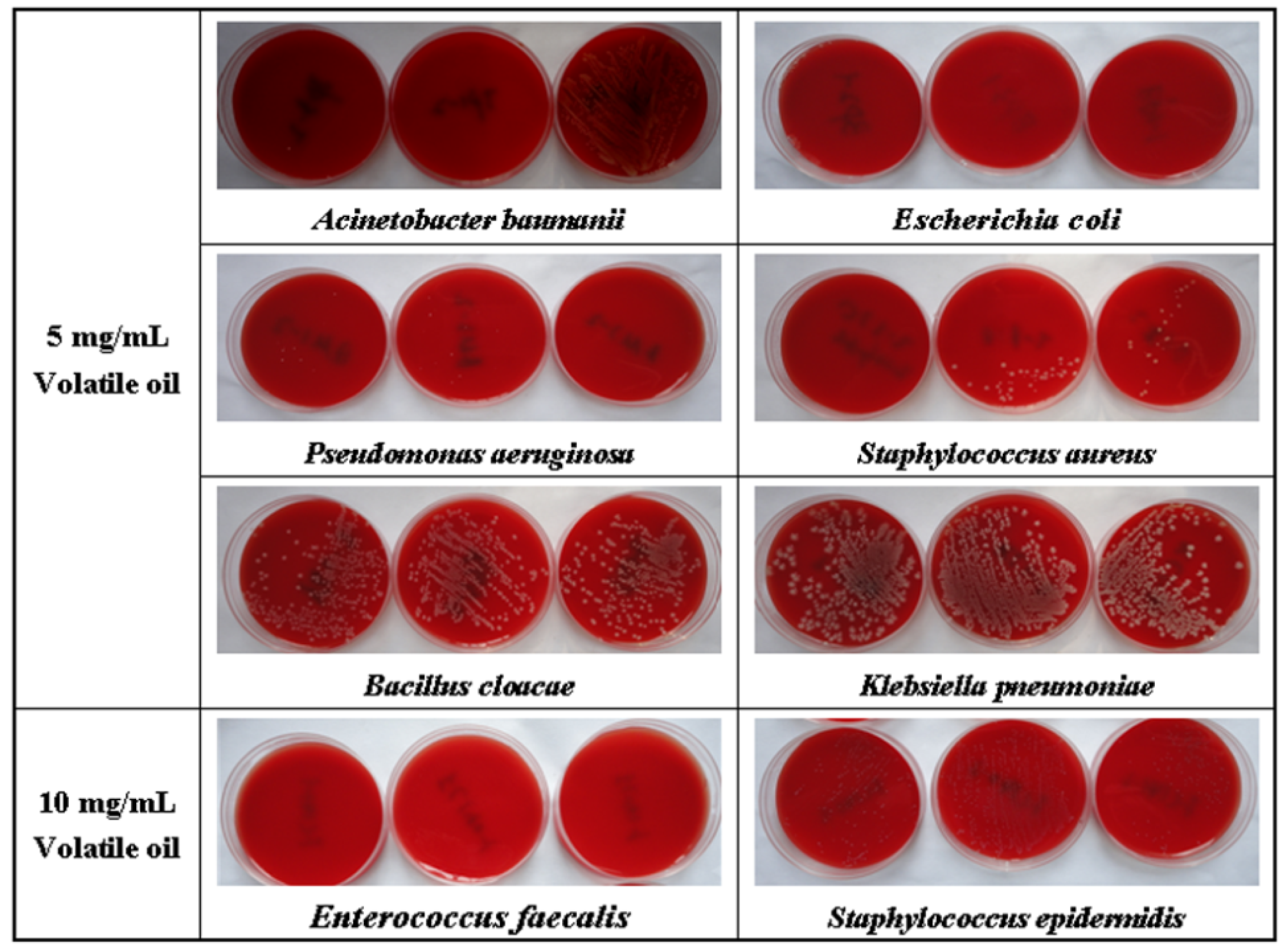
2.2. Antibacterial Activity of Rp Volatile Oil
Minimum inhibitory concentration (MIC) is commonly used to evaluate antibacterial activity. In our study, the MICs of Rp volatile oil on several bacterial strains were tested to determine their antibacterial potential. We found that the Rp volatile oil exhibited antibacterial activity (Table 2 and Figure 2).
Table 2.
Antibacterial activity of volatile oilextracted from Rubus parvifolius L. leaves.
| Bacterial species | Source | MIC (mg/mL) | MBC (mg/mL) |
|---|---|---|---|
| Gram-positive bacteria | |||
| Staphylococcus aureus | CIS a | 5 | > 5 |
| Staphylococcus aureus | ATCC b29213 | 5 | 10 |
| Staphylococcus epidermidis | CIS | 10 | 10 |
| Enterococcus faecalis | CIS | 10 | 10 |
| Gram-negative bacteria | |||
| Escherichia coli | CIS | 5 | 5 |
| Escherichia coli | ATCC 25922 | 5 | 5 |
| Pseudomonas aeruginosa | CIS | > 2.5 | 5 |
| Pseudomonas aeruginosa | ATCC 27853 | 2.5 | 5 |
| Acinetobacter baumanii | CIS | > 2.5 | 5 |
| Bacillus cloacae | CIS | 5 | 10 |
| Klebsiella pneumoniae | CIS | 5 | > 10 |
a MIC: Minimal inhibitory concentration; bMBC: Mimimal bactericidal concentration; c CIS: Clinically Isolated Strains, Department of Pharmacy, Southwestern Hospital (China); d ATCC: American Type Culture Collection (USA).
Figure 2.
Minimal bactericidal concentration of Rp volatile oil.
The MICs of Rp volatile oil were 2.5 mg/mL against clinically isolated bacterial strains of Pseudomonas aeruginosa and Acinetobacter baumanii, 5 mg/mL against Staphylococcus aureus, Escherichia coli, Bacillus cloacae and Klebsiella pneumoniae, and 10 mg/mL against Staphylococcus epidermidis and Enterococcus faecalis. Our results, unlike previous reports [,], revealed that Rp volatile oil possessed a broad spectrum of antibacterial activity, possibly due to the differences in the origins of Rp used in the studies and the method used to extract the oil.
2.3. Components of the Rp Volatile Oil
In order to define the components of the Rp volatile oil, gas chromatography-mass spectroscopy (GC/MS) was applied. Previous phytochemical analysis based on high-performance liquid chromatography/tandem mass spectrometry (HPLC-MS/MS) revealed the presence of various phenolic compounds in Rp volatile oil, including caffeic acid conjugates, ellagic acid glycosides, and flavonol glycosides. Organic acids represent the highest content in the Rp volatile oil (66.92%), followed by formaldehyde (5.97%), alcohol-phenol (4.72%), alkanes (1.26%), ketones (1.26%), lactone (0.43%) and olefin (0.21%). The content of palmitic acid (32.67%) is the highest. In one previous study [], only 20 components were found from the Rp volatile oil, including hexadecanoic acid (32.67%) and elaidic acid (21.20%). In our study, 29 compounds were identified using the NIST08 mass spectral database. A total ion chromatogram (TIC) trace of the volatile oil extracted from Rp leaves is shown in Figure 3. By comparing the retention times, the selected ion current (m/z = 51, 77, 107 and 135) profile, and the possible fragmentation mechanisms, we calculated that the structure of compound with retention time of 14.114 min was 4-hydroxy-3-methoxystyrene, which may be derived from decarboxylation of ferulic acid [,]. It has been reported to possess anti-inflammatory and anti-oxidative activity []. 4-Hydroxy-3-methoxystyrene levels increased with increasing boiling times and approximated the threshold value of 0.3 mg/L after 3 h [].
Figure 3.
TIC-trace of Rp volatile oil.
Table 3 summarizes the chemical composition of the Rp volatile oil. Our results showed that the alcohol-phenol constituents accounted for the highest content (81.09%) with 66.05% as 4-hydroxy-3-methoxystyrene (Table 4 and Figure 4). The yield of Rp volatile by our extraction method (0.36%, w/w) is far more better than the previous studies (<0.2%, w/w) [,]. Because of our uniform distribution experimental design, we identified more components (Table 3) that were not reported in previous reports [,].
Table 3.
Chemical composition of the volatile oilextracted from Rubus parvifolius L. leaves.
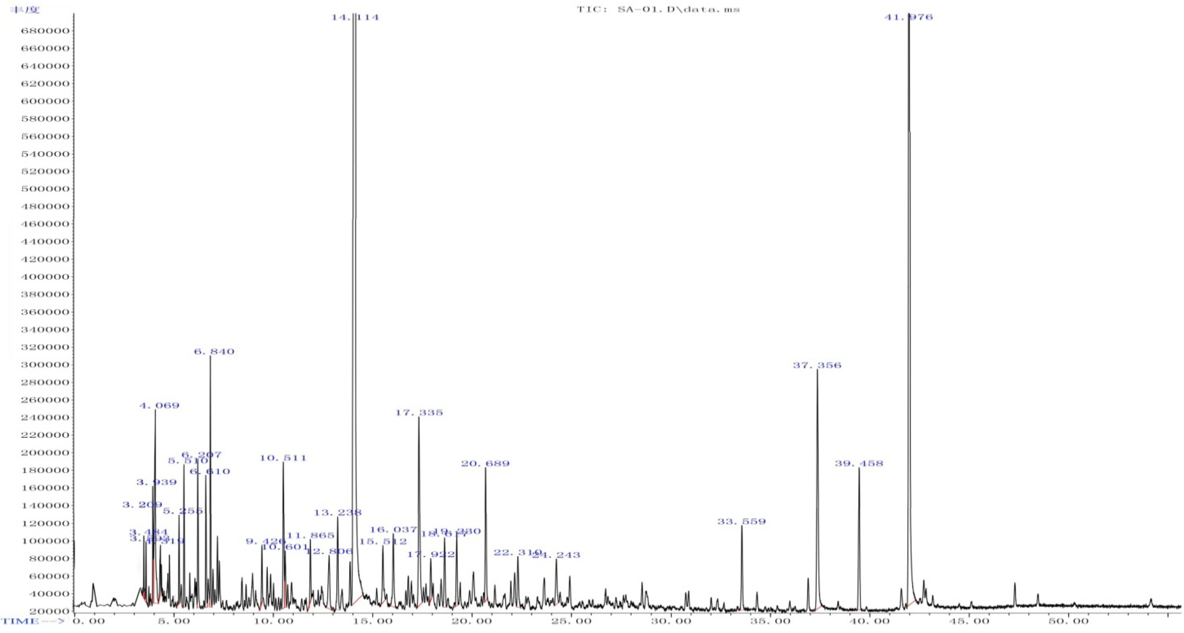
| No | RT (min) | Compound ID | Structural Formula | Formula | Content% |
|---|---|---|---|---|---|
| 1 | 3.209 | 2-Acetylfuran | ![]() | C6H6O2 | 0.28 |
| 2 | 3.484 | Butyl formate | | C5H10O2 | 0.46 |
| 3 | 3.939 | 5-Methyl furfural | | C6H6O2 | 0.55 |
| 4 | 4.069 | Hexanoic acid | | C6H12O2 | 2.03 |
| 5 | 4.319 | trans-3-Hexenoic acid | | C6H10O2 | 0.32 |
| 6 | 5.255 | Benzyl alcohol | | C7H8O | 0.45 |
| 7 | 5.510 | Phenyl acetaldehyde | | C8H8O | 0.77 |
| 8 | 6.207 | cis-α,α,5-Trimethyl-5-vinyl-tetrahydrofuran-2-methanol | | C10H18O2 | 0.88 |
| 9 | 6.610 | α-Methyl-α-[4-methyl-3-pentenyl]oxiranemethanol
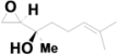
 | ![]() | C10H18O2 | 0.78 |
| 10 | 6.840 | Linalool | | C10H18O | 1.39 |
| 11 | 9.426 | Naphthalene | | C10H8 | 0.37 |
| 12 | 10.511 | 2,3-Dihydrobenzofuran | | C8H8O | 1.21 |
| 13 | 10.601 | α,4-Dimethyl-3-cyclohexene-1-acetaldehyde | | C10H16O | 0.41 |
| 14 | 11.865 | Nerol | | C10H18O | 0.42 |
| 15 | 12.806 | Phenylephrine
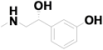
 | ![]() | C9H13NO2 | 0.69 |
| 16 | 13.238 | 2-Methylnaphthalene | | C11H10 | 0.81 |
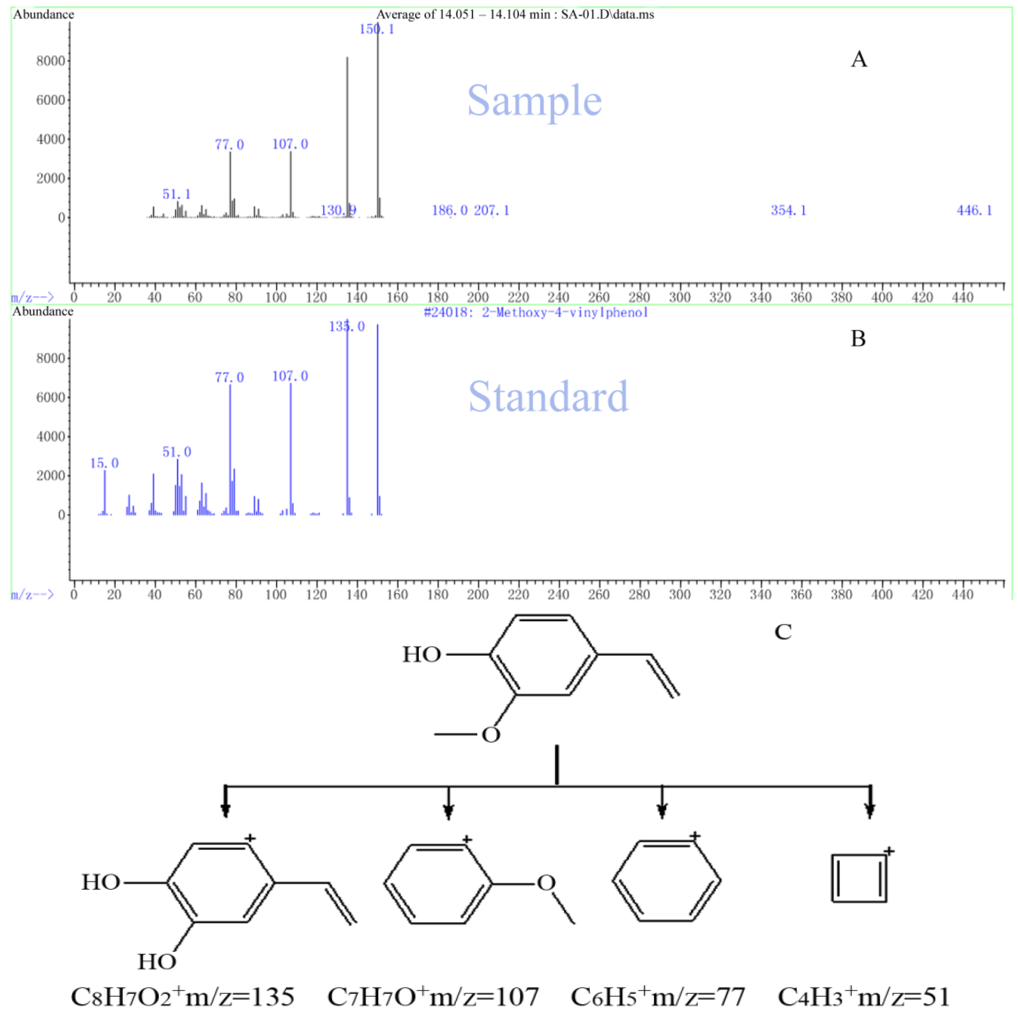
| 17 | 14.114 | 4-Hydroxy-3-methoxystyrene | ![]() | C9H10O2 | 66.05 |
| 18 | 15.512 | 1,1,6-Trimethyl-1,2-dihydronaphthalene | | C13H16 | 0.57 |
| 19 | 16.037 | Methyl 4-formylbenzoate
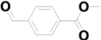
 | | C9H8O3 | 0.77 |
| 20 | 17.335 | 4-tert-Butylbenzoic acid | | C11H14O2 | 2.22 |
| 21 | 17.922 | Dimethylnaphthalene | | C12H12 | 0.37 |
| 22 | 18.617 | α,2,6-Trimethyl-benzeneethanamine | ![]() | C11H17N | 0.65 |
| 23 | 19.230 | 2-Methoxy-4-(prop-1-enyl)phenol | | C10H12O2 | 0.64 |
| 24 | 20.689 | Irisone | ![]() | C13H20O | 1.17 |
| 25 | 22.310 | Dihydroactinidiolide | ![]() | C11H16O2 | 0.52 |
| 26 | 24.243 | 2-(1,3-Butadienyl)mesitylene | ![]() | C13H16 | 0.52 |
| 27 | 33.559 | Hexahydrofarnesylacetone
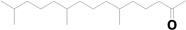
 | | C18H36O | 0.8 |
| 28 | 39.458 | Kaur-16-ene
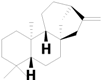
 | ![]() | C20H32 | 1.36 |
| 29 | 41.976 | 3,7,11,15-Tetramethyl-2-hexadecen-1-ol
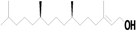
 | | C20H40O | 9.79 |
Table 4.
Components of Rp volatile oil.
| Compound types | Content% | Amount |
|---|---|---|
| Alcohol-phenol | 81.09 | 9 |
| Carboxylic acid | 4.57 | 3 |
| Ester | 1.52 | 3 |
| Ketone | 2.25 | 3 |
| Aldehyde | 1.73 | 3 |
| Other | 5.86 | 8 |
Figure 4.
Selected ion current profile for Rp volatile oil and measured using GC/MS.(A) the sample with retention time of 14.114 min; (B) 4-hydroxy-3-methoxystyrene; (C) Proposed fragmentation mechanism for 4-hydroxy-3-methoxystyrene.
3. Experimental
3.1. Plant Materials
Rubus parvifolius L. (Rp) was obtained from the Chongqing Market of Traditional Chinese Herbs and authenticated at the Chongqing Institute of Chinese Materia Medica.
3.2. Other Reagents
Colombia blood agar plates [] and turbidity tubes were purchased from Chongqing Pangtong Medical Apparatus and Instrument Co., Ltd, Chongqing, China. Purified water was prepared by the Department of Pharmacy, Southwestern Hospital, Chongqing, China. Dimethyl sulfoxide (DMSO, analytical grade) was purchased from Chengdu Kelong Chemical Factory, Chengdu, China. Physiological saline was purchased from Tiansheng Pharmaceutical Group Co., Ltd., Chongqing, China.
3.3. Volatile Rp Oil Extraction
We extracted the volatile oil using a Clevenger apparatus in an attempt to improve the yield. A uniform distribution design was used (Table 1) to optimize extraction efficiency []. Rp dried leaves (200 g) were placed in a Clevenger apparatus and the powdered parts were hydro-distillated for 10 h with 2 L of purified water. Ether was added in the side arm of Clevenger apparatus to dissolve Rp volatile oil. The volatile oil was concentrated under reduced pressure to remove the ether, dehydrated by anhydrous sodium sulfate, and kept in air tight glass bottles in a refrigerator for further experiments.
3.4. Bacterial Strains
Three bacterial strains, Staphylococcus aureus (ATCC29213), Escherichia coli (ATCC25922) and Pseudomonas aeruginosa (ATCC27853) were obtained from the American Type Culture Collection (ATCC, Rockefeller, MD USA). The other bacterial strains were clinically isolated and obtained from the Department of Pharmacy, Southwestern Hospital, Chongqing, China. These strains included Gram-positive (G+) bacteria: Staphylococcus aureus, Staphylococcus epidermidis and Enterococcus faecalis; and Gram-negative (G−) bacteria: Escherichia coli, Pseudomonas aeruginosa, Acinetobacter baumanii, Bacillus cloacae and Klebsiella pneumoniae (Table 2).
3.5. Antibacterial Tests
To test the antibacterial effects of the volatile Rp oil, 0.048 M BaCl2 (1.17% w/v BaCl2·2H2O, 0.5 mL) was mixed with 0.18 M H2SO4 (1%, v/v, 99.5 mL) with constant stirring. Bacterial strains were grown on blood agar plates. Bacteria were prepared from 24 h MH broth cultures and adjusted to 0.5 McFarland turbidity equivalents. MIC of volatile oil of Rp leaves was determined by the standard CLSI (Clinical Laboratory Standards Institute) method []. Volatile Rp oil was weighed and dissolved in DMSO to a concentration of 10 mg/100 μL, and were then distributed at various concentrations in triplicate with a volume of 100 µL in turbidity tubes. Bacterial suspension (5 µL) with a density of 106/mL in Mueller-Hinton (MH) broth was added to each tube. Positive control was a suspension of bacteria in 1 mL of MH broth, and negative control was medium without bacteria. After an incubation period of 20~24 h at 37 °C, the optical values were measured by the microdilution MH broth susceptibility assay as recommended. The lowest concentration of the test agent that prevented the appearance of turbidity was considered as the MIC. At this dilution, the test agent is bacteriostatic. Minimal bactericidal concentration (MBC) is defined as the maximal dilution of the oil that will kill a microorganism.
3.6. Analysis of Components
An Agilent 7890A/5975C Gas Chromatography-Mass Spectroscopy system equipped with a GC-MSD was used for analysis with ionization achieved by electron impact at 70 eV. The capillary column used was a J&W Scientific DB-1 (methylsiloxane, 30 m × 0.25 mm I.D., 0.25 µm-thick film). Experimental conditions for GC analysis of volatile Rp oil were: injection port temperature, 200 °C; interface temperature, 200 °C; column oven temperature, 80 °C for 3 min, then programmed at 3 °C/ min to 220 °C and held for 10 min; Helium (purity 99.995%) as carrier gas at a flow rate of 1.0 mL/min, and 0.20 µL injection volume. The split/splitless injector was operated in the splitless mode for 1.5 min after injection of the sample. For identification of components, the mass spectrum of each peak was recorded in the total ion current mode of the mass spectrometer within a mass range of 35 to 450 []. Identification of oil constituents was achieved using NIST08 mass spectral database.
3.7. Statistical Analysis
The data were analyzed by using uniform distribution experimental design programmed by Matlab []. p value less than 0.05 was considered statistically significant.
4. Conclusions
Volatile oil extracted from Rp significantly inhibits microbial growth. Rp volatile oil may represent an economic and effective antiseptic topical treatment. Taking into account that new forms of bacterial resistance to antibacterials are rising unconstrained and that the problem of pathogenic bacteria showing multi-drug resistance, our findings could be of particular useful for human and animal health. Rp volatile oil may have potential antibacterial applications. Although further studies are needed, the use of Rp volatile oil against microbial growth, especially opportunistic and pathogenic microorganisms, seems a valuable alternative to antibiotics or antibacterial compounds, especially in the case of dealing with antibiotic resistance.
Acknowledgments
This research project was supported by grants from National Science and Technology Major Projects (No. 2011ZXJ09106–06B), and National Natural Science Foundation of China (NSFC, No. 30873034 and 30973493). We thank Feng Pan (Central Laboratory, Third Military Medical University, Chongqing, China) for the technical assistance in Gas Chromatography-Mass Spectroscopy analysis.
References and Notes
- Wang, Y.; He, J.M.; Chen, H.; Zhang, D.S.; Cai, H.; Shao, H.B. Analysis of flavones in Rubus parvifolius Linn by high performance liquid chromatography combined with electrospray ionization-mass spectrometry and thin-layer chromatography combined with Fourier transform surface enhanced Raman spectroscopy. Fen Xi Hua Xue 2006, 34, 1073–1077. [Google Scholar]
- Gao, J.; Sun, C.R.; Yang, J.H.; Shi, J.M.; Du, Y.G.; Zhang, Y.Y.; Li, J.H.; Wan, H.T. Evaluation of the hepatoprotective and antioxidant activities of Rubus parvifolius L. J. Zhejiang Univ. Sci. B 2011, 12, 135–142. [Google Scholar] [CrossRef]
- Zhu, Z.H.; Zhang, H.Q.; Yuan, M.J. Pharmacological study of Rubus parvifolius L. Zhongguo Zhong Yao Za Zhi 1990, 15, 427–447. [Google Scholar]
- Wang, J.S.; Qiu, Z.Y.; Xia, Y.P.; Li, H.Z.; Ren, L.Y.; Zhang, L. The protective effects of total glycosides Rubus parviflolius on cerebral ischemia in rat. Zhongguo Zhong Yao Za Zhi 2006, 2, 138–141. [Google Scholar]
- Wang, J.S.; Li, H.Z.; Qiu, Z.Y.; Xia, Y.P.; Ren, L.Y.; Zhou, C.L. Protective effects of aqueous extract of Rubus parviflolius on middle cerebral artery occlusion and reperfusion injury in rats. Chin. J. New Drugs Clin. Remed. s 2006, 12, 920–923. [Google Scholar]
- Deighton, N.; Brennan, R.; Finn, C.; Davies, H.V. Antioxidant properties of domesticated and wild Rubus species. J. Sci. Food Agric. 2000, 80, 1307–1313. [Google Scholar] [CrossRef]
- Hsiao, G.; Shen, M.Y.; Lin, K.H.; Lan, M.H.; Wu, L.Y.; Chou, D.S.; Lin, C.H.; Su, C.H.; Sheu, J.R. Antioxidative and hepatoprotective effects of Antrodia camphorata extract. J. Agric. Food Chem. 2003, 51, 3302–3308. [Google Scholar] [CrossRef]
- Gowri Shankar, N.L.; Manavalan, R.; Venkappayya, D.; David Raj, C. Hepatoprotective and antioxidant effects of Commiphora berryi (Arn) Engl bark extract against CCl4-induced oxidative damage in rats. Food Chem. Toxicol. 2008, 46, 3182–3185. [Google Scholar] [CrossRef]
- Zeashan, H.; Amresh, G.; Singh, S.; Rao, C.V. Hepatoprotective and antioxidant activity of Amaranthus spinosus against CCl4 induced toxicity. J. Ethnopharmarcol. 2009, 125, 364–366. [Google Scholar] [CrossRef]
- Ho, C.L.; Tseng, Y.H.; Wang, E.I.; Liao, P.C.; Chou, J.C.; Lin, C.N.; Su, Y.C. Composition, antioxidant and antibacterial activities of the seed essential oil of Calocedrus formosana from Taiwan. Nat. Prod. Commun. 2011, 6, 133–136. [Google Scholar]
- Hamill, F.A.; Apio, S.; Mubiru, N.K.; Mosango, M.; Bukenya-Ziraba, R.; Maganyi, O.W.; Soejarto, D.D. Traditional herbal drugs of southern Uganda. Part III: Isolation and methods for physical characterization of bioactive alkanols from Rubus apetalus. J. Ethnopharmarcol. 2003, 87, 15–19. [Google Scholar] [CrossRef]
- Tan, M.X.; Wang, H.S.; Li, S.; Yang, Y. Studies on the chemical constituents of the volatile oil from the leaves of Rubus parviflolius. Tianran Chanwu Yanjiu Yu Kaifa 2003, 1, 32–33. [Google Scholar]
- Thiem, B.; Goslinska, O. Antimicrobial activity of Rubus chamaemorus leaves. Fitoterapia 2004, 75, 93–95. [Google Scholar] [CrossRef]
- Nissen, L.; Zatta, A.; Stefanini, I.; Grandi, S.; Sgorbati, B.; Biavati, B.; Monti, A. Characterization and antimicrobial activity of essential oils of industrial hemp varieties (Cannabis sativa L.). Fitoterapia 2010, 81, 413–419. [Google Scholar] [CrossRef]
- Coghe, S.; Benoot, K.; Delvaux, F.; Vanderhaegen, B.; Delvaux, F.R. Ferulic acid release and 4-vinylguaiacol formation during brewing and fermentation: indications for feruloyl esterase activity in Saccharomyces cerevisiae. J. Agric. Food Chem. 2004, 52, 602–608. [Google Scholar] [CrossRef]
- Dugelay, I.; Gunata, Z.; Sapis, J.C.; Baumes, R.; Bayonove, C. Role of cinnamoyl esterase activities from enzyme preparations on the formation of volatile phenols during winemaking. J. Agric. Food Chem. 1993, 41, 2092–2096. [Google Scholar] [CrossRef]
- Pongprayoon, U.; Bohlin, L.; Soonthornsaratune, P.; Wasuwat, S. Anti-inflammatory activity of Ipomoea pes-caprae (L.) R. Br. Phytother. Res. 1991, 5, 63–66. [Google Scholar] [CrossRef]
- Morton, C.E.G.; Holt, H.A. A problem encountered using staphylococcus/streptococcus supplement. Med. Lab. Sci. 1989, 46, 72–73. [Google Scholar]
- Clinical and Laboratory Standards Institute (CLSI), Methods for Dilution Antimicrobial Susceptibility Tests for Bacteria that Grow Aerobically: Approved Guideline, 7th edCLSI: Wayne, NE, USA, 2006; Approved Standard M7–A7.
- Fang, K.T.; Ma, C.X. Orthogonal and Uniform Experimental Design, 1st ed; Science press: Beijing, China, 2001. [Google Scholar]
- Sample Availability: Not available.
© 2012 by the authors; licensee MDPI, Basel, Switzerland. This article is an open-access article distributed under the terms and conditions of the Creative Commons Attribution license (http://creativecommons.org/licenses/by/3.0/).


