Antibacterial Activity of Aristolochia brevipes against Multidrug-Resistant Mycobacterium tuberculosis

Abstract
1. Introduction
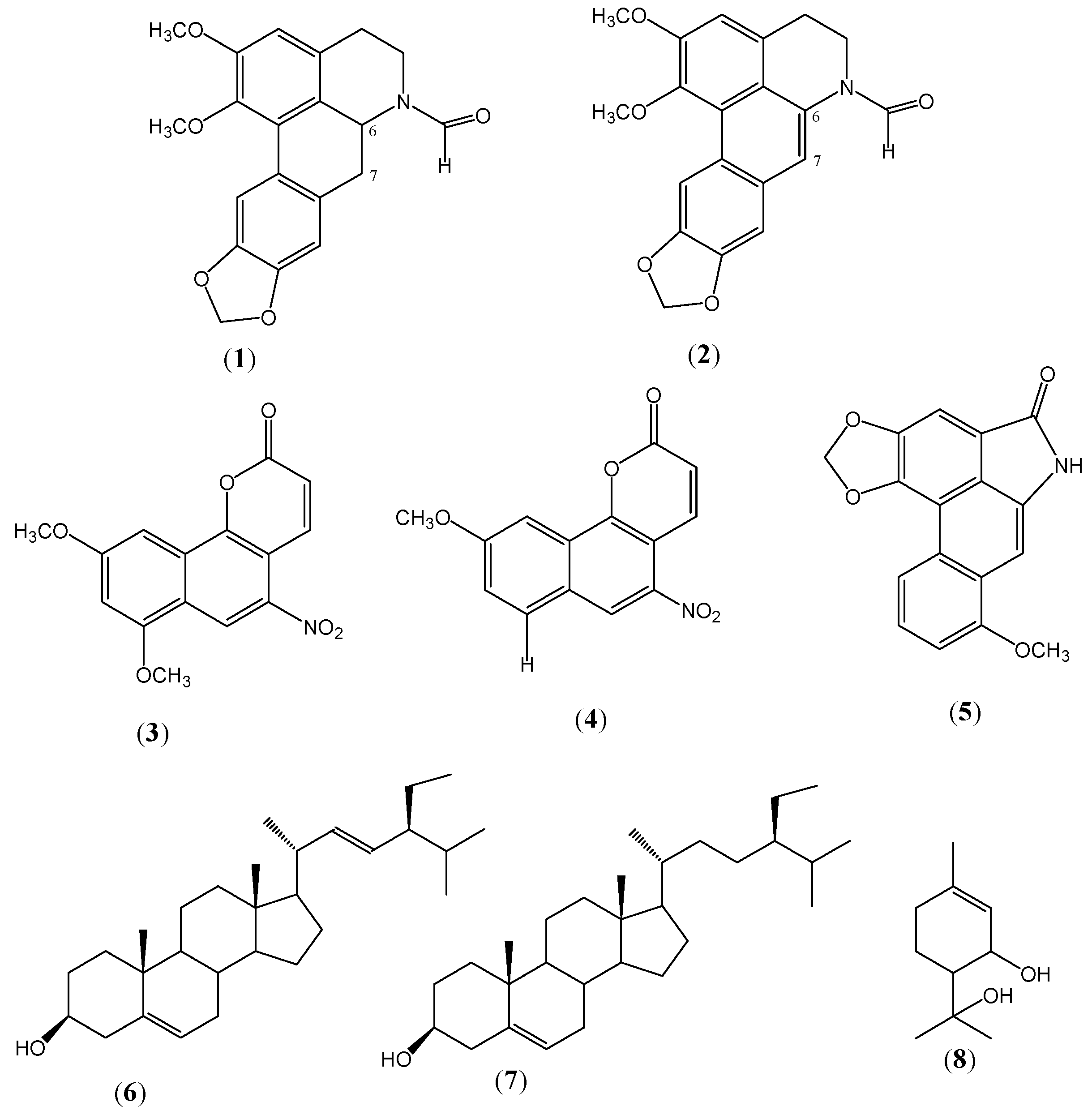
2. Results and Discussion


| Compound | (1) b | (2) | (3) | (4) | (5) | (6) |
|---|---|---|---|---|---|---|
| Microorganism | ||||||
| H37Rv a | >50 c | >50 | 25.0 | 25.0 | 12.5 | 12.5 |
| H37RvEr | >50 | >50 | 50.0 | 25.0 | 12.5 | NT d |
| H37RvIr | >50 | >50 | 12.5 | 50.0 | 25.0 | NT |
| H37RvRr | >50 | >50 | 50.0 | 50.0 | 25.0 | NT |
| H37RvSr | >50 | >50 | 25.0 | 25.0 | 25.0 | NT |
| MMDO | >50 | NT | 50.0 | 50.0 | 25.0 | NT |
| MTY147 | >50 | NT | 50.0 | 25.0 | 12.5 | NT |
| SIN452 | >50 | NT | 25.0 | 25.0 | 12.5 | NT |
3. Experimental
3.1. Collection of Plant Material
3.2. General Procedure and Equipment Utilized
3.3. Extract Preparation, Separation, and Isolation of Secondary Metabolites
3.4. Mycobacterium Strains and Clinical Isolates
3.5. Inoculum Preparation for Biological Assay
3.6. Antimycobacterial Activity Determination by Fluorometric Microplate Alamar Blue Assay
4. Conclusions
Acknowledgments
- Sample Availability: Not available.
References and Notes
- World Health Organization, Tuberculosis Facts, 2009 Update; World Health Organization: Geneva, Switzerland, 2009.
- Quinto Informe de la Secretaría de Salud Pública México; Subsecretaría de Prevención y Promoción a la Salud: D.F., Mexico, Mexico, 2005; Chapter 3.
- Murray, C.J.L.; Stylblok, C.; Rouillon, A. Tuberculosis in developing countries: Burden, intervention and cost. Bull. Int. Union against Tuberculosis Lung Dis. 1990, 65, 1–120. [Google Scholar]
- Barstian, I.; Portaels, F. Introduction. In In Multidrug-resistant Tuberculosis, 1st ed; Kluwer Academic: Dordrecht, The Netherlands, 2000; pp. 1–12. [Google Scholar] [Green Version]
- Copp, B.R. Antimycobacterial natural products. Nat. Prod. Rep. 2003, 20, 535–557. [Google Scholar] [CrossRef]
- Newman, D.J.; Cragg, G.; Snader, K.M. The influence of natural products upon drug discovery. Nat. Prod. Rep. 2000, 17, 215–234. [Google Scholar] [CrossRef]
- Newman, D.J.; Cragg, G.; Snader, K.M. Natural products as sources of new drugs over the period 1981-2002. J. Nat. Prod. 2003, 66, 1022–1037. [Google Scholar] [CrossRef]
- Martínez, M. Las Plantas Medicinales de México, 6th ed; Botas: D.F. Mexico, Mexico, 1991; p. 270. [Google Scholar]
- Tian-Shung, W.; Amooru, D.; Chung-Ren, S.; Ping-Chung, K. Terpenoids of Aristolochia and their biological activities. Nat. Prod. Rep. 2004, 21, 594–624. [Google Scholar] [CrossRef]
- Achenbach, H.; Waibel, R.; Zwanzger, M.; Domínguez, X.; Espinosa, G.; Verde, J.; Sánchez, H. 9-methoxy-and 7,9-dimethoxytariacuripyrone, natural nitro-compounds with a new basic skeleton from Aristolochia brevipes. J. Nat. Prod. 1992, 55, 918–922. [Google Scholar] [CrossRef]
- Achenbach, H.; Waibel, R.; Zwanzger, M.; Domínguez, X.; Espinosa, G.; Verde, J.; Sánchez, H.; Guajardo, E. 6a,7-dehydro-N-formylnornatenine and other constituents from Aristolochia brevipes. Planta Med. 1995, 61, 189–190. [Google Scholar] [CrossRef]
- Tantisewie, B.; Pharadai, T.; Pandhuganont, M.; Guinaudeau, H.; Freyer, A.; Shamma, M. N-Formylnornantenine, a new aporphine alkaloid from Cyclea atjehensis. J. Nat. Prod. 1989, 52, 652–654. [Google Scholar] [CrossRef]
- Che, C.; Ahmed, M.; Kang, S.; Waller, D.; Bingel, A.; Martin, A.; Rajamahendran, P.; Bunyapraphatsara, N.; Lankin, D.; Cordell, G.; et al. Studies on Aristolochia III. Isolation and biological evaluation of constituents of Aristolochia indica roots for fertility-regulation activity. J. Nat. Prod. 1984, 47, 331–341. [Google Scholar] [CrossRef]
- Rastrelli, L.; Capasso, A.; Pizza, C.; De Tommasi, N.; Sorrentino, L. New protopine and benzyltetrahydroprotoberberine alkaloids from Aristolochia constricta and their activity on isolated guinea-pig ileum. J. Nat. Prod. 1997, 60, 1065–1069. [Google Scholar] [CrossRef]
- Tian-shung, W.; Amooru, D.; Chung-Ren, S.; Ping-Chung, K. Chemical Constituents and Pharmacology of Aristolochia Species. In Studies in Natural Products Chemistry. Bioactive Natural Products (Part L); Elsevier: Amsterdam, The Netherlands, 2005; pp. 855–1018. [Google Scholar]
- Schimmer, O.; Drewello, U. 9-methoxytariacuripyrone, a naturally occurring nitro-aromatic compound with strong mutagenicity in Salmonella typhimurium. Mutagenesis 1994, 9, 547–551. [Google Scholar] [CrossRef]
- Akasu, M.; Itokawa, H.; Fujita, M. Four new fluorescent components isolated from the callus tissue of Stephania cepharanta. Tetrahedron Lett. 1974, 15, 3609–3612. [Google Scholar] [CrossRef]
- Dyke, S.F.; Emery, G.E. The structure of alkaloid Y from Schefferomitra subaequalis. (Abstract). Phytochemistry 1978, 17, 599. [Google Scholar]
- Tian-Shung, W.; Li-Fei, O.; Che-Ming, T. Aristolochic acids, aristolactam alkaloids and amides from Aristolochia kankauensis. Phytochemistry 1994, 36, 1063–1068. [Google Scholar]
- Stiboroba, M.; Frei, E.; Schmeiser, H.H.; Wiessler, M. Cytochrome P-450 and peroxidase oxidize detoxication products of carcinogenic aristolochic acids (aristolactams) to reactive metabolites binding to DNA in vitro. Collect Czech Chem. Commun. 1995, 60, 2189. [Google Scholar] [CrossRef]
- Luna-Herrera, J.; Costa, M.C.; González, H.G.; Rodriguez, A.I.; Castilho, P.C. Synergistic antimycobacterial activities of sesquiterpene lactones from Laurus spp. J. Antimicrob. Chemother. 2007, 59, 548–552. [Google Scholar] [CrossRef]
© 2011 by the authors; licensee MDPI, Basel, Switzerland. This article is an open access article distributed under the terms and conditions of the Creative Commons Attribution license ( http://creativecommons.org/licenses/by/3.0/).
Share and Cite
Navarro-García, V.M.; Luna-Herrera, J.; Rojas-Bribiesca, M.G.; Álvarez-Fitz, P.; Ríos, M.Y. Antibacterial Activity of Aristolochia brevipes against Multidrug-Resistant Mycobacterium tuberculosis. Molecules 2011, 16, 7357-7364. https://doi.org/10.3390/molecules16097357
Navarro-García VM, Luna-Herrera J, Rojas-Bribiesca MG, Álvarez-Fitz P, Ríos MY. Antibacterial Activity of Aristolochia brevipes against Multidrug-Resistant Mycobacterium tuberculosis. Molecules. 2011; 16(9):7357-7364. https://doi.org/10.3390/molecules16097357
Chicago/Turabian StyleNavarro-García, Víctor Manuel, Julieta Luna-Herrera, Ma. Gabriela Rojas-Bribiesca, Patricia Álvarez-Fitz, and María Yolanda Ríos. 2011. "Antibacterial Activity of Aristolochia brevipes against Multidrug-Resistant Mycobacterium tuberculosis" Molecules 16, no. 9: 7357-7364. https://doi.org/10.3390/molecules16097357
APA StyleNavarro-García, V. M., Luna-Herrera, J., Rojas-Bribiesca, M. G., Álvarez-Fitz, P., & Ríos, M. Y. (2011). Antibacterial Activity of Aristolochia brevipes against Multidrug-Resistant Mycobacterium tuberculosis. Molecules, 16(9), 7357-7364. https://doi.org/10.3390/molecules16097357