Effects of Quercetin on Mushroom Tyrosinase and B16-F10 Melanoma Cells

Abstract
:Introduction

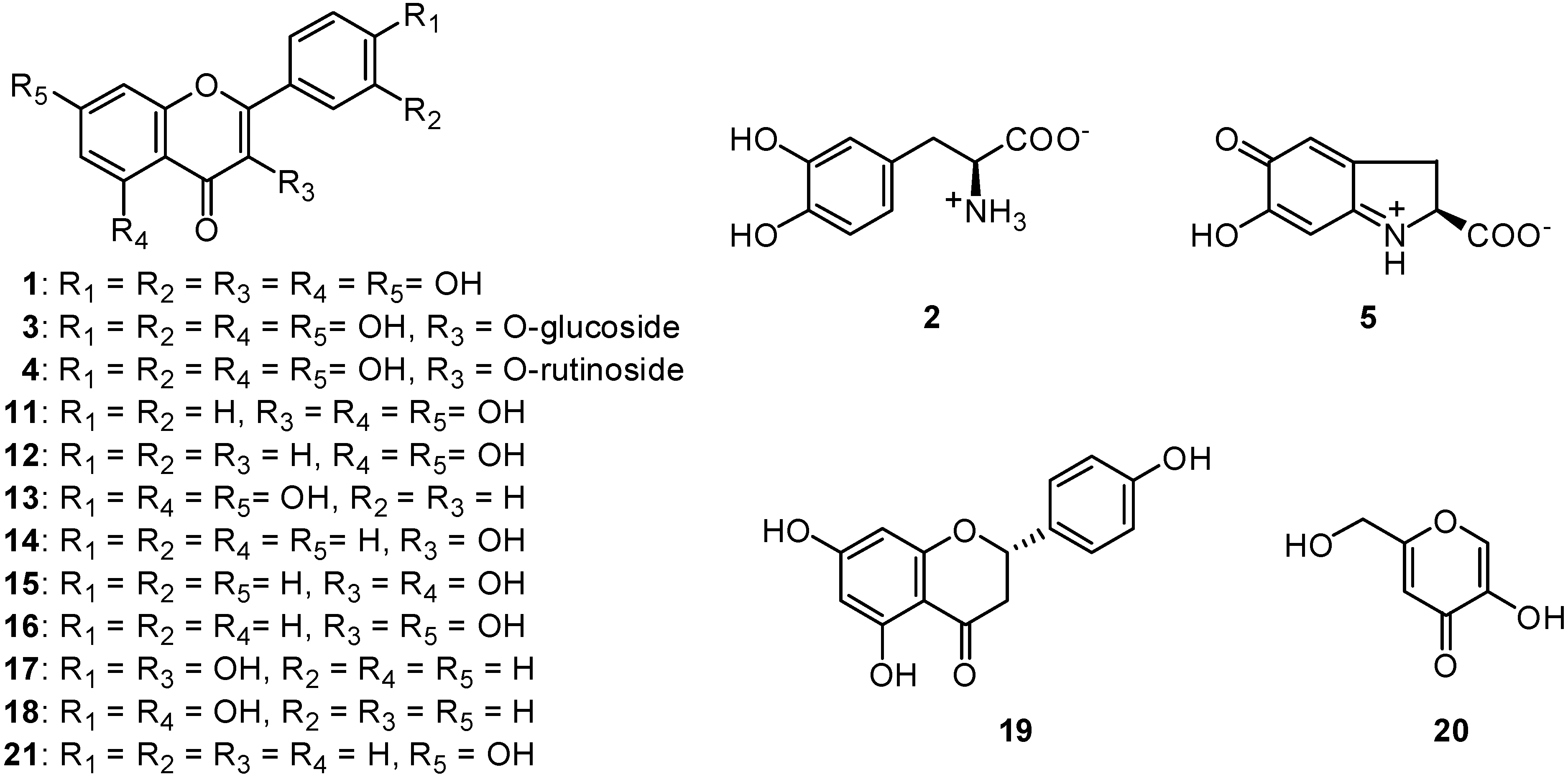
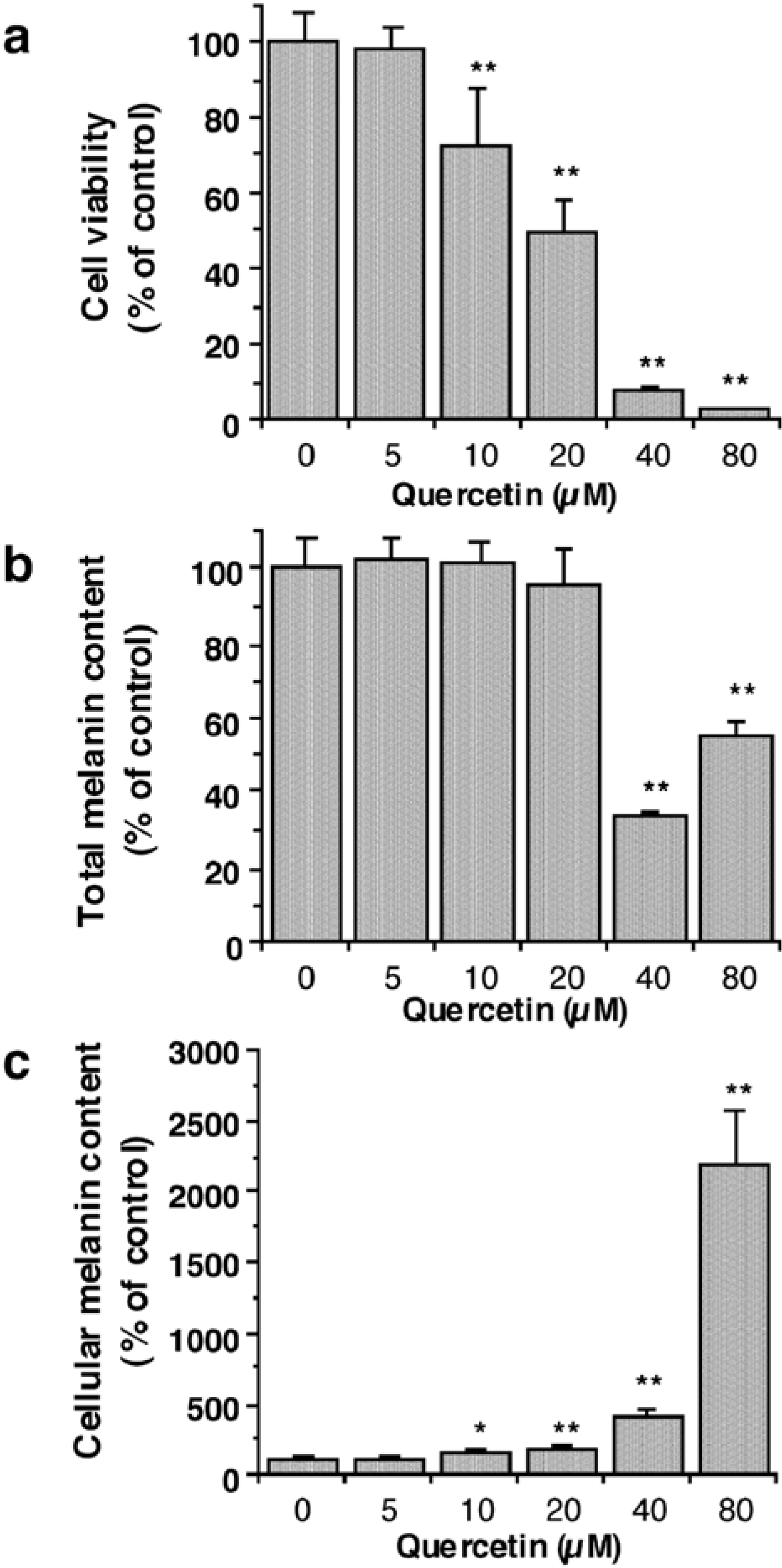
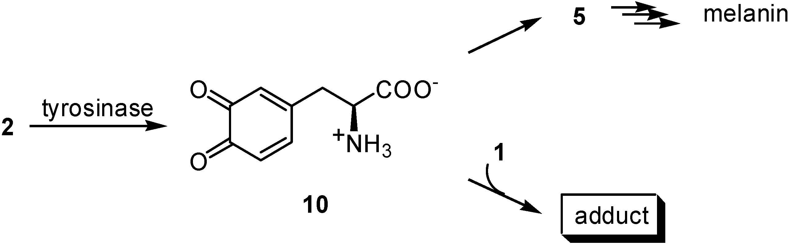
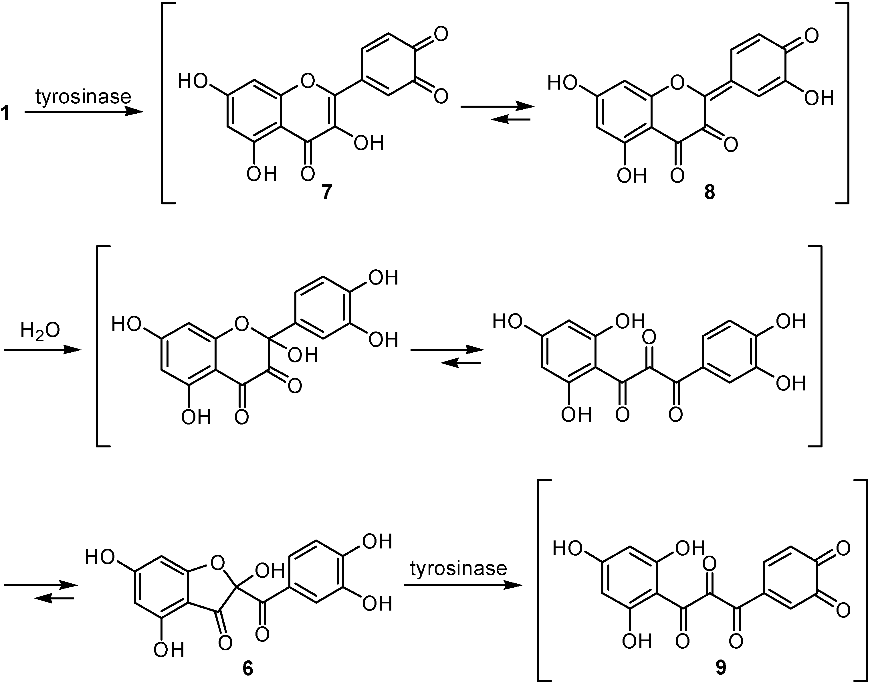
Results and Discussion


Experimental
General
Enzyme Assay
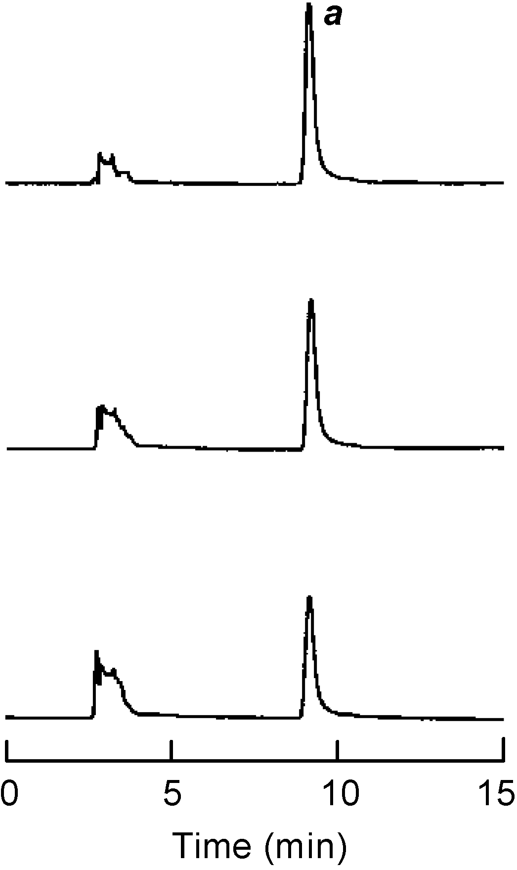
HPLC Analysis
Cell Culture
Cell Viability Assays
Melanin Assay
Acknowledgments
References
- Holme, S. A.; Varma, S.; Chowdhury, M. M.; Roberts, D. L. Audit of a melanoma screening day in the U.K.: clinical results, participant satisfaction and perceived value. Br. J. Dermatol. 2001, 145, 784–788. [Google Scholar] [CrossRef]
- Prezioso, J. A.; Epperly, M. W.; Wang, N.; Bloomer, W. D. Effects of tyrosinase activity on the cytotoxicity of 4-S-cysteaminylphenol and N-acetyl-4-S-cysteaminylphenol in melanoma cells. Cancer Lett. 1992, 63, 73–79. [Google Scholar] [CrossRef]
- Xu, Y.; Stokes, A. H.; Freeman, W. M.; Kumer, S. C.; Vogt, B. A.; Vrana, K. E. Tyrosinase mRNA is expressed in human substantia nigra. Mol. Brain Res. 1997, 45, 159–162. [Google Scholar] [CrossRef]
- Kubo, I.; Kinst-Hori, I.; Ishiguro, K.; Chaudhuri, S. K.; Sanchez, Y.; Ogura, T. Tyrosinase inhibitory flavonoids from Heterotheca inuloides and their structural functions. Bioorg. Med. Chem. Lett. 1994, 4, 1443–1446. [Google Scholar] [CrossRef]
- Kubo, I.; Kinst-Hori, I.; Chaudhuri, S. K.; Sanchez, Y.; Ogura, T. Flavonols from Heterotheca inuloides. Tyrosinase inhibitory activity and structural criteria. Bioorg. Med. Chem. 2000, 8, 1585–1591. [Google Scholar]
- Matinez, M. Catálogo de Nombres Vulgares y Cientificos de Plantas Mexicanas; Fondo de Cultura Economica: Mexico, 1984; pp. 145–146. [Google Scholar]
- Matsuda, H.; Morikawa, T.; Ando, S.; Toguchida, I.; Yoshikawa, M. Structural requirements of flavonoids for nitric oxide production inhibitory activity and mechanism of action. Bioorg. Med. Chem. 2003, 11, 1995–2000. [Google Scholar] [CrossRef]
- Nagatsu, A.; Zhang, H. L.; Mizukami, H.; Sakakibara, J.; Tokuda, H.; Nishino, H. Tyrosinase inhibitory and anti-tumor promoting activities of compounds isolated from safflower (Carthamus tinctorius L.) and cotton (Gossypium hirsutum L.) oil cakes. Nat. Prod. Lett. 2000, 14, 153–158. [Google Scholar] [CrossRef]
- Xie, L. P.; Chen, Q. Y.; Huang, H.; Wang, H. Z.; Zhang, R. Q. Inhibitory effects of some flavonoids on the activity of mushroom tyrosinase. Biochemistry (Moscow) 2003, 68, 487–491. [Google Scholar]
- Jeong, C. H.; Shim, K. H. Tyrosinase inhibitor isolated from the leaves of Zanthoxylum piperitum. Biosci. Biotechnol. Biochem. 2004, 68, 1984–1987. [Google Scholar]
- Kubo, I.; Kinst-Hori, I. Flavonols from saffron flower: Tyrosinase inhibitory activity and inhibition mechanism. J. Agric. Food Chem. 1999, 47, 4121–4125. [Google Scholar] [CrossRef]
- Kubo, I.; Nihei, K.; Shimizu, K. Oxidation products of quercetin by mushroom tyrosinase. Bioorg. Med. Chem. 2004, 12, 5343–5347. [Google Scholar] [CrossRef]
- Mayer, A. M. Polyphenol oxidases in plants - Recent progress. Phytochemistry 1987, 26, 11–20. [Google Scholar] [CrossRef]
- Boersma, M. G.; Vervoort , J.; Szymusiak, H.; Lemanska, K.; Tyrakowska, B.; Cenas, N.; Segura-Aguilar, J.; Rietjens, I. M. Regioselectivity and reversibility of the glutathione conjugation of quercetin quinone methide. Chem Res Toxicol. 2000, 13, 185–191. [Google Scholar]
- Awad, H. M.; Boersma, M. G.; Vervoort, J.; Rietjens, I. M. C. M. Peroxidase-catalyzed formation of quercetin quinone methide-glutathione adducts. Arch. Biochem. Biophys. 2000, 378, 224–233. [Google Scholar] [CrossRef]
- Chen, Q. X.; Kubo, I. Kinetics of tyrosinase inhibition by quercetin. J. Agric. Food Chem. 2002, 50, 4108–4112. [Google Scholar] [CrossRef]
- Sánchez-Ferrer, A.; Rodríguez-López, J. N.; Garcia-Cánovas, F.; García-Carmona, F. Tyrosinase: A comprehensive review of its mechanism. Biochem. Biophys. Acta 1995, 1247, 1–11. [Google Scholar]
- Maeda, K.; Fukuda, M. Arbutin: Mechanism of its depigmenting action in human melanocyte culture. J. Pharm. Exp. Therap. 1996, 276, 765–769. [Google Scholar]
- Tayama, K.; Takahama, M. Depigmenting action of phenylhydroquinone, an o-phenylphenol metabolite, on the skin of JY-4 black guinea-pigs. Pigment Cell Res. 2002, 15, 447–453. [Google Scholar] [CrossRef]
- Nagata, H.; Takekoshi, S.; Takeyama, R.; Homma, T. Quercetin enhances melanogenesis by increasing the activity and synthesis of tyrosinase in human melanoma cells and in normal human melanocytes. Pigment Cell Res. 2004, 17, 66–73. [Google Scholar] [CrossRef]
- Metodiewa, D.; Jaiswal, A. K.; Cenas, N.; Dickancaite, E.; Segura-Aguilar, J. Quercetin may act as a cytotoxic prooxidant after its metabolic activation to semiquinone and quinoid product. Free Radic. Biol. Med. 1999, 26, 107–116. [Google Scholar] [CrossRef]
- Bolton, J. L.; Trush, M. A.; Penning, T. M.; Dryhurst, G.; Monks, T. J. Role of quinones in toxicology. Chem. Res. Toxicol. 2000, 13, 135–160. [Google Scholar] [CrossRef]
- Kaldas, M. I.; Walle, U. K.; van der Woude, H.; McMillan, J. M.; Walle, T. Covalent binding of the flavonoid quercetin to human serum albumin. J. Agric. Food Chem. 2005, 53, 4194–4197. [Google Scholar] [CrossRef]
- Le Bail, J. C.; Laroche, T.; Marre-Fournier, F.; Habrioux, G. Aromatase and 17β-hydroxysteroid dehydrogenase inhibition by flavonoids. Cancer Lett. 1998, 133, 101–106. [Google Scholar]
- Sanderson, J. T.; Hordijk, J.; Denison, M. S.; Springsteel, M. F.; Nantz, M. H.; van den Berg, M. Induction and inhibition of aromatase (CYP19) activity by natural and synthetic flavonoid compounds in H295R human adrenocortica carciboma cells. Toxicol. Sci. 2004, 82, 70–79. [Google Scholar] [CrossRef]
- Yáñez, J.; Vicente, V.; Alcaraz, M.; Castillo, J.; Benavente-Garcćia, O.; Canteras, M.; Teruel, J. A. L. Cytotoxicity and antiproliferative activities of several phenolic compounds against three melanocytes cell lines: relationship between structure and activity. Nutr. Cancer 2004, 49, 192–199. [Google Scholar]
- Peng, L.; Wang, B.; Ren, P. Reduction of MTT by flavonoids in the absence of cells. Colloids Surf., B 2005, 45, 108–111. [Google Scholar] [CrossRef]
- Mueller, H.; Kassack, M.; Wiese, M. Comparison of the usefulness of the MTT, ATP, and calcein assays to predict the potency of cytotoxic agents in various human cancer cell lines. J. Biomolec. Screen. 2004, 9, 506–515. [Google Scholar]
- Fidler, I. J. Selection of successive tumor lines for metastasis. Nature 1973, 242, 148–149. [Google Scholar]
- Poste, G.; Fidler, I. J. The pathogenesis of cancer metastasis. Nature 1980, 283, 139–146. [Google Scholar] [CrossRef]
- Eisenthal, A.; Cameron, R. B.; Uppenkamp, I.; Resenberg, S. A. Effect of combined therapy with lymphokine activated killer cells, interleukin 2 and specific monoclonal antibody on established B16 melanoma lung metastasis. Cancer Res. 1988, 48, 7140–7145. [Google Scholar]
- Menon, L. G.; Kuttan, R.; Kuttan, G. Inhibition of lung metastasis in mice induced by B16F10 melanoma cells by polyphenolic compounds. Cancer Lett. 1995, 95, 221–225. [Google Scholar]
- Espín, J. C.; Wichers, H. J. Slow-binding inhibition of mushroom (Agaricus bisporus) tyrosinase isoforms by tropolone. J. Agric. Food Chem. 1999, 47, 2638–2644. [Google Scholar] [CrossRef]
- Rodríguez-López, J. N.; Ros-Martínez, J. R.; Varón, R.; García-Cánovas, F. Calibration of a clark-type oxygen electrode by tyrosinase-catalyzed oxidation of 4-tert-butylcatechol. Anal. Biochem. 1992, 202, 356–360. [Google Scholar] [CrossRef]
- Nihei, K.; Kubo, I. Identification of oxidation product of arbutin in mushroom tyrosinase assay system. Bioorg. Med. Chem. Lett. 2003, 13, 2409–2412. [Google Scholar] [CrossRef]
- Kageyama, A.; Oka, M.; Okada, T.; Nakamura, S.; Ueyama, T.; Saito, N.; Hearing, V. J.; Ichihashi, M.; Nishigori, C. Down-regulation of melanogenesis by phospholipase D2 through ubiquitin proteasome-mediated degradation of tyrosinase. J. Biol. Chem. 2004, 279, 27774–27780. [Google Scholar] [CrossRef]
- Venkatasamy, R.; Faas, L.; Young, A. R.; Raman, A.; Hider, R. C. Effects of piperine analogues on stimulation of melanocyte proliferation and melanocyte differentiation. Bioorg. Med. Chem. 2004, 12, 1905–1920. [Google Scholar] [CrossRef]
- Sample Availability: Contact the authors.
© 2007 by MDPI (http://www.mdpi.org). Reproduction is permitted for noncommercial purposes.
Share and Cite
Kubo, I.; Nitoda, T.; Nihei, K.-i. Effects of Quercetin on Mushroom Tyrosinase and B16-F10 Melanoma Cells. Molecules 2007, 12, 1045-1056. https://doi.org/10.3390/12051045
Kubo I, Nitoda T, Nihei K-i. Effects of Quercetin on Mushroom Tyrosinase and B16-F10 Melanoma Cells. Molecules. 2007; 12(5):1045-1056. https://doi.org/10.3390/12051045
Chicago/Turabian StyleKubo, Isao, Teruhiko Nitoda, and Ken-ichi Nihei. 2007. "Effects of Quercetin on Mushroom Tyrosinase and B16-F10 Melanoma Cells" Molecules 12, no. 5: 1045-1056. https://doi.org/10.3390/12051045
APA StyleKubo, I., Nitoda, T., & Nihei, K.-i. (2007). Effects of Quercetin on Mushroom Tyrosinase and B16-F10 Melanoma Cells. Molecules, 12(5), 1045-1056. https://doi.org/10.3390/12051045