Article

An Improved NMR Study of Liposomes Using 1-Palmitoyl-2-oleoyl-sn-glycero-3-phospatidylcholine as Model

by
Oscar Cruciani
1,
Luisa Mannina
1,*,
Anatoli P. Sobolev
1,
Cesare Cametti
3 and
AnnaLaura Segre
1
1
Istituto di Metodologie Chimiche, CNR, 00016 Monterotondo Stazione, Roma, Italy
2
Dipartimento S.T.A.A.M, Università degli Studi del Molise, 86100 Campobasso, Italy
3
Dipartimento di Fisica, Università di Roma "La Sapienza", piazz. A. Moro, 2, 00185 Roma, Italy and Istituto Nazionale per la Fisica della Materia (INFM), Unità di Roma1, Roma, Italy
*
Author to whom correspondence should be addressed.
Molecules 2006, 11(5), 334-344; https://doi.org/10.3390/11050334
Submission received: 15 April 2006 / Accepted: 9 May 2006 / Published: 10 May 2006

Abstract

:
In this paper we report a comparative characterization of Small Unilamellar Vesicles (SUVs), Large Unilamellar Vesicles (LUVs) and Multilamellar Vesicles (MLVs) prepared from 1-palmitoyl-2-oleoyl-sn-glycero-3-phospatidylcholine (POPC), carried out using two NMR techniques, namely High Resolution NMR in solution and High Resolution–Magic Angle Spinning (HR-MAS). The size and size distributions of these vesicles were investigated using the dynamic light scattering technique. An improved assignment of the 1H-NMR spectrum of MLVs is also reported.

Introduction

Liposomes, or vesicles, are self-closed spherical or ellipsoidal structures where one or more phospholipid bilayers entrap part of the solvent into their interior [1,2]. They are widely [3] studied not only as a model for cell membranes and transport phenomena across membranes but also for their great potential in applications such as drug delivery vehicles [4,5]. Liposomes can be distinguished on the basis of both their physical characteristics and the method of preparation [6]. According to the size and to the number of bilayers, liposomes are classified as small unilamellar vesicles (SUVs), with a diameter in the 20-100 nm range, large unilamellar vesicles (LUVs) with a diameter larger than 100 nm, and finally multilamellar vesicles (MLVs) whose size can span from 0.1 to several dozens of μm [7]. These three classes of liposomes have very different structural properties: due to their small dimensions, SUVs are characterized both by a high curvature and by a marked packing asymmetry of the inner and outer monolayers [8,9,10]; due to these structural properties SUVs are significantly different from biological cell membranes. On the other hand, due to their smaller curvature and their unilamellarity, LUVs constitute a good model for biological membranes and are particularly advantageous for therapeutic delivery applications [6]. Finally, MLVs have a very low curvature, extremely close to that of real biological membranes and are therefore extremely useful for membrane characterization [11,12]; the limitation in the use of MLVs arises when unilamellarity is a fundamental requirement.
NMR spectroscopy is a valid tool to study aggregated systems [13,14] and, in particular, liposomes [15]. Due to the presence of residual static dipolar interactions between different protons of the lipid bilayers, high-resolution 1H-NMR spectra both of natural membranes and of model membranes are often not very informative; in fact, 1H-NMR spectra of liposomes obtained with static (no spinning) methods are usually very broad and do not allow either the assignment of all chemically significant groups or the study of the dynamic of membranes to be obtained through relaxation experiments.
Due to their characteristic structures, SUVs, LUVs and MLVs show a different NMR resolution. In the case of SUVs, the rapid molecular tumbling averages the anisotropic dipolar interactions and allows classical NMR spectra with good resolution to be obtained [16,17,18]. On the contrary, in the case of LUVs and MLVs [19], the time scale of the molecular tumbling is too slow to average out the anisotropic interactions and therefore the corresponding NMR spectra appear broad and unresolved [19]. The anisotropic interactions can however be averaged [20] using the High-Resolution Magic Angle Spinning technique (HR-MAS). Due to their intrinsically different molecular motion during the rotation around the magic angle [21], LUVs and MLVs show HR-MAS spectra with a different resolution.
In this paper, we compare the results obtained using High-Resolution NMR and HR-MAS on SUVs, LUVs and MLVs prepared from 1-palmitoyl-2-oleoyl-sn-glycero-3-phospatidylcholine (POPC) (see Figure 1). We also report the great improvements obtained in the NMR characterization of the POPC MLVs.
Figure 1. POPC.
Figure 1. POPC.
Molecules 11 00334 g001

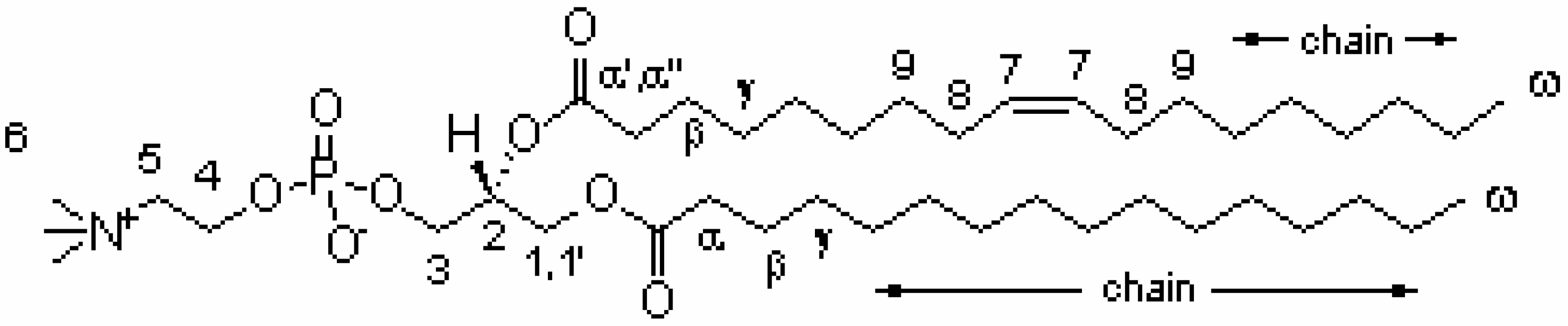
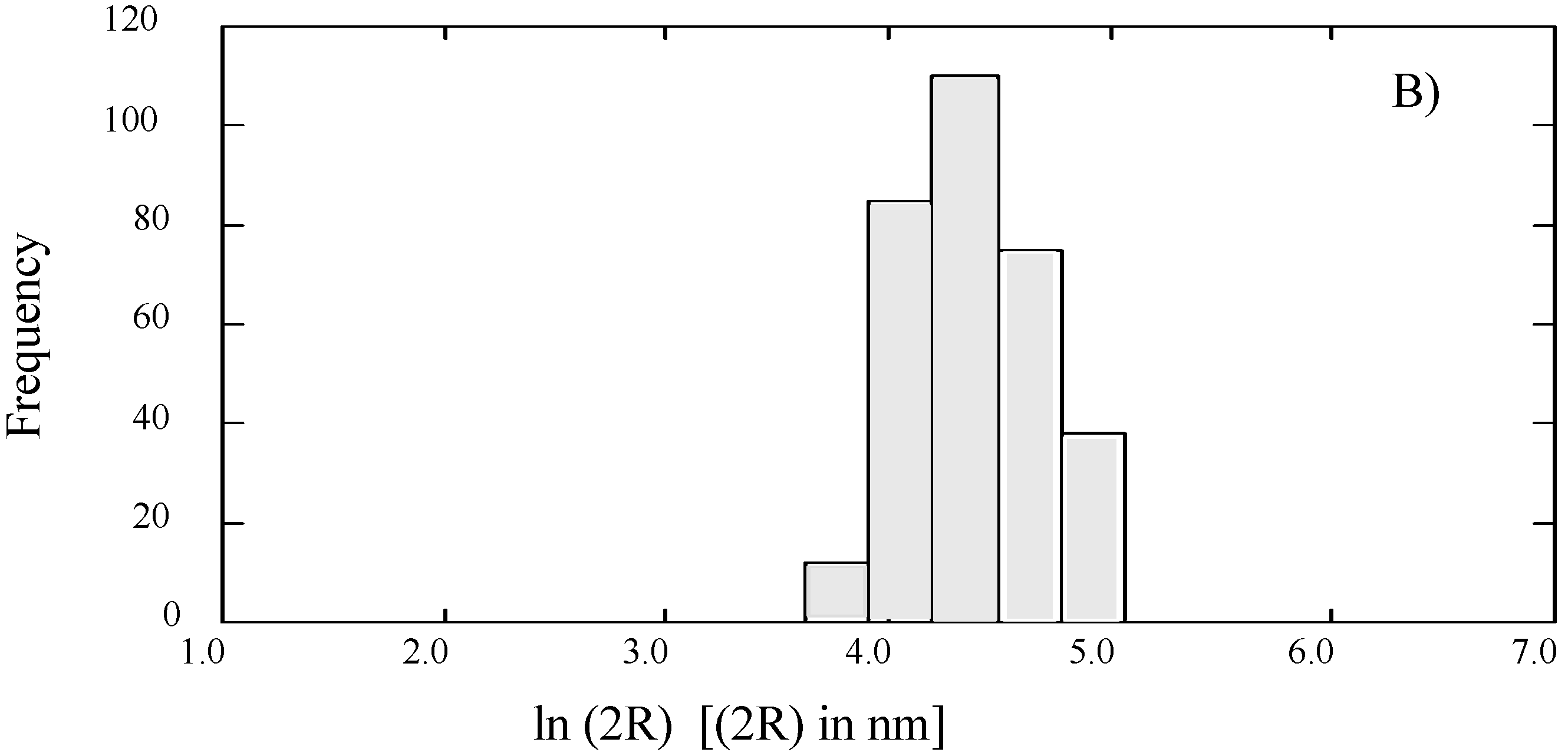
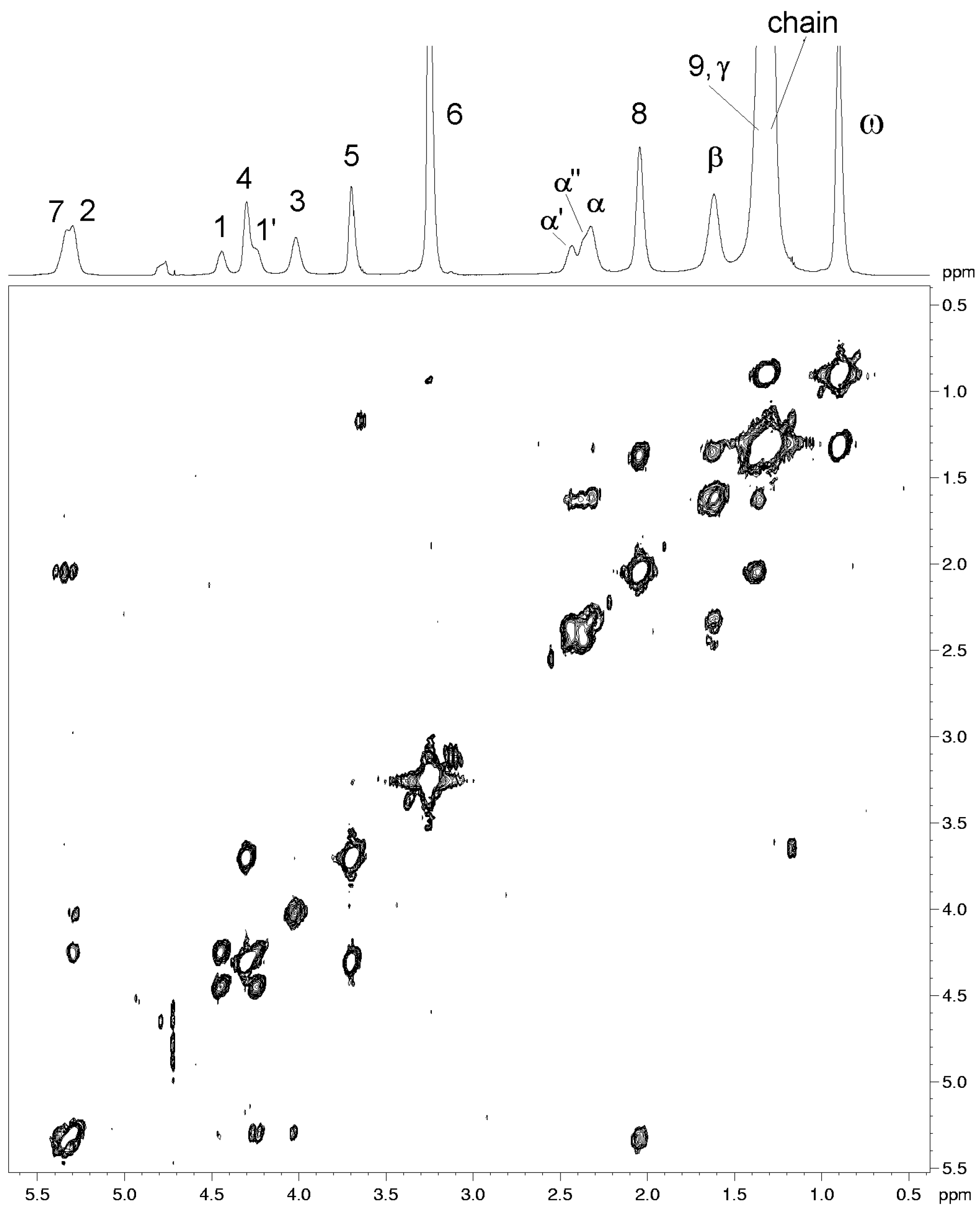
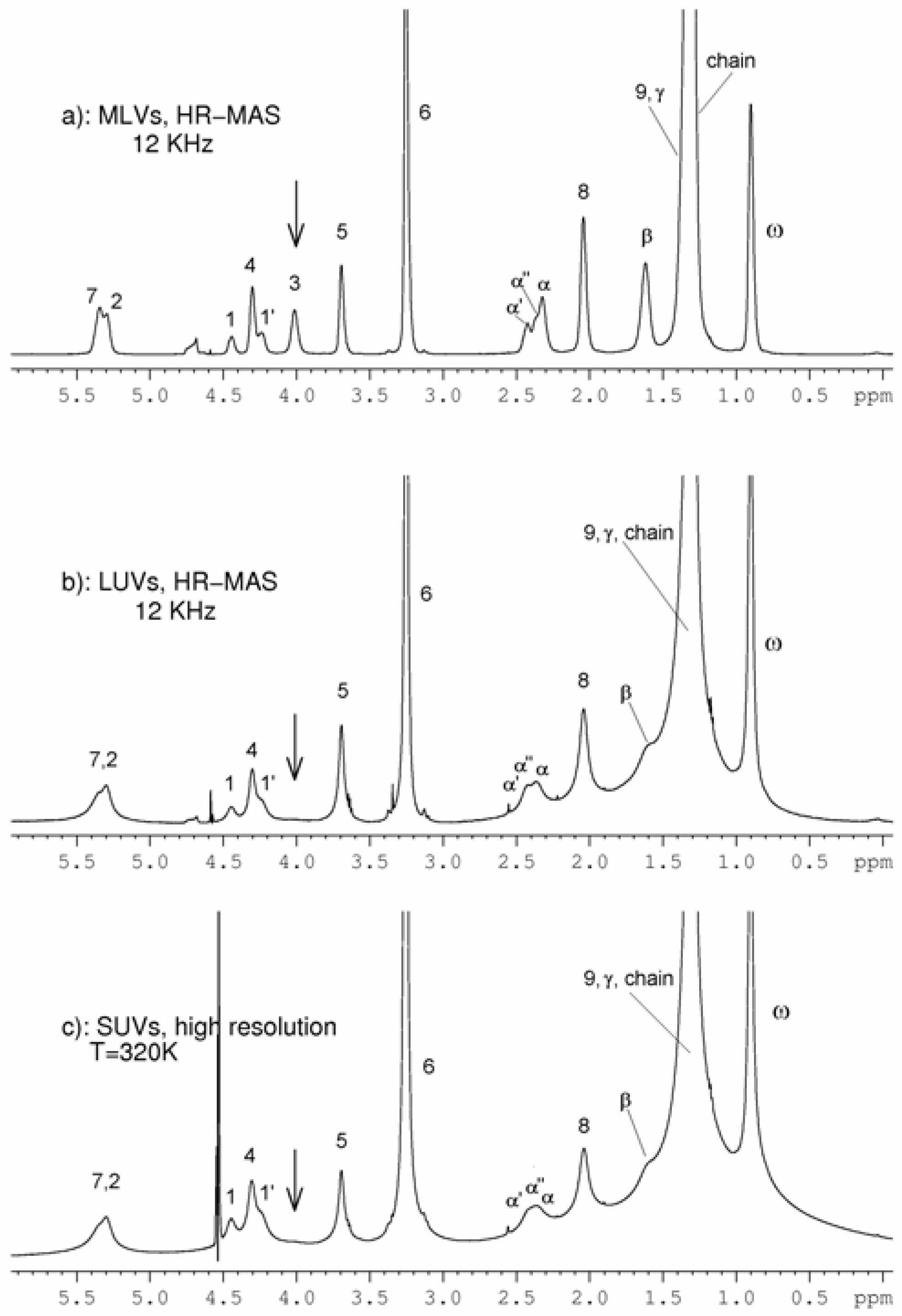
Results and Discussion

The size and the size distribution of SUVs, LUVs and MLVs were investigated using dynamic light scattering methods. The Laplace inversion results yield a monomodal distribution where a single population of liposomes with some degree of polydispersity is present; the case of LUVs extruded to 100 nm is shown in Figure 2. The average dimensions obtained for the different liposomes were: 60 ± 3 nm for SUVs extruded to 50 nm; 145 ± 6 nm for LUVs extruded to 100 nm (see Figure 2) and 890 ± 10 nm for MLVs. Here, the results obtained for SUVs, LUVs and MLVs using classical NMR conditions in solution and HR-MAS conditions will be discussed separately.
Figure 2. CONTIN analysis of the autocorrelation function for the sample of POPC LUVs: a monomodal distribution with an average hydrodynamic diameter of 145 ± 6 nm is obtained.
Molecules 11 00334 g002

SUVs

Due to the rapid molecular tumbling which averages the anisotropic dipolar interactions, SUVs can be studied using the classical NMR approach in solution [16,17,18]. The resolution of SUVs spectra depends on the size of the vesicles: the larger the size, the worse the resolution. We chose to study SUVs with a 60 nm diameter, obtained by the extrusion process, since these vesicles give rise to NMR spectra in solution with sufficient resolution; moreover the effect of curvature in vesicles of this size does not seem so severe as in the case of small SUVs obtained by sonication. In the case of very small SUVs of dipalmitoyl-sn-glycero-3-phospatidylcholine (DPPC) obtained by sonication, Schuh et al. reported a splitting effect [8] on some NMR resonances attributed to the packing asymmetry of the inner and outer part of the bilayer; in our experiments performed on 60 nm SUVs, this effect was not present. An increase in temperature, and, therefore, in molecular tumbling, only improved slightly the resolution, as reported in Table 1, where the line width values relative to some 1H resonances measured at half height is reported.

LUVS and MLVs

As rationalized by Maricq and Waugh using the average Hamiltonian theory [22], the problem of obtaining high resolution spectra of dynamic systems is due to the presence of two sources of dynamic effects, namely the random molecular motion of the spin system and the mechanical spinning of the sample. The inhomogeneous and anisotropic interactions can be averaged using the HR-MAS technique.
LUVs display a characteristic molecular tumbling in the intermediate time scale regime [19]. Consequently, their molecular motion interferes with the coherent perturbation applied by the MAS and reduces the efficiency of the averaging of the inhomogeneous and anisotropic interactions giving rise to a broadening of the spectral lines [23]. In our specific case, the potential of HR-MAS for the characterization of LUVs was verified on LUVs of 145 nm, considered unaffected by curvature effects [24]. As reported by Devaux et al. [19], the molecular tumbling of LUVs can be reduced by adding glycerol and by lowering the temperature; in this fashion they obtained 31P HR-MAS spectra of LUVs with a significant improvement of the resolution. However, this method turned out to be incompatible with the observation of 1H-NMR spectra; in fact, 1H HR-MAS experiments performed on a sample of LUVs in the presence of deuterated glycerol ( 50% in v/v) showed an extremely broad HOD signal (up to 100 Hz) which hampered the use of any water-suppression sequence.
Table 1. Line width (in Hz) of some resonances, measured at half height in different samples of POPC liposomes. The error is ± 2 Hz.
SampleSpinning speed (kHz)T (K)aωchainβ865341
UVs n.s.300.067109**28****
SUVs n.s.320.03368*1041534*76*
LUVs3.0301.83984**18**70*
LUVs6.0303.93274*901537*52*
LUVs8.0307.03070*781333*48*
LUVs12.0314.82458*501024*3433
MLVs3.0301.8244247371821342932
MLVs6.0303.9233943281721342631
MLVs8.0307.0233641271721322530
MLVs12.0314.8223339261620292328
* = Not measurable ; a Values as obtained by the temperature calibration setting the temperature at 300.0 K
Therefore, the 145 nm LUVs were analyzed under HR-MAS conditions at different spinning speeds. The analysis of the spectra showed that the resolution was increased by raising the spinning speed. The values, reported in Table 1, show that even at intermediate spinning rates (6-8 kHz) the line widths of some NMR signals of LUVs were sensibly narrowed with respect to those observed in the case of SUVs in solution at 320K. Therefore, the resolution achievable for LUVs using “normal” HR-MAS conditions, i.e. non-extremely high spinning speeds, was better than that obtained in the case of SUVs at higher temperature.
MLVs have an extremely slow molecular tumbling and so an essentially infinite correlation time [21]. Therefore, in the case of these liposomes, the molecular tumbling does not interfere with the coherent perturbation applied by the MAS and gives rise to an efficient average of the inhomogeneous and anisotropic interactions contributing. A sample of MLVs was analyzed using the HR-MAS technique at different spinning speeds, see Table 1. As previously discussed, in the case of MLVs which have an extremely slow molecular tumbling [21], the gain in resolution obtained by raising the spinning speed, is more evident than in the case of LUVs which, in turn, have a molecular tumbling in an intermediate time scale regime [19].
Figure 3. 600 MHz 1H-NMR spectra of different classes of POPC (50mg/mL) liposomes: a) HR-MAS spectrum of MLVs, 12kHz at 314.8 K; b) HR-MAS spectrum of LUVs, 12kHz at 314.8 K; c) 1H NMR spectrum of SUVs, no spinning at 320.0K. The arrows indicate the chemical shift position of CH2-3.
Molecules 11 00334 g003
Under the same spinning speed and temperature conditions, the 1H HR-MAS spectrum of MLVs showed a better resolution than the HR-MAS spectrum of LUVs; in fact, the line widths of the resonances in the spectra of MLVs are always smaller than the corresponding resonances in the spectra of LUVs, see Table 1. Moreover, in the case of MLVs the values obtained for the line widths by spinning the sample at 3 kHz are compare well to those registered for LUVs at 12 kHz. These results confirm that in the case of MLVs the rotation at the magic angle fully averages the interactions whereas in the case of LUVs only a partial average is obtained.
The improved resolution in the 1H MAS spectra of MLVs allowed more detailed spectral information to be obtained. For instance, the resonance at 4.01 ppm of the CH2-3 of the glycerol moiety, barely observable both in HR-MAS spectra of LUVs and in solution NMR spectra of SUVs (Figure 3) is readily detectable in the spectrum of MLVs.
The better resolution obtained for MLVs under HR-MAS conditions allows a more complete assignment of MLVs to be obtained with respect to that reported in the literature [25,26,27]. The assignment was obtained by means of 1H-1H COSY and 1H-1H NOESY experiments as reported in Table 2 and Figure 3a). In particular, the good resolution allowed the 1H chemical shifts of different methylene groups of fatty acids chains, previously [25,26,27] reported as coincident at 1.3 ppm, to be discriminated.
Table 2. Assignment of 1H HR-MAS spectrum of POPC MLVs (50 mg/mL) 1.
Resonance1H (ppm)
(±0.02 ppm)2
COSY cross-peaksNOESY cross-peaks
CH-75.3388, 9
CH-25.301', 1, 31', 1
CH2- 14.441', 21', 2, 3, 6
  1'4.241, 21, 2 , 3,6
CH2- 44.3055, 6
CH2- 34.0121', 1, 2, 6
CH2- 53.6944, 6
CH3- 63.25------4, 5, 1, 1', 3,2
CH2- α'2.43β, α'' α'', γ, β
  α''2.36β, α'α', γ, β
CH22.32βγ, β
CH2-82.047, 97, 9
CH21.62α, α', α'', γα, α', α'', γ
CH2-91.3787, 8
CH2-γ1.35ββ
chain1.30ωω
CH3 - ω0.90chainchain
1 Spinning frequency = 6 kHz and T= 303.9 K.2 Chemical shifts are reported with respect to the ω-CH3 at 0.90 ppm.
A detailed description was also obtained for the methylene groups in the α position with respect to the carbonyls of the fatty acids: well resolved signals are present at 2.32 ppm, 2.36 ppm and 2.43 ppm. In the 1H-1H COSY (see Figure 4) and the 1H-1H NOESY (not shown) maps the signals at 2.36 ppm and at 2.43 ppm have a strong cross-peak typical of a geminal coupling. Therefore, these signals are due to non equivalent protons of a methylene in the α position with respect to a carbonyl. Keeping in consideration the more restricted motion of the oleoyl chain with respect to the palmitoyl one, the non equivalent protons labelled as α' and α'' can be attributed to the oleoyl moiety whereas the signal at 2.32 ppm can be attributed to the palmitoyl moiety having a less restricted motion. Non-equivalent chemical shifted protons for both the methylene groups in α position with respect to the carbonyls were reported Previously for liposomes obtained from derivatives of POPC (1-palmitoyl-2-oleoyl-sn-glycero-3-phospatidylcytidine and 1-palmitoyl-2-oleoyl-sn-glycero-3-phospatidylinosine) [28].
Figure 4. 1H-1H COSY map of POPC MLVs obtained using a spinning rate of 6 kHz. The calibrated temperature value of 303.9 K was obtained setting the temperature at 300.0 K.
Molecules 11 00334 g004
Other resonances previously reported as coincident [25,26,27] were well separated: for instance, the resonance at 4.30 ppm due to the CH2-4 of the choline moiety was separated from that of the glycerol proton 1' at 4.24 ppm and also the resonance at 5.33 ppm due to the olefinic CH - 7 of the oleoyl chain from that of the glycerol CH- 2 at 5.30 ppm.
The 1H-1H NOESY map confirmed the assignment obtained through the 1H-1H COSY map and gave further information (see Table 2). For instance, the presence of NOESY cross peaks between the signal of the three CH3 of the choline moiety and the signals due to the CH2-4 and to the CH2-5 of the choline as well as the NOESY cross peaks between the signal due to the three CH3 of the choline and the signals due to the protons 1, 1', 3, 2 of glycerol, indicate that the choline tends to turn back toward the glycerol moiety.

Conclusions

The screening performed on the three different classes of POPC liposomes confirmed that currently the best structural results are obtained studying the MLVs with HR-MAS. In fact, due to the particular motion of the vesicles, in the case of LUVs the use of HR-MAS methods does not allow one to attain the same spectral quality obtained for MLVs and yields only a small resolution improvement with respect to the case of SUVs. However, for an NMR characterization of unilamellar liposomes, in consideration of the better likeness of LUVs to the real membranes with respect to the SUVs, the study of LUVs by HR-MAS is preferable.

Experimental Section

Materials

1-Palmitoyl-2-oleoyl-sn-glycero-3-phospatidylcholine (POPC) was purchased from Avanti Polar Lipids (Alabaster, AL., USA) and used without further purification. D2O (99.9% atom D) was purchased from Fluka. Glycerol-1,1,2,3,3-d5 (98 % atom D) was purchased from Isotec, Inc. All the other reagents of the highest purity grade were obtained from Fluka, Sigma, or Merck.

Sample preparation

Liposome preparation has been described extensively elsewhere [29]. MLVs from POPC were produced by hydrating a dry film of the lipid deposited from a CHCl3 solution on a glass wall in an appropriate buffer solution (20 mM deuterated sodium phosphate buffer, 20 mM KCl, 0.2 mM EDTA, pD = 8). The final lipid concentration was 50 mg/mL. The unilamellar liposome dispersions were formed by pressure extrusion. In order to reduce the lamellarity of the lipid dispersions before the extrusion process, MLVs were subjected to five freeze-thaw cycles. LUVs with diameters larger than 100 nm were obtained by repeated extrusions of the multilamellar liposomes at room temperature through two stacked polycarbonate membranes with a pore size of 200 nm, followed by extrusion through 100 nm pore size membranes. In order to obtain SUVs with a diameter of approximately 50 nm an additional extrusion through 50 nm pore size membranes was carried out. All extrusions were made by using "The Extruder" system (Lipex Biomembranes, Vancouver, Canada) and Nucleopore polycarbonate membranes [30]. The LUVs/glycerol-d5 sample was obtained by mixing equal volumes of solutions of glycerol-d5 and LUVs.

Dynamic Light Scattering (DLS) [31, 32]

Dynamic light scattering measurements were performed at 293 K, using a 90° scattering angle on a home-made photo-goniometer equipped with a logarithmic correlator (Brookhaven, mod BI900 AT). The light source was a 10 mW He-Ne laser (λ = 632.8 nm). Samples of SUVs, LUVs and MLVs were dispersed in a aqueous suspension at a 0.2 mg/mL concentration and were investigated in the 0.1 μs to 1 s temporal range. The results obtained as autocorrelation functions were analysed using the Laplace inversion function performed with the CONTIN algorithm.

NMR spectroscopy

Two different NMR techniques were used: high resolution NMR at different temperatures (T>Tm being Tm the temperature for the transition of the phospholipids from a crystalline, or solid, state to a fluid analogue state) and different spinning speeds for the samples of LUVs and MLVs.

1. High resolution NMR on SUVs

All high resolution NMR experiments were performed on a Bruker AVANCE AQS600 spectrometer operating at 600.13 MHz and using a Bruker z-gradient reverse probe head. The 1H-NMR experiments were performed at 300.0 K and 320.0 K accumulating 800 FIDs with 16K data points and a spectral width of 12.2 kHz. A 90° excitation pulse (11-12 μs) was used with a relaxation delay of 2s. Prior to the Fourier transformation, the data were zero filled to 16K points and apodized using an exponential line broadening of 1 Hz. A standard pulse sequence with a soft water suppression by a presaturation of the HDO resonance during a relaxation delay was used.

2. HR-MAS

All HR-MAS experiments were performed on a Bruker AVANCE 600 spectrometer operating at 600.13 MHz using a Bruker HR-MAS probe head. Samples were loaded in 4 mm ZrO2 cylindricalrotors with spherical inserts (internal volume of 12 μL) and spun at different spinning rates in range from 3 to 12 kHz. All the experiments were performed after a sufficient equilibration time. The 1H HR-MAS NMR experiments on LUVs, LUVs/glycerol and MLVs samples were performed setting the temperature to 300.0 K and using different spinning speeds (3, 6, 8 and 12 KHz); 128 FIDs with 32K data points and a spectral width of 6.5 kHz were accumulated. In order to verify that the rotation at high speed, i.e. 12 kHz, does not modify the liposomes, after each HR-MAS spectrum at 12 KHz, an HR-MAS spectrum at low speeds was carried out.
In the case of LUVs and MLVs samples, a standard pulse sequence with a soft water suppression by a presaturation of the HDO resonance during a relaxation delay was used. A 90° excitation pulse (4-5 μs) was used with a relaxation delay of 2s. Prior to Fourier transformation, the data were zero filled to 32K points and apodized using an exponential line broadening of 1 Hz. In order to assign the MLV MAS spectrum, 1H-1H COSY and 1H-1H NOESY experiments were performed [33] setting the temperature at 300.0 K and with a spinning speed of 6 kHz. The 1H-1H COSY experiment was performed using a spectral width of 6 kHz in both dimensions, 256 increments, 10 scans, 2K data points and 2 s relaxation delay. 1H-1H -NOESY spectra were obtained in TPPI phase-sensitive mode, using a spectral width of 6 kHz width in both dimensions, 512 increments, 32 scans, 1K data points, a relaxation delay of 2 s and mixing times of 15 ms and 200 ms. Due to the presence of spin diffusion effects, the assignment of the spectrum was obtained using the 2D NOESY with a short mixing time (15 ms): in fact, at this mixing time no spin diffusion effect were observed.

3. Temperature calibration in the HR-MAS experiments

In order to know the real temperature value in the case of the HR-MAS experiments a calibration of temperature is necessary. The calibration can be obtained by measuring the HOD chemical shift at different spinning frequencies in HR-MAS conditions: from this value it is possible to obtain the correct temperature using a calibration curve (chemical shift as function of the temperature) obtained in high resolution condition. Therefore, the temperature was calibrated at MAS spinning frequencies (3, 6, 8, 12 KHz) by measuring the chemical shift difference between the HOD signal and the ω-CH3 signal in the samples of POPC LUVs and of POPC MLVs loaded in 4 mm ZrO2 cylindrical rotors with spherical inserts (internal volume of 12 μL). The chemical shift as a function of the temperature was measured on the POPC LUVs sample in a 5 mm tube with a high resolution probe whose temperature had been precisely calibrated with a thermocouple. The corrected temperature values are reported in Table 1. No temperature difference was registered for the samples of LUVs and MLVs during the MAS experiments at the same spinning speed, as reported in column 3 of Table 1.

References and Notes

  1. Bangham, A.D.; Horne, R.W. Negative Staining of Phospholipids and their Structural Modification by Surface-active Agents as observed in the Electron Microscope. J. Mol. Biol. 1964, 8, 660–668. [Google Scholar] [CrossRef] [PubMed]
  2. Papahadjopoulos, D. (Ed.) Liposomes and their uses in biology and medicine. Ann. NY Acad. Sci. 1978, 308, 1–462. [CrossRef] [PubMed]
  3. Lasic, D.D. Liposomes from Physics to Applications; Elsevier: Amsterdam, 1993. [Google Scholar]
  4. Wasan, K.M.; Lopez-Berestein, G. The past, present, and future uses of liposomes in treating infectious diseases. Immunopharm. Immunotox. 1995, 17, 1–15. [Google Scholar] [CrossRef]
  5. Gregoriadis, G. Engineering liposomes for drug delivery: progress and problems. TIBTECH 1995, 13, 527–537. [Google Scholar]
  6. Vemuri, S.; Rhodes, C.T. Preparation and characterization of liposomes as therapeutic delivery systems: a review. Pharmaceut. Acta Helv. 1995, 70, 95–111. [Google Scholar] [CrossRef]
  7. Lasic, D.D. The mechanism of vesicle formation. Biochem. J. 1988, 256, 1–11. [Google Scholar] [PubMed]
  8. Schuh, J.R.; Banerjee, U.; Müller, L.; Chan, S.I. The phospholipid packing arrangement in small bilayer vesicles as revealed by proton magnetic resonance studies at 500 MHz. BBA– Biomembranes 1982, 687, 219–225. [Google Scholar]
  9. Longmuir, K.J.; Dahlquist, F.W. Direct Spectroscopic Observation of Inner and Outer Hydrocarbon Chains of Lipid Bilayer Vesicles. Proc. Natl Acad. Sci. USA 1976, 73, 2716–2719. [Google Scholar]
  10. Tauskela, J.S.; Thompson, M. A 31P-NMR spin-lattice relaxation and 31P{1H} nuclear Overhauser effect study of sonicated small unilamellar phosphatidylcholine vesicles. BBA–Biomembranes 1992, 1104, 137–146. [Google Scholar]
  11. Smith, R.L.; Oldfield, E. Dynamic structure of membranes by deuterium NMR. Science 1984, 225, 280–288. [Google Scholar] [CrossRef] [PubMed]
  12. Forbes, J.; Bowers, J.; Shan, X.; Moran, L.; Oldfield, E.; Moscatello, M.A. Some new developments in solid-state nuclear magnetic resonance spectroscopic studies of lipids and biological membranes, including the effects of cholesterol in model and natural systems. J. Chem. Soc. Faraday Trans.1 1988, 84, 3821–3850. [Google Scholar] [CrossRef]
  13. Dong, R.Y. Nuclear Magnetic Resonance of Liquid Crystals; Springer: New York, 1997. [Google Scholar]
  14. Evans, F.D.; Wennerstrom, H. The Colloidal Domain, where Physics, Chemistry, Biology and Technology Meet; VCH Publishers: New York, 1994. [Google Scholar]
  15. Lasic, D.D. Magnetic Resonance Methods in The Studies of Liposomes. Bull. Magn. Reson. 1991, 13, 3–13. [Google Scholar]
  16. Chapman, D.; Kamat, V.B.; De Gier, J.; Penkett, S.A. Nuclear magnetic resonance studies of erythrocyte membranes. J. Mol. Biol. 1968, 31, 101–114. [Google Scholar] [CrossRef] [PubMed]
  17. Bhamidipati, S.P.; Hamilton, J.A. Interactions of Lyso 1-Palmitoylphosphatidylcholine with Phospholipids: A 13C and 31P NMR Study. Biochemistry 1995, 34, 5666–5677. [Google Scholar] [CrossRef] [PubMed]
  18. Swairjo, M.A.; Seaton, B.A.; Roberts, M.F. Effect of vesicle composition and curvature on the dissociation of phosphatidic acid in small unilamellar vesicles - a 31P-NMR study. BBA – Biomembranes 1994, 1191, 354–361. [Google Scholar]
  19. Traikia, M.; Langlais, B.D.; Cannarozzi, G.; Devaux, P.F. High-Resolution Spectra of Liposomes Using MAS NMR. The Case of Intermediate-Size Vesicles. J. Magn. Reson. 1997, 125, 140–144. [Google Scholar]
  20. Oldfield, E.; Bowers, J.L.; Forbes, J. High-resolution proton and carbon-13 NMR of membranes: why sonicate? Biochemistry 1987, 26, 6919–6923. [Google Scholar] [CrossRef]
  21. Bloom, M.; Burnell, E.E.; MacKay, A.l.; Nichol, C.P.; Valic, M.I.; Weeks, G. Fatty acyl chain order in lecithin model membranes determined from proton magnetic resonance. Biochemistry 1978, 17, 5750–5762. [Google Scholar] [CrossRef] [PubMed]
  22. Maricq, M.M.; Waugh, J.S. NMR in rotating solids. J. Chem. Phys. 1979, 70, 3300–3316. [Google Scholar] [CrossRef]
  23. Long, J.R.; Sun, B.Q.; Bowen, A.; Griffin, R.G. Molecular Dynamics and Magic Angle Spinning NMR. J. Am. Chem. Soc. 1994, 116, 11950–11956. [Google Scholar] [CrossRef]
  24. Sheetz, M.P.; Chan, S.I. Effect of sonication on the structure of lecithin bilayers. Biochemistry 1972, 11, 4573–4581. [Google Scholar]
  25. Hong, M.; Schimidt-Rohr, K.; Nanz, D. Study of phospholipid structure by 1H, 13C, and 31P dipolar couplings from two-dimensional NMR. Biophys. J. 1995, 69, 1939–1950. [Google Scholar] [CrossRef] [PubMed]
  26. Gawrish, K.; Yau, W.M. Lateral Lipid Diffusion Dominates NOESY Cross-Relaxation in Membranes. J. Am. Chem. Soc. 2000, 122, 3971–3972. [Google Scholar] [CrossRef]
  27. Volke, F.; Pampel, A. Membrane hydration and structure on a subnanometer scale as seen by high resolution solid state nuclear magnetic resonance: POPC and POPC/C12EO4 model membranes. Biophys. J. 1995, 68, 1960–1965. [Google Scholar] [CrossRef] [PubMed]
  28. Cruciani, O.; Mannina, L.; Sobolev, A.P.; Segre, A.L.; Luisi, P.L. Multilamellar Liposomes Formed by Phosphatidyl Nucleosides: An NMR-HR-MAS Characterization. Langmuir 2004, 20, 1144–1151. [Google Scholar] [CrossRef] [PubMed]
  29. Szoka, F.; Papahadjopoulos, D. Comparative Properties and Methods of Preparation of Lipid Vesicles (Liposomes). Annu. Rev. Biophys. Bio. 1980, 9, 467–508. [Google Scholar] [CrossRef]
  30. Hope, M.J.; Bally, M.B.; Webb, G.; Cullis, P.R. Production of large unilamellar vesicles by a rapid extrusion procedure. Characterization of size distribution, trapped volume and ability to maintain a membrane potential. BBA– Biomembranes 1985, 812, 55–65. [Google Scholar]
  31. Schmitz, K.S. Dynamic light scattering by macromolecules; Academic Press Inc.: San Diego, CA, 1990. [Google Scholar]
  32. Brown, W. (Ed.) Dynamic Light scattering: The method and some applications; Oxford Science Pub., Clarendon Press: Oxford, 1993.
  33. Braun, S.; Kalinowski, H.O.; Berger, S. 150 and More Basic NMR Experiments. A practical course; Wiley-VCH: Weinhiem, Germany, 1998; Chapter 10. [Google Scholar]
  • Sample Availability: Available from authors.

