
Electrochemical Sensors Based on the Electropolymerized Natural Phenolic Antioxidants and Their Analytical Application


Abstract
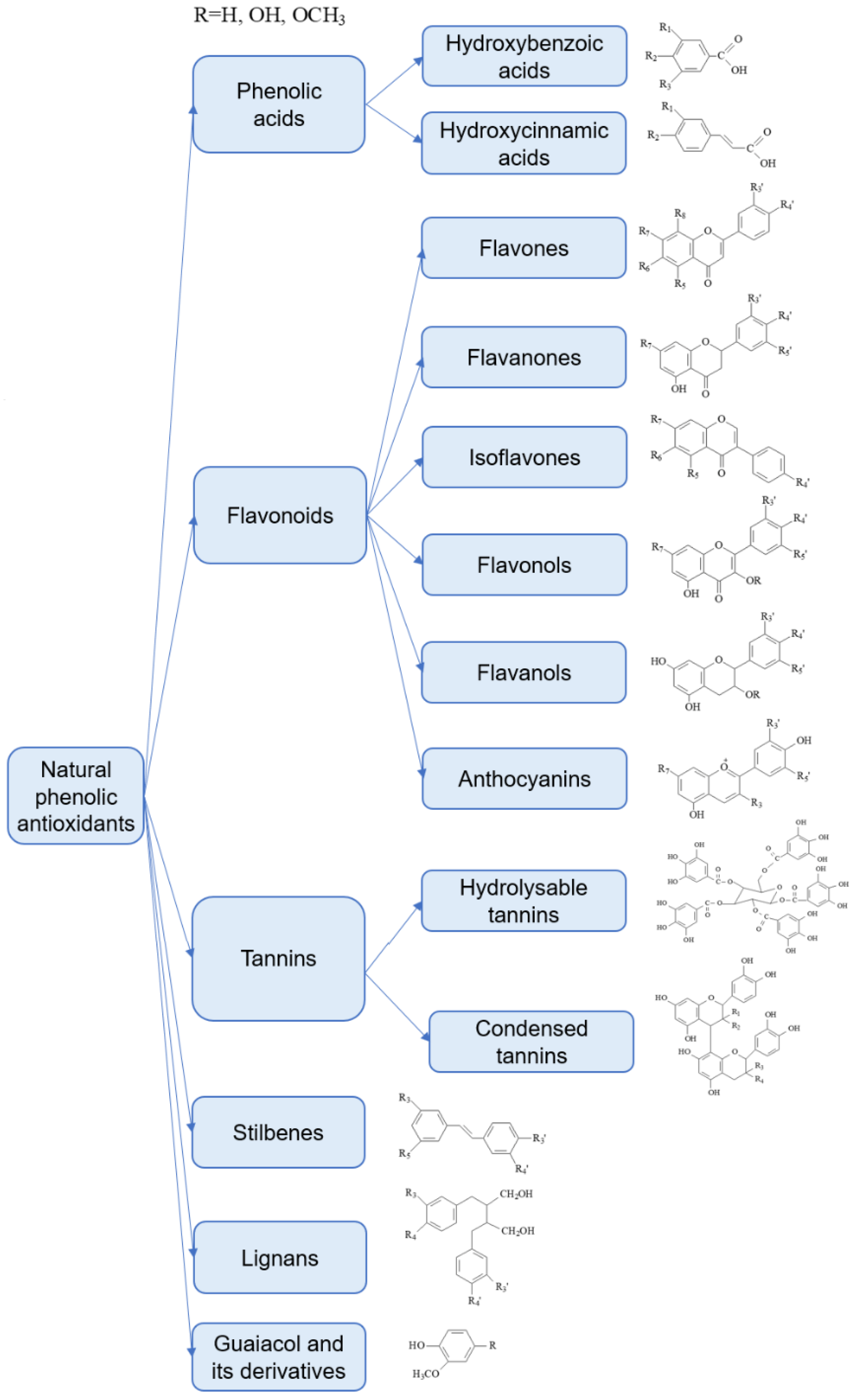
1. Introduction
2. Electropolymerization of Phenolic Compounds as a Route for the Sensor Surface Modification
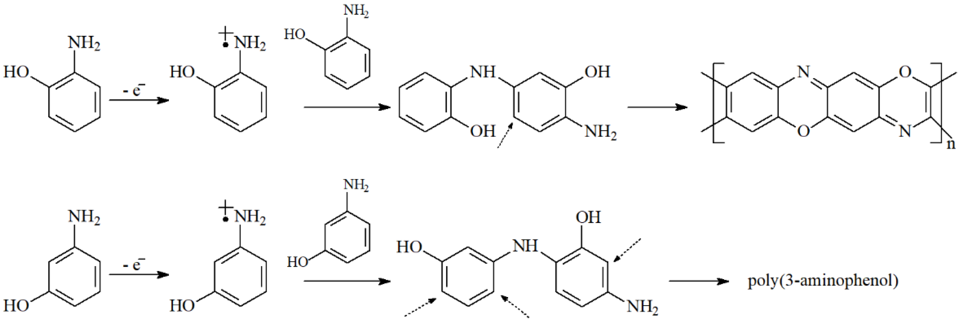
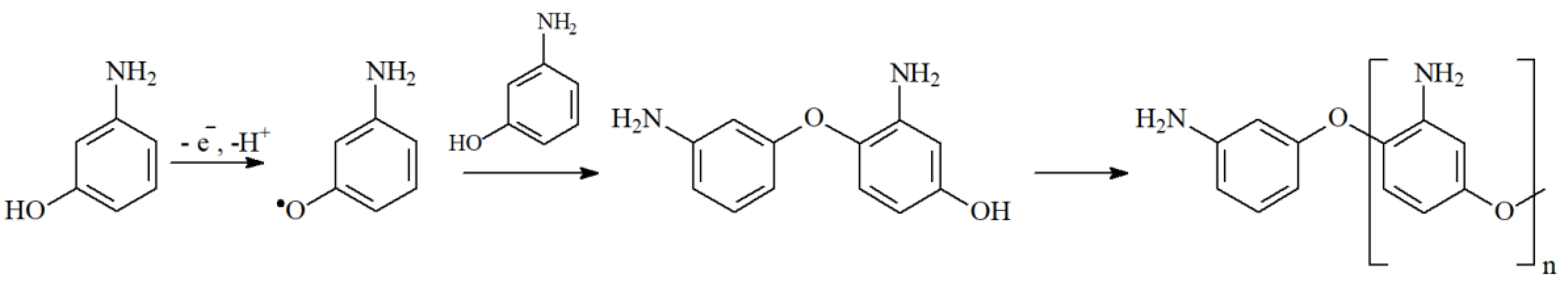
2.1. Electropolymerization of Phenols and Aminophenols
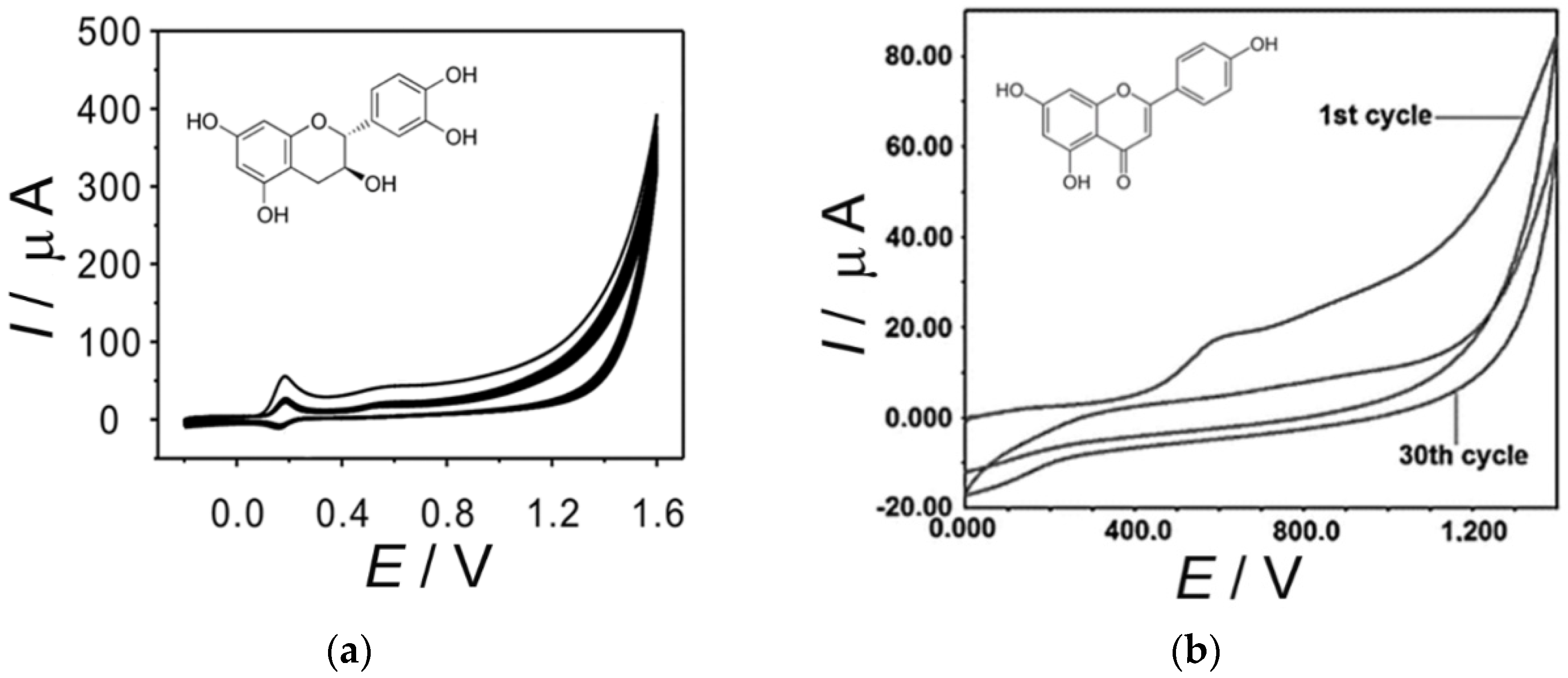
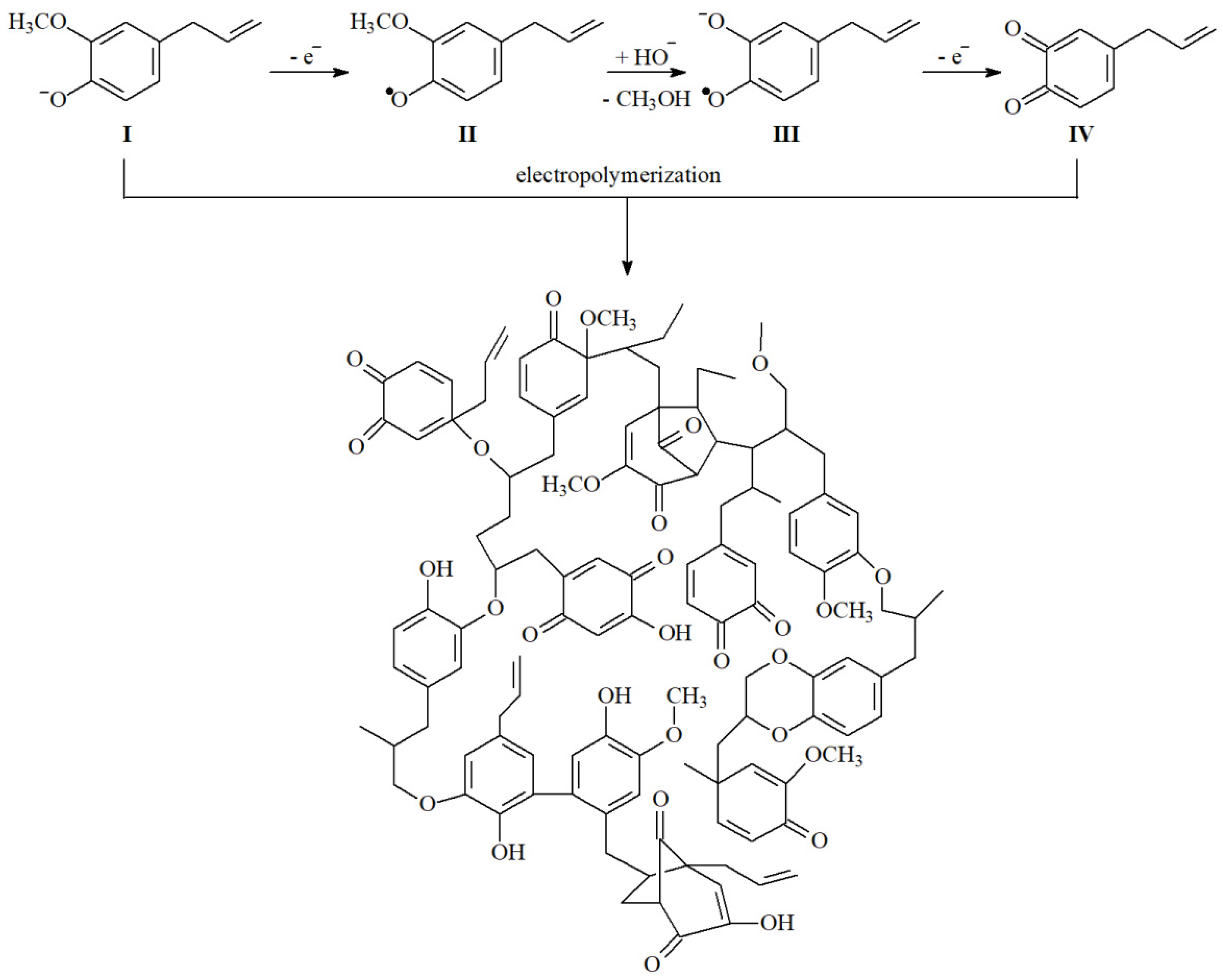
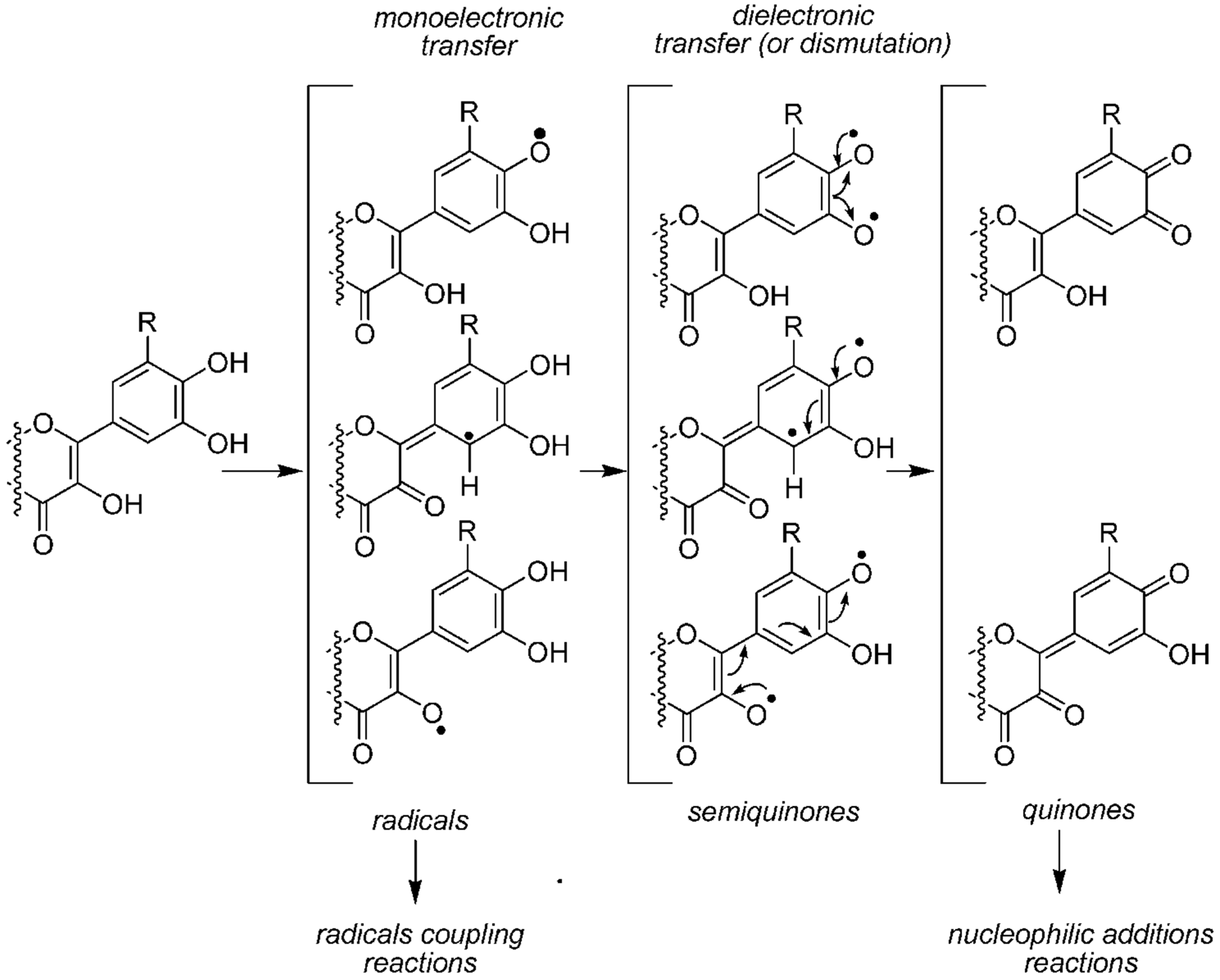
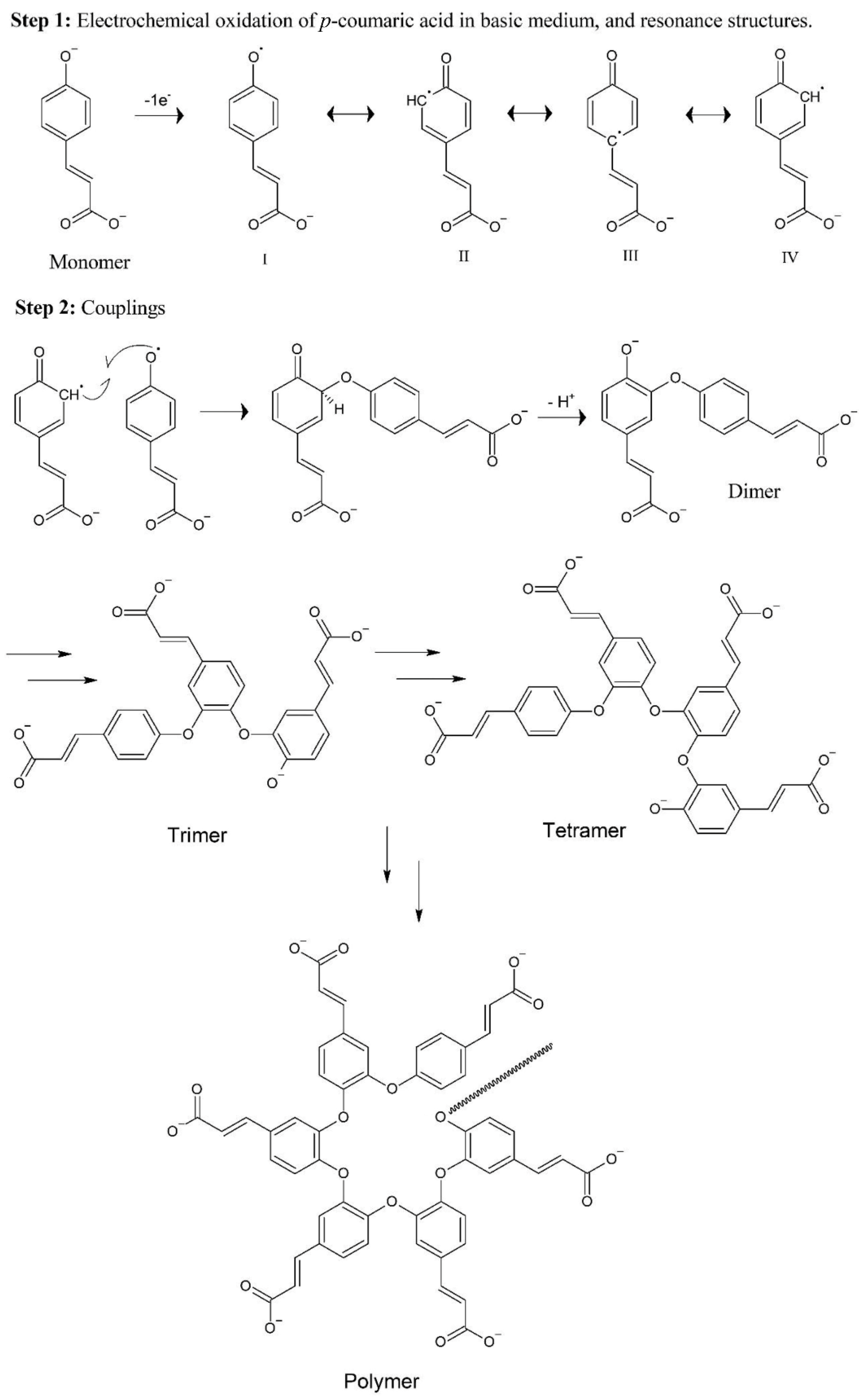
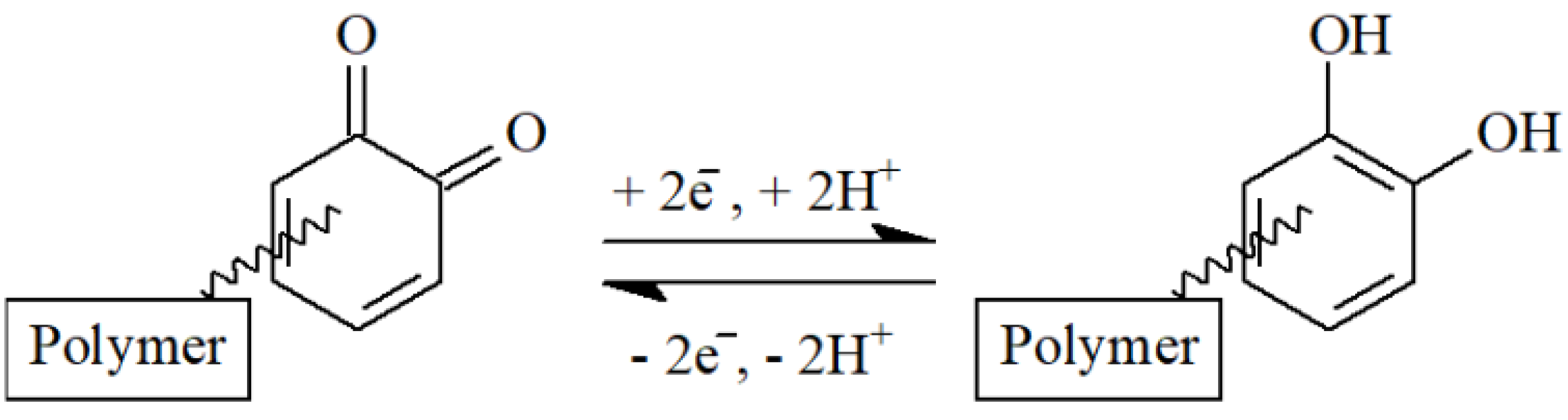
2.2. Electropolymerization of Natural Phenolic Antioxidants
- Phenoxyl radical polymerization with poly(oxyphenylene) film [27];
- Diels–Alder reaction between IV and double bond forming 1,4-benzenedioxane fragment [21];
- Dimerization of phenoxyl radicals, and oxidative coupling of I with the formation of lignan-like structures [28];
- Hydroxylation of IV giving p-quinone fragments [29].
- Via phenoxyl radical formation and its following reactions;
- Via a conjugated double bond.
3. Analytical Application of the Sensors Based on the Electropolymerized Natural Phenolics
3.1. Application in Organic Analysis
3.2. Application in Inorganic Analysis
3.3. Electropolymerized Natural Phenolic Antioxidants as Protective Coatings
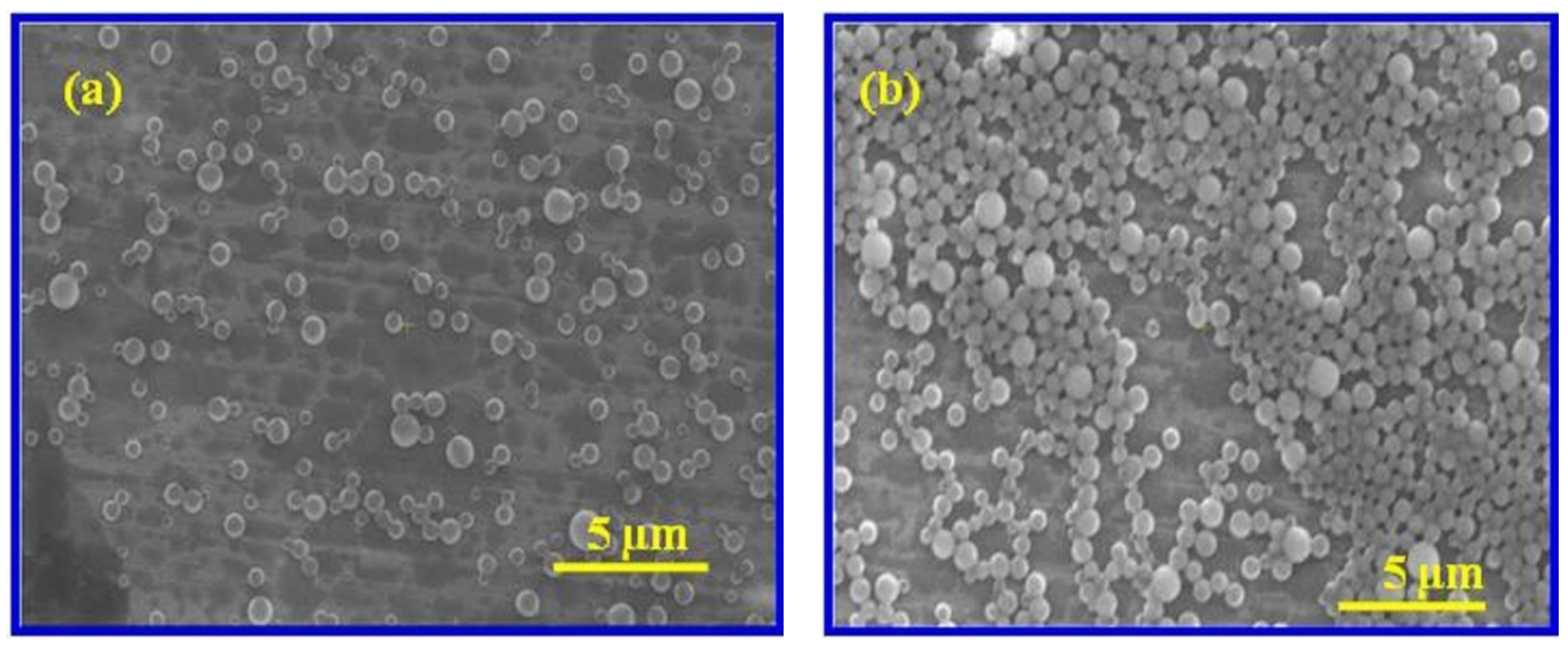
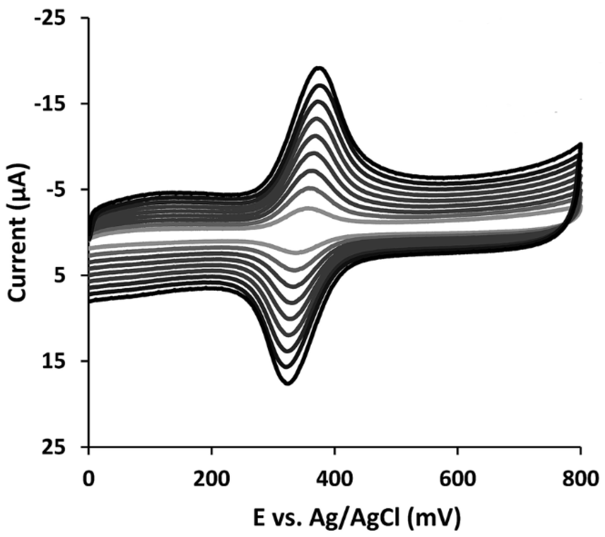
4. Sensors Based on the Combination of Nanomaterials and Electropolymerized Natural Phenolic Antioxidants and Their Analytical Capabilities
5. Electropolymerized Natural Phenolics as a Platform for Immobilization of Other Modifiers
- The formation of a positively charged choline layer on the bare platinum electrode using cyclic voltammetry in a 1.0 mM choline solution using 0.01 M LiClO4 as supporting electrolyte;
- The drop-casting of carboxylated-by-acid treatment MWCNTs via electrostatic adsorption and evaporation of the solvent to dryness;
- The potentiodynamic electropolymerization of quercetin from its 1 mM solution in a 0.1 M phosphate-buffered saline (pH 7.0);
- The chelation and adsorption of silver ions on the polyquercetin surface from its 1.0 mM solution in 0.1 M LiNO3 for 30 min;
- The electrodeposition of silver nanoparticles by the voltammetric reduction in 0.1 M LiNO3.
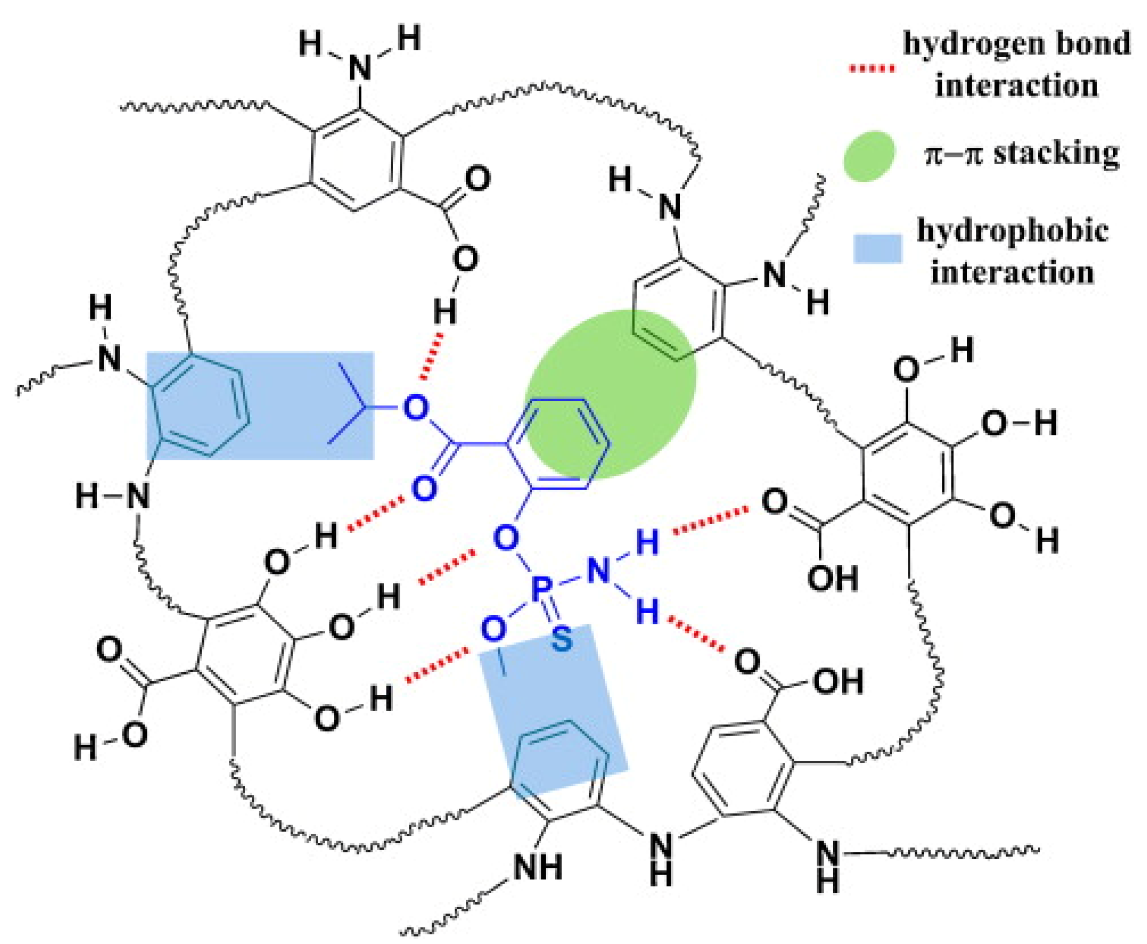
6. Molecularly Imprinted Polymers Based on the Electropolymerized Natural Phenolics as a Sensitive Layer of Electrochemical Sensors
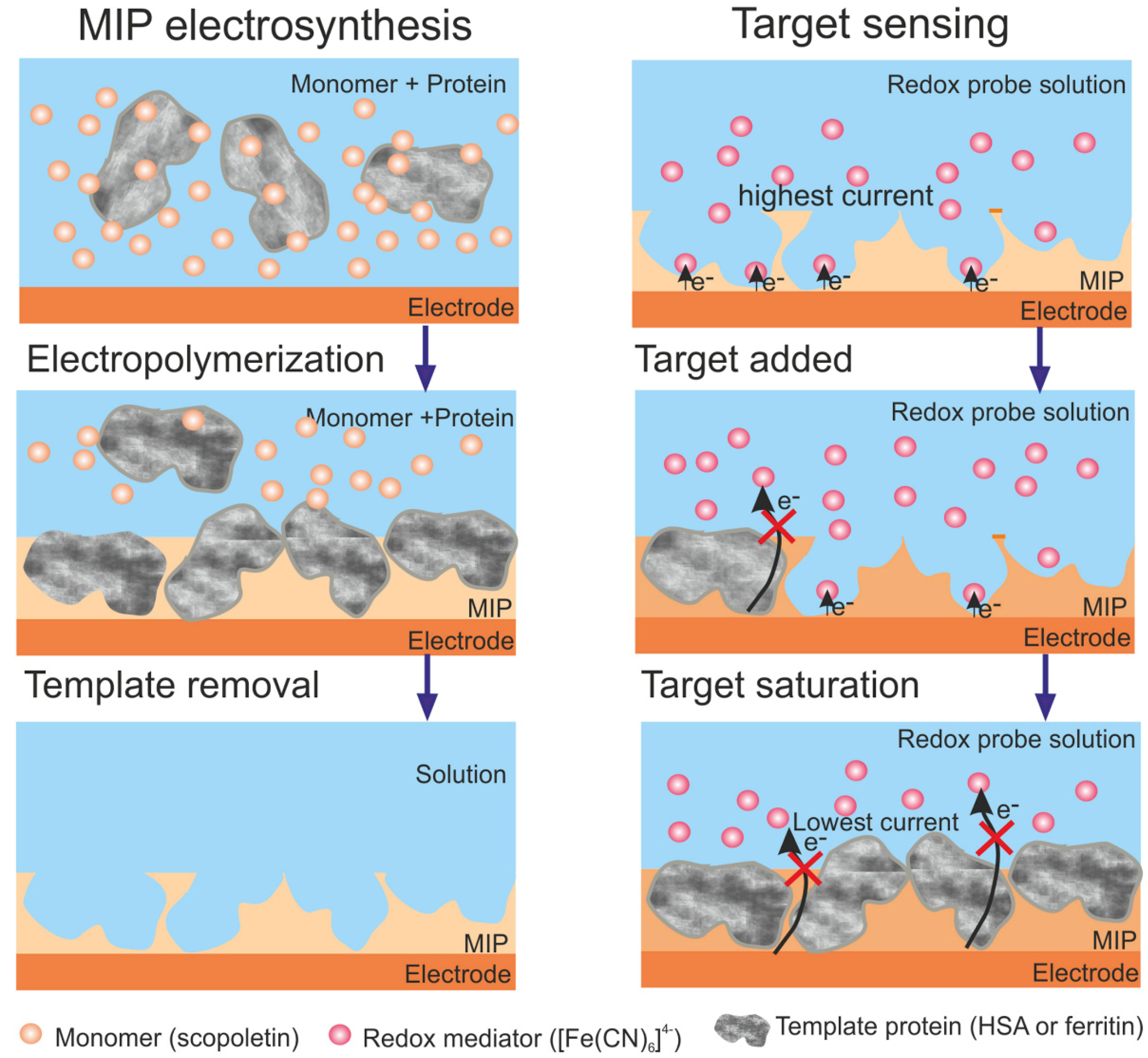
6.1. Protein Imprinted Polymers
- Electrodeposition on the gold or fluorine-doped indium tin oxide after preliminary incubation in the protein template solution, followed by scopoletin electropolymerization in the solution using repeated chronoamperometry by applying multiple alternating oxidation and reduction pulses of 1 s at 0.9 V and 5 s at 0 V [101,102];
- Electrodeposition on the gold electrode covered with a self-assembled monolayer of mercaptoundecanoic acid from the solution containing scopoletin and a protein template by single redox potential pulses at 0 V for 15 s and 0.5 V for 35 s [103];
- Electrodeposition on the gold electrode covered with a self-assembled monolayer of mercaptoundecanoic acid after preliminary incubation in the protein template solution, followed by scopoletin electropolymerization in a solution using a single potential pulse at 0.7 V for 5 s and 0 V for 5 s [104].
6.2. Low-Molecular Organic Compounds Imprinted Polymers
7. Problems in the Field of Electrochemical Sensors Based on Electropolymerized Natural Phenolic Antioxidants
8. Conclusions
- An investigation of the polymeric coverage structure and clarification of the electropolymerization reaction mechanisms and schemes;
- An application of other nanomaterials (metal and metal oxide nanoparticles, other nanostructured compounds, etc.) as a platform for the electrodeposition of natural phenolics based polymers;
- The development of the electrochemical sensors based on the copolymerization of natural phenolic antioxidants of the same or different classes;
- The further development of the electrochemical sensors using electropolymerized natural phenolic antioxidants as a platform for the immobilization of other modifiers;
- The creation of the sensors based on the electrochemically synthesized MIPs for the low molecular biologically active compounds, other than organophosphate pesticides and melamine;
- The application of mathematical design methods and machine learning for the optimization of electropolymerization conditions and the choice of monomers for sensor creation, including MIPs-based ones;
- The fabrication of sensors characterized by a high long-term stability of the response, allowing for their commercial production, storage, and application in real practice.
Author Contributions
Funding
Institutional Review Board Statement
Informed Consent Statement
Data Availability Statement
Conflicts of Interest
References
- Qin, X.; Xiao-Ya, H.; Shi-Rong, H. Electrochemical sensors based on electropolymerized films. In Electropolymerization; Schab-Balcerzak, E., Ed.; In TechOpen: London, UK, 2011; pp. 187–198. [Google Scholar]
- Wallace, G.G.; Spinks, G.M.; Kane-Maguire, L.A.P.; Teasdale, P.R. Conductive Electroactive Polymers: Intelligent Materials Systems; CRC Press: Boca Raton, FL, USA, 2003. [Google Scholar]
- Inzelt, G. Conducting Polymers: A New Era in Electrochemistry; Springer: New York, NY, USA, 2012. [Google Scholar]
- Pauliukaite, R.; Ghica, M.E.; Barsan, M.M.; Brett, C.M.A. Phenazines and polyphenazines in electrochemical sensors and biosensors. Anal. Lett. 2009, 43, 1588–1608. [Google Scholar] [CrossRef]
- Tucceri, R. Non-conducting poly(o-aminophenol) films in the field of the bioelectrochemistry. Am. J. Anal. Chem. 2013, 4, 13–26. [Google Scholar] [CrossRef]
- Samet, Y.; Kraiem, D.; Abdelhédi, R. Electropolymerization of phenol, o-nitrophenol and o-methoxyphenol on gold and carbon steel materials and their corrosion protection effects. Prog. Org. Coat. 2010, 69, 335–343. [Google Scholar] [CrossRef]
- Tahar, N.B.; Savall, A. Electropolymerization of phenol on a vitreous carbon electrode in alkaline aqueous solution at different temperatures. Electrochim. Acta 2009, 55, 465–469. [Google Scholar] [CrossRef][Green Version]
- Yuqing, M.; Jianrong, C.; Xiaohua, W. Using electropolymerized non-conducting polymers to develop enzyme amperometric biosensors. Trends Biotechnol. 2004, 22, 227–231. [Google Scholar] [CrossRef]
- Nakabayashi, Y.; Wakuda, M.; Imai, H. Amperometric glucose sensors fabricated by electrochemical polymerization of phenols on carbon paste electrodes containing ferrocene as an electron transfer mediator. Anal. Sci. 1998, 14, 1069–1076. [Google Scholar] [CrossRef]
- Nakabayashi, Y.; Yoshikawa, H. Amperometric biosensors for sensing of hydrogen peroxide based on electron transfer between horseradish peroxidase and ferrocene as a mediator. Anal. Sci. 2000, 16, 609–613. [Google Scholar] [CrossRef]
- Ezerskis, Z.; Jusys, Z. Electropolymerization of chlorinated phenols on a Pt electrode in alkaline solution. Part IV: A gas chromatography mass spectrometry study. J. Appl. Electrochem. 2002, 32, 543–550. [Google Scholar] [CrossRef]
- Iotov, P.I.; Kalcheva, S.V. Mechanistic approach to the oxidation of phenol at a platinum/gold electrode in an acid medium. J. Electroanal. Chem. 1998, 442, 19–26. [Google Scholar] [CrossRef]
- Sayyah, S.M.; Khaliel, A.B.; Azooz, R.E.; Mohamed, F. Electropolymerization of some ortho-substituted phenol derivatives on Pt-electrode from aqueous acidic solution; kinetics, mechanism, electrochemical studies and characterization of the polymer obtained. In Electropolymerization; Schab-Balcerzak, E., Ed.; In TechOpen: London, UK, 2011; pp. 21–52. [Google Scholar]
- Gattrell, M.; Kirk, D.W. A study of electrode passivation during aqueous phenol electrolysis. J. Electrochem. Soc. 1993, 140, 903–911. [Google Scholar] [CrossRef]
- Matsushita, Y.; Sekiguchi, T.; Ichino, R.; Fukushima, K. Electropolymerization of coniferyl alcohol. J. Wood Sci. 2009, 55, 344–349. [Google Scholar] [CrossRef]
- Zhang, Z.; Liu, H.; Deng, J. A glucose biosensor based on immobilization of glucose oxidase in electropolymerized o-aminophenol film on platinized glassy carbon electrode. Anal. Chem. 1996, 68, 1632–1638. [Google Scholar] [CrossRef]
- Salavagione, H.J.; Arias, J.; Garces, P.; Morallon, E.; Barbero, C.; Vazquez, J.L. Spectroelectrochemical study of the oxidation of aminophenols on platinum electrode in acid medium. J. Electroanal. Chem. 2004, 565, 375–383. [Google Scholar] [CrossRef]
- Tucceri, R. Poly(o-aminophenol) Film Electrodes. Synthesis, Transport Properties and Practical Applications; Springer: New York, NY, USA, 2013. [Google Scholar]
- Guenbour, A.; Kacemi, A.; Benbachir, A.; Aries, L. Electropolymerization of 2-aminophenol: Electrochemical and spectroscopic studies. Prog. Org. Coat. 2000, 38, 121–126. [Google Scholar] [CrossRef]
- Ziyatdinova, G.; Budnikov, H. Natural phenolic antioxidants in bioanalytical chemistry: State of the art and prospects of development. Russ. Chem. Rev. 2015, 84, 194–224. [Google Scholar] [CrossRef]
- Ciszewski, A.; Milczarek, G. Polyeugenol-modified platinum electrode for selective detection of dopamine in the presence of ascorbic acid. Anal. Chem. 1999, 71, 1055–1061. [Google Scholar] [CrossRef]
- Toniolo, R.; Dossi, N.; Pizzariello, A.; Susmel, S.; Bontempelli, G. Simultaneous detection of ascorbic acid and hydrogen peroxide by flow-injection analysis with a thin layer dual-electrode detector. Electroanalysis 2011, 23, 628–636. [Google Scholar] [CrossRef]
- Ciszewski, A.; Milczarek, G. Preparation and general properties of chemically modified electrodes based on electrosynthesized thin polymeric films derived from eugenol. Electroanalysis 2001, 13, 860–867. [Google Scholar] [CrossRef]
- Okumura, L.L.; Stradiotto, N.R.; Rees, N.V.; Compton, R.G. Modifying glassy carbon (GC) electrodes to confer selectivity for the voltammetric detection of l-cysteine in the presence of Dl-homocysteine and glutathione. Electroanalysis 2008, 20, 916–918. [Google Scholar] [CrossRef]
- Paul, D.W.; Prajapati, I.; Reed, M.L. Electropolymerized eugenol: Evaluation as a protective film for oxygen sensing. Sens. Actuators B Chem. 2013, 183, 129–135. [Google Scholar] [CrossRef]
- El Qouatli, S.; Ngono, R.T.; Najih, R.; Chtaini, A. Eugenol modified titanium electrode for the analysis of carbocysteine. Zaštita Materijala 2011, 52, 242–246. [Google Scholar]
- MacTylor, C.E.; Ewing, A.G. Characterization of the effects of varying the pH and monomer concentrations of poly(oxyphenylene) insulating films on carbon fiber electrodes. Electroanalysis 1997, 9, 755–758. [Google Scholar] [CrossRef]
- Ban, Y.; Iwasaki, T.; Ohmizu, H. Natural products and pharmaceuticals. In Organic Electrochemistry, 3rd ed.; Lund, H., Baizer, M.M., Eds.; Marcel Dekker: New York, NY, USA, 1991; pp. 765–786. [Google Scholar]
- Ueda, C.; Tse, D.C.-S.; Kuwana, T. Stability of catechol modified carbon electrodes for electrocatalysis of dihydronicotinamide adenine dinucleotide and ascorbic acid. Anal. Chem. 1982, 54, 850–856. [Google Scholar] [CrossRef]
- Es-Safi, N.-E.; Ghidouche, S.; Ducrot, P.H. Flavonoids: Hemisynthesis, reactivity, characterization and free radical scavenging activity. Molecules 2007, 12, 2228–2258. [Google Scholar] [CrossRef] [PubMed]
- Da Silva, L.V.; de Almeida, A.K.A.; Xavier, J.A.; Lopes, C.B.; dos Santos Silva, F.A.; Lima, P.R.; dos Santos, N.D.; Kubota, L.T.; Goulart, M.O.F. Phenol based redox mediators in electroanalysis. J. Electroanal. Chem. 2018, 827, 230–252. [Google Scholar] [CrossRef]
- Ziyatdinova, G.; Guss, E.; Budnikov, H. Poly(gallic acid)/MWNT-modified electrode for the selective and sensitive voltammetric determination of quercetin in medicinal herbs. J. Electroanal. Chem. 2018, 821, 73–81. [Google Scholar] [CrossRef]
- Silva, L.V.d.; Silva, F.A.S.; Kubota, L.T.; Lopes, C.B.; Lima, P.R.; Costa, E.O.; Pinho Júnior, W.; Goulart, M.O.F. Amperometric sensor based on carbon nanotubes and electropolymerized vanillic acid for simultaneous determination of ascorbic acid, dopamine, and uric acid. J. Solid State Electrochem. 2016, 20, 2389–2393. [Google Scholar] [CrossRef]
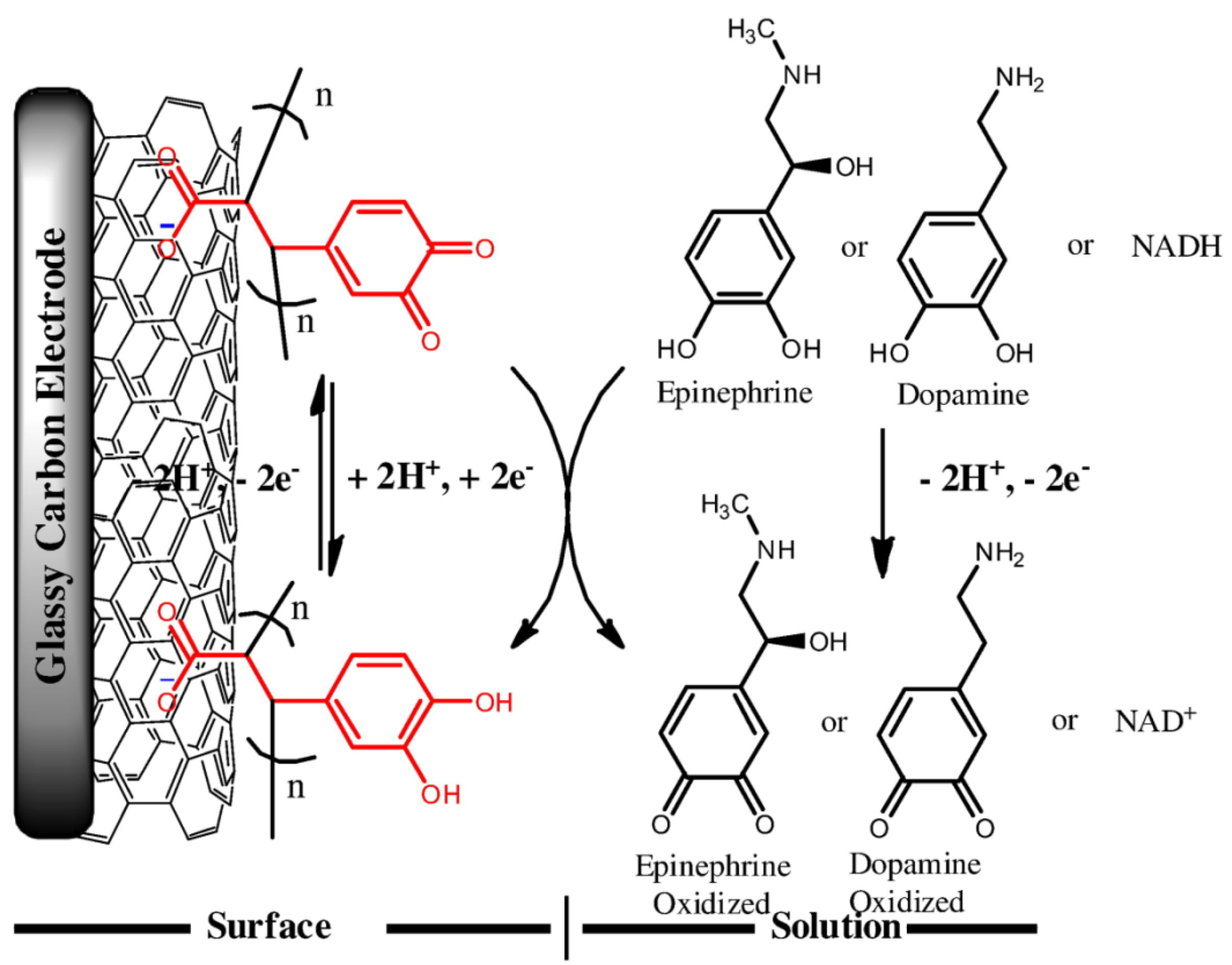
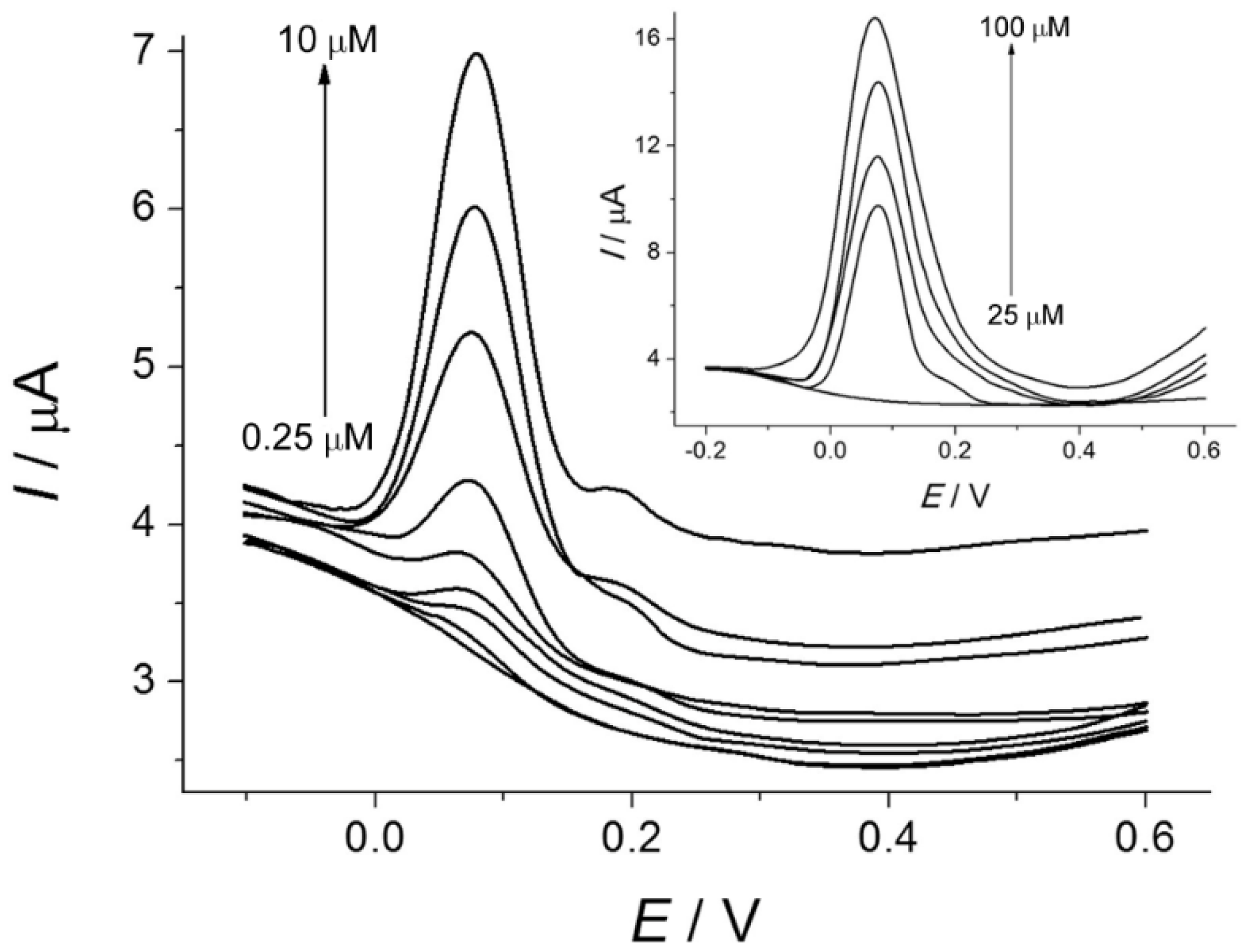
- Da Silva, L.V.; Lopes, C.B.; da Silva, W.C.; de Paiva, Y.G.; dos Santos Silva, F.A.; Lima, P.R.; Kubota, L.T.; Goulart, M.O.F. Electropolymerization of ferulic acid on multi-walled carbon nanotubes modified glassy carbon electrode as a versatile platform for NADH, dopamine and epinephrine separate detection. Microchem. J. 2017, 133, 460–467. [Google Scholar] [CrossRef]
- Ziyatdinova, G.; Guss, E.; Budnikov, H. Selective electrochemical sensor based on the electropolymerized p-coumaric acid for the direct determination of L-cysteine. Electrochim. Acta 2018, 270, 369–377. [Google Scholar] [CrossRef]
- Matsushita, Y.; Nakamura, A.; Aoki, D.; Yagami, S.; Fukushima, K. Bio-based polymer from ferulic acid by electropolymerization. BioResources 2016, 11, 9789–9802. [Google Scholar] [CrossRef][Green Version]
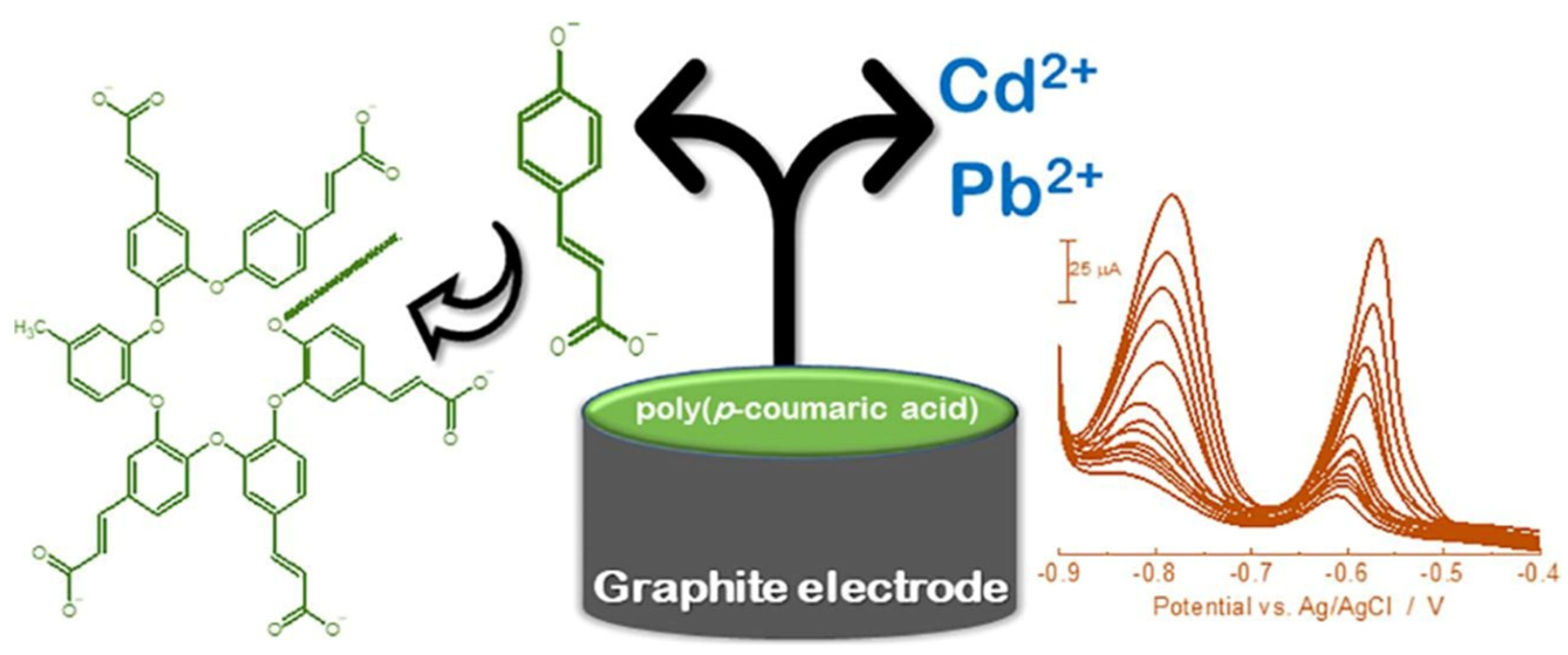
- Lima, T.M.; Soares, P.I.; Nascimento, L.A.; Franco, D.L.; Pereira, A.C.; Ferreira, L.F. A novel electrochemical sensor for simultaneous determination of cadmium and lead using graphite electrodes modified with poly(p-coumaric acid). Microchem. J. 2021, 168, 106406. [Google Scholar] [CrossRef]
- Jin, G.-P.; Chen, Q.-Z.; Ding, Y.-F.; He, J.-B. Electrochemistry behavior of adrenalin, serotonin and ascorbic acid at novel poly rutin modified paraffin-impregnated graphite electrode. Electrochim. Acta 2007, 52, 2535–2541. [Google Scholar] [CrossRef]
- Madhuri, C.; Reddy, Y.V.M.; Saritha, D.; Venu, M.; Kiranmai, S.; Rao, V.P.; Madhavi, G. Electrochemical behavior of poly (rutin) modified carbon paste electrode for the determination of uric acid in the presence of ascorbic acid and dopamine. Chem. Sci. Rev. Lett. 2016, 5, 136–143. [Google Scholar]
- Manjunatha, J.G.; Swamy, B.E.K.; Deraman, M.; Mamatha, G.P. Simultaneous voltammetric measurement of ascorbic acid and dopamine at poly (vanillin) modified carbon paste electrode: A cyclic voltammetric study. Der. Pharm. Chem. 2012, 4, 2489–2497. [Google Scholar]
- Madhuchandra, H.D.; Swamy, B.E.K. Poly (vanillin) modified carbon paste electrode for the determination of adrenaline: A voltammetric study. Mater. Sci. Energy Technol. 2019, 2, 697–702. [Google Scholar] [CrossRef]
- Manjunatha, J.G.; Swamy, B.E.K.; Mamatha, G.P.; Gilbert, O.; Chandrashekar, B.N.; Sherigara, B.S. Electrochemical studies of dopamine and epinephrine at a poly (tannic acid) modified carbon paste electrode: A cyclic voltammetric study. Int. J. Electrochem. Sci. 2010, 5, 1236–1245. [Google Scholar]
- Devadas, B.; Rajkumar, M.; Chen, S.M. Electropolymerization of curcumin on glassy carbon electrode and its electrocatalytic application for the voltammetric determination of epinephrine and p-acetoaminophenol. Colloids Surf. B 2014, 116, 674–680. [Google Scholar] [CrossRef]
- Wei, J.; He, J.; Chen, C.; Wang, X. A catechin-modified carbon paste electrode for electrocatalytic determination of neurotransmitters. Anal. Methods 2015, 7, 5641–5648. [Google Scholar] [CrossRef]
- Oztekin, Y.; Yazicigil, Z.; Ramanaviciene, A.; Ramanavicius, A. Polyphenol-modified glassy carbon electrodes for copper detection. Sensors Actuat. B 2011, 152, 37–48. [Google Scholar] [CrossRef]
- Oztekin, Y.; Yazicigil, Z.; Ramanaviciene, A.; Ramanavicius, A. Square wave voltammetry based on determination of copper (II) ions by polyluteolin- and polykaempferol-modified electrodes. Talanta 2011, 85, 1020–1027. [Google Scholar] [CrossRef]
- Mülazımoğlu, I.E.; Solak, A.O. A novel apigenin modified glassy carbon sensor electrode for the determination of copper ions in soil samples. Anal. Methods 2011, 3, 2534–2539. [Google Scholar] [CrossRef]
- Kumar, K.K.; Devendiran, M.; Kalaivani, R.A.; Narayanan, S.S. Polycurcumin nanospheres modified electrode for nanoscale detection of mercury ions in seawater. Chem. Phys. Lett. 2021, 781, 138974. [Google Scholar] [CrossRef]
- Li, N.B.; Ren, W.; Luo, H.Q. Simultaneous voltammetric measurement of ascorbic acid and dopamine on poly(caffeic acid)-modified glassy carbon electrode. J. Solid State Electrochem. 2008, 12, 693–699. [Google Scholar] [CrossRef]
- Filik, H.; Avan, A.A.; Aydar, S.; Çetintaş, G. Determination of acetaminophen in the presence of ascorbic acid using a glassy carbon electrode modified with poly(caffeic acid). Int. J. Electrochem. Sci. 2014, 9, 148–160. [Google Scholar]
- Rohanifar, A.; Devasurendra, A.M.; Young, J.A.; Kirchhoff, J.R. Determination of L-DOPA at an optimized poly(caffeic acid) modified glassy carbon electrode. Anal. Methods 2016, 8, 7891–7897. [Google Scholar] [CrossRef]
- Abdel-Hamid, R.; Newair, E.F. Voltammetric determination of polyphenolic content in pomegranate juice using a poly(gallic acid)/multiwalled carbon nanotube modified electrode. Beilstein J. Nanotechnol. 2016, 7, 1104–1112. [Google Scholar] [CrossRef]
- Karabiberoğlu, Ş.U.; Koçak, Ç.C.; Dursun, Z. An over-oxidized poly(rutin) modified electrode for selective and sensitive determination of catechol and hydroquinone. J. Electroanal. Chem. 2019, 850, 113415. [Google Scholar] [CrossRef]
- Herrero-Martínez, J.M.; Sanmartin, M.; Rosés, M.; Bosch, E.; Ràfols, C. Determination of dissociation constants of flavonoids by capillary electrophoresis. Electrophoresis 2005, 26, 1886–1895. [Google Scholar] [CrossRef] [PubMed]
- Ziyatdinova, G.K.; Guss, E.V.; Yakupova, E.N.; Budnikov, H.C. An electrode based on electropolymerized naringin for voltammetry. Uchenye Zap. Kazan. Univ. Seriya Estestv. Nauk. 2019, 161, 5–19. [Google Scholar] [CrossRef]
- Ziyatdinova, G.; Kozlova, E.; Budnikov, H. Electropolymerized eugenol-MWNT-based electrode for voltammetric evaluation of wine antioxidant capacity. Electroanalysis 2015, 27, 1660–1668. [Google Scholar] [CrossRef]
- Marwati, S.; Siwanta, D.; Trisunaryanti, W.; Louise, I.S.Y. Polyeugenol-modified graphite electrode for determination of hydroquinone in cosmetic. J. Phys. Conf. Ser. 2020, 1485, 012034. [Google Scholar] [CrossRef]
- Zhou, J.; Wei, P.; Pan, J.; Mei, Q.; Tong, Y.; Zhai, H. A simple and sensitive electrochemical sensor with A-PCA film modified electrode for the determination of metanephrine. New J. Chem. 2019, 43, 14368–14376. [Google Scholar] [CrossRef]
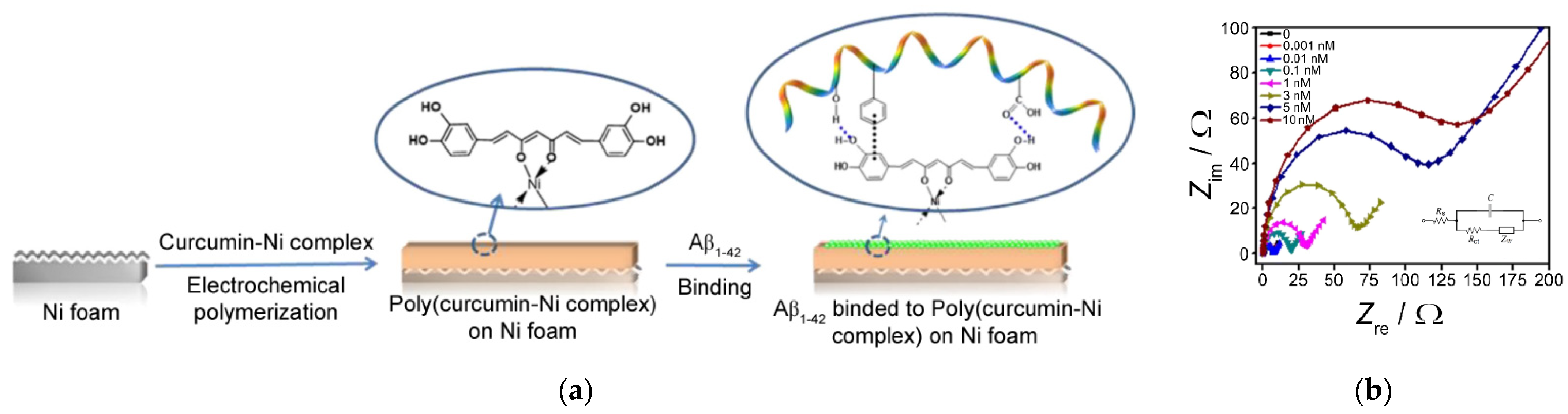
- Qin, J.; Park, J.S.; Joc, D.G.; Cho, M.; Lee, Y. Curcumin-based electrochemical sensor of amyloid-β oligomer for the early detection of Alzheimer’s disease. Sens. Actuators B Chem. 2018, 273, 1593–1599. [Google Scholar] [CrossRef]
- Mukwevho, E.; Ferreira, Z.; Ayeleso, A. Potential role of sulfur-containing antioxidant systems in highly oxidative environments. Molecules 2014, 19, 19376–19389. [Google Scholar] [CrossRef]
- Duran, S.T.; Hassine, C.B.A.; Burç, M.; Güngör, Ö. Voltammetric determination of α-lipoic acid using poly(vanillin) modified platinum electrode. Anal. Bioanal. Electrochem. 2020, 12, 857–869. [Google Scholar]
- Ziyatdinova, G.K.; Budnikov, G.K.; Pogorel’tsev, V.I. Electrochemical determination of lipoic acid. J. Anal. Chem. 2004, 59, 288–290. [Google Scholar] [CrossRef]
- Burç, M.; Güngör, Ö.; Duran, S.T. Voltammetric Determination of curcumin in spices using platinum electrode electrochemically modified with poly(vanillin-co-caffeic acid). Anal. Bioanal. Electrochem. 2020, 12, 625–643. [Google Scholar]
- Ziyatdinova, G.K.; Nizamova, A.M.; Budnikov, H.C. Voltammetric determination of curcumin in spices. J. Anal. Chem. 2012, 67, 591–594. [Google Scholar] [CrossRef]
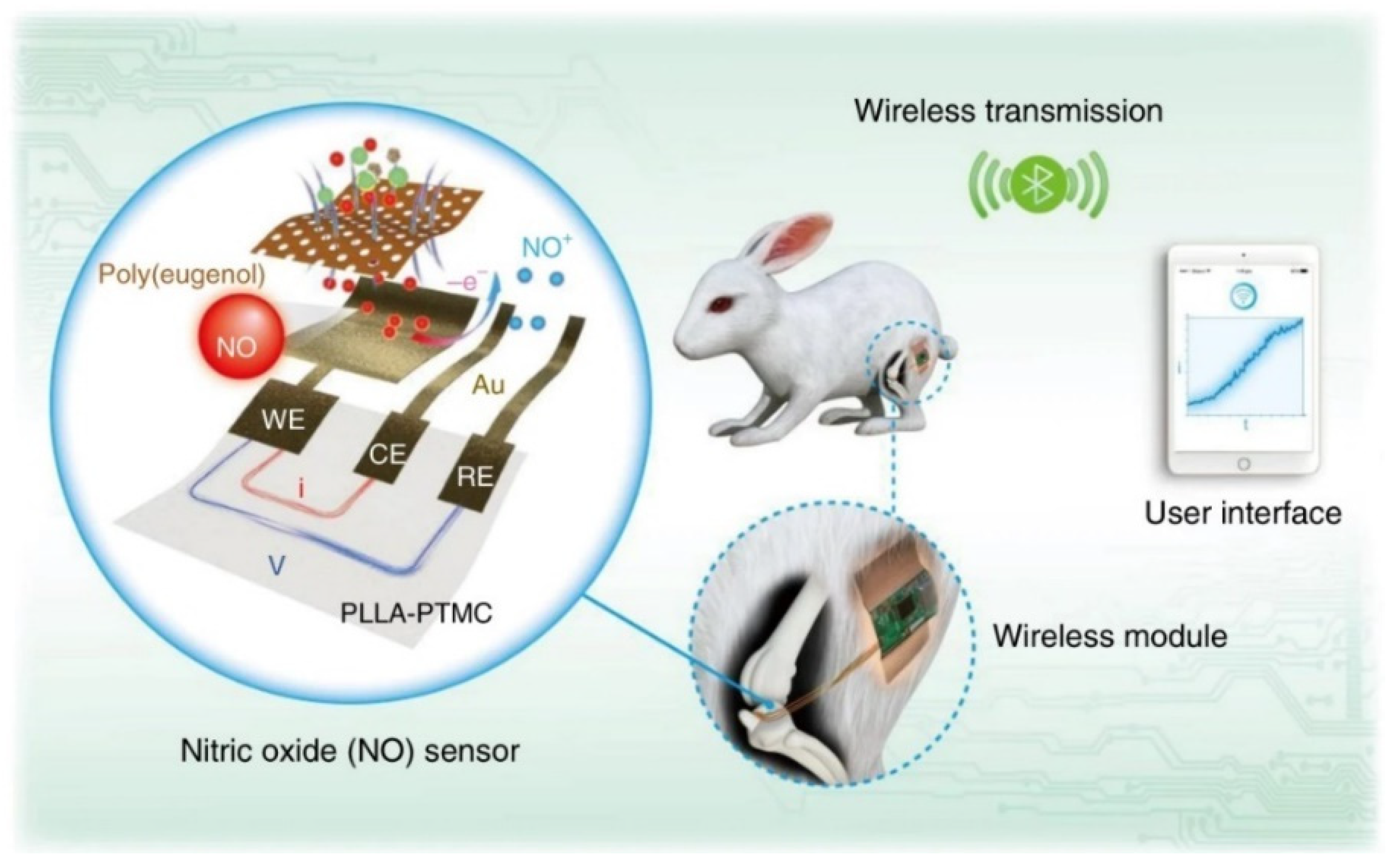
- Ciszewski, A.; Milczarek, G. A new Nafion-free bipolymeric sensor for selective and sensitive detection of nitric oxide. Electroanalysis 1998, 10, 791–793. [Google Scholar] [CrossRef]
- Ciszewski, A.; Milczarek, G. Electrochemical detection of nitric oxide using polymer modified electrodes. Talanta 2003, 61, 11–26. [Google Scholar] [CrossRef]
- Quinton, D.; Girard, A.; Thi Kim, L.T.; Raimbault, V.; Griscom, L.; Razan, F.; Griveau, S.; Bedioui, F. On-chip multi-electrochemical sensor array platform for simultaneous screening of nitric oxide and peroxynitrite. Lab Chip 2011, 11, 1342–1350. [Google Scholar] [CrossRef] [PubMed]
- Oliveira, R.; Sella, C.; Souprayen, C.; Ait-Yahiatene, E.; Slim, C.; Griveau, S.; Thouin, L.; Bedioui, F. Development of a flow microsensor for selective detection of nitric oxide in the presence of hydrogen peroxide. Electrochim. Acta 2018, 286, 365–373. [Google Scholar] [CrossRef]
- Li, R.; Qi, H.; Ma, Y.; Deng, Y.; Liu, S.; Jie, Y.; Jing, J.; He, J.; Zhang, X.; Wheatley, L.; et al. A flexible and physically transient electrochemical sensor for real-time wireless nitric oxide monitoring. Nature Commun. 2020, 11, 3207. [Google Scholar] [CrossRef] [PubMed]
- Milczarek, G. Selective and sensitive detection of nitrite based on NO sensing on a polymer-coated rotating disc electrode. J. Electroanal. Chem. 2007, 610, 199–204. [Google Scholar] [CrossRef]
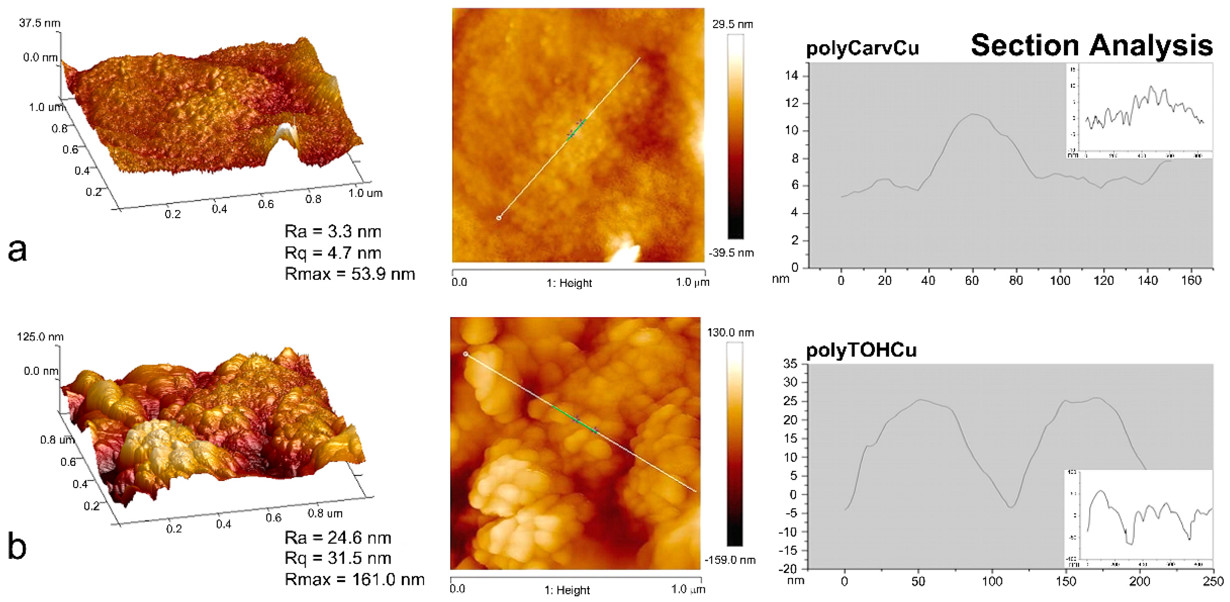
- Bertuola, M.; Pissinisa, D.E.; Ruberta, A.A.; Prietoa, E.D.; de Mele, M.A.F.L. Impact of molecular structure of two natural phenolic isomers on the protective characteristics of electropolymerized nanolayers formed on copper. Electrochim. Acta 2016, 215, 289–297. [Google Scholar] [CrossRef]
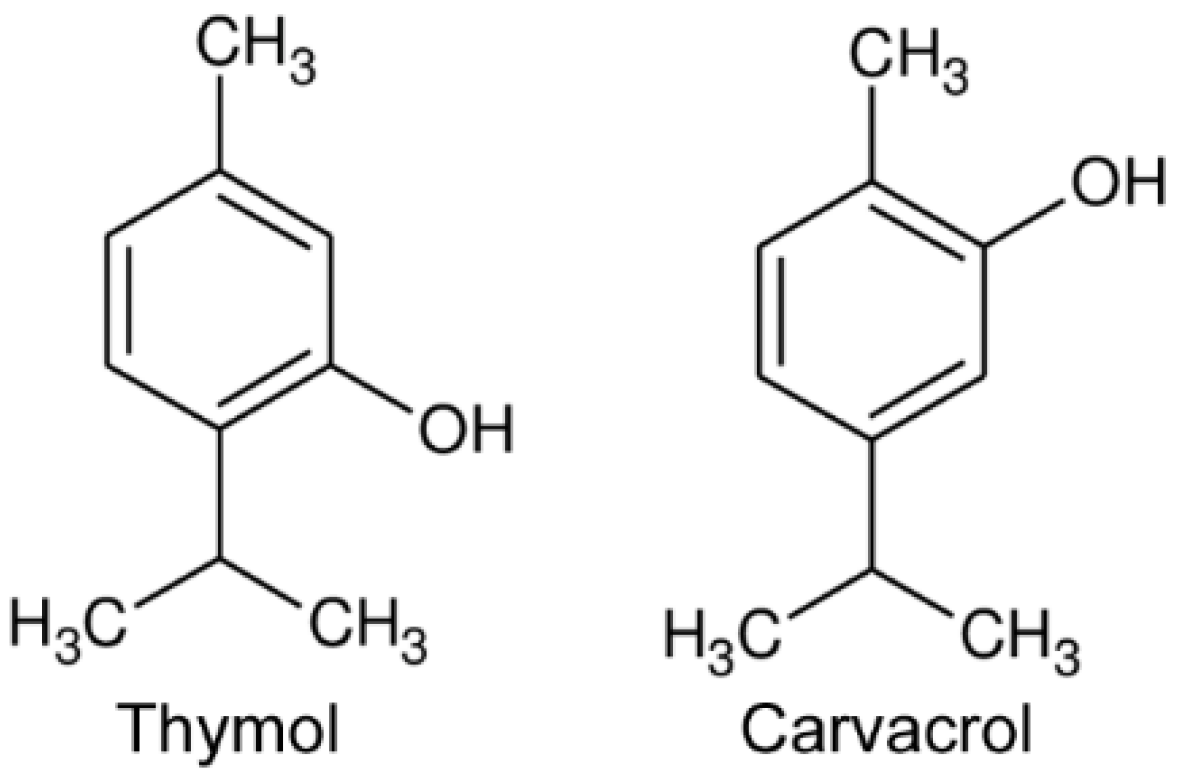
- Bertuola, M.; Fagali, N.; de Mele, M.A.F.L. Detection of carvacrol in essential oils by electrochemical polymerization. Helyon 2020, 6, e03714. [Google Scholar] [CrossRef] [PubMed]
- Ziyatdinova, G.; Kozlova, E.; Morozova, E.; Budnikov, H. Chronocoulometric method for the evaluation of antioxidant capacity of medicinal plant tinctures. Anal. Methods 2018, 10, 4995–5003. [Google Scholar] [CrossRef]
- Ziyatdinova, G.; Kozlova, E.; Budnikov, H.; Davletshin, R. Selective determination of total capsaicinoids in plant material using poly(gallic acid)-modified electrode. Electroanalysis 2019, 31, 222–230. [Google Scholar] [CrossRef]
- Ziyatdinova, G.; Yakupova, E.; Guss, E.; Budnikov, H. The selective electrochemical sensing of naringin using electropolymerized ellagic acid film. J. Electrochem. Soc. 2020, 167, 107502. [Google Scholar] [CrossRef]
- Ziyatdinova, G.; Kozlova, E.; Budnikov, H. Polyquercetin/MWNT-modified electrode for the determination of natural phenolic antioxidants. Electroanalysis 2017, 29, 2610–2619. [Google Scholar] [CrossRef]
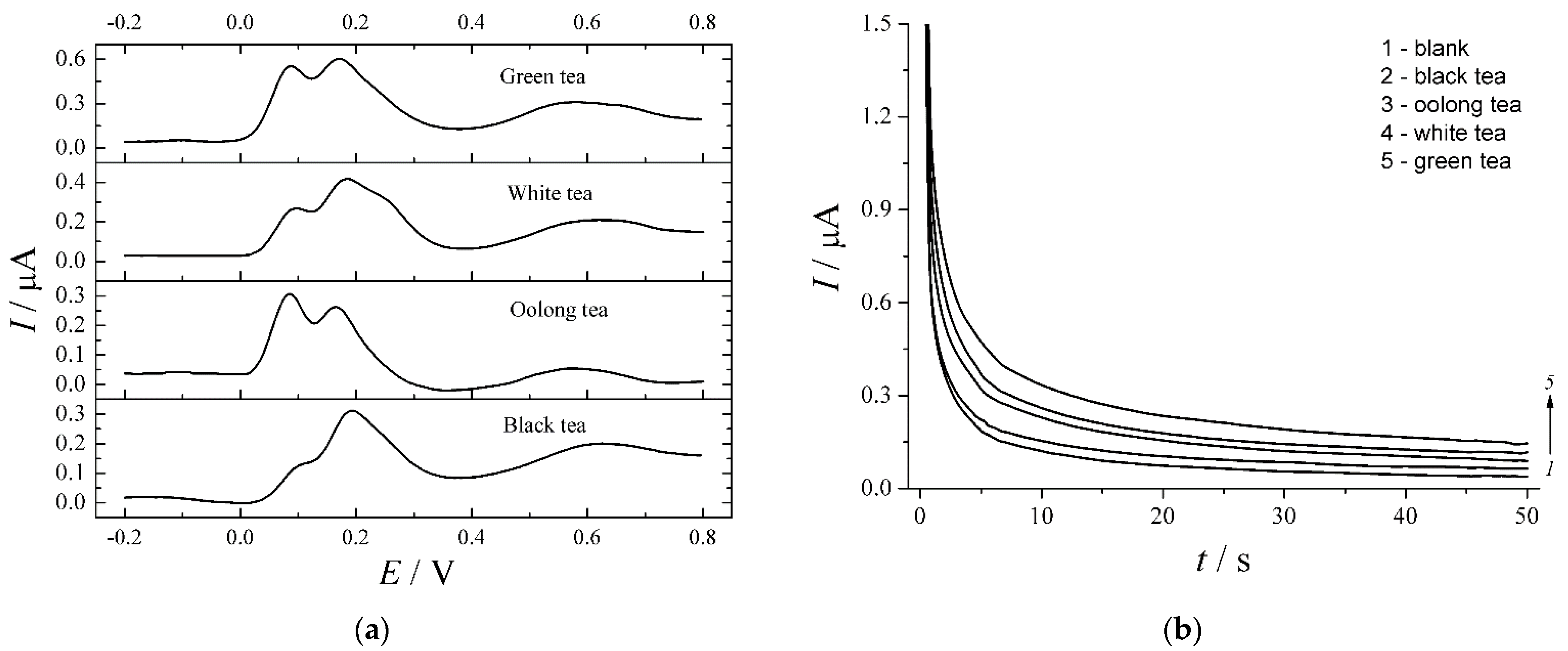
- Ziyatdinova, G.K.; Guss, E.V.; Budnikov, H.C. Voltammetric evaluation of polyphenol–protein interactions and their influence on the antioxidant capacity of tea. J. Anal. Chem. 2020, 75, 685–690. [Google Scholar] [CrossRef]
- Ziyatdinova, G.K.; Kozlova, E.V.; Budnikov, H.C. Chronoamperometric evaluation of the antioxidant capacity of tea on a polyquercetin-modified electrode. J. Anal. Chem. 2017, 72, 382–389. [Google Scholar] [CrossRef]
- Zheng, L.; Song, J.-f. Curcumin multi-wall carbon nanotubes modified glassy carbon electrode and its electrocatalytic activity towards oxidation of hydrazine. Sens. Actuators B Chem. 2009, 135, 650–655. [Google Scholar] [CrossRef]
- Kumar, K.K.; Devendiran, M.; Jyothithamizhanban, S.S. Curcumin/MWCNT modified graphite electrode for electrochemical determination of BHA. Intern. J. Innov. Res. Sci. Eng. 2014, 2, 654–659. [Google Scholar]
- Raoof, J.B.; Amiri-Aref, R.O.M.; Baghayeri, M. Electrodeposition of quercetin at a multi-walled carbon nanotubes modified glassy carbon electrode as a novel and efficient voltammetric sensor for simultaneous determination of levodopa, uric acid and tyramine. Sens. Actuators B Chem. 2012, 166–167, 508–518. [Google Scholar] [CrossRef]
- Lee, P.T.; Ward, K.R.; Tschulik, K.; Chapman, G.; Compton, R.G. Electrochemical detection of glutathione using a poly(caffeic acid) nanocarbon composite modified electrode. Electroanalysis 2014, 26, 366–373. [Google Scholar] [CrossRef]
- Zanardi, C.; Ferrari, E.; Pigani, L.; Arduini, F.; Seeber, R. Development of an electrochemical sensor for NADH determination based on a caffeic acid redox mediator supported on carbon black. Chemosensors 2015, 3, 118–128. [Google Scholar] [CrossRef]
- Sundaram, S.; Kadir, M.R.A. A new highly conducting carbon black (CL-08) modified electrode functionalized with syringic acid for sensitive and selective L-cysteine electrocatalysis at low potential. Electrochim. Acta 2017, 224, 475–486. [Google Scholar] [CrossRef]
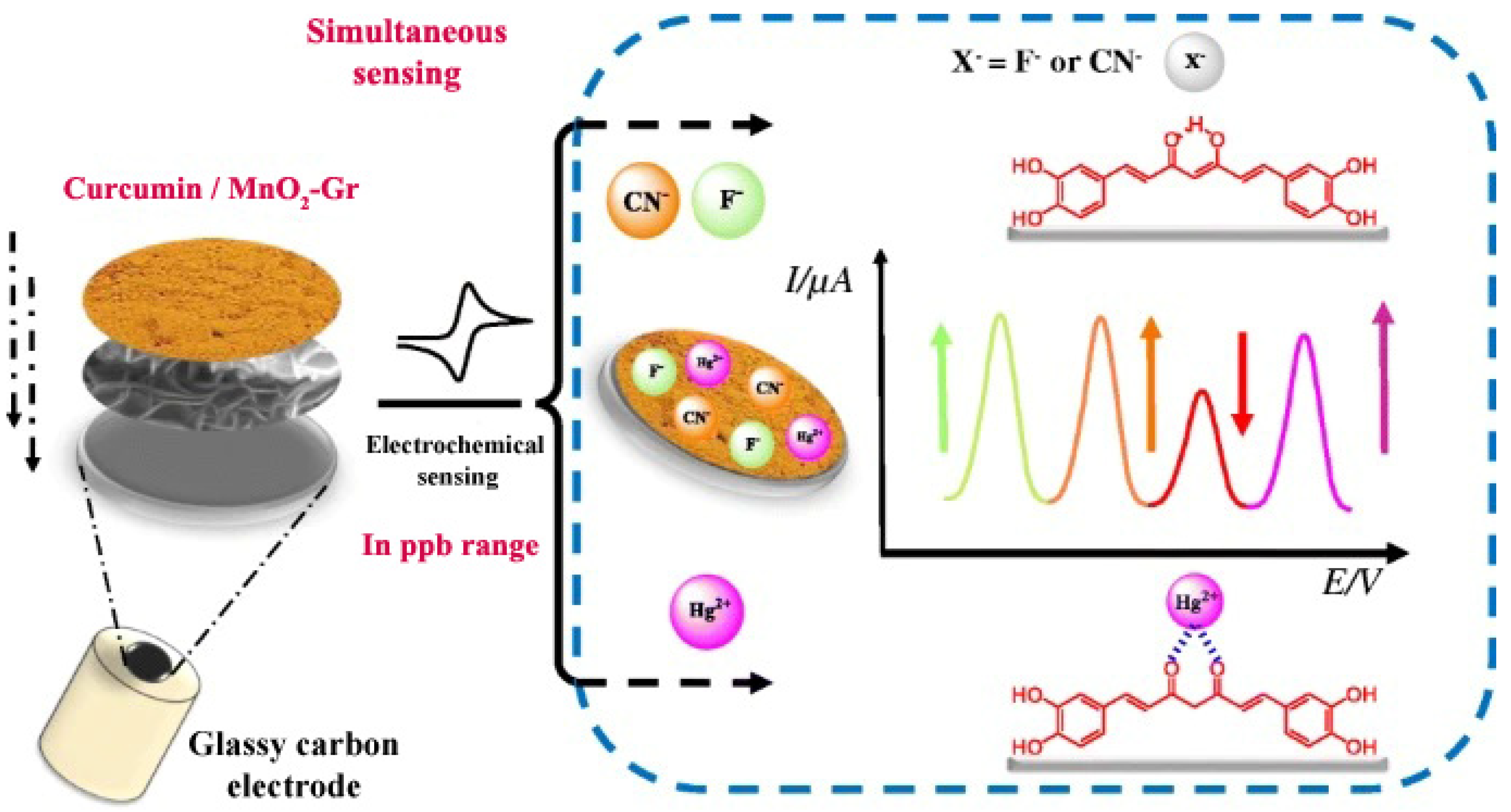
- Mejri, A.; Mars, A.; Elfil, H.; Hamzaoui, A.H. Graphene nanosheets modified with curcumin-decorated manganese dioxide for ultrasensitive potentiometric sensing of mercury(II), fluoride and cyanide. Microchim. Acta 2018, 185, 529. [Google Scholar] [CrossRef]
- Zheng, D.; Hu, C.; Peng, Y.; Hu, S. A carbon nanotube/polyvanillin composite film as an electrocatalyst for the electrochemical oxidation of nitrite and its application as a nitrite sensor. Electrochim. Acta 2009, 54, 4910–4915. [Google Scholar] [CrossRef]
- Corrêa, C.C.; Santhiago, M.; e Silva, C.C.C.; Formiga, A.L.B.; Kubota, L.T. Synthesis and electrochemical characterization of poly(2-methoxy-4-vinylphenol) with MWCNTs. Electroanalysis 2011, 23, 2562–2568. [Google Scholar] [CrossRef]
- Heras, A.; Vulcano, F.; Garoz-Ruiz, J.; Porcelli, N.; Terzi, F.; Colina, A.; Seeber, R.; Zanardi, C. A flexible platform of electrochemically functionalized carbon nanotubes for NADH sensors. Sensors 2019, 19, 518. [Google Scholar] [CrossRef] [PubMed]
- Li, T.; Xu, J.; Zhao, L.; Shen, S.; Yuan, M.; Liu, W.; Tu, Q.; Yu, R.; Wang, J. Au nanoparticles/poly(caffeic acid) composite modified glassy carbon electrode for voltammetric determination of acetaminophen. Talanta 2016, 159, 356–364. [Google Scholar] [CrossRef] [PubMed]
- Jin, G.-P.; Peng, X.; Chen, Q.-Z. Preparation of novel arrays silver nanoparticles modified polyrutin coat-paraffin-impregnated graphite electrode for tyrosine and tryptophan’s oxidation. Electroanalysis 2008, 20, 907–915. [Google Scholar] [CrossRef]
- Jin, G.-P.; Chen, L.-L.; Hang, G.-P.; Yang, S.-Z.; Wu, X.-J. Stripping chronopotentiometric analysis of cysteine on nano-silver coat polyquercetin–MWCNT modified platinum electrode. J. Solid State Electrochem. 2010, 14, 1163–1169. [Google Scholar] [CrossRef]
- Wei, Y.; Wang, A.; Liu, Y. Development of a glassy carbon electrode modified with graphene/Au nanoparticles for determination of acetaminophen in pharmaceutical preparation. Russ. J. Electrochem. 2018, 54, 1141–1147. [Google Scholar] [CrossRef]
- Bui, M.-P.N.; Li, C.A.; Han, K.N.; Pham, X.-H.; Seong, G.H. Determination of acetaminophen by electrochemical co-deposition of glutamic acid and gold nanoparticles. Sens. Actuators B Chem. 2012, 174, 318–324. [Google Scholar] [CrossRef]
- Li, R.; Zhai, T.; Zhao, L.; Zhang, N.; He, M.; Tan, L. Preparation of poly(caffeic acid)-CoP nanoparticle film on electrode surface and sensitive voltammetric detection of acetaminophen. Colloids Surf. A 2021, 627, 127173. [Google Scholar] [CrossRef]
- Mejri, A.; Mars, A.; Elfil, H.; Hamzaoui, A.H. Curcumin graphite pencil electrode modified with molybdenum disulfide nanosheets decorated gold foams for simultaneous quantification of nitrite and hydrazine in water samples. Anal. Chim. Acta 2020, 1137, 19–27. [Google Scholar] [CrossRef]
- Crapnell, R.D.; Hudson, A.; Foster, C.W.; Eersels, K.; Grinsven, B.v.; Cleij, T.J.; Banks, C.E.; Peeters, M. recent advances in electrosynthesized molecularly imprinted polymer sensing platforms for bioanalyte detection. Sensors 2019, 19, 1204. [Google Scholar] [CrossRef]
- Rebelo, T.S.C.R.; Pereira, C.M.; Sales, M.G.F.; Noronha, J.P.; Silva, F. Protein imprinted material electrochemical sensor for determination of Annexin A3 in biological samples. Electrochim. Acta 2016, 190, 887–893. [Google Scholar] [CrossRef]
- Rebelo, T.S.C.R.; Pereira, C.M.; Sales, M.G.F.; Noronha, J.P.; Silva, F. Protein imprinted materials designed with charged binding sites on screen-printed electrode for microseminoprotein-beta determination in biological samples. Sens. Actuators B Chem. 2016, 223, 846–852. [Google Scholar] [CrossRef]
- Stojanovic, Z.; Erdőssy, J.; Keltai, K.; Scheller, F.W.; Gyurcsányi, R.E. Electrosynthesized molecularly imprinted polyscopoletin nanofilms for human serum albumin detection. Anal. Chim. Acta 2017, 977, 1–9. [Google Scholar] [CrossRef] [PubMed]
- Yarman, A. electrosynthesized molecularly imprinted polymer for laccase using the inactivated enzyme as the target. Bull. Korean Chem. Soc. 2018, 39, 483–488. [Google Scholar] [CrossRef]
- Yarman, A.; Urlacher, V.B.; Kielb, P.; Scheller, F.W.; Wollenberger, U.; Weidinger, I.M.; Fischer, A.; Jetzschmann, K.J.; Rustam, L. Molecular LEGO by domain-imprinting of cytochrome P450 BM3. Colloids Surf. B Biointerfaces 2018, 164, 240–246. [Google Scholar]
- Zhang, X.; Yarman, A.; Erdossy, J.; Katz, S.; Zebger, I.; Jetzschmann, K.J.; Altintas, Z.; Wollenberger, U.; Gyurcsányi, R.E.; Scheller, F.W. Electrosynthesized MIPs for transferrin: Plastibodies or nano-filters? Biosens. Bioelectron. 2018, 105, 29–35. [Google Scholar] [CrossRef]
- Bosserdt, M.; Gajovic-Eichelman, N.; Scheller, F.W. Modulation of direct electron transfer of cytochrome c by use of a molecularly imprinted thin film. Anal. Bioanal. Chem. 2013, 405, 6437–6444. [Google Scholar] [CrossRef]
- Peng, L.; Yarman, A.; Jetzschmann, K.J.; Jeoung, J.-H.; Schad, D.; Dobbek, H.; Wollenberger, U.; Scheller, F.W. Molecularly imprinted electropolymer for a hexameric heme protein with direct electron transfer and peroxide electrocatalysis. Sensors 2016, 16, 272. [Google Scholar] [CrossRef]
- Yan, X.; Deng, J.; Xu, J.; Li, H.; Wang, L.; Chen, D.; Xie, J. A novel electrochemical sensor for isocarbophos based on a glassy carbon electrode modified with electropolymerized molecularly imprinted terpolymer. Sens. Actuators B Chem. 2012, 171–172, 1087–1094. [Google Scholar] [CrossRef]
- Deng, J.; Ju, S.; Liu, Y.; Xiao, N.; Xie, J.; Zhao, H. Highly sensitive and selective determination of melamine in milk using glassy carbon electrode modified with molecularly imprinted copolymer. Food Anal. Methods 2015, 8, 2437–2446. [Google Scholar] [CrossRef]
- Torkashvand, M.; Gholivand, M.B.; Taherkhani, F. Fabrication of an electrochemical sensor based on computationally designed molecularly imprinted polymer for the determination of mesalamine in real samples. Mater. Sci. Eng. C 2015, 55, 209–217. [Google Scholar] [CrossRef] [PubMed]
- Wang, F.-R.; Lee, G.-J.; Haridharan, N.; Wu, J.J. Electrochemical sensor using molecular imprinting polymerization modified electrodes to detect methyl parathion in environmental media. Electrocatalysis 2018, 9, 1–9. [Google Scholar] [CrossRef]
- Li, H.; Wang, Z.; Wu, B.; Liu, X.; Xue, Z.; Lu, X. Rapid and sensitive detection of methyl-parathion pesticide with an electropolymerized, molecularly imprinted polymer capacitive sensor. Electrochim. Acta 2012, 62, 319–326. [Google Scholar] [CrossRef]
- Pinczewska, A.; Sosna, M.; Bloodworth, S.; Kilburn, J.D.; Bartlett, P.N. High-throughput synthesis and electrochemical screening of a library of modified electrodes for NADH oxidation. J. Am. Chem. Soc. 2012, 134, 18022–18033. [Google Scholar] [CrossRef] [PubMed]


| Monomer | Electrode | cmonomer | Supporting Electrolyte | Polarization Window (V) | υ (mV s−1) | Number of Scans | Refs. |
|---|---|---|---|---|---|---|---|
| Eugenol | Pt | 0.0–2.2 | 100 | 10 | [21] | ||
| 0.0–0.70 | 5 | 10 | [22] | ||||
| GCE | 0.0–2.0 | 100 | 10 | [23] | |||
| Au | 10 mM | 0.1 M NaOH | −1.0–1.0 | 50 | 5 | [24] | |
| GCE | −1.0–0.60 | 100 | 10 | [24] | |||
| Au | 0.0–0.70 | 100 | 20 | [25] | |||
| Ti | −0.60–1.5 | 10 | 2 | [26] | |||
| p-Coumaric acid | Graphite electrode | 2.5 mM | 0.1 M NaOH | −0.25–0.8 | 50 | 25 | [37] |
| Rutin | Paraffin-impregnated graphite electrode | 1.0 mM | 0.1 M phosphate buffer pH 7.0 | 0.0–1.4 | 50 | 50 | [38] |
| CPE | 1.0 mM | 0.1 M phosphate buffer pH 7.0 | −0.8–1.2 | 50 | 30 | [39] | |
| Vanillin | CPE | 1.0 mM | 0.01 M NaOH | −0.40–1.2 | 100 | 10 | [40] |
| 10 μM | 0.1 M NaOH | −0.40–1.2 | 50 | 15 | [41] | ||
| Tannin | CPE | 1.0 mM | 0.01 M NaOH | −0.40–1.2 | 100 | 10 | [42] |
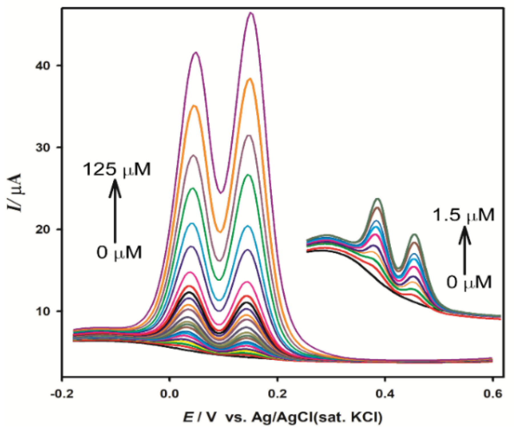
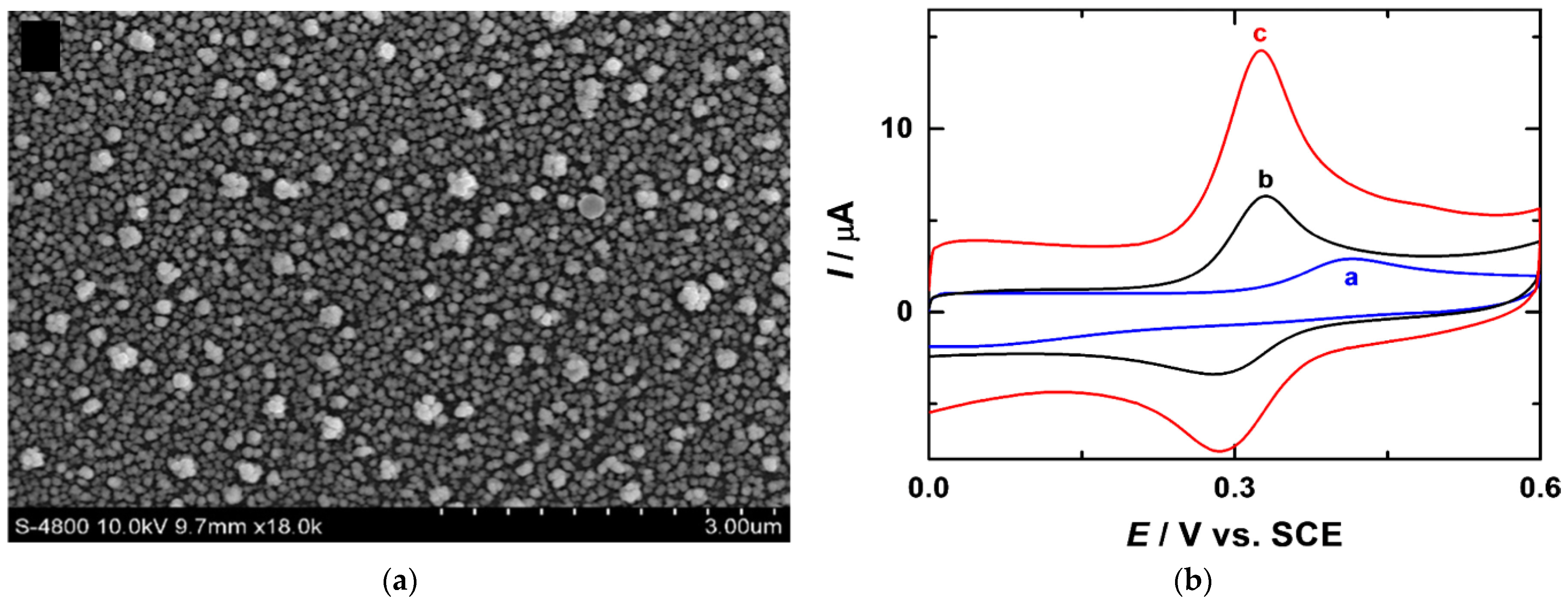
| Curcumin | Electroactivated GCE | 50 μM | 0.1 M phosphate buffer pH 8.0 | 0.15–0.55 | 100 | 16 | [43] |
| Catechin | Activated CPE | 1.0 mM | 0.1 M phosphate buffer pH 7.4 | 0.20–1.6 | 50 | 15 | [44] |
| Luteolin Kaempferol | GCE | 1.0 mM | 0.1 M phosphate buffer pH 7.5 | 0.10–0.90 | 100 | 30 | [45,46] |
| Apigenin | GCE | 1.0 mM | 0.1 M phosphate buffer pH 7.0 | 0.0–1.4 | 100 | 30 | [47] |
| Sensor | Method | Analyte | Linear Dynamic Range (µM) | Detection Limit (µM) | Refs. |
|---|---|---|---|---|---|
| Polyeugenol/Pt | DPV 1 | Dopamine | 0.1–50 | 0.1 | [21] |
| Polyrutin/PIGE 2 | DPV | Epinephrine | 3.0–90.0 | 0.8 | [38] |
| Serotonin | 0.3–9.0 | 0.1 | |||
| Ascorbic acid | 2.0–60.0 | 1.0 | |||
| Polyvanillin/CPE | Cyclic voltammetry | Dopamine | 1000–3000 | - | [40] |
| Epinephrine | 10–60 | 5.4 | [41] | ||
| Uric acid | 10–60 | 5.40 | |||
| Polytannin/CPE | Cyclic voltammetry | Dopamine | 1000–3500 | - | [42] |
| Polycurcumin/GCE | Linear sweep voltammetry | Epinephrine | 4.97–230.76 | 0.054 | [43] |
| p-Acetoaminophenol | 0.99–230.76 | ||||
| Polycatechin/CPE | Amperometry | Dopamine | 0.010–0.78 | 0.0005 | [44] |
| Serotonin | 0.030–2.34 | 0.003 | |||
| Poly(caffeic acid)/GCE | CV | Dopamine | 1.0–40 | 0.40 | [49] |
| Ascorbic acid | 20–1200 | 9.0 | |||
| DPV | L-DOPA | 1.0–50 | 0.14 | [51] |
| Electrode | Sensitive Layer | Method | Analyte | Detection Limit (µM) | Linear Dynamic Range (µM) | Sample | Refs. |
|---|---|---|---|---|---|---|---|
| GCE/MWCNTs | Poly(vanillic acid) | Amperometry | Urine | [33] | |||
| 0.100 V | Ascorbic acid | 3.5 | 5–120 | ||||
| 0.225 V | Dopamine | 1.5 | 5–120 | ||||
| 0.325 V | Uric acid | 4.5 | 5–120 | ||||
| Poly(ferulic acid) | Amperometry 0.2 V | NADH | 17.73 | 59.1–1560 | Pharmaceutical dosage forms | [34] | |
| Dopamine | 2.210 | 5.00–120.0 | |||||
| Epinephrine | 22.28 | 73.0–1406 | |||||
| Poly(p-coumaric acid) | DPV | L-cysteine | 1.1 | 7.5–50; 50–1000 | Human urine | [35] | |
| Poly(gallic acid) | DPV | Quercetin | 0.054 | 0.075–25; 25–100 | Medicinal herbs | [32] | |
| AdASWV 1 | Gallic acid | 3.22 | 4.97–33.8 | Model solutions | [52] | ||
| Chronocoulometry 1.0 V | Quercetin | 0.0029 | 0.010–0.25; 0.25–250 | Medicinal plant tinctures | [73] | ||
| DPV | Capsaicin | 0.0029 | 0.010–1.0; 1.0–50 | Red hot pepper spices and Capsicum annuum L. tinctures | [74] | ||
| Dihydrocapsaicin | 0.0059 | 0.025–0.75; 0.75–75 | |||||
| Nonivamide | 0.0061 | 0.025–5.0; 5.0–75 | |||||
| Poly(ellagic acid) | DPV | Naringin | 0.014 | 0.050–1.0; 1.0–100 | Grapefruit juices | [75] | |
| Polyeugenol | DPV | Catechin | 0.21 | 1.00–250 | Antioxidant capacity of wine | [56] | |
| Polyquercetin | DPV | Gallic acid | 0.10 | 0.50–10; 10–750 | Antioxidant capacity of tea | [76,77] | |
| Catechin | 0.024 | 0.10–10; 10–250 | |||||
| Epigallocatechin gallate | 0.014 | 0.050–10; 10–100 | |||||
| Chronoamperometry 0.2 V | Gallic acid | 0.063 | 0.25–750 | Antioxidant capacity of tea | [78] | ||
| Polycurcumin | Amperometry 0.25 V | Hydrazine | 1.4 | 2–44 | - | [79] | |
| PIGE/MWCNTs | Polycurcumin | CV | Butylated hydroxyanisole | 0.23 | 3.37–332 | - | [80] |
| GCE/Carboxylated MWCNTs | Polyquercetin | DPV | L-DOPA | 0.381 | 0.90–85.0 | Biosamples and pharmaceutical dosage forms | [81] |
| Uric acid | 0.575 | 1.0–125 | |||||
| Tyramine | 0.647 | 0.70–75 | |||||
| GCE/CNTs 2 | Poly(caffeic acid) | CV | Glutathione | 0.5 | 50–5000 | - | [82] |
| GCE/Carbon black | Poly(caffeic acid) | Amperometry 0.25 V | NADH | 3.7 | - | - | [83] |
| GCE/Highly conductive carbon black | Poly(syringic acid) | Chronoamperometry 0.04 V | L-cysteine | 0.639 | 20–100; 100–1000 | Simulated blood serum and chicken samples | [84] |
| Functional Monomer | Template/Analyte | Electrode Material | Detection Method | Detection Limit | Linear Dynamic Range | Refs. |
|---|---|---|---|---|---|---|
| Caffeic acid | Annexin A3 | Screen-printed carbon electrode | SWV | 0.095 ng mL−1 | 0.10–200 ng mL−1 | [97] |
| Microseminoprotein-beta | SWV | 0.12 ng mL−1 | 0.50–100 ng mL−1 | [98] | ||
| Scopoletin | Human serum albumin | Au | CV | 3.7 mg L−1 | 20–100 mg L−1 | [99] |
| Ferritin | 10.7 mg L−1 | 120–360 mg L−1 |
| Functional Monomer | Template/Analyte | Electrode Material | Detection Method | Detection Limit | Linear Dynamic Range | Refs. |
|---|---|---|---|---|---|---|
| Gallic acid + o-phenylenediamine | Melamine | GCE | SWV | 1.4 nM | 5.0–100.0 nM | [106] |
| Gallic acid + o-phenylenediamine + m-aminobenzoic acid | Isocarbophos | GCE | DPV | 0.0201 µM | 0.075–50; 50–100 µM | [105] |
| Gallic acid + o-phenylenediamine + m-aminobenzoic acid (and further electrodeposited Ag nanodendrites) | Mesalamine | GCE | Anodic stripping SWV | 0.015 µM | 0.05–100 µM | [107] |
| Quercetin + resorcinol | Methyl parathion | Au nanoparticles/GCE | CV | 0.01 μM | 0.05–15 µM | [108] |
| EIS 1 | 0.34 nM | 70–1000 nM | [109] |
Publisher’s Note: MDPI stays neutral with regard to jurisdictional claims in published maps and institutional affiliations. |
© 2021 by the authors. Licensee MDPI, Basel, Switzerland. This article is an open access article distributed under the terms and conditions of the Creative Commons Attribution (CC BY) license (https://creativecommons.org/licenses/by/4.0/).
Share and Cite
Ziyatdinova, G.; Guss, E.; Yakupova, E. Electrochemical Sensors Based on the Electropolymerized Natural Phenolic Antioxidants and Their Analytical Application. Sensors 2021, 21, 8385. https://doi.org/10.3390/s21248385
Ziyatdinova G, Guss E, Yakupova E. Electrochemical Sensors Based on the Electropolymerized Natural Phenolic Antioxidants and Their Analytical Application. Sensors. 2021; 21(24):8385. https://doi.org/10.3390/s21248385
Chicago/Turabian StyleZiyatdinova, Guzel, Ekaterina Guss, and Elvira Yakupova. 2021. "Electrochemical Sensors Based on the Electropolymerized Natural Phenolic Antioxidants and Their Analytical Application" Sensors 21, no. 24: 8385. https://doi.org/10.3390/s21248385
APA StyleZiyatdinova, G., Guss, E., & Yakupova, E. (2021). Electrochemical Sensors Based on the Electropolymerized Natural Phenolic Antioxidants and Their Analytical Application. Sensors, 21(24), 8385. https://doi.org/10.3390/s21248385