Article

Production, Optimization and Characterization of Polylactic Acid Microparticles Using Electrospray with Porous Structure

1
Center for Nanotechnology & Biomaterials Application and Research (NBUAM), Marmara University, İstanbul 34000, Turkey
2
Faculty of Medicine, Marmara University, İstanbul 34000, Turkey
3
Department of Moleculer Biology and Genetics, Faculty of Engineering, Istanbul Technical University, İstanbul 34467, Turkey
4
Department of Bioengineering, Faculty of Engineering, Yildiz Technical University, İstanbul 34000, Turkey
5
Department of Bioengineering, Faculty of Engineering, Uskudar University, İstanbul 34000, Turkey
6
Department of Bioengineering, Faculty of Engineering, Celal Bayar University, Manisa 45000, Turkey
7
Department of Metallurgical and Materials Engineering, Institute of Pure and Applied Sciences, Marmara University, İstanbul 34000, Turkey
8
Department of Bioengineering, Institute of Pure and Applied Sciences, Marmara University, İstanbul 34000, Turkey
9
Department of Chemical and Materials Engineering, Tunghai University, Taichung 40704, Taiwan
10
Deparment of Biomedical Engineering, Indian Institute of Technology, Banaras Hindu University, Varanasi 221005, India
11
Department of Inorganic Chemistry, Physical Chemistry and Electrochemistry, Faculty of Applied Chemistry and Materials Science, University POLITEHNICA of Bucharest, 1-7 GhPolizu Street, 060042 Bucharest, Romania
12
National Centre for Micro- and Nanomaterials, University POLITEHNICA of Bucharest, 1-7 GhPolizu Street, 060042 Bucharest, Romania
13
Department of Science and Engineering of Oxide Materials and Nanomaterials, Faculty of Applied Chemistry and Materials Science, University POLITEHNICA of Bucharest, 1-7 GhPolizu Street, 060042 Bucharest, Romania
14
Academy of Romanian Scientists, Ilfovst. 3, 060042 Bucharest, Romania
15
Department of Metallurgical and Materials Engineering, Faculty of Technology, Marmara University, İstanbul 34000, Turkey
*
Author to whom correspondence should be addressed.
Appl. Sci. 2021, 11(11), 5090; https://doi.org/10.3390/app11115090
Submission received: 4 March 2021 / Revised: 24 March 2021 / Accepted: 30 March 2021 / Published: 31 May 2021
(This article belongs to the Special Issue Nanoscale Characterization of Bioceramics)

Abstract

:
Polymeric microparticles with controlled morphologies and sizes are being studied by researchers in many applications, such as for drug release, healthcare and cosmetics. Herein, spherical and porous polymeric microparticles of different sizes and morphologies by electrospray technique have been developed as a viable alternative. In this work, polylactic acid (PLA) microparticles with a spherical shape and porous morphology were successfully produced via an electrospray technique in a single step. Molecular interactions between the components and the effect of parameters, such as varying solvent compositions, flow rates and voltage on microparticle morphology, were investigated over the particle formation. It was observed that the type of solvents used is the most effective parameter in terms of particle morphology, size and distribution. When the optical microscopy and SEM images of the microparticles were examined, 3 wt.% PLA in dichloromethane (DCM) solution concentration with an applied voltage of 18 kV and a flow rate of 20 µL/min was found to be the optimum parameter combination to achieve the desired spherical and porous micron-size particles. The average diameter of the particles achieved was 3.01 ± 0.58 µm. DCM was found to be a more suitable solvent for obtaining microparticles compared to the other solvents used. Finally, particles that are obtained by electrospraying of PLA–DCM solution are porous and monodisperse. They might have excellent potential as a carrier of drugs to the targeted sides and can be used in different biomedical applications.

1. Introduction

Polymeric nano and micron-sized particles are extensively used in various areas, such as for paints and coatings, oil and gas explorations, adhesives, composites, cosmetics, personal grooming products, medicine and for fabrication of medical devices, life sciences, biotechnology, etc. [1] Particularly in recent years, the uses of submicro-/microparticles derived from natural and synthetic polymers have been investigated more for biomedical applications. The production methods of polymer microparticles have become increasingly important for applications such as controlled drug delivery, medical diagnostic tests, obtaining superhydrophobic surfaces, optimum design of toughened polymeric composites and food technology [2,3,4]. Suspension or emulsion polymerization, solvent evaporation, spray drying, spraying a polymer solution through a small hole and the Shirasu Porous Glass (SPG) membrane emulsification technique are the methods used to produce polymeric microparticles. However, conventional manufacturing methods have many drawbacks such as the fact that they are time-consuming, the coalescence among particles and obtaining of non-homogeneous particle sizes and non-uniformity of shapes [5]. These limitations can be overcome by introducing the electrospray method. In addition to this, the electrospray method has huge advantages over traditional methods in terms of generating little residue, using fewer solvents, being low-cost and using high molecular weight polymers [6].
The electrospray method became widespread with the use of ions as a method for mass spectrometry analysis [7]. Electrospray is an electrohydrodynamic process in which monodisperse droplets are formed by passing a liquid of sufficient electrical conductivity through a capillary channel or nozzle to a high potential [8]. Further, the electrospray technique is capable of producing droplets of different sizes and shapes from solutions with high viscosity. The production of the droplets with the desired size is a vital processing step in advanced engineering applications such as dispersion and particle production [9]. Electrospraying can be influenced by many variable parameters, such as electrostatic field strength, needle size solution flow rate and conductivity, physical properties and concentration [10]. The microparticles produced by these techniques are usually monodisperse, and the size of these particles can be easily controlled by varying the flow rates [11].
Additionally, it is a potential process to fabricate particles with a small amount of material with minimum losses. Thus, it has features such as simplicity and easy applicability [12]. Compared to traditional production methods, the electrospray method has several unique superiorities, including an improved encapsulation ability of hydrophilic and hydrophobic agents, strict control over particle size, excellent particle reproducibility and obtaining particle size micron to the submicron range [13]. The electrospray method is a remarkable role in pharmacological applications due to its ability to form smaller particles, better size distribution and less agglomeration [14]. In the previous report, Fantini et al. demonstrated a method of producing monodispersed polymeric microparticles with uniform size and surface morphology using low molecular weight polystyrene with the electrospray method [5]. In another work, Ardila et al. produced micro- and nanoparticles of chitosan using a one-step electrospray method. Nanospheres with a size of around 128 nm were obtained from a 22 G needle under an effective voltage of 33 kV with a 0.2 mL/s flow rate [15].
Biodegradable polymeric porous microparticles are used in tissue regenerative medicine; various applications in pharmaceutical science, for example, use biomedical applications, such as for controlled drug delivery [16]. Pore structure and porosity are the main factors that govern controlled drug release in particular applications. Porous microparticles have very low mass densities and large surface areas. This provides excellent drug adsorption and controls the release kinetics [17]. Polylactic acid (PLA) is a synthetic biopolymer commonly used in agriculture, pharmaceutical, packaging and biomedical applications due to its unique physical properties, such as biodegradability, biocompatibility, high strength and modulus, good thermoplasticity and processability [18,19]. PLA is frequently used in biomedical applications, especially in the fields of drug delivery, wound dressing, artificial skin and bone and tissue engineering, because of its low cost and availability from renewable sources [20,21]. It has been widely reported in the literature that PLA-based microparticles can be used for a variety of drug delivery systems with different biological molecules such as cytostatics, anti-inflammatory agents, peptides, hormones, etc. [22]. In previous studies, Xu et al. investigated the effect of PLA microparticle formations in various solvents by the electrospray method. Their results demonstrated that the microparticles with spherical and uniform morphology could be generated using dichloromethane (DCM) as a solvent [10]. Mai et al. produced PLA microparticles containing curcumin using the electrospray method. They reported that PLA-based microcapsules have a high drug loading capability and may act as a potential substrate for encapsulating therapeutic agents [23]. Ikeuchi et al. obtained nanofibrous microcapsules using PLA–chloroform–ethanol solution. They showed that the shape, surface porosity and diameter of the microparticles depend on the solution mixture, ambient humidity and flow rate [24].
There are studies about forming particles with PLA and different solvents, including DCM as mentioned before, and these articles describe the obtaining of specific drug delivery systems [10,23]. The aim of this work is to understand the fundamentals of PLA microparticles and the optimization of their size/morphology without loading any drugs. This is essential in order to design PLA micro-/nanoparticles with the desired characteristics. Thus, to achieve the main objective, the present research work concentrates on the formation of polymer particles using three different PLA solutions (using the solvent chloroform, dichloromethane and chloroform- ethanol mixture) to achieve uniform size particles after the electrospray method has been employed. During the experiment, all the parameters were controlled to get the best results. For all batches of samples, rheological properties such as viscosity, density, surface tension and electrical conductivity were measured. The morphology of the electrosprayed microparticles formed with different solvents was investigated and analyzed via scanning electron microscopy (SEM).
In this study, the PLA microparticles formed with DCM solvents were found to be highly porous with uniform surface roughness and monodisperse structure. Further, microparticles prepared in this work with a defined structure could be promising candidates for the loading of different drugs and could be potential candidates in drug delivery and for therapeutic applications. We are committed to further exploiting the outcome of this study and to developing specific drug delivery systems with predefined characteristics and desired release kinetics.

2. Materials and Methods

Poly (L-lactic acid) (PLA) 2003D was purchased from Nature Works LLC, Minnetonka, MN. Solvents chloroform (CHCl3), ethanol (C2H5OH) and dichloromethane (DCM, CH2Cl2) were purchased from Sigma Aldrich (St. Louis, MO, USA) and used without further purification.

2.1. Preparation of Electrospray Solutions

PLA was dissolved in different solvents (see Table 1) such as DCM, chloroform and an ethanol–chloroform mixture. Solutions were stirred with a magnetic stirrer (600 rpm) (Wise Stir®, MSH-20 A, Wertheim, Germany) at room temperature (25 °C) for 1 h. Firstly, PLA and chloroform solution were obtained following the procedure. Solutions of 2 wt.% and 4 wt.% of PLA were prepared by dissolving in 20 mL chloroform. Secondly, 3 wt.% of PLA was dissolved in 19 mL chloroform, and then, 1 mL ethanol was added drop-wise and mixed with a magnetic stirrer. Finally, 3 wt.% of PLA solution was prepared by dissolving a suitable amount of PLA in 20 mL of DCM.

2.2. Characterization of Physical Properties of Solutions

The physical characterization of electrospray solutions was carried out for four different solutions with a viscometer to measure the surface tension, electrical conductivity and density. The coefficient of viscosity of solutions was measured by a digital viscometer (DV-E, Brookfield AMETEK, Middleborough, MA, USA). The surface tensions of the solutions were gauged using a force tensiometer (Sigma 703D, Attention, Darmstadt, Germany) with a platinum ring. The electrical conductivity was measured with a conductivity probe (Cond 3110 SET 1, WTW, Weilheim, Germany). The density was gauged using a standard density bottle (10 mL), DIN ISO 3507-Gay-Lussac (Boru Cam Inc., Istanbul, Turkey). All measurements were made at room temperature (23 °C).

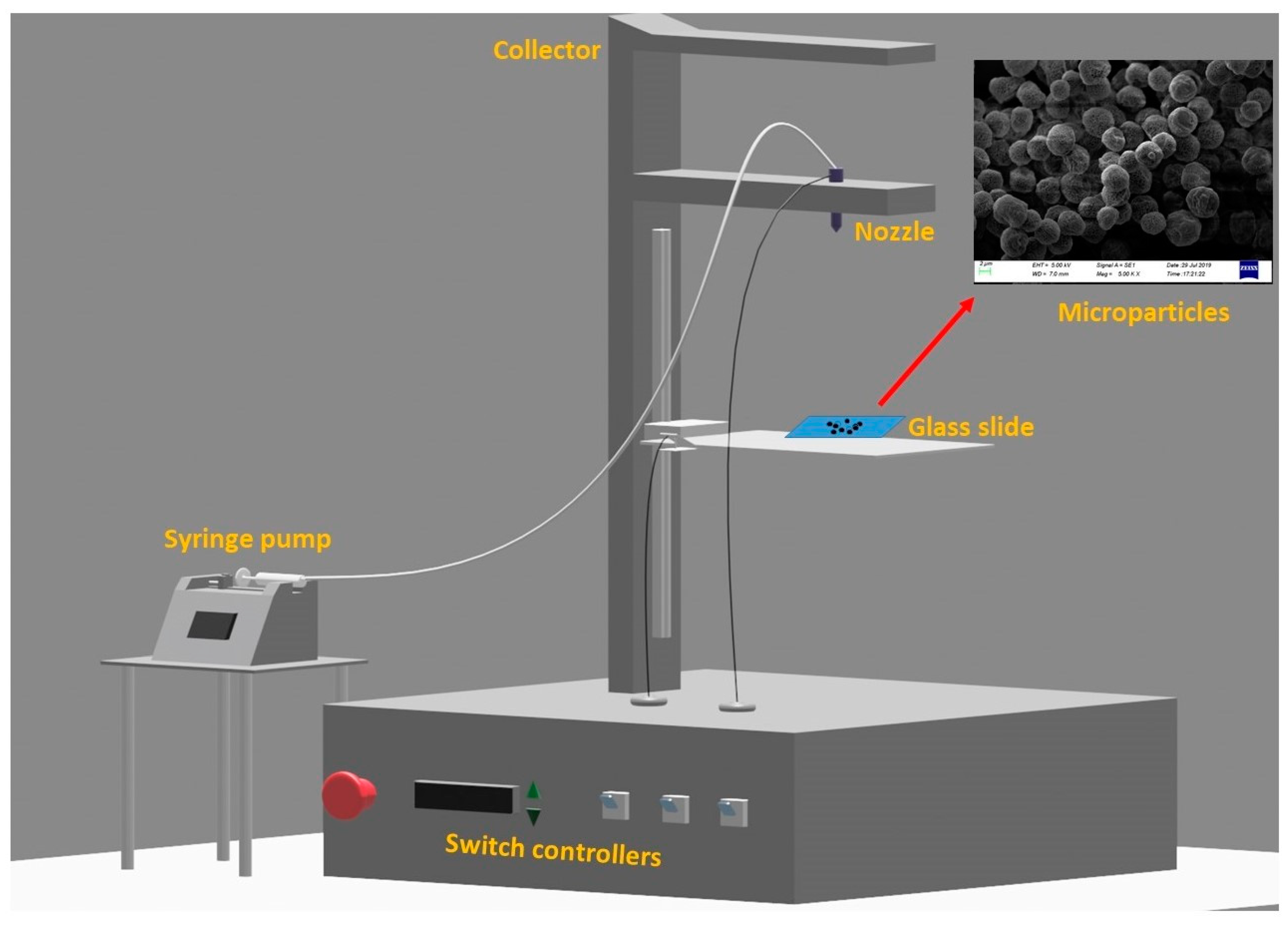
2.3. Fabrication of the Microparticles via the Electrospray Method

Microparticles were manufactured using a Basic System (Inovenso, İstanbul, Turkey) electrospraying process (Figure 1). The electrospraying apparatus was formed of a grounded target electrode made of an aluminum plate. First, 20 mL of the prepared solutions was pulled into a 20 mL plastic syringe. The experimental process for electrospray was as follows: flow rate was applied by the syringe pump (IPS-12, Inovenso, Turkey), and the PLA solutions were pushed through the metallic needle with an outer and inner diameter of 0.82 mm and 0.51 mm, respectively. The operating voltages were set up in between 10 and 30 kV, and the flow rate was used in the range of 12 to 60 µL/min. The distance between the collector and the needle was set to 12, 15 and 18 cm, and a glass coverslip was placed on top of the collector to collect microparticles. The microparticles were dried properly in a vacuum.

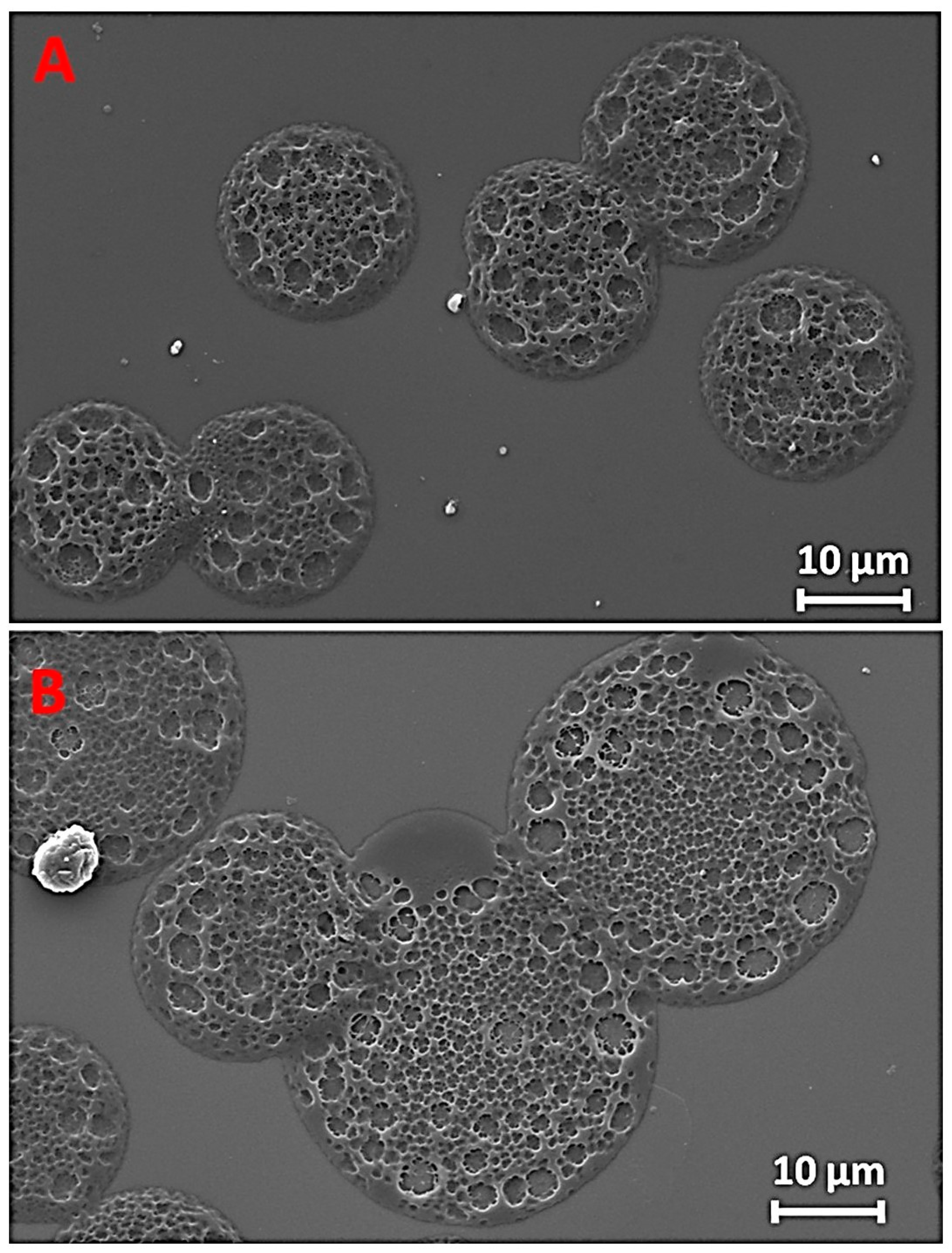
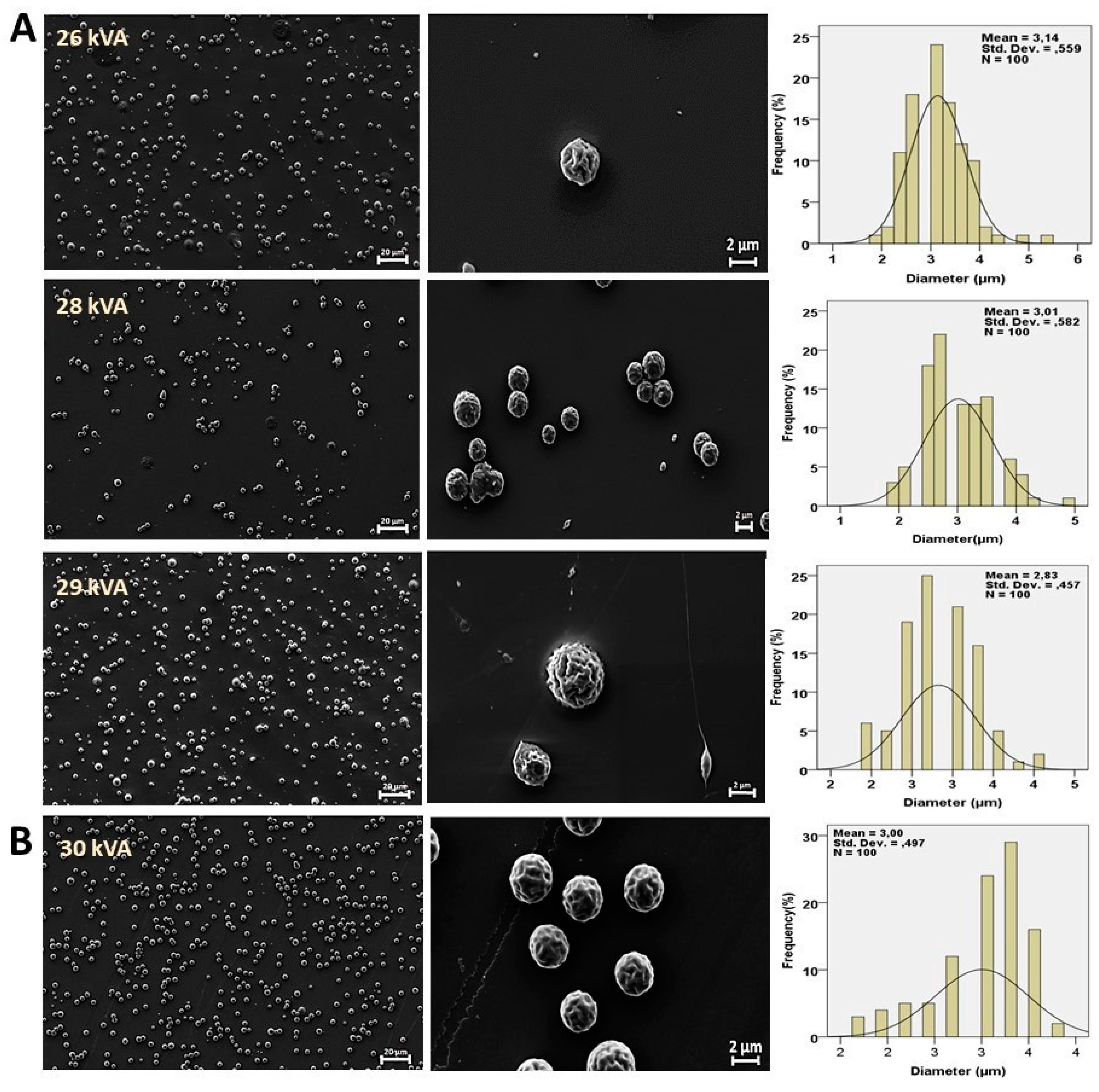
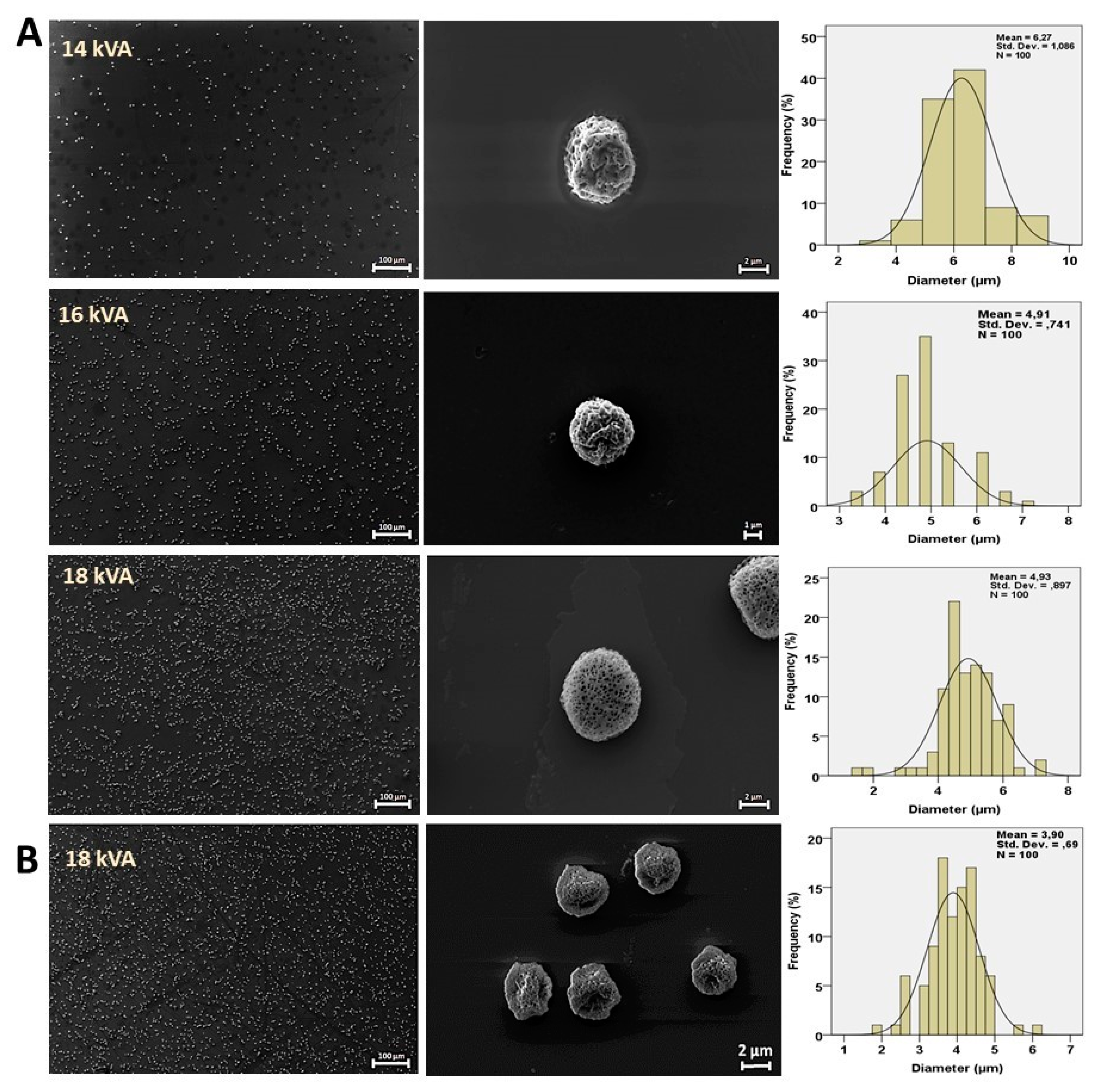
2.4. Optical Microscopy and Scanning Electron Microscopy (SEM)

An optical microscope (Olympus AnalySIS, Tokyo, Japan) was used to examine the structure and average particle diameter of the obtained microparticles. The morphology of the particles was characterized by a scanning electron microscope (SEM, EVO LS 10, ZEISS Istanbul, Turkey). Before acquiring images, the surface of the sample was sputter-coated with Au-Pd for 120 s using Quorum SC7620 Mini Sputter Coater. The acceleration voltage of 10 kV was kept fixed for all the experiments.

2.5. Fourier Transform Infrared Spectroscopy (FTIR)

Fourier-transformed infrared spectroscopy (FTIR) analysis was performed using a Jasco FT/IR-4700 spectrometer to examine the chemical structure, bonding and functional groups present in pure PLA and for the microparticles. The measurements were carried out at room temperature using transmission mode over the range of 500–4000 cm−1 and averaged over 32 scans with 4 cm−1 resolution.

3. Results and Discussion

Electrospray parameters, e.g., concentration, the distance between nozzle and glass and the voltage were found to be fundamental factors for producing microparticles products as previously described [23]. Many parameters were studied to provide microparticles. One of the most vital factors was the type of solvent used [25]. The effects of the solvent type on the morphology of the PLA microparticles were studied using chloroform, DCM, ethanol and a mixture of these organic solvents [26]. In the process of standardization, firstly, chloroform was chosen as a solvent for dissolving PLA. According to the previously reported literature, the size and morphology of the microparticles have a strong correlation to the set distance between the nozzle and the glass slide [11]. The most obvious reason may be to keep the distance parameter. In this work, the experiment was started with 1 wt.% of PLA with a collection distance set at 12, 15 and 18 cm. Smaller and homogenous particles were produced from a 15 cm distance with an excellent morphology, and therefore, a 15 cm distance was kept fixed for all experiments conducted with varying concentrations. In addition to distance, morphology and homogeneity of the particle may directly be affected by the solvent concentration [27].
Table 1 shows the concentrations of PLA–chloroform solution, voltage and flow rate parameters that were used in our optimization experiments. The alteration of particle morphology was evaluated using an optical microscope by considering parameter changes. Then, the two best optical images were chosen to perform SEM experiments considering the monodispersed structure and morphology of particles. Both particles were selected according to the optical microscopy results that were sprayed on a 40 µL/min flow rate, and their concentrations are 2 wt.% and 4 wt.%, respectively. SEM micrographs of particles that formed from 2 wt.% (Figure 2A) and 4 wt.% (Figure 2B) concentrated solutions are porous in nature and microscale in size.
Furthermore, from Figure 2A, it can be observed that the diameter of the particles increased as the PLA concentration increased. Freitas et al. examined the effect of PLA prepared in different concentrations varying between 0.05% and 0.15% (w/v) in organic solvents on the size of microparticles. They found that the particle diameter increased with high polymer concentrations [28]. Aside from these, neither fibers nor beaded fibrous morphologies were observed in our samples. SEM images also indicated that the chain entanglement point for fiber production did not reach what was stated in our previous work [27]. Despite all the different parameters that were chosen in combination, the desired particle could not be reached. As a result, the optimization of microparticles using chloroform is challenging. Therefore, it was decided to use a different material for this solution.
Secondly, ethanol was added to the PLA–chloroform solution as in the previous studies [24]. Since it was decided to use 15 cm for distance, the effects of all other parameters were investigated, except distance. All the samples were examined by optical microscopy, and the best ones were chosen for the SEM study. Therefore, SEM images were taken for the 3 wt.% PLA ethanol–chloroform solution with 12 and 15 µL/min flow rates (Figure 3). As seen in Figure 3A, by increasing the voltage from 26 to 29 kV, the diameter of the particles was decreased from 3.14 µm to 2.81 µm with a 12 µL/min flow rate. However, spherical morphology was found to be disrupted. Additionally, it was observed that beaded nanofibers were formed in higher voltage. Then, the effects of flow rate and voltage on particle morphology and sizes were investigated by keeping other parameters fixed (Figure 3B). It was observed that both were increased with the flow rate, and voltage resulted in microparticles with monodispersed properties and a minimum diameter. From these results, it can be concluded that the effects of voltage and flow rates played a promising role in particle size and morphology. Presumably, the reasons for the homogeneous distribution of polymer particles are that the applied voltage and flow rate affected the evaporation time and chain entanglement. It can be noted that SEM experiments were performed for those samples, which exhibited spherical and uniform shapes that were observed under the optical microscopy experiments. Further, the effects of flow rate and voltage on particle morphology were not studied separately in this work.
As mentioned, evaporation of the solvent has a crucial role in the electrospraying technique to obtain monodispersed and uniform-sized particles. Dichloromethane (DCM) is a volatile solvent and was used for obtaining PLA microparticles. While electrospraying, after droplet formation occurred, the DCM was evaporated slowly until the droplet reached a glass slide and porous particles were obtained.
Some microfiber structures were also obtained as the concentration of the polymer was increased to 4 wt.% of PLA (which was checked with the optic microscope; an optical microscopy image is not shown). For the 4 wt.% PLA–DCM group, the flow of the biopolymer solution was unstable, and the Taylor cone was not formed. Additionally, microfiber formed on the nozzle tip, which is not a suitable condition for the production of microparticles. When polymer concentration decreased to 1 wt.% PLA, turbulence flow occurred and resulted in the less uniform and regular distribution of the particles. The concentration of PLA was fixed at 3 wt.% according to the above optimization and previous research [12,23,24,29]. The relationship between flow rate and voltage was investigated to find the best PLA microparticles. Saadipour et al. prepared PLA samples in different solvents and could not obtain uniform and monodisperse microparticles in the 5% PLA–DCM sample [30]. However, in this study, when SEM images of the 3 wt.% PLA–DCM sample were analyzed, a homogeneous, spherical and porous microparticle was obtained as shown in Figure 4A, at 18 kV, with a flow rate of 20 µL/min. However, at 18 kV, the flow rate of 15 µL/min and the non-spherical microparticle were formed (see Figure 4B). These results obtained might be because the flow rate was not critical enough to obtain the spherical droplets. This can be interpreted as the flow rate of the PLA–DCM solution being slow. Therefore, the applied electrical field was not enough to obtain microspheres [29,31,32]. Since a 20 µL/min flow rate gives more suitable particles, it was decided to investigate the effects of voltage on the 20 µL/min flow rate. In this work, three voltages (14 kV, 16 kV and 18 kV) were applied to the PLA–DCM solution in order to show the effects of applied voltage on the morphological features of the microparticles generated. From the obtained results, it is seen that 18 kV was the most appropriate voltage setting for the production of particles with the best morphology as is seen in Figure 4A. The morphology of the microparticles was disrupted at 14 kV and 16 kV. Furthermore, the intensity of microparticles was smaller in number for 14 kV and 16 kV as seen in Figure 4A.
The possible explanation for this is that the low voltage might not be enough for the surface tension of the solution [29,31,32]. When the voltage was decreased, the electrical field might be less to obtain spherical droplets. As a result, the voltage rate was determined to be 18 kVA.
After all SEM images were acquired and the results were compared, it could be concluded that the formation of the microparticle is strongly dependent on the type of solvent used in the experiment. As shown in Figure 5A, the average diameter of the microparticles for 3 wt.% of PLA–DCM solution was obtained at ϕ = 3.00 ± 0.49 µm. From Figure 5B, it is clearly observed that the average diameter of the microparticles of the 3 wt.% PLA–5 wt.% ethanol solution was found to be ϕ = 3.01 ± 0.58 µm, and the microparticle radius for 2 wt.% PLA was obtained at ϕ = 19.70 ± 1.94 µm (Figure 5C). In Figure 5C, the size and morphological properties of the microparticles were obtained as desired. According to these results, the diameter of the PLA–DCM microparticles was quite small and porous as desired. The surface tension played an important role at the interface of the viscous fluids for the formation of the spherical microparticles. It is known that DCM has higher surface tension compared to the other solvent used in this work, resulting in the formation of a better particle size and morphology. Further, DCM has a lower boiling point than that of chloroform and ethanol. Ma et al. prepared PLA with DCM at very high concentrations of 10% to 20% (w/w) by adding various surfactants and obtained smooth, uniform microparticles [33]. The low boiling point of the solvent strongly controls the evaporation rate and the stability of particle formation during the electrospray process, which can further be easily controlled. Thus, the PLA–DCM solution has more suitable properties for the production of microparticles than the PLA–ethanol and PLA–chloroform solutions. In a previous study, undesirable structures such as cracked and irregular sunken surfaces were obtained in PLA microparticles produced with DCM solvent [23]. However, in this study, it was observed that PLA microparticles formed with DCM solvent have rough surfaces with highly porous or monodisperse structures. Therefore, using DCM as a solvent produced the best result among the solvents. The differences between the physical properties of the solvents, such as density, surface tension and viscosity, are seen in Table 2.
The chemical configuration of pure PLA was confirmed through FTIR spectra scanned in the range of 4000 cm−1–500 cm−1 (see Figure 6A,B). The characteristic absorption bands appeared for the C=O vibration band at 1749 cm−1; the CH3 asymmetrical scissoring band appeared at 1453 cm−1; for C–O and C–O–C, the stretching band appeared at 1080 cm−1; the C–CH3 stretching band appeared at 1042 cm−1; and for C–COO, the stretching band appeared at 867 cm−1 [34,35,36]. Comparing the spectrum of pure PLA and the results for microparticles obtained after electrospraying process, it was found that the spectra of pure PLA and the PLA microparticles obtained matched well to each other (Figure 6A). Further, these results matched well with the FTIR results reported for pure PLA in literature. Figure 6B shows the FTIR results of the PLA solutions in different solvents before the drying process. For chloroform, the characteristic absorption band appeared at 1214 cm−1, which was assigned to CH-bending (ν4) modes [37]. The spectrum of PLA–ethanol–chloroform showed a negligible shift compared to the PLA–chloroform spectrum. For dichloromethane, broadband with a branch can be observed at 1261 cm−1 [38]. As shown in Figure 6B, the FTIR results for novel microparticles revealed that the PLA microparticles are free from solvents, as no additional characteristic bands appeared for the solvents that were used during the formation of the microparticles, and also no chemical modifications of the PLA structure occurred during the fabrication of microparticles.

4. Conclusions

In this work, PLA microparticles were successfully produced via the electrospray technique using different solvents including chloroform, DCM and ethanol–chloroform mixtures. The sizes and morphology of the prepared microparticles were optimized via well-controlled flow rates, applied voltages and solvents. The particle size, shape and morphology for the particles obtained from the optimized method were characterized by optical and electron microscopy. It is observed that the process that involved 30 kVA applied voltage and 15 µL/min flow rates with 3 wt.% of PLA is suitable to obtain the desired spherical and porous microparticles. For PLA in a chloroform–ethanol mixture, 18 kVA applied voltage and 20 µL/min flow rate were optimized for 3 wt%. PLA in DCM solvent produced the best results, and particles formed with an average diameter of 3.00 µm. Thus, DCM was found to be a more suitable solvent for obtaining microparticles with a uniform spherical shape and a porous structure. In conclusion, particles that are obtained by electrospraying of PLA–DCM solution are porous and monodisperse in nature. The polymer particles achieved in this work will - have excellent potential as carriers of drugs to targeted sites and can be used in different biomedical and therapeutic applications.

Author Contributions

Methodology, M.E.T., B.D., E.T., A.G., D.F. and R.B.S.; Investigation, Visualization, S.C.; Investigation, E.I. and A.F.; Writing—review & editing, C.-C.L. and P.P.; Conceptualization, O.G. All authors have read and agreed to the published version of the manuscript.

Funding

This research received no external funding.

Acknowledgments

The authors would like to acknowledge the financial supports of Marmara University (Grant No.FEN-B-121218-0614).

Conflicts of Interest

The authors declare that they have no financial interests in this paper.

References

  1. Campos, E.; Branquinho, J.; Carreira, A.S.; Carvalho, A.; Coimbra, P.; Ferreira, P.; Gil, M.H. Designing polymeric microparticles for biomedical and industrial applications. Eur. Polym. J. 2016, 49, 2005–2021. [Google Scholar] [CrossRef]
  2. Sinha, V.R.; Bansal, K.; Kaushik, R.; Kumria, R.; Trehan, A. Poly-ε-caprolactone microspheres and nanospheres: An overview. Int. J. Pharm. 2004, 278, 1–23. [Google Scholar] [CrossRef]
  3. Freiberg, S.; Zhu, X.X. Polymer microspheres for controlled drug release. Int. J. Pharm. 2004, 282, 1–18. [Google Scholar] [CrossRef]
  4. Soares, R.M.D.; Siqueira, N.M.; Prabhakaram, M.P. Materials Science & Engineering C Electrospinning and electrospray of bio-based and natural polymers for biomaterials development. Mater. Sci. Eng. C 2018, 92, 969–982. [Google Scholar] [CrossRef]
  5. Fantini, D.; Zanetti, M.; Costa, L. Polystyrene microspheres and nanospheres produced by electrospray. Macromol. Rapid Commun. 2006, 27, 2038–2042. [Google Scholar] [CrossRef]
  6. Tapia-Hernández, J.A.; Torres-Chávez, P.I.; Ramírez-Wong, B.; Rascón-Chu, A.; Plascencia-Jatomea, M.; Barreras-Urbina, C.G.; Rangel-Vázquez, N.A.; Rodríguez-Félix, F. Micro- and Nanoparticles by Electrospray: Advances and Applications in Foods. J. Agric. Food Chem. 2015, 63, 4699–4707. [Google Scholar] [CrossRef] [PubMed]
  7. Dole, M.; Mack, L.L.; Hines, R.L.; Mobley, R.C.; Ferguson, L.D. Molecular Beams of Macroions. J. Chem. Phys. 1968, 49, 2240–2249. [Google Scholar] [CrossRef]
  8. Notz, P.K.; Basaran, O.A. Dynamics of drop formation in an electric field. J. Colloid Interface Sci. 1999. [Google Scholar] [CrossRef]
  9. Zakeri, M.; Moghadam, H.; Samimi, A.; Mohebbi-Kalhori, D. Optimization of calcium alginate beads production by electrospray using response surface methodology. Mater. Res. Express 2019, 6, 095412. [Google Scholar] [CrossRef]
  10. Xu, Y.; Hanna, M.A. Electrospray encapsulation of water-soluble protein with polylactide. Effects of formulations on morphology, encapsulation efficiency and release profile of particles. Int. J. Pharm. 2006, 320, 30–36. [Google Scholar] [CrossRef] [PubMed]
  11. Kim, W.; Kim, S.S. Synthesis of biodegradable triple-layered capsules using a triaxial electrospray method. Polymer 2011, 52, 3325–3336. [Google Scholar] [CrossRef]
  12. Valo, H.; Peltonen, L.; Vehviläinen, S.; Karjalainen, M.; Kostiainen, R.; Laaksonen, T.; Hirvonen, J. Electrospray encapsulation of hydrophilic and hydrophobic drugs in poly(L-lactic acid) nanoparticles. Small 2009, 5, 1791–1798. [Google Scholar] [CrossRef] [PubMed]
  13. Chen, J.; Cao, L.; Cui, Y.; Tu, K.; Wang, H.; Wang, L. Colloids and Surfaces B: Biointerfaces The exploration of endocytic mechanisms of PLA-PEG nanoparticles prepared by coaxialtri-capillary electrospray-template removal method. Colloids Surf. B Biointerfaces 2018, 161, 10–17. [Google Scholar] [CrossRef] [PubMed]
  14. Cam, M.E.; Zhang, Y.; Edirisinghe, M. Electrosprayed microparticles: A novel drug delivery method. Expert Opin. Drug Deliv. 2019, 16, 895–901. [Google Scholar] [CrossRef]
  15. Ardila, N.; Ajji, Z.; Heuzey, M.C.; Ajji, A. Chitosan electrospraying: Mapping of process stability and micro and nanoparticle formation. J. Aerosol Sci. 2018, 126, 85–98. [Google Scholar] [CrossRef]
  16. Amoyav, B.; Benny, O. Microfluidic based fabrication and characterization of highly porous polymeric microspheres. Polymers (Basel) 2019, 11, 419. [Google Scholar] [CrossRef] [PubMed] [Green Version]
  17. Ghosh Dastidar, D.; Saha, S.; Chowdhury, M. Porous microspheres: Synthesis, characterisation and applications in pharmaceutical & medical fields. Int. J. Pharm. 2018, 548, 34–48. [Google Scholar] [PubMed]
  18. Ilhan, E.; Ulag, S.; Sahin, A.; Yilmaz, B.K.; Ekren, N.; Kilic, O.; Sengor, M.; Kalaskar, D.M.; Oktar, F.N.; Gunduz, O. Fabrication of tissue-engineered tympanic membrane patches using 3D-Printing technology. J. Mech. Behav. Biomed. Mater. 2020, 114, 104219. [Google Scholar] [CrossRef]
  19. Cam, M.E.; Cesur, S.; Taskin, T.; Erdemir, G.; Kuruca, D.S.; Sahin, Y.M.; Kabasakal, L.; Gunduz, O. Fabrication, characterization and fibroblast proliferative activity of electrospun Achillea lycaonica-loaded nanofibrous mats. Eur. Polym. J. 2019. [Google Scholar] [CrossRef]
  20. Cesur, S.; Oktar, F.N.; Ekren, N.; Kilic, O.; Alkaya, D.B.; Seyhan, S.A.; Ege, Z.R.; Lin, C.-C.; Kuruca, S.E.; Erdemir, G.; et al. Preparation and characterization of electrospun polylactic acid/sodium alginate/orange oyster shell composite nanofiber for biomedical application. J. Aust. Ceram. Soc. 2019. [Google Scholar] [CrossRef]
  21. Ilhan, E.; Ulag, S.; Sahin, A.; Ekren, N.; Kilic, O.; Oktar, F.N.; Gunduz, O. Production of 3D-Printed Tympanic Membrane Scaffolds as a Tissue Engineering Application. In Lecture Notes in Computer Science (Including Subseries Lecture Notes in Artificial Intelligence and Lecture Notes in Bioinformatics); Springer Nature Switzerland AG: Cham, Switerland, 2020. [Google Scholar]
  22. Lassalle, V.; Ferreira, M.L.; Aires, B. PLA Nano- and Microparticles for Drug Delivery: An Overview of the Methods of Preparation. Macromol. Biosci. 2007, 767–783. [Google Scholar] [CrossRef] [PubMed]
  23. Mai, Z.; Chen, J.; He, T.; Hu, Y.; Dong, X.; Zhang, H.; Huang, W.; Ko, F.; Zhou, W. Electrospray biodegradable microcapsules loaded with curcumin for drug delivery systems with high bioactivity. RSC Adv. 2017, 7, 1724–1734. [Google Scholar] [CrossRef] [Green Version]
  24. Ikeuchi, M.; Tane, R.; Ikuta, K. Electrospray deposition and direct patterning of polylactic acid nanofibrous microcapsules for tissue engineering. Biomed. Microdevices 2012, 14, 35–43. [Google Scholar] [CrossRef] [PubMed] [Green Version]
  25. Megelski, S.; Stephens, J.S.; Bruce Chase, D.; Rabolt, J.F. Micro- and nanostructured surface morphology on electrospun polymer fibers. Macromolecules 2002. [Google Scholar] [CrossRef]
  26. Viet Linh, N.V. Fabrication of biodegradable polyester microspheres by electrospraying methods for drug carrier application. Vietnam J. Sci. Technol. 2018. [Google Scholar] [CrossRef]
  27. Gupta, P.; Elkins, C.; Long, T.E.; Wilkes, G.L. Electrospinning of linear homopolymers of poly(methyl methacrylate): Exploring relationships between fiber formation, viscosity, molecular weight and concentration in a good solvent. Polymer 2005. [Google Scholar] [CrossRef]
  28. Freitas, S.; Merkle, H.P.; Gander, B. Microencapsulation by solvent extraction/evaporation: Reviewing the state of the art of microsphere preparation process technology. J. Control. Release 2005, 102, 313–332. [Google Scholar] [CrossRef]
  29. Xu, Y.; Skotak, M.; Hanna, M. Electrospray encapsulation of water-soluble protein with polylactide. I. Effects of formulations and process on morphology and particle size. J. Microencapsul. 2006. [Google Scholar] [CrossRef] [PubMed]
  30. Saadipour, M.; Karkhaneh, A.; Haghbin Nazarpak, M. An investigation into curcumin release from PLA particles loaded in PCL-GELATIN fibers for skin application. Int. J. Polym. Mater. Polym. Biomater. 2020. [Google Scholar] [CrossRef]
  31. Xie, J.; Ng, W.J.; Lee, L.Y.; Wang, C.H. Encapsulation of protein drugs in biodegradable microparticles by co-axial electrospray. J. Colloid Interface Sci. 2008. [Google Scholar] [CrossRef] [PubMed]
  32. Tang, K.; Gomez, A. Monodisperse electrosprays of low electric conductivity liquids in the cone-jet mode. J. Colloid Interface Sci. 1996. [Google Scholar] [CrossRef] [PubMed]
  33. Ma, G.; Nagai, M.; Omi, S. Preparation of uniform poly(lactide) microspheres by employing the Shirasu Porous Glass (SPG) emulsification technique. Colloids Surf. A Physicochem. Eng. Asp. 1999, 153, 383–394. [Google Scholar] [CrossRef]
  34. Cam, M.E.; Hazar-Yavuz, A.N.; Cesur, S.; Ozkan, O.; Alenezi, H.; Turkoglu Sasmazel, H.; Sayip Eroglu, M.; Brako, F.; Ahmed, J.; Kabasakal, L.; et al. A novel treatment strategy for preterm birth: Intra-vaginal progesterone-loaded fibrous patches. Int. J. Pharm. 2020. [Google Scholar] [CrossRef] [PubMed]
  35. Feng, C.; Yuan, X.; Chu, K.; Zhang, H.; Ji, W.; Rui, M. Preparation and optimization of poly (lactic acid) nanoparticles loaded with fisetin to improve anti-cancer therapy. Int. J. Biol. Macromol. 2019. [Google Scholar] [CrossRef] [PubMed]
  36. Cesur, S.; Ulag, S.; Ozak, L.; Gumussoy, A.; Arslan, S.; Yilmaz, B.K.; Ekren, N.; Agirbasli, M.; Kalaskar, D.M.; Gunduz, O. Production and characterization of elastomeric cardiac tissue-like patches for Myocardial Tissue Engineering. Polym. Test. 2020, 90, 106613. [Google Scholar] [CrossRef]
  37. Nishida, J.; Shigeto, S.; Yabumoto, S.; Hamaguchi, H. Anharmonic coupling of the CH-stretch and CH-bend vibrations of chloroform as studied by near-infrared electroabsorption spectroscopy. J. Chem. Phys. 2012, 137, 234501. [Google Scholar] [CrossRef] [PubMed]
  38. Chen, M.; Guo, R.; Zhao, Y.; Weng, S.; Xu, Y.; Noda, I. Investigation on the intermolecular interaction between diethyl ether and dichloromethane in gaseous phase by using the DAOSD approach. J. Mol. Struct. 2016, 1124, 244–248. [Google Scholar] [CrossRef]
Figure 1. Electrospray set-up.
Applsci 11 05090 g001
Figure 2. SEM micrographs of prepared microparticles. (A) 2 wt.% polylactic acid (PLA)–chloroform and (B) 4 wt.% PLA–chloroform solution.
Applsci 11 05090 g002
Figure 3. SEM images and particle size distribution profile of 3 wt.% PLA ethanol–chloroform group. (A) The effect of voltage to particle shape in a constant 12 µL/min flow rate. Each line contains both SEM images and a size distribution profile of 26, 27 and 28 kVA, respectively. (B) SEM images and size distribution profile of 15 µL/min flow rate 30 kVA particles.
Applsci 11 05090 g003
Figure 4. SEM images and diameter distribution profile of 3%PLA— dichloromethane (DCM) solution. (A) Each line contains both a SEM image and diameter distribution graphic of 14, 16 and 18 kVA, respectively; the flow rate is fixed at 20 µL/min. (B) SEM images and diameter distribution profile of 15 µL/min at 18 kVA.
Applsci 11 05090 g004
Figure 5. (A) SEM images of the 3 wt.% PLA–DCM group sample. (B) SEM images and of the 3 wt.% PLA ethanol–chloroform group sample. (C) SEM images of the 2 wt.% PLA–chloroform group sample.
Applsci 11 05090 g005
Figure 6. FTIR transmittance spectra of (A, a) pure PLA, (A, b) PLA-chloroform (A, c) PLA-ethanol-chloroform and (A, d) PLA-DCM microparticles. FTIR transmittance spectra of (B, a) pure PLA, (B, b) PLA-chloroform (B, c) PLA-ethanol-chloroform and (B, d) PLA-DCM solutions.
Applsci 11 05090 g006
Table 1. Fabrication parameters for electrospray microparticles. (* Different voltages were applied as mentioned).
SolventPLA Concentration (%)Flow Rate (µL/min)Voltage (kVA) * Distance (cm)
Chloroform1–4406065 10.2–17.5121518
Ethanol–Chloroform1–31215405012–30121518
DCM31520 11–30121518
Table 2. Physical characteristics of different solutions.
SolutionDensity (g/dL)Viscosity (cP)Surface Tension (Mn/m)
3 % PLA – chloroform1.4815318.85
3 % PLA – 5 % ethanol– chloroform1.4151023.31
3 % PLA – DCM 1.32151.826.30
Publisher’s Note: MDPI stays neutral with regard to jurisdictional claims in published maps and institutional affiliations.

