
Phytochemical Profile and In Vitro Antioxidant, Antimicrobial, Vital Physiological Enzymes Inhibitory and Cytotoxic Effects of Artemisia jordanica Leaves Essential Oil from Palestine


Abstract
1. Introduction
2. Results
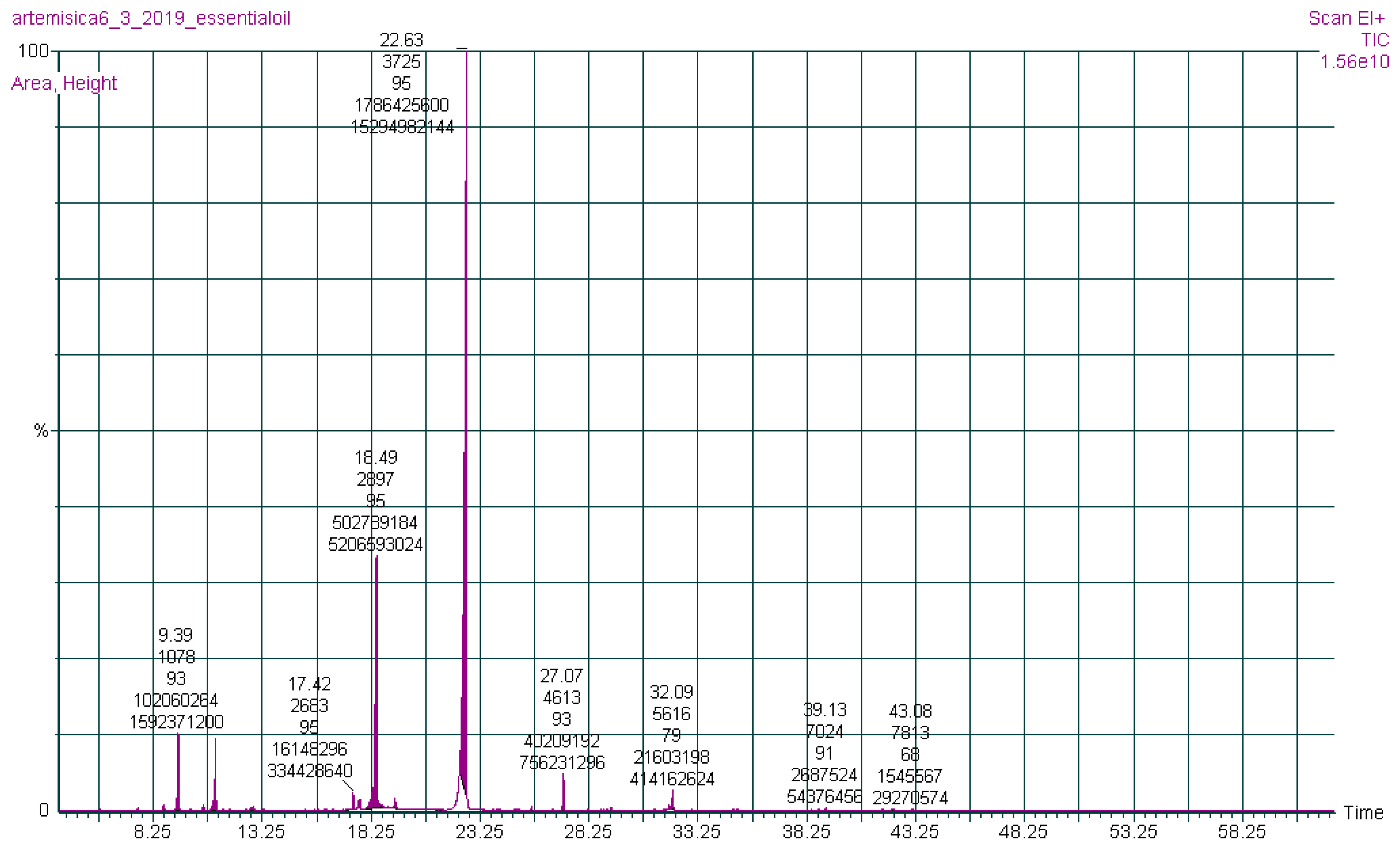
2.1. Phytochemistry
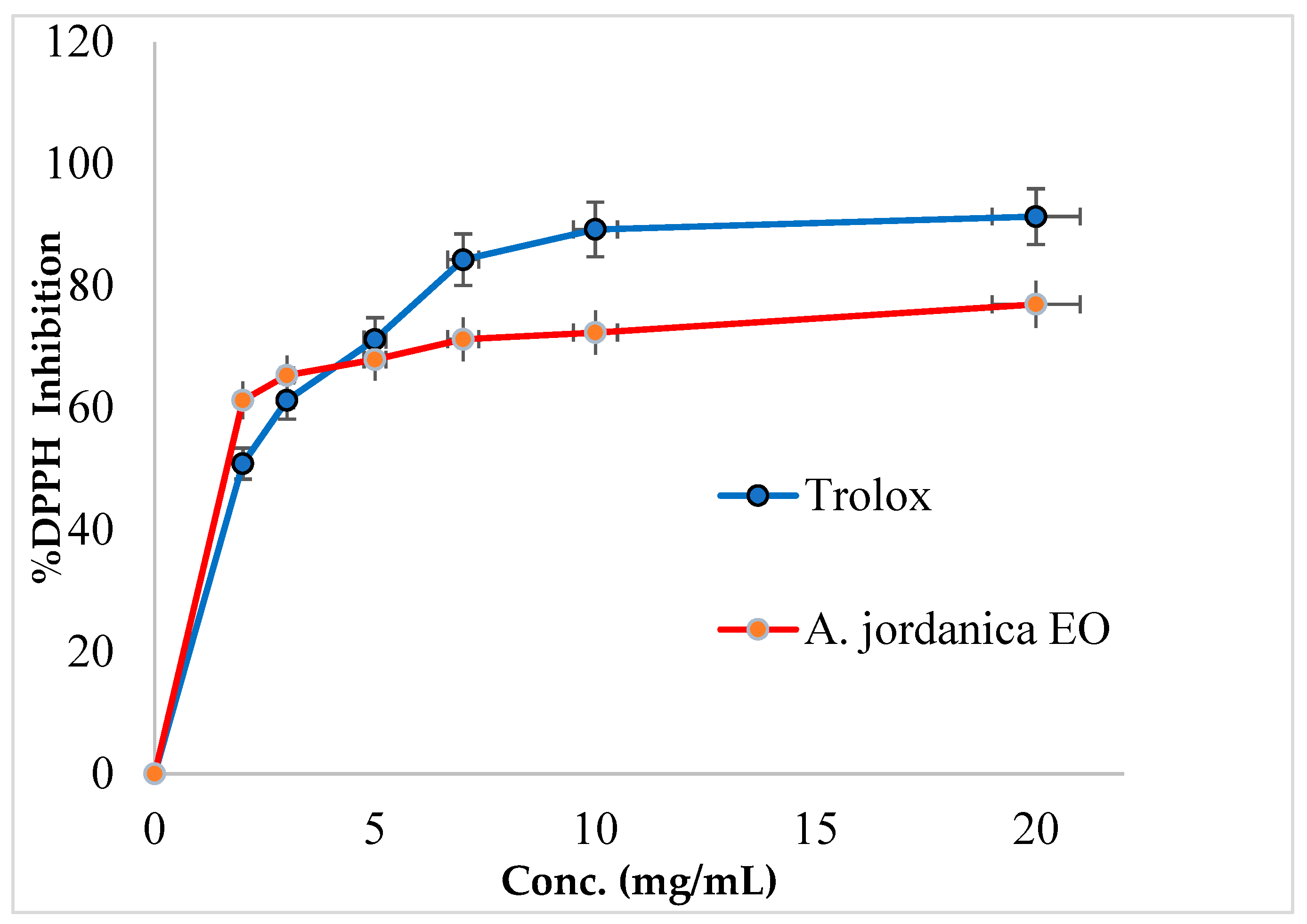
2.2. Antioxidant Activity
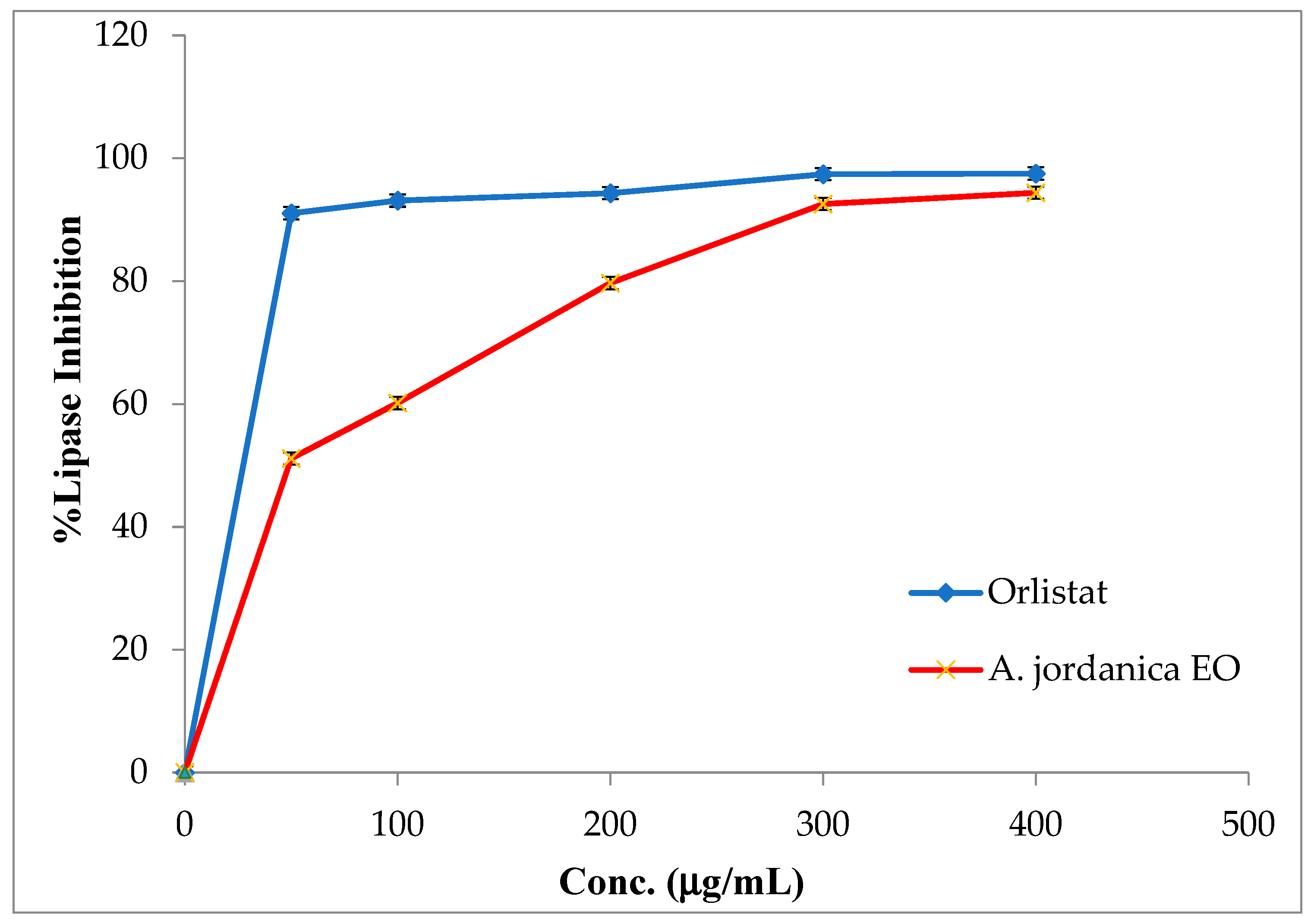
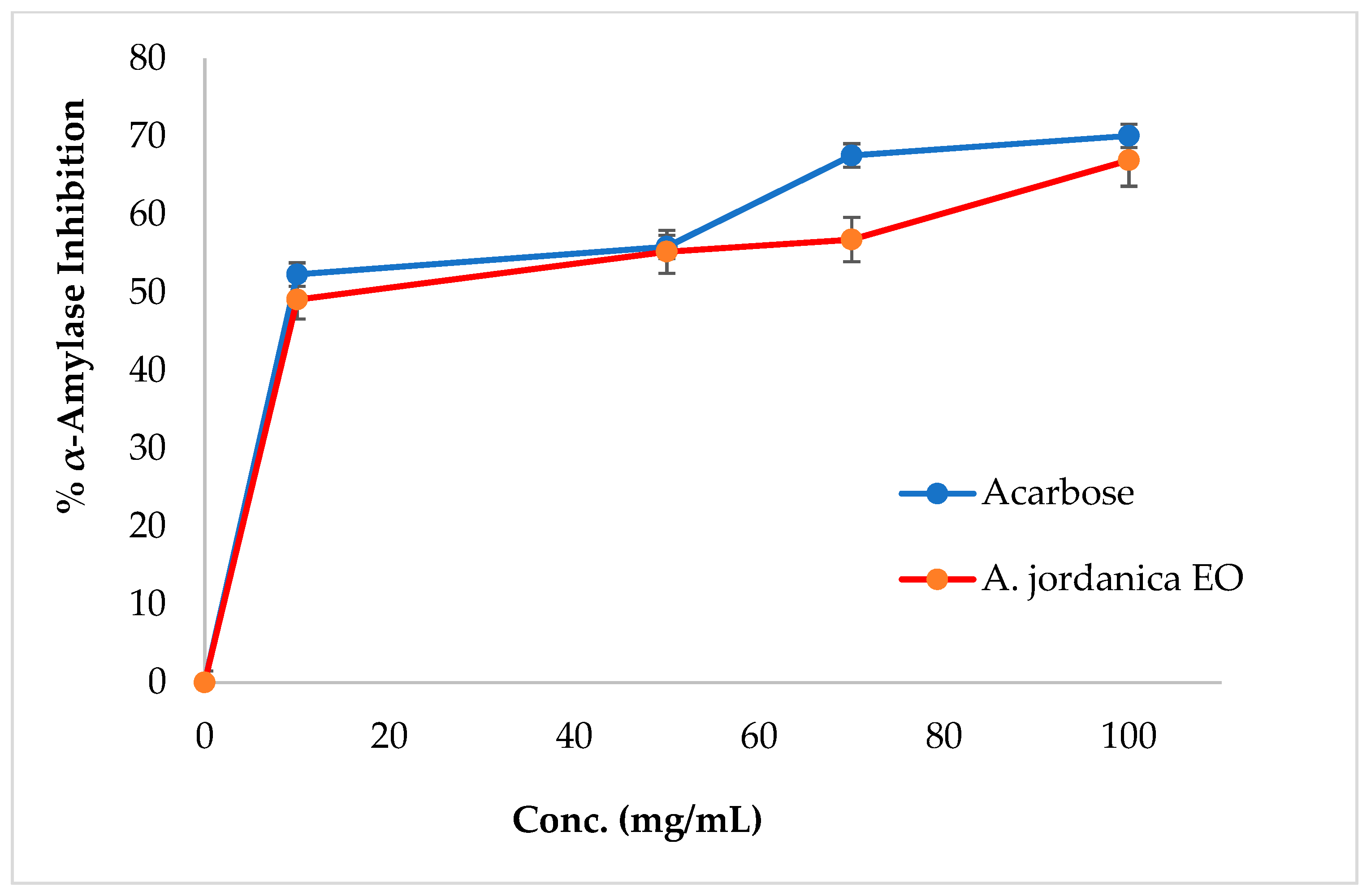
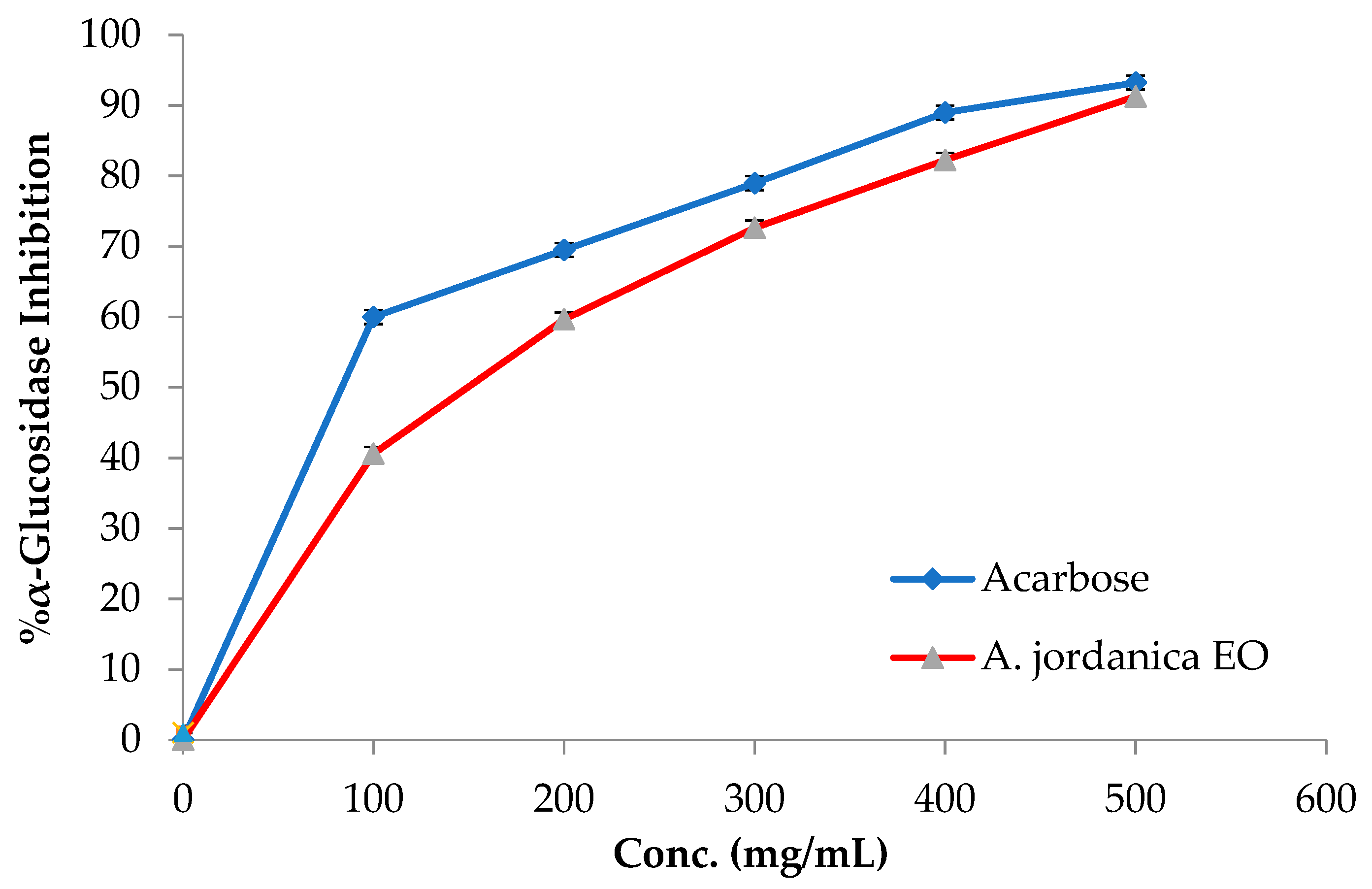
2.3. Target Metabolic Enzyme Inhibitory Activity
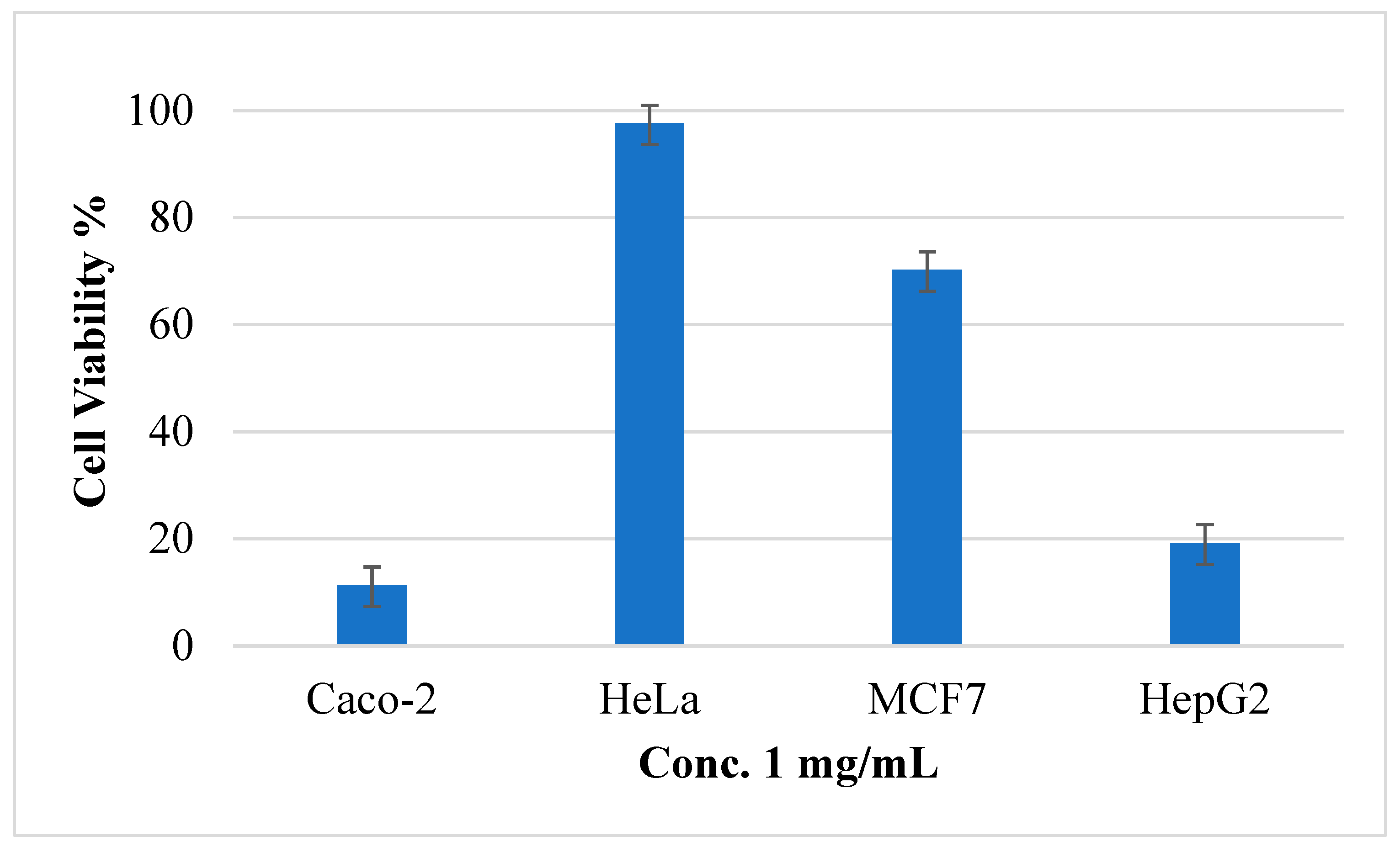
2.4. Cytotoxicity
2.5. Antimicrobial Effect
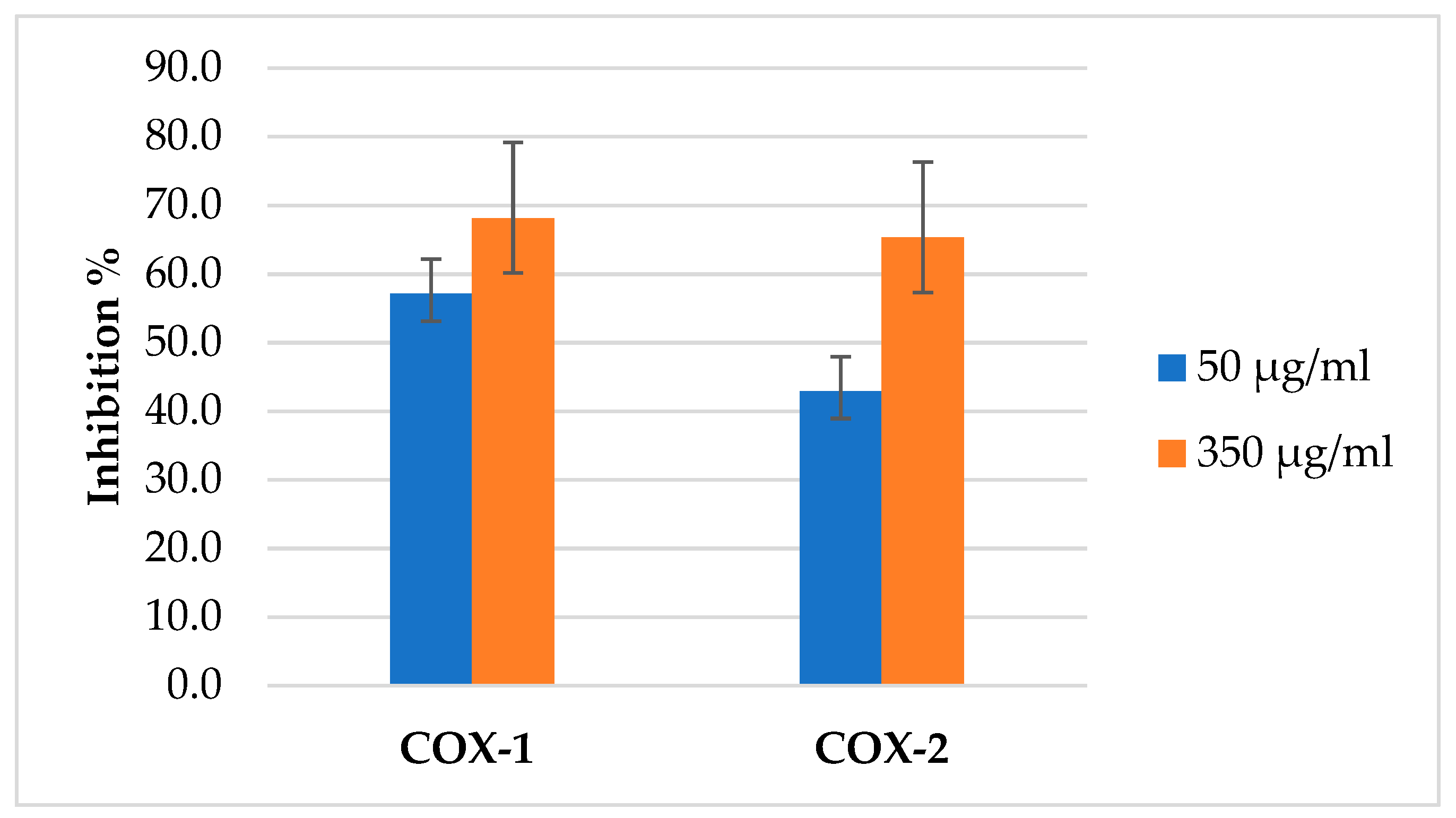
2.6. COX Inhibitory Activity
3. Discussion
3.1. Phytochemical Constituents
3.2. Antioxidant Activity
3.3. Metabolic Lipase, α-Amylase, and α-Glucosidase Inhibitory Activities
3.4. Cytotoxicity
3.5. Antimicrobial Activity
3.6. Cyclooxygenase Inhibitory Activity
4. Material and Methods
4.1. Collection and Drying of the Plant
4.2. Isolation of Artemisia jordanica Essential Oil
4.3. Characterization of Artemisia jordanica Essential Oil
4.4. Free Radical Scavenging Activity
4.5. Porcine Pancreatic Lipase Inhibitory Activity
4.6. α-Amylase Inhibitory Activity
4.7. α-Glucosidase Inhibitory Activity
4.8. Cell Culture and Cytotoxicity Assay
4.9. Microbial Strains, Culture Media, and Antimicrobial Assay
4.10. COX Inhibitory Assay
4.11. Statistical Analysis
5. Conclusions
Funding
Institutional Review Board Statement
Informed Consent Statement
Data Availability Statement
Acknowledgments
Conflicts of Interest
Sample Availability
References
- Yuan, H.; Ma, Q.; Ye, L.; Piao, G. The traditional medicine and modern medicine from natural products. Molecules 2016, 21, 559. [Google Scholar] [CrossRef]
- Wink, M. Modes of action of herbal medicines and plant secondary metabolites. Medicines 2015, 2, 251–286. [Google Scholar] [CrossRef] [PubMed]
- Dhifi, W.; Bellili, S.; Jazi, S.; Bahloul, N.; Mnif, W. Essential oils’ chemical characterization and investigation of some biological activities: A critical review. Medicines 2016, 3, 25. [Google Scholar] [CrossRef] [PubMed]
- Sarkic, A.; Stappen, I. Essential oils and their single compounds in cosmetics—A critical review. Cosmetics 2018, 5, 11. [Google Scholar] [CrossRef]
- Amorati, R.; Foti, M.C. Mode of Antioxidant Action of Essential Oils; Wiley Online Library: Hoboken, NJ, USA, 2017; pp. 267–291. [Google Scholar]
- Costa, G.; Gidaro, M.C.; Vullo, D.; Supuran, C.T.; Alcaro, S. Active components of essential oils as anti-obesity potential drugs investigated by in silico techniques. J. Agric. Food Chem. 2016, 64, 5295–5300. [Google Scholar] [CrossRef] [PubMed]
- Rashed, A.A.; Mohd Nawi, M.N.; Sulaiman, K. Assessment of essential oil as a potential anti-obesity agent: A narrative review. J. Essent. Oil Res. 2017, 29, 1–10. [Google Scholar] [CrossRef]
- Collaboration, E.R.F. Diabetes mellitus, fasting blood glucose concentration, and risk of vascular disease: A collaborative meta-analysis of 102 prospective studies. Lancet 2010, 375, 2215–2222. [Google Scholar] [CrossRef]
- Mrabti, H.N.; Sayah, K.; Jaradat, N.; Kichou, F.; Ed-Dra, A.; Belarj, B.; Cherrah, Y.; Faouzi, M.E.A. Antidiabetic and protective effects of the aqueous extract of Arbutus unedo L. in streptozotocin-nicotinamide-induced diabetic mice. J. Complement. Integr. Med. 2018, 15. [Google Scholar] [CrossRef] [PubMed]
- NCD Risk Factor Collaboration. Worldwide trends in diabetes since 1980: A pooled analysis of 751 population-based studies with 4.4 million participants. Lancet 2016, 387, 1513–1530. [Google Scholar] [CrossRef]
- Akolade, J.O.; Usman, L.A.; Okereke, O.E.; Muhammad, N.O. Antidiabetic potentials of essential oil extracted from the leaves of Hoslundia opposita Vahl. J. Med. Food 2014, 17, 1122–1128. [Google Scholar] [CrossRef]
- Abdollahi, M.; Salehnia, A.; Mortazavi, S.H.R.; Ebrahimi, M.; Shafiee, A.; Fouladian, F.; Keshavarz, K.; Sorouri, S.; Khorasani, R.; Kazemi, A. Antioxidant, antidiabetic, antihyperlipidemic, reproduction stimulatory properties and safety of essential oil of Satureja Khuzestanica in rat in vivo: A toxicopharmacological study. Med. Sci. Monit. 2003, 9, BR331–BR335. [Google Scholar]
- Benkhayal, F.A.; El-Ageeli, W.; Ramesh, S.; Farg-hamd, M. Anti-hyperglycaemic effects of volatile oils extracted from Rosemarinus officinalis and Artemisia cinae in diabetic rats. J. Vet. Anim. Sci. 2009, 5, 216–218. [Google Scholar]
- Hichri, F.; Omri, A.; Hossan, A.S.M.; Ben Jannet, H. Alpha-glucosidase and amylase inhibitory effects of Eruca vesicaria subsp. longirostris essential oils: Synthesis of new 1,2,4-triazole-thiol derivatives and 1,3,4-thiadiazole with potential inhibitory activity. Pharm. Biol. 2019, 57, 564–570. [Google Scholar] [CrossRef]
- Cabral, C.; Efferth, T.; Pires, I.M.; Severino, P.; Lemos, M.F. Natural products as a source for new leads in cancer research and treatment. Evid.-Based Complement. Altern. Med. 2018, 2018. [Google Scholar] [CrossRef]
- Nazzaro, F.; Fratianni, F.; De Martino, L.; Coppola, R.; De Feo, V. Effect of essential oils on pathogenic bacteria. Pharmaceuticals 2013, 6, 1451–1474. [Google Scholar] [CrossRef]
- Arora, M.; Choudhary, S.; Singh, P.K.; Sapra, B.; Silakari, O. Structural investigation on the selective COX-2 inhibitors mediated cardiotoxicity: A review. Life Sci. 2020, 251, 117631. [Google Scholar] [CrossRef]
- An, B.-S.; Kang, J.-H.; Yang, H.; Jung, E.-M.; Kang, H.-S.; Choi, I.-G.; Park, M.-J.; Jeung, E.-B. Anti-inflammatory effects of essential oils from Chamaecyparis obtusa via the cyclooxygenase-2 pathway in rats. Mol. Med. Rep. 2013, 8, 255–259. [Google Scholar] [CrossRef] [PubMed]
- Borges, R.S.; Ortiz, B.L.S.; Pereira, A.C.M.; Keita, H.; Carvalho, J.C.T. Rosmarinus officinalis essential oil: A review of its phytochemistry, anti-inflammatory activity, and mechanisms of action involved. J. Ethnopharmacol. 2019, 229, 29–45. [Google Scholar] [CrossRef] [PubMed]
- Nigam, M.; Atanassova, M.; Mishra, A.P.; Pezzani, R.; Devkota, H.P.; Plygun, S.; Salehi, B.; Setzer, W.N.; Sharifi-Rad, J. Bioactive compounds and health benefits of Artemisia species. Nat. Prod. Commun. 2019, 14. [Google Scholar] [CrossRef]
- Feinbrun-Dothan, N. Flora Palaestina; Academy of Sciences and Human: Jerusalem, Israel, 1978. [Google Scholar]
- PubChem NIST Mass Spectrometry Data Center. Available online: https://pubchem.ncbi.nlm.nih.gov/source/NIST%20Mass%20Spectrometry%20Data%20Center (accessed on 13 January 2021).
- Sharifi-Rad, J.; Sureda, A.; Tenore, G.C.; Daglia, M.; Sharifi-Rad, M.; Valussi, M.; Tundis, R.; Sharifi-Rad, M.; Loizzo, M.R.; Ademiluyi, A.O. Biological activities of essential oils: From plant chemoecology to traditional healing systems. Molecules 2017, 22, 70. [Google Scholar] [CrossRef]
- Bendary, E.; Francis, R.; Ali, H.; Sarwat, M.; El Hady, S. Antioxidant and structure–activity relationships (SARs) of some phenolic and anilines compounds. Ann. Agric. Sci. 2013, 58, 173–181. [Google Scholar] [CrossRef]
- Burt, S. Essential oils: Their antibacterial properties and potential applications in foods—A review. Int. J. Food Microbiol. 2004, 94, 223–253. [Google Scholar] [CrossRef] [PubMed]
- Pino, J.A.; Rosado, A.; Fuentes, V. Chemical composition of the essential oil of Artemisia absinthium L. from Cuba. J. Essent. Oil Res. 1997, 9, 87–89. [Google Scholar] [CrossRef]
- Nezhadali, A.; Akbarpour, M.; Shirvan, B.Z. Chemical composition of the essential oil from the aerial parts of Artemisia herba. J. Chem. 2008, 5, 557–561. [Google Scholar] [CrossRef]
- Korolyuk, E.; Tkachev, A. Chemical composition of the essential oil from two wormwood species Artemisia frigida and Artemisia argyrophylla. Russ. J. Bioorganic Chem. 2010, 36, 884–893. [Google Scholar] [CrossRef]
- Verma, M.K.; Anand, R.; Chisti, A.M.; Kitchlu, S.; Chandra, S.; Shawl, A.S.; Khajuria, R.K. Essential oil composition of Artemisia dracunculus L.(tarragon) growing in Kashmir-India. J. Essent. Oil-Bear. Plants 2010, 13, 331–335. [Google Scholar] [CrossRef]
- Juteau, F.; Masotti, V.; Bessière, J.M.; Dherbomez, M.; Viano, J. Antibacterial and antioxidant activities of Artemisia annua essential oil. Fitoterapia 2002, 73, 532–535. [Google Scholar] [CrossRef]
- Kordali, S.; Kotan, R.; Mavi, A.; Cakir, A.; Ala, A.; Yildirim, A. Determination of the chemical composition and antioxidant activity of the essential oil of Artemisia dracunculus and of the antifungal and antibacterial activities of Turkish Artemisia absinthium, A. dracunculus, Artemisia santonicum, and Artemisia spicigera essential oils. J. Agric. Food Chem. 2005, 53, 9452–9458. [Google Scholar]
- Ćavar, S.; Maksimović, M.; Vidic, D.; Parić, A. Chemical composition and antioxidant and antimicrobial activity of essential oil of Artemisia annua L. from Bosnia. Ind. Crops Prod. 2012, 37, 479–485. [Google Scholar] [CrossRef]
- Diniz do Nascimento, L.; Moraes, A.A.B.D.; Costa, K.S.D.; Pereira Galúcio, J.M.; Taube, P.S.; Costa, C.M.L.; Neves Cruz, J.; de Aguiar Andrade, E.H.; Faria, L.J.G.D. Bioactive natural compounds and antioxidant activity of essential oils from spice plants: New findings and potential applications. Biomolecules 2020, 10, 988. [Google Scholar] [CrossRef]
- Kim, S.; Lee, S.; Hong, C.; Gwak, K.; Park, M.; Smith, D.; Choi, I. Whitening and antioxidant activities of bornyl acetate and nezukol fractionated from Cryptomeria japonica essential oil. Int. J. Cosmet. Sci. 2013, 35, 484–490. [Google Scholar] [CrossRef]
- El Jemli, M.; Kamal, R.; Marmouzi, I.; Doukkali, Z.; Bouidida, E.H.; Touati, D.; Nejjari, R.; El Guessabi, L.; Cherrah, Y.; Alaoui, K. Chemical composition, acute toxicity, antioxidant and anti-inflammatory activities of Moroccan Tetraclinis articulata L. J. Tradit. Complement. Med. 2017, 7, 281–287. [Google Scholar] [CrossRef]
- Cutillas, A.-B.; Carrasco, A.; Martinez-Gutierrez, R.; Tomas, V.; Tudela, J. Composition and antioxidant, antienzymatic and antimicrobial activities of volatile molecules from Spanish Salvia lavandulifolia (Vahl.) essential oils. Molecules 2017, 22, 1382. [Google Scholar] [CrossRef] [PubMed]
- Bhupathiraju, S.N.; Hu, F.B. Epidemiology of obesity and diabetes and their cardiovascular complications. Circ. Res. 2016, 118, 1723–1735. [Google Scholar] [CrossRef] [PubMed]
- Lega, I.C.; Lipscombe, L.L. Diabetes, Obesity, and Cancer—Pathophysiology and Clinical Implications. Endocr. Rev. 2020, 41, 33–52. [Google Scholar] [CrossRef] [PubMed]
- Roberts, M.F. Alkaloids: Biochemistry, Ecology, and Medicinal Applications; Springer: Berlin/Heidelberg, Germany, 2013. [Google Scholar]
- Jaradat, N.; Qadi, M.; Abualhasan, M.N.; Al-lahham, S.; Al-Rimawi, F.; Hattab, S.; Hussein, F.; Zakarneh, D.; Hamad, I.; Sulayman, I. Carbohydrates and lipids metabolic enzymes inhibitory, antioxidant, antimicrobial and cytotoxic potentials of Anchusa ovata Lehm. from Palestine. Eur. J. Integr. Med. 2020, 34, 101066. [Google Scholar] [CrossRef]
- Jaradat, N. Ethnopharmacological survey of natural products in Palestine. An-Najah Univ. J. Res. 2005, 19, 13–67. [Google Scholar]
- Jugran, A.K.; Rawat, S.; Devkota, H.P.; Bhatt, I.D.; Rawal, R.S. Diabetes and plant-derived natural products: From ethnopharmacological approaches to their potential for modern drug discovery and development. Phytother. Res. 2020, 2020, 1–23. [Google Scholar] [CrossRef] [PubMed]
- Jaradat, N.A. Medical plants utilized in Palestinian folk medicine for treatment of diabetes mellitus and cardiac diseases. Al-Aqsa Univ. J. 2005, 9, 1–28. [Google Scholar]
- Cho, Y.-Y.; Baek, N.-I.; Chung, H.-G.; Jeong, T.-S.; Lee, K.T.; Jeon, S.-M.; Kim, H.-J.; McGregor, R.A.; Choi, M.-S. Randomized controlled trial of Sajabalssuk (Artemisia princeps Pampanini) to treat pre-diabetes. Eur. J. Integr. Med. 2012, 4, e299–e308. [Google Scholar] [CrossRef]
- Olennikov, D.N.; Chirikova, N.K.; Kashchenko, N.I.; Nikolaev, V.M.; Kim, S.-W.; Vennos, C. Bioactive phenolics of the genus Artemisia (Asteraceae): HPLC-DAD-ESI-TQ-MS/MS profile of the Siberian species and their inhibitory potential against α-amylase and α-glucosidase. Front. Pharmacol. 2018, 9, 756. [Google Scholar] [CrossRef]
- Hoey, J. Resistance movement. Aust. Pharm. 2018, 37, 30–39. [Google Scholar]
- Osuntokun, O.; Ogunleye, A. Prospects of essential oils in drug discovery. Adv. Cytol. Pathol. 2017, 2, 17–19. [Google Scholar] [CrossRef][Green Version]
- Skała, E.; Rijo, P.; Garcia, C.; Sitarek, P.; Kalemba, D.; Toma, M.; Szemraj, J.; Pytel, D.; Wysokińska, H.; Śliwiński, T. The essential oils of Rhaponticum carthamoides hairy roots and roots of soil-grown plants: Chemical composition and antimicrobial, anti-inflammatory, and antioxidant activities. Oxid. Med. Cell Longev. 2016, 2016. [Google Scholar] [CrossRef] [PubMed]
- Calo, J.R.; Crandall, P.G.; O’Bryan, C.A.; Ricke, S.C. Essential oils as antimicrobials in food systems—A review. Food Control 2015, 54, 111–119. [Google Scholar] [CrossRef]
- Hyldgaard, M.; Mygind, T.; Meyer, R.L. Essential oils in food preservation: Mode of action, synergies, and interactions with food matrix components. Front. Microbiol. 2012, 3, 12–19. [Google Scholar] [CrossRef]
- Amor, G.; Caputo, L.; La Storia, A.; De Feo, V.; Mauriello, G.; Fechtali, T. Chemical composition and antimicrobial activity of Artemisia herba-alba and Origanum majorana essential oils from Morocco. Molecules 2019, 24, 4021. [Google Scholar] [CrossRef]
- Aati, H.Y.; Perveen, S.; Orfali, R.; Al-Taweel, A.M.; Aati, S.; Wanner, J.; Khan, A.; Mehmood, R. Chemical composition and antimicrobial activity of the essential oils of Artemisia absinthium, Artemisia scoparia, and Artemisia sieberi grown in Saudi Arabia. Arab. J. Chem. 2020, 13, 8209–8217. [Google Scholar] [CrossRef]
- Das, S.; Vörös-Horváth, B.; Bencsik, T.; Micalizzi, G.; Mondello, L.; Horváth, G.; Kőszegi, T.; Széchenyi, A. Antimicrobial activity of different Artemisia essential oil formulations. Molecules 2020, 25, 2390. [Google Scholar] [CrossRef]
- Dorman, H.D.; Deans, S.G. Antimicrobial agents from plants: Antibacterial activity of plant volatile oils. J. Appl. Microbiol. 2000, 88, 308–316. [Google Scholar] [CrossRef]
- Rabib, H.; Elagdi, C.; Hsaine, M.; Fougrach, H.; Koussa, T.; Badri, W. Antioxidant and Antibacterial Activities of the Essential Oil of Moroccan Tetraclinis articulata (Vahl.) Masters. Biochem. Res. Int. 2020, 2020. [Google Scholar] [CrossRef]
- Nathan, C. Points of control in inflammation. Nature 2002, 420, 846–852. [Google Scholar] [CrossRef]
- Reddy, C.M.; Bhat, V.B.; Kiranmai, G.; Reddy, M.N.; Reddanna, P.; Madyastha, K. Selective inhibition of cyclooxygenase-2 by C-phycocyanin, a biliprotein from Spirulina platensis. Biochem. Biophys. Res. Commun. 2000, 277, 599–603. [Google Scholar] [CrossRef]
- Jaradat, N. Quantitative estimations for the volatile oil by using hydrodistillation and microwave accelerated distillation methods from Ruta graveolens L. and Ruta chalepensis L. leaves from Jerusalem area/Palestine. Mor. J. Chem. 2016, 4, 1–6. [Google Scholar]
- Tumuluru, J.S.; Sokhansanj, S.; Wright, C.T.; Kremer, T. GC Analysis of Volatiles and Other Products from Biomass Torrefaction Process; IntechOpen: London, UK, 2012; pp. 211–234. [Google Scholar]
- Jaradat, N.A.; Abualhasan, M. Comparison of phytoconstituents, total phenol contents and free radical scavenging capacities between four Arum species from Jerusalem and Bethlehem. Pharm. Sci. 2016, 22. [Google Scholar] [CrossRef]
- Hawash, M.; Eid, A.M.; Jaradat, N.; Abualhasan, M.; Amer, J.; Naser Zaid, A.; Draghmeh, S.; Daraghmeh, D.; Daraghmeh, H.; Shtayeh, T.; et al. Synthesis and Biological Evaluation of Benzodioxole Derivatives as Potential Anticancer and Antioxidant agents. Heterocycl. Commun. 2020, 26, 157–167. [Google Scholar] [CrossRef]
- Jaradat, N.; Zaid, A.; Hussein, F.; Zaqzouq, M.; Aljammal, H.; Ayesh, O. Anti-lipase potential of the organic and aqueous extracts of ten traditional edible and medicinal plants in Palestine; a comparison study with orlistat. Medicines 2017, 4, 89. [Google Scholar] [CrossRef]
- Ali, H.; Houghton, P.; Soumyanath, A. α-Amylase inhibitory activity of some Malaysian plants used to treat diabetes; with particular reference to Phyllanthus amarus. J. Ethnopharmacol. 2006, 107, 449–455. [Google Scholar] [CrossRef] [PubMed]
- Hawash, M.; Jaradat, N.; Elaraj, J.; Hamdan, A.; Lebdeh, S.A.; Halawa, T. Evaluation of the hypoglycemic effect of seven wild folkloric edible plants from Palestine: (Antidiabetic effect of seven plants from Palestine). J. Altern. Complement. Med. 2019, 17. [Google Scholar] [CrossRef]
- Jaradat, N.A.; Al-lahham, S.; Zaid, A.N.; Hussein, F.; Issa, L.; Abualhasan, M.N.; Hawash, M.; Yahya, A.; Shehadi, O.; Omair, R.; et al. Carlina curetum plant phytoconstituents, enzymes inhibitory and cytotoxic activity on cervical epithelial carcinoma and colon cancer cell lines. Eur. J. Integr. Med. 2019, 30, 100933. [Google Scholar] [CrossRef]
- Hawash, M.; Kahraman, D.C.; Cetin-Atalay, R.; Baytas, S. Induction of Apoptosis in Hepatocellular Carcinoma Cell Lines by Novel Indolylacrylamide Derivatives: Synthesis and Biological Evaluation. Chem. Biodivers. 2021. [Google Scholar] [CrossRef]
- Balouiri, M.; Sadiki, M.; Ibnsouda, S.K. Methods for in vitro evaluating antimicrobial activity: A review. J. Pharm. Anal. 2016, 6, 71–79. [Google Scholar] [CrossRef]
- Khalil, A.; Jaradat, N.; Hawash, M.; Issa, L. In vitro biological evaluation of benzodioxol derivatives as antimicrobial and antioxidant agents. Arab. J. Sci. Eng. 2021, 2021, 1–7. [Google Scholar] [CrossRef]
- Jaradat, N.; Hawash, M.; Abualhasan, M. Synthesis and Biological Evaluation of Benzodioxol Derivatives as Cyclooxygenase Inhibitors. Lett. Drug Des. Discov. 2020, 17, 1117–1125. [Google Scholar] [CrossRef]
- Hawash, M.; Jaradat, N.; Hameedi, S.; Mousa, A. Design, synthesis and biological evaluation of novel benzodioxole derivatives as COX inhibitors and cytotoxic agents. BMC Chem. 2020, 14, 1–9. [Google Scholar] [CrossRef]


| Name | Retention Indexcalculated | Retention IndexLitreture [22] | Essential Oil (%) |
|---|---|---|---|
| Thujene | 912 | 911 | 0.13 |
| a-Pinene | 936 | 936 | 0.28 |
| Camphene | 953 | 954 | 3.62 |
| Sabinene | 979 | 979 | 0.26 |
| 2,3-Dehydro-1,8-Cineole | 991 | 991 | 3.56 |
| Cineole | 1040 | 1041 | 0.20 |
| Camphor | 1142 | 1141 | 0.57 |
| Nirol oxide | 1161 | 1160 | 0.23 |
| Terpinen-4-ol | 1163 | 1162 | 0.27 |
| Endo-borneol | 1178 | 1178 | 17.75 |
| Bornyl acetate | 1280 | 1280 | 63.40 |
| Caryophyllene | 1420 | 1421 | 1.43 |
| Germacrene | 1489 | 1489 | 0.14 |
| Caryophyllene oxide | 1574 | 1574 | 0.14 |
| Geranyl isovalerate | 1602 | 1602 | 7.67 |
| 14-Hydroxy-(Z)-caryophelene | 1667 | 1667 | 0.07 |
| Germacra-4(15),5,10(14)-trien-1α-ol | 1674 | 1674 | 0.14 |
| Farnesyl | 1803 | 1803 | 0.10 |
| Phytochemical Classes | |||
| Monoterpene hydrocarbon | 4.28 | ||
| Oxygenated monoterpenoid | 85.98 | ||
| Sesquiterpene hydrocarbon | 1.66 | ||
| Oxygenated sesquiterpenoid | 8.01 | ||
| Antioxidant, Target Metabolic Enzymes, and Cancer Cells Lines | IC50 (µg/mL) | |
|---|---|---|
| Artemisia jordanica Essential Oil | Positive Controls | |
| DPPH | 2.18 ± 0.24 | 1.58 ± 1.49 a |
| Lipase | 51.41 ± 0.91 | 0.13 ± 0.86 b |
| α-Amylase | 14.17 ± 0.39 | 8.53 ± 0.72 c |
| α-Glucosidase | 144.45 ± 0.88 | 62.36 ± 1.05 c |
| Caco-2 | 379.12 ± 1.98 | 0.37 ± 0.08 d |
| HeLa | 15412 ± 2.2 | 0.84 ± 0.03 d |
| MCF7 | 2550 ± 2.11 | 0.43 ± 0.06 d |
| HepG2 | 440.12 ± 3.11 | 1.21 ± 0.05 d |
| Tested Samples | Microbial Strains | ||||||
|---|---|---|---|---|---|---|---|
| MRSA | S. aureus | E. coli | K. pneumoniae | P. vulgaris | P. aeruginosa | C. albicans | |
| Fluconazole | − | − | − | − | − | − | 1.56 |
| Ampicillin | R | 3.12 | 3.12 | 1 | 18 | R | − |
| Ciprofloxacin | 12.5 | 0.78 | 1.56 | 0.125 | 15 | 3.12 | − |
| Artemisia jordanica essential oil | 0.625 | 0.625 | R | 2.5 | 0.625 | R | 0.156 |
| Name | IC50 (µg/mL) | Selectivity Ratio for COX-2 | |
|---|---|---|---|
| COX-1 | COX-2 | ||
| Ketoprofen | 7.89 ± 0.96 | 40.18 ± 1.09 | 0.196 |
| Artemisia jordanica essential oil | 15.64 ± 0.67 | 91.91 ± 1.91 | 0.170 |
Publisher’s Note: MDPI stays neutral with regard to jurisdictional claims in published maps and institutional affiliations. |
© 2021 by the author. Licensee MDPI, Basel, Switzerland. This article is an open access article distributed under the terms and conditions of the Creative Commons Attribution (CC BY) license (https://creativecommons.org/licenses/by/4.0/).
Share and Cite
Jaradat, N. Phytochemical Profile and In Vitro Antioxidant, Antimicrobial, Vital Physiological Enzymes Inhibitory and Cytotoxic Effects of Artemisia jordanica Leaves Essential Oil from Palestine. Molecules 2021, 26, 2831. https://doi.org/10.3390/molecules26092831
Jaradat N. Phytochemical Profile and In Vitro Antioxidant, Antimicrobial, Vital Physiological Enzymes Inhibitory and Cytotoxic Effects of Artemisia jordanica Leaves Essential Oil from Palestine. Molecules. 2021; 26(9):2831. https://doi.org/10.3390/molecules26092831
Chicago/Turabian StyleJaradat, Nidal. 2021. "Phytochemical Profile and In Vitro Antioxidant, Antimicrobial, Vital Physiological Enzymes Inhibitory and Cytotoxic Effects of Artemisia jordanica Leaves Essential Oil from Palestine" Molecules 26, no. 9: 2831. https://doi.org/10.3390/molecules26092831
APA StyleJaradat, N. (2021). Phytochemical Profile and In Vitro Antioxidant, Antimicrobial, Vital Physiological Enzymes Inhibitory and Cytotoxic Effects of Artemisia jordanica Leaves Essential Oil from Palestine. Molecules, 26(9), 2831. https://doi.org/10.3390/molecules26092831