
Potential Antioxidant and Anti-Inflammatory Effects of Spilanthes acmella and Its Health Beneficial Effects: A Review

 , , ,
, , ,  , ,
, ,  ,
,
Abstract
1. Introduction
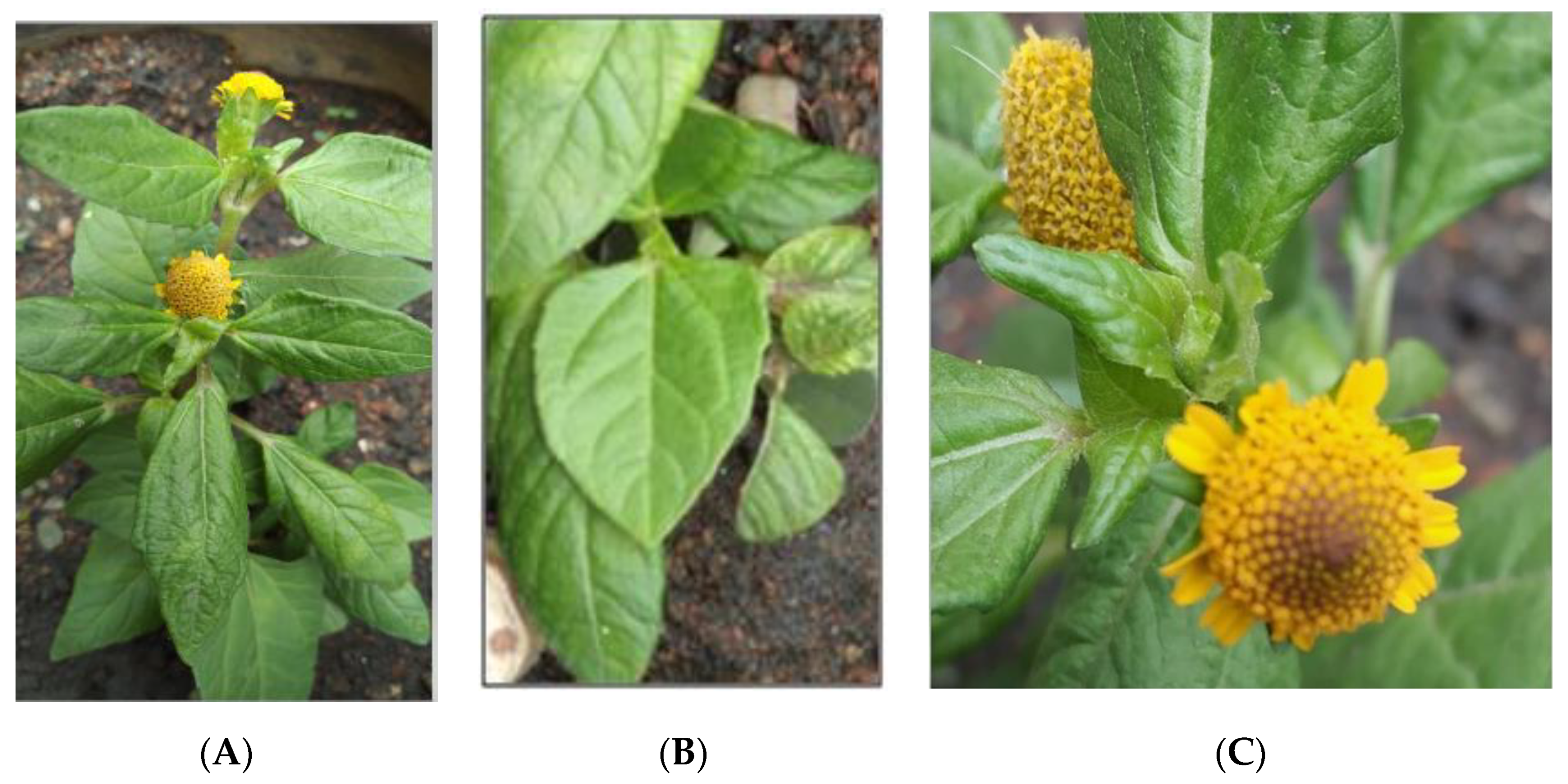
2. Spilanthes acmella
2.1. Traditional Medicinal Uses
2.2. Phytochemical
3. Anti-Inflammatory Effects of S. acmella
3.1. In Vitro Study
3.2. In Vivo Study
4. Antioxidant Properties of S. acmella
4.1. Antioxidant Activity in a Cell-Free System
4.2. Antioxidant Activity in Cell System (Cellular Method)
5. Conclusions
Supplementary Materials
Author Contributions
Funding
Institutional Review Board Statement
Informed Consent Statement
Acknowledgments
Conflicts of Interest
References
- Verhamme, P.; Hoylaerts, M.F. Hemostasis and inflammation: Two of a kind? Thromb. J. 2009, 7, 15. [Google Scholar] [CrossRef]
- Hold, G.L.; El-Omar, M.E. Genetic aspects of inflammation and cancer. Biochem. J. 2008, 410, 225–235. [Google Scholar] [CrossRef] [PubMed]
- Gutteridge, J.M. Lipid peroxidation and antioxidants as biomarkers of tissue damage. Clin. Chem. 1995, 41, 1819–1828. [Google Scholar] [CrossRef] [PubMed]
- Winrow, V.R.; Winyard, P.G.; Morris, C.J.; Blake, D.R. Free radicals in inflammation: Second messengers and mediators of tissue destruction. Br. Med. Bull. 1993, 49, 506–522. [Google Scholar] [CrossRef] [PubMed]
- Iwakiri, R. NSAIDs and its gastrointestinal side effects: Relation of NSAIDs variety and influence of concomitant medicine. Nihon Rinsho Jpn. J. Clin. Med. 2007, 65, 1776–1780. [Google Scholar]
- Conforti, F.; Sosa, S.; Marrelli, M.; Menichini, F.; Statti, G.A.; Uzunov, D.; Tubaro, A.; Menichini, F.; Della Loggia, R. In vivo anti-inflammatory and in vitro antioxidant activities of Mediterranean dietary plants. J. Ethnopharmacol. 2008, 116, 144–151. [Google Scholar] [CrossRef]
- Majolo, F.; De Oliveira Becker Delwing, L.K.; Marmitt, D.J.; Bustamante-Filho, I.C.; Goettert, M.I. Medicinal plants and bioactive natural compounds for cancer treatment: Important advances for drug discovery. Phytochem. Lett. 2019, 31, 196–207. [Google Scholar] [CrossRef]
- Newman, D.J.; Cragg, G.M. Natural Products as Sources of New Drugs from 1981 to 2014. J. Nat. Prod. 2016, 79, 629–661. [Google Scholar] [CrossRef]
- Dias, D.A.; Urban, S.; Roessner, U. A Historical Overview of Natural Products in Drug Discovery. Metabolites 2012, 2, 303–336. [Google Scholar] [CrossRef]
- Mueller, M.; Hobiger, S.; Jungbauer, A. Anti-inflammatory activity of extracts from fruits, herbs and spices. Food Chem. 2010, 122, 987–996. [Google Scholar] [CrossRef]
- Paulraj, J.; Govindarajan, R.; Palpu, P. The Genus Spilanthes Ethnopharmacology, Phytochemistry, and Pharmacological Properties: A Review. Adv. Pharmacol. Pharm. Sci. 2013, 2013, 510298. [Google Scholar]
- Jansen, R.K. Systematics of Spilanthes (Compositae: Heliantheae). Syst. Bot. 1981, 6, 231. [Google Scholar] [CrossRef]
- Van Steenis, C.G.G.J.; Willis, J.C. A Dictionary of the Flowering Plants and Ferns. J. Ecol. 1967, 55, 864–865. [Google Scholar] [CrossRef]
- Fu, Z.-X.; Jiao, B.-H.; Nie, B.; Zhang, G.-J.; Gao, T.-G.; China Phylogeny Consortium. A comprehensive generic-level phylogeny of the sunflower family: Implications for the systematics of Chinese Asteraceae. J. Syst. Evol. 2016, 54, 416–437. [Google Scholar] [CrossRef]
- Michel, J.; Abd Rani, N.Z.; Husain, K. A Review on the Potential Use of Medicinal Plants from Asteraceae and Lamiaceae Plant Family in Cardiovascular Diseases. Front. Pharmacol. 2020, 11, 852. [Google Scholar] [CrossRef] [PubMed]
- Ramsewak, R. Bioactive N-isobutylamides from the flower buds of Spilanthes acmella. Phytochemistry 1999, 51, 729–732. [Google Scholar] [CrossRef]
- Wongsawatkul, O.; Prachayasittikul, S.; Isarankura-Na-Ayudhya, C.; Satayavivad, J.; Ruchirawat, S.; Prachayasittikul, V. Vasorelaxant and Antioxidant Activities of Spilanthes acmella Murr. Int. J. Mol. Sci. 2008, 9, 2724–2744. [Google Scholar] [CrossRef]
- Dias, A.; Santos, P.V.F.; Seabra, I.; Júnior, R.; Braga, M.E.M.; De Sousa, H. Spilanthol from Spilanthes acmella flowers, leaves and stems obtained by selective supercritical carbon dioxide extraction. J. Supercrit. Fluids 2012, 61, 62–70. [Google Scholar] [CrossRef]
- Abd Jalil, M.A.; Shuid, A.N.; Muhammad, N. Role of Medicinal Plants and Natural Products on Osteoporotic Fracture Healing. Evid. Based Complement. Altern. Med. 2012, 2012, 1–7. [Google Scholar] [CrossRef]
- Schottenfeld, D.; Beebe-Dimmer, J. Chronic Inflammation: A Common and Important Factor in the Pathogenesis of Neoplasia. CA Cancer J. Clin. 2006, 56, 69–83. [Google Scholar] [CrossRef]
- Choy, K.W.; Murugan, D.; Leong, X.-F.; Abas, R.; Alias, A.; Mustafa, M.R. Flavonoids as Natural Anti-Inflammatory Agents Targeting Nuclear Factor-Kappa B (NFκB) Signaling in Cardiovascular Diseases: A Mini Review. Front. Pharmacol. 2019, 10, 1295. [Google Scholar] [CrossRef]
- Hotamisligil, G.S. Inflammation, metaflammation and immunometabolic disorders. Nature 2017, 542, 177–185. [Google Scholar] [CrossRef]
- Heinrich, M. Ethnobotany and its role in drug development. Phytother. Res. 2000, 14, 479–488. [Google Scholar] [CrossRef]
- Schultes, R.E. Conservation of Medicinal Plants, Edited by Olayiwola Akerele, Vernon Heywood & Hugh Synge. Cambridge University Press, Cambridge, England, UK: xiii + 362 pp., 20 × 15 × 3 cm, no price indicated, 1991. Environ. Conserv. 1993, 20, 93. [Google Scholar] [CrossRef]
- Hind, N.; Biggs, N. Plate 460. Acmella oleracea Compositae. Curtis’s Bot. Mag. 2003, 20, 31–39. [Google Scholar] [CrossRef]
- Blagden, C.O. A Dlctionary of the Economic Products of the Malay Peninsula. By J. H. Burkill, with contributions by William Birtwistle, Frederick W. Foxworthy, J. B. Scrivenor, and J. G. Watson. 2 vols. 9 × 6. pp. xi +2402. London: Published on behalf of the Governments of the Straits Settlements and Federated Malay States by the Crown Agents for the Colonies, 1935. 30s. J. R. Asiat. Soc. 1937, 69, 134–135. [Google Scholar] [CrossRef]
- Storey, C.; Salem, J.I. Lay use of amazonian plants for the treatment of tuberculosis. Acta Amaz. 1997, 27, 175–182. [Google Scholar] [CrossRef]
- Baruah, R.N.; Leclercq, P.A. Characterization of the Essential Oil from Flower Heads of Spilanthes acmella. J. Essent. Oil Res. 1993, 5, 693–695. [Google Scholar] [CrossRef]
- Hossan, S.; Agarwala, B.; Sarwar, S.; Karim, M.; Jahan, R.; Rahmatullah, M. Traditional use of medicinal plants in Bangladesh to treat urinary tract infections and sexually transmitted diseases. Ethnobot. Res. Appl. 2010, 8, 61–74. [Google Scholar] [CrossRef]
- Dubey, S.; Maity, S.; Singh, M.; Saraf, S.A.; Saha, S. Phytochemistry, Pharmacology and Toxicology of Spilanthes acmella: A Review. Adv. Pharmacol. Sci. 2013, 2013, 423750. [Google Scholar] [CrossRef]
- Nakatani, N.; Nagashima, M. Pungent Alkamides from Spilanthes acmella L. var. oleracea Clarke. Biosci. Biotechnol. Biochem. 1992, 56, 759–762. [Google Scholar] [CrossRef] [PubMed]
- Boonen, J.; Baert, B.; Burvenich, C.; Blondeel, P.; De Saeger, S.; De Spiegeleer, B. LC–MS profiling of N-alkylamides in Spilanthes acmella extract and the transmucosal behaviour of its main bio-active spilanthol. J. Pharm. Biomed. Anal. 2010, 53, 243–249. [Google Scholar] [CrossRef]
- Krishnaswamy, N.; Prasanna, S.; Seshandri, T.; Vedantham, T. α- and β-Amyrin esters and sitosterol glucoside from Spilanthes acmella. Phytochemistry 1975, 14, 1666–1667. [Google Scholar] [CrossRef]
- Prachayasittikul, V.; Prachayasittikul, S.; Ruchirawat, S.; Prachayasittikul, V. High therapeutic potential of Spilanthes acmella: A review. EXCLI J. 2013, 12, 291–312. [Google Scholar] [CrossRef] [PubMed]
- Greger, H. Alkamides: A critical reconsideration of a multifunctional class of unsaturated fatty acid amides. Phytochem. Rev. 2016, 15, 729–770. [Google Scholar] [CrossRef]
- Yasuda, I.; Takeya, K.; Itokawa, H. The geometric structure of spilanthol. Chem. Pharm. Bull. 1980, 28, 2251–2253. [Google Scholar] [CrossRef]
- Leng, T.C.; Ping, N.S.; Lim, B.P.; Keng, C.L. Detection of Bioactive Compounds from Spilanthes Acmella (L.) Plants and Its Various In Vitro Culture Products. J. Med. Plant Res. 2011, 5, 371–378. [Google Scholar] [CrossRef]
- Prachayasittikul, S.; Suphapong, S.; Worachartcheewan, A.; Lawung, R.; Ruchirawat, S.; Prachayasittikul, V. Bioactive Metabolites from Spilanthes acmella Murr. Molecules 2009, 14, 850–867. [Google Scholar] [CrossRef]
- Chakraborty, A.; Devi, B.; Sanjebam, R.; Khumbong, S.; Thokchom, I. Preliminary studies on local anesthetic and antipyretic activities of Spilanthes acmella Murr. in experimental animal models. Indian J. Pharmacol. 2010, 42, 277–279. [Google Scholar] [CrossRef]
- Ratnasooriya, W.; Pieris, K.; Samaratunga, U.; Jayakody, J. Diuretic activity of Spilanthes acmella flowers in rats. J. Ethnopharmacol. 2004, 91, 317–320. [Google Scholar] [CrossRef] [PubMed]
- Rani, S.; Murty, S. Antifungal potential of flower head extract of Spilanthes acmella Linn. Afr. J. Biomed. Res. 2009, 9, 67–69. [Google Scholar] [CrossRef]
- Mbeunkui, F.; Grace, M.H.; Lategan, C.; Smith, P.J.; Raskin, I.; Lila, M.A. Isolation and identification of antiplasmodial N-alkylamides from Spilanthes acmella flowers using centrifugal partition chromatography and ESI-IT-TOF-MS. J. Chromatogr. B 2011, 879, 1886–1892. [Google Scholar] [CrossRef]
- Arora, S.; Vijay, S.; Kumar, D. Phytochemical and Antimicrobial Studies on the Leaves of Spilanthes Acmella. J. Chem. Pharm. Res. 2011, 3, 145–150. [Google Scholar]
- Sharma, A.; Kumar, V.; Rattan, R.S.; Kumar, N.; Singh, B. Insecticidal Toxicity of Spilanthol from Spilanthes acmella Murr. against Plutella xylostella L. Am. J. Plant Sci. 2012, 3, 1568–1572. [Google Scholar] [CrossRef]
- Ahmed, S.; Rahman, A.; Muslim, T.; Sohrab, M.; Akbor, M.; Siraj, S.; Sultana, N.; Al-Mansur, M. Antimicrobial cytotoxicity and phytochemical activities of Spilanthes acmella. Bangladesh J. Sci. Ind. Res. 2013, 47, 437–440. [Google Scholar] [CrossRef]
- Arif, M.; Juyal, D.; Joshi, A. A Review on Pharmacognostic and Phytochemical Study of a Plant Spilanthes Acmella Murr. Phrma Innov. J. 2017, 6, 172–177. [Google Scholar]
- Jirovetz, L.; Buchbauer, G.; Wobus, A.; Shafi, M.P.; Abraham, G.T. Essential Oil Analysis of Spilanthes acmella Murr. Fresh Plants from Southern India. J. Essent. Oil Res. 2005, 17, 429–431. [Google Scholar] [CrossRef]
- Lalthanpuii, P.B.; Lalchhandama, K. Intestinal cestodes of chicken are effectively killed by quinoline-rich extract of Spilanthes acmella. Vet. World 2020, 13, 821–826. [Google Scholar] [CrossRef] [PubMed]
- Widyowati, R.; Sulistyowaty, M.I.; Uyen, N.H.; Sugimoto, S.; Yamano, Y.; Otsuka, H.; Matsunami, K. New Methyl Threonolactones and Pyroglutamates of Spilanthes acmella (L.) L. and Their Bone Formation Activities. Molecules 2020, 25, 2500. [Google Scholar] [CrossRef] [PubMed]
- Widyowati, R.; Ekasari, W.; Purwitasari, N. Amine Derivative from the Aerial Part of Spilanthes acmella Murr. and their Alkaline Phosphatase Activity. Nat. Prod. J. 2020, 10, 571–577. [Google Scholar] [CrossRef]
- Liehn, E.A.; Cabrera-Fuentes, H.A. Inflammation between defense and disease: Impact on tissue repair and chronic sickness. Discoveries 2015, 3, e42. [Google Scholar] [CrossRef] [PubMed]
- Zhang, H.; Tsao, R. Dietary polyphenols, oxidative stress and antioxidant and anti-inflammatory effects. Curr. Opin. Food Sci. 2016, 8, 33–42. [Google Scholar] [CrossRef]
- Lin, W.-W.; Karin, M. A cytokine-mediated link between innate immunity, inflammation, and cancer. J. Clin. Investig. 2007, 117, 1175–1183. [Google Scholar] [CrossRef] [PubMed]
- Lamkanfi, M.; Dixit, V.M. Inflammasomes and Their Roles in Health and Disease. Annu. Rev. Cell Dev. Biol. 2012, 28, 137–161. [Google Scholar] [CrossRef] [PubMed]
- Adebayo, S.A.; Dzoyem, J.P.; Shai, L.J.; Eloff, J.N. The anti-inflammatory and antioxidant activity of 25 plant species used traditionally to treat pain in southern African. BMC Complement. Altern. Med. 2015, 15, 1–10. [Google Scholar] [CrossRef]
- Wu, L.-C.; Fan, N.-C.; Lin, M.-H.; Chu, I.-R.; Huang, S.-J.; Hu, C.-Y.; Han, S.-Y. Anti-inflammatory Effect of Spilanthol from Spilanthes acmella on Murine Macrophage by Down-Regulating LPS-Induced Inflammatory Mediators. J. Agric. Food Chem. 2008, 56, 2341–2349. [Google Scholar] [CrossRef]
- Wu, M.J.; Wang, L.; Ding, H.Y.; Weng, C.Y.; Yen, J.H. Glossogyne Tenuifolia Acts to Inhibit Inflammatory Mediator Production in a Macrophage Cell Line by Downregulating LPS-Induced NF-IκB. J. Biomed. Sci. 2004, 11, 186–199. [Google Scholar] [CrossRef]
- Kim, K.H.; Kim, E.J.; Kwun, M.J.; Lee, J.Y.; Bach, T.T.; Eum, S.M.; Choi, J.Y.; Cho, S.; Kim, S.-J.; Jeong, S.-I.; et al. Suppression of lung inflammation by the methanol extract of Spilanthes acmella Murray is related to differential regulation of NF-κB and Nrf2. J. Ethnopharmacol. 2018, 217, 89–97. [Google Scholar] [CrossRef]
- Lu, M.-C.; Ji, J.-A.; Jiang, Z.-Y.; You, Q.-D. The Keap1-Nrf2-ARE Pathway As a Potential Preventive and Therapeutic Target: An Update. Med. Res. Rev. 2016, 36, 924–963. [Google Scholar] [CrossRef]
- Umemura, K.; Itoh, T.; Hamada, N.; Fujita, Y.; Akao, Y.; Nozawa, Y.; Matsuura, N.; Iinuma, M.; Ito, M. Preconditioning by sesquiterpene lactone enhances H2O2-induced Nrf2/ARE activation. Biochem. Biophys. Res. Commun. 2008, 368, 948–954. [Google Scholar] [CrossRef]
- Wang, R.; Zhang, C.Y.; Bai, L.P.; Pan, H.D.; Shu, L.M.; Kong, A.-N.T.; Leung, E.L.-H.; Liu, L.; Li, T. Flavonoids derived from liquorice suppress murine macrophage activation by up-regulating heme oxygenase-1 independent of Nrf2 activation. Int. Immunopharmacol. 2015, 28, 917–924. [Google Scholar] [CrossRef]
- Amin, F.U.; Shah, S.A.; Kim, M.O. Vanillic acid attenuates Aβ1-42-induced oxidative stress and cognitive impairment in mice. Sci. Rep. 2017, 7, 40753. [Google Scholar] [CrossRef] [PubMed]
- Dilshara, M.G.; Lee, K.-T.; Jayasooriya, R.G.P.T.; Kang, C.-H.; Park, S.R.; Choi, Y.H.; Choi, I.-W.; Hyun, J.-W.; Chang, W.-Y.; Kim, Y.-S.; et al. Downregulation of NO and PGE2 in LPS-stimulated BV2 microglial cells by trans-isoferulic acid via suppression of PI3K/Akt-dependent NF-κB and activation of Nrf2-mediated HO-1. Int. Immunopharmacol. 2014, 18, 203–211. [Google Scholar] [CrossRef] [PubMed]
- Chang, W.-C.; Wu, S.-C.; Xu, K.-D.; Liao, B.-C.; Wu, J.-F.; Cheng, A.-S. Scopoletin Protects against Methylglyoxal-Induced Hyperglycemia and Insulin Resistance Mediated by Suppression of Advanced Glycation Endproducts (AGEs) Generation and Anti-Glycation. Molecules 2015, 20, 2786–2801. [Google Scholar] [CrossRef] [PubMed]
- Chakraborty, A.; Devi, R.K.B.; Rita, S.; Sharatchandra, K.; Singh, T.I. Preliminary Studies on Antiinflammatory and Analgesic Activities of Spilanthes acmella in Experimental Animal Models. Indian J. Pharmacol. 2004, 36, 148–150. [Google Scholar]
- Ayertey, F.; Ofori-Attah, E.; Antwi, S.; Amoa-Bosompem, M.; Djameh, G.; Lartey, N.L.; Ohashi, M.; Kusi, K.A.; Appiah, A.A.; Appiah-Opong, R.; et al. Anti-inflammatory activity and mechanism of action of ethanolic leaf extract of Morinda lucida Benth. J. Tradit. Complement. Med. 2020. [Google Scholar] [CrossRef]
- Bakondi, E.; Singh, S.B.; Hajnády, Z.; Nagy-Pénzes, M.; Regdon, Z.; Kovács, K.; Hegedűs, C.; Madácsy, T.; Maléth, J.; Hegyi, P.; et al. Spilanthol Inhibits Inflammatory Transcription Factors and iNOS Expression in Macrophages and Exerts Anti-inflammatory Effects in Dermatitis and Pancreatitis. Int. J. Mol. Sci. 2019, 20, 4308. [Google Scholar] [CrossRef]
- Huang, W.-C.; Huang, C.-H.; Hu, S.; Peng, H.-L.; Wu, S.-J. Topical Spilanthol Inhibits MAPK Signaling and Ameliorates Allergic Inflammation in DNCB-Induced Atopic Dermatitis in Mice. Int. J. Mol. Sci. 2019, 20, 2490. [Google Scholar] [CrossRef]
- Huang, W.-C.; Peng, H.-L.; Hu, S.; Wu, S.-J. Spilanthol from Traditionally Used Spilanthes acmella Enhances AMPK and Ameliorates Obesity in Mice Fed High-Fat Diet. Nutrients 2019, 11, 991. [Google Scholar] [CrossRef]
- Huang, W.-C.; Wu, L.-Y.; Hu, S.; Wu, S.-J. Spilanthol Inhibits COX-2 and ICAM-1 Expression via Suppression of NF-κB and MAPK Signaling in Interleukin-1β-Stimulated Human Lung Epithelial Cells. Inflammation 2018, 41, 1934–1944. [Google Scholar] [CrossRef]
- Birben, E.; Sahiner, U.M.; Sackesen, C.; Erzurum, S.; Kalayci, O. Oxidative Stress and Antioxidant Defense. World Allergy Organ. J. 2012, 5, 9–19. [Google Scholar] [CrossRef]
- Pham-Huy, L.A.; He, H.; Pham-Huy, C. Free Radicals, Antioxidants in Disease and Health. Int. J. Biomed. Sci. 2008, 4, 89–96. [Google Scholar] [PubMed]
- Moreno, M.A.; Zampini, I.C.; Isla, M.I. Antifungal, anti-inflammatory and antioxidant activity of bi-herbal mixtures with medicinal plants from Argentinean highlands. J. Ethnopharmacol. 2020, 253, 112642. [Google Scholar] [CrossRef] [PubMed]
- Wu, Q.; Song, R.; Zhao, L.; Yun, Z. Advances in cellular evaluation and standard of antioxidant activity. E3S Web Conf. 2019, 131, 01008. [Google Scholar] [CrossRef]
- Özyürek, M.; Güçlü, K.; Apak, R. The main and modified CUPRAC methods of antioxidant measurement. TrAC Trends Anal. Chem. 2011, 30, 652–664. [Google Scholar] [CrossRef]
- Dudonné, S.; Vitrac, X.; Coutière, P.; Woillez, M.; Mérillon, J.M. Comparative study of antioxidant properties and total phenolic content of 30 plant extracts of industrial interest using DPPH, ABTS, FRAP, SOD, and ORAC assays. J. Agric. Food Chem. 2009, 57, 1768–1774. [Google Scholar] [CrossRef]
- Esmaeili, M.A.; Sonboli, A. Antioxidant, free radical scavenging activities of Salvia brachyantha and its protective effect against oxidative cardiac cell injury. Food Chem. Toxicol. 2010, 48, 846–853. [Google Scholar] [CrossRef]
- Tanwer, B.S.; Choudhary, R.; Vijayvergia, R. In Vitro and In Vivo Comparative Study of Primary Metabolites and Antioxidant Activity in Spilanthes acmella Murr. Int. J. Biotechnol. Biochem. 2010, 6, 819–825. [Google Scholar]
- Nabi, N.G.; Shrivastava, M. Estimation of Total Flavonoids and Antioxidant Activity of Spilanthes acmella Leaves. Pharm. Biosci. J. 2016, 4, 29. [Google Scholar] [CrossRef]
- Swargiary, A.; Daimari, M.; Roy, M.; Haloi, D.; Ramchiary, B. Evaluation of phytochemical properties and larvicidal activities of Cynodon dactylon, Clerodendrum viscosum, Spilanthes acmella and Terminalia chebula against Aedes aegypti. Asian Pac. J. Trop. Med. 2019, 12, 224. [Google Scholar] [CrossRef]
- Boontha, S.; Thoedyotin, T.; Saengtabtim, T.; Im-Erb, P.; Chaniad, N.; Buranrat, B.; Pitaksuteepong, T. Cytotoxic, colony formation and anti-migratory effects of Spilanthes acmella (Asteraceae) aerial extract on MCF-7 cells and its cream formulation. Trop. J. Pharm. Res. 2020, 19, 17–24. [Google Scholar] [CrossRef]
- Thakur, S.; Sagar, A.; Prakash, V. Studies on Antibacterial and Antioxidant Activity of Different Extracts of Spilanthes acmella L. Plant Arch. 2019, 19, 1711–1717. [Google Scholar]
- Suwanjang, W.; Khongniam, B.; Srisung, S.; Prachayasittikul, S.; Prachayasittikul, V. Neuroprotective effect of Spilanthes acmella Murr. on pesticide-induced neuronal cells death. Asian Pac. J. Trop. Med. 2017, 10, 35–41. [Google Scholar] [CrossRef]
- Gay, N.H.; Phopin, K.; Suwanjang, W.; Songtawee, N.; Ruankham, W.; Wongchitrat, P.; Prachayasittikul, S.; Prachayasittikul, V. Neuroprotective Effects of Phenolic and Carboxylic Acids on Oxidative Stress-Induced Toxicity in Human Neuroblastoma SH-SY5Y Cells. Neurochem. Res. 2018, 43, 619–636. [Google Scholar] [CrossRef] [PubMed]
- Kelsey, N.A.; Wilkins, H.M.; Linseman, D.A. Nutraceutical Antioxidants as Novel Neuroprotective Agents. Molecules 2010, 15, 7792. [Google Scholar] [CrossRef]
- Pietta, P.-G. Flavonoids as Antioxidants. J. Nat. Prod. 2000, 63, 1035–1042. [Google Scholar] [CrossRef]


| Kingdom | Plantae |
|---|---|
| Subkingdom | Tracheobiont |
| Phylum | Tracheophyta |
| Division | Magnoliophyta |
| Superdivision | Spermatophyte |
| Class | Magnoliopsida |
| Sub Class | Asteridae |
| Order | Asterales |
| Family | Asteraceae |
| Subfamily | Mimosoideae |
| Genus | Spilanthes |
| Species | Acmella |
| Groups | Parts | Compounds | References |
|---|---|---|---|
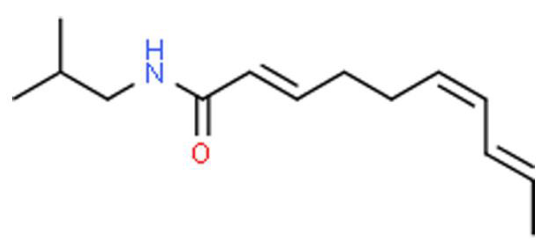
| Alkyl amide | Flower | (2Z)-N-isobutyl-2-nonene-6,8-diynamide(1), N-phenethyl-2, 3-epoxy-6,8-nonadiynamide(2), (2E,4Z)-N-isobutyl-2,4-undecadiene-8,10-diynamide(3), (2E)-N-isobutyl-2-undecene-8,10-diynamide(4), (2E,6Z,8E)-N-isobutyl-2,6,8-decatrienamide/ Spilanthol(5), (2E)-N-(2-methylbutyl)-2-undecene-8,10-diynamide(6), (2E,7Z)-N-isobutyl-2,7-tridecadiene-10,12-diynamide(7), (2E,7Z)-N-isobutyl-2,7-decadienamide(8), (2E,6Z,8E)-N-(2-methylbutyl)-2,6,8-decatrienamide(9), (2E,4E,8Z,10Z)-N- isobutyl-dodeca-2,4,8,10-tetraenamide(10), undeca-2E,7Z,9E-trienoic acid isobutylamide(11) | [16,31,32] |
| Fatty acid esters | Leaf | α–amyrin acetates(12), β–amyrin acetates(13), | [33,45] |
| Stigmastane | Aerial | (24ξ)-Stigmast-4-en-3-one(16) | [38] |
| Glucoside | Aerial | Stigmasteryl glucoside(18), | |
| Phenolics | Aerial | Vanilic acid(17), trans-ferulic acid(19), Pentacyclic, trans-isoferulic acid(22) | |
| Triterpenoid | Aerial, Leaf | β-sitosterone(23), 3-acetylaleuritolic acid (21), Stigmasterol (15) | [38,45] |
| Fatty acids | Whole plant | Lauric acid(24), Myristic acid(25), Palmitic acid(26), Linoleic acid(27) | [46] |
| Essential oils (Fatty alcohols) | Whole plant | Myricyl alcohol(14), (E)-2-hexenol(28), 2-tridecanone(29), hexanol(30), (Z)-3-hexanol(32) | [33,47] |
| Aromatic quinone | Aerial | 2,2,4-trimethyl-1,2-dihydroquinoline(31) | [48] |
| Glycosides | Whole plant | 2-C-methyl-D-threono-1,4-lactone-3-O-β-D-glucopyranoside(33), 2-C-methyl-D-threono-1,4-lactone-2-O-α-D-fructofuranoside(34) | [49] |
| Pyroglutamate | Whole plant | 1,3-butanediol,1-pyroglutamate(35), 1,3-butanediol,3-pyroglutamate (36), 2-C-methyl-d-threono-1,4-lactone(37), 2-deoxy-D-ribono-1,4-lactone(38), Dendranthemoside A(39), Dendranthemoside B(40), Ampelopsisionoside(41), Icariside B1(42), Benzyl-α-l-arabinopyranosyl-(1→6)-β-D-glucopyranoside(43), Uridine(45), methyl pyroglutamate(46) | [49,50] |
| Coumarine | Aerial | Chicoriin(44), Scopoletin(20) | [38] |
| Essential oils (Terpene) | Flower head | α-pinene(47), Sabinene(48), β-pinene(49), Myrcene(50), D-3-carene(51), Limonene(52), (Z)-β-ocimene(53), (E)-β-ocimene(54), γ-terpinene(55), terpinen-4-ol(56), β-elemene(57), β-caryophyllene(58), α-humulene(59), germacrene D(60), (E)-β-farnesene(61), (Z,E)-α-farnesene(62), β-bisabolene(63), β-sesquiphellandrene (64) | [28] |
| Part of Plant Used | Experimental Model | Major Findings | References |
|---|---|---|---|
| Whole plant, leaves | Carrageenan induced paw edema | Suppressed hind paw edema; increased pain threshold | [65] |
| Flowers | RAW264.7 cell lines | Inhibited NO, iNOS, COX-2 protein production; reduced pro-inflammatory mediator production (IL-1β, IL-6, TNF-α); reduced NF-κB binding activity | [56] |
| Whole plant | RAW264.7 cell lines | Suppressed NF-κB nuclear localization, NF-κB dependent cytokine genes; increased Nrf2 level, Nrf2 dependent cytokine genes; suppressed Nrf2 ubiquitination | [58] |
| Whole plant | LPS-induced lung injury | Suppressed expression of IL-1β, IL-6, TNF-α; suppressed MPO activity | [58] |
| Flower, leaf, stem | RAW264.7 cell lines | Suppressed NO production; inhibit iNOS mRNA and protein expression; inhibit NF-κB activation | [67] |
| Flower, leaf, stem | PMA-induced dermatitis; Cerulean-induced acute pancreatitis | Ameliorated histological signs of acute inflammation in contact dermatitis model; reduced leukocyte migration in an acute pancreatitis mouse model | [67] |
| Active compound (spilanthol) | DNCB-induced atopic dermatitis | Reduced serum IgE, IgG2a levels; suppressed iNOS, COX-2 expression; inhibited MAPK signaling reduced epidermal thickness, collagen accumulation; inhibited mast cells and eosinophils infiltration | [68] |
| Active compound (spilanthol) | 3T3-L1 cells | Suppressed COX-2, phospho-p38, phosphor-JNK; Promoted protein HO-1 expression; inhibited phosphorylation of MAPK | [69] |
| Active compound (spilanthol) | A549 cells | Downregulate COX-2 production; decrease TNF-α and MCP-1 production; decreased phosphorylation of IκBα and MAPK pathways; promoted HO-1 protein; | [70] |
| Part of Plant Used | Experimental Model | Major Findings | References |
|---|---|---|---|
| Aerial parts | Phenylephrine-induced male Sprague-Dawley rats | Chloroform extract—highest SOD activity and vasorelaxation effect; ethyl acetate extract—most potent radical scavenging activity with immediate vasorelaxation effect. | [17] |
| Callus, Root, Stem, Leaves | Quantitative estimation of primary metabolites and antioxidant activity | Comparable antioxidative activity to BHA (all plant parts); methanol extract—Highest superoxide radical scavenging activity (stem), highest DPPH scavenging activity (leaves). | [78] |
| Leaves | Quantitative estimation of phytochemicals and antioxidant activity | Ethanol extract—strong antioxidant activity with lowest IC50 value for DPPH and superoxide scavenging activity. | [79] |
| Whole plant | Quantitative estimation of phytochemicals and antioxidant activity | Methanol extract—high TPC, DPPH, TBARS, SOD; High TPC contribute to the high antioxidant activity | [80] |
| Aerial parts | Quantitative estimation of phytochemicals and antioxidant activity | Ethanol extract- TPC and TFC contribute to high DPPH scavenging activity | [81] |
| Stem, Leaves, Flowers | Quantitative estimation of phytochemicals and antioxidant activity | Methanol extract- high DPPH scavenging activity | [82] |
| Aerial parts | Neuronal cell death in SH-SY5Y cell lines | Attenuation of cell viability reduction in pirimicarb-induced in SH-SY5Y cell lines; hexane extract—strongest protective effect in SH-SY5Y cells induced with H2O2 | [83] |
| Active compound (vanillic acid, trans-ferulic acid) | Neuronal cell death in SH-SY5Y cell lines | Attenuate cell death on SH-SY5Y caused by H202-induced toxicity; upregulate H2O2-induced depletion of the SIRT1, FoxO3a expressions; induced superoxide dismutase 2, catalase, and induced anti-apoptotic Bcl-2 proteins | [84] |
Publisher’s Note: MDPI stays neutral with regard to jurisdictional claims in published maps and institutional affiliations. |
© 2021 by the authors. Licensee MDPI, Basel, Switzerland. This article is an open access article distributed under the terms and conditions of the Creative Commons Attribution (CC BY) license (http://creativecommons.org/licenses/by/4.0/).
Share and Cite
Abdul Rahim, R.; Jayusman, P.A.; Muhammad, N.; Mohamed, N.; Lim, V.; Ahmad, N.H.; Mohamad, S.; Abdul Hamid, Z.A.; Ahmad, F.; Mokhtar, N.; et al. Potential Antioxidant and Anti-Inflammatory Effects of Spilanthes acmella and Its Health Beneficial Effects: A Review. Int. J. Environ. Res. Public Health 2021, 18, 3532. https://doi.org/10.3390/ijerph18073532
Abdul Rahim R, Jayusman PA, Muhammad N, Mohamed N, Lim V, Ahmad NH, Mohamad S, Abdul Hamid ZA, Ahmad F, Mokhtar N, et al. Potential Antioxidant and Anti-Inflammatory Effects of Spilanthes acmella and Its Health Beneficial Effects: A Review. International Journal of Environmental Research and Public Health. 2021; 18(7):3532. https://doi.org/10.3390/ijerph18073532
Chicago/Turabian StyleAbdul Rahim, Rohanizah, Putri Ayu Jayusman, Norliza Muhammad, Norazlina Mohamed, Vuanghao Lim, Nor Hazwani Ahmad, Sharlina Mohamad, Zuratul Ain Abdul Hamid, Fairus Ahmad, Norfilza Mokhtar, and et al. 2021. "Potential Antioxidant and Anti-Inflammatory Effects of Spilanthes acmella and Its Health Beneficial Effects: A Review" International Journal of Environmental Research and Public Health 18, no. 7: 3532. https://doi.org/10.3390/ijerph18073532
APA StyleAbdul Rahim, R., Jayusman, P. A., Muhammad, N., Mohamed, N., Lim, V., Ahmad, N. H., Mohamad, S., Abdul Hamid, Z. A., Ahmad, F., Mokhtar, N., Shuid, A. N., & Mohamed, I. N. (2021). Potential Antioxidant and Anti-Inflammatory Effects of Spilanthes acmella and Its Health Beneficial Effects: A Review. International Journal of Environmental Research and Public Health, 18(7), 3532. https://doi.org/10.3390/ijerph18073532