Article

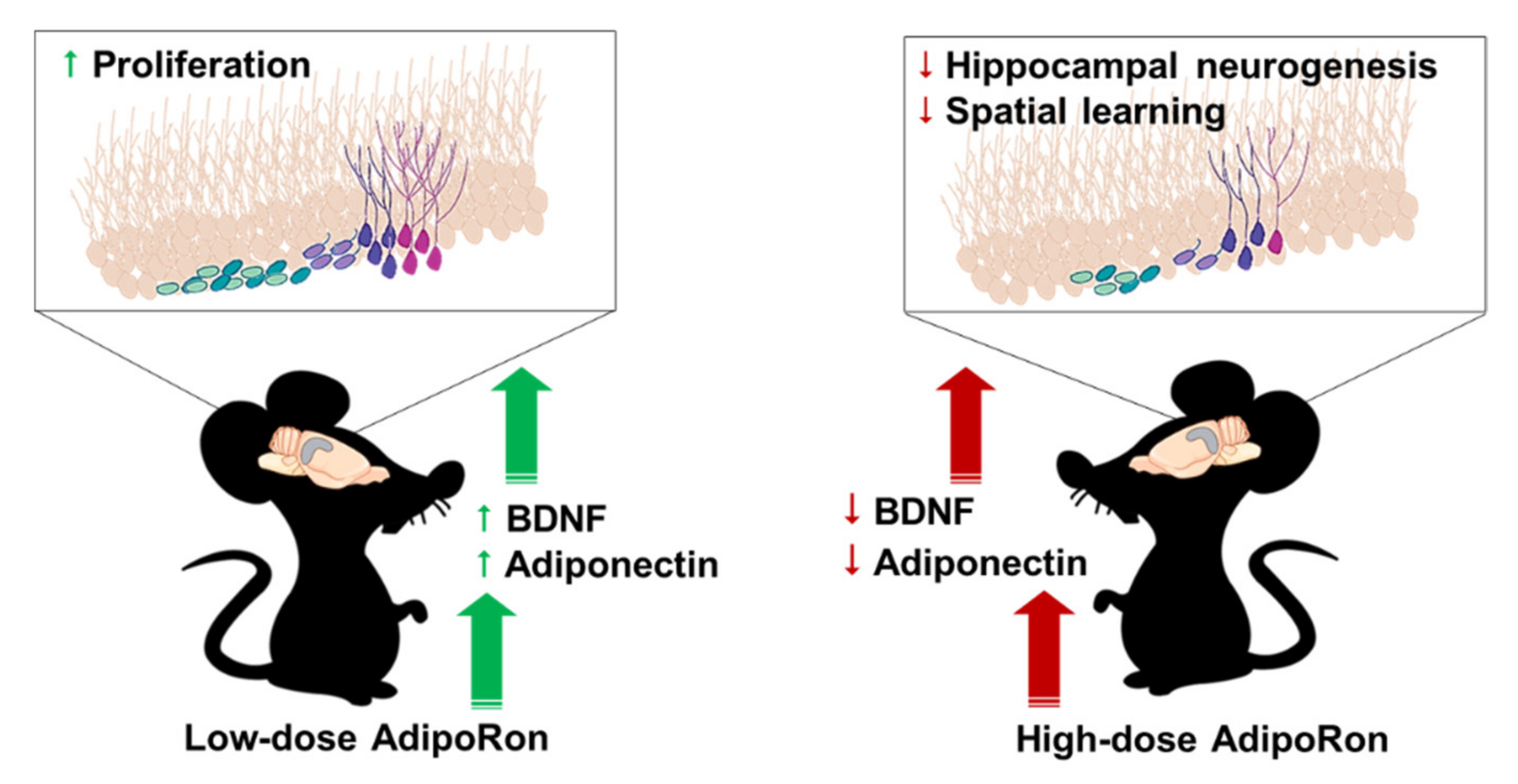
AdipoRon Treatment Induces a Dose-Dependent Response in Adult Hippocampal Neurogenesis

1
Department of Rehabilitation Sciences, Faculty of Health and Social Sciences, The Hong Kong Polytechnic University, Hong Kong
2
Division of Biomedical Sciences, University of Victoria, Victoria, BC V8P 5C2, Canada
3
FAU Brain Institute and Charles E. Schmidt College of Medicine, Florida Atlantic University, Jupiter, FL 33431, USA
4
Department of Affective Disorder, Guangzhou Brain Hospital, The Brain Affiliated Hospital of Guangzhou Medical University, Guangzhou 510370, China
5
Division of Kinesiology, School of Public Health, The University of Hong Kong, Hong Kong
6
Department of Medicine, The University of Hong Kong, Hong Kong
7
Department of Pharmacology and Pharmacy, The University of Hong Kong, Hong Kong
8
The State Key Laboratory of Pharmacology, The University of Hong Kong, Hong Kong
9
Guangdong-Hong Kong-Macau Institute of CNS Regeneration, Jinan University, Guangzhou 510632, China
10
State Key Laboratory of Brain and Cognitive Sciences, The University of Hong Kong, Hong Kong
11
Department of Ophthalmology, The University of Hong Kong, Hong Kong
*
Author to whom correspondence should be addressed.
Int. J. Mol. Sci. 2021, 22(4), 2068; https://doi.org/10.3390/ijms22042068
Submission received: 11 January 2021 / Revised: 1 February 2021 / Accepted: 5 February 2021 / Published: 19 February 2021
(This article belongs to the Section Molecular Endocrinology and Metabolism)

Abstract

:
AdipoRon, an adiponectin receptor agonist, elicits similar antidiabetic, anti-atherogenic, and anti-inflammatory effects on mouse models as adiponectin does. Since AdipoRon can cross the blood-brain barrier, its chronic effects on regulating hippocampal function are yet to be examined. This study investigated whether AdipoRon treatment promotes hippocampal neurogenesis and spatial recognition memory in a dose-dependent manner. Adolescent male C57BL/6J mice received continuous treatment of either 20 mg/kg (low dose) or 50 mg/kg (high dose) AdipoRon or vehicle intraperitoneally for 14 days, followed by the open field test to examine anxiety and locomotor activity, and the Y maze test to examine hippocampal-dependent spatial recognition memory. Immunopositive cell markers of neural progenitor cells, immature neurons, and newborn cells in the hippocampal dentate gyrus were quantified. Immunosorbent assays were used to measure the serum levels of factors that can regulate hippocampal neurogenesis, including adiponectin, brain-derived neurotrophic factor (BDNF), and corticosterone. Our results showed that 20 mg/kg AdipoRon treatment significantly promoted hippocampal cell proliferation and increased serum levels of adiponectin and BDNF, though there were no effects on spatial recognition memory and locomotor activity. On the contrary, 50 mg/kg AdipoRon treatment impaired spatial recognition memory, suppressed cell proliferation, neuronal differentiation, and cell survival associated with reduced serum levels of BDNF and adiponectin. The results suggest that a low-dose AdipoRon treatment promotes hippocampal cell proliferation, while a high-dose AdipoRon treatment is detrimental to the hippocampus function.

1. Introduction

Adiponectin is the most abundant adipokine secreted by the adipose tissue [1]. It circulates as a trimer, hexamer, and multimer, but only trimeric and hexameric forms are detectable in human cerebrospinal fluid [2,3]. Rodent studies show that adiponectin can cross the blood-brain barrier [4,5]. Adiponectin levels in the central nervous system are approximately 1000 times lower than those in the circulation [2]. Human subjects with dementia [6,7,8] and depressive disorder [9,10] have significantly lower adiponectin levels in the circulation and the cerebrospinal fluid (CSF).
Adiponectin exerts its effects by binding to adiponectin receptor 1, receptor 2, or T-cadherin [11]. Both receptors 1 and 2 are expressed in the hippocampus [12], in which their levels are downregulated in Alzheimer’s disease (AD) patients [8]. Adiponectin plays a role in regulating hippocampal plasticity, including adult neurogenesis [13], dendritic arborization [14], and synaptic plasticity [15,16]. In 8–12 weeks old mice, adiponectin haploinsufficiency increases susceptibility to stress [13] while adiponectin deficiency impairs fear extinction [17]. Chronic adiponectin deficiency also impairs learning and causes memory deficits in older mice. In 12 months old mice, adiponectin deficiency impairs learning and memory performance in fear conditioning, object recognition, and Y-maze tests [16,18]. Up to 18 months old, these mice further display spatial memory impairment in the water maze task [18]. On the other hand, activating adiponectin signaling in the brain improves depressive behaviors [5,13] and hippocampal-dependent learning and memory deficits [16,17,19], in which hippocampal plasticity is involved. These findings collectively suggest that upregulating adiponectin expression or activating adiponectin signaling could be an effective strategy to improve hippocampal function by enhancing hippocampal neuroplasticity.
Hippocampal plasticity is strongly linked to the metabolic condition of adipose tissue [20,21]. Adolescence is a critical window for shaping both adipose tissue properties and hippocampal plasticity. The proliferation of adipocyte is observed from childhood to adolescence with higher rates in obese individuals [22]. The adipocyte number remains constant in adulthood, but higher in obese adults [23]. Towards brain health, the adolescent brain is more vulnerable to metabolic stimuli than the adult brain, such as high-fat diet [24,25] and voluntary running exercise [26,27].
Circulating adiponectin levels are decreased in individuals with obesity and Type 2 diabetes [28]. Adiponectin possesses antidiabetic property [29,30,31] by promoting fatty acid oxidation through AdipoR1/AMPK or AdipoR2/PPAR-α pathway [32]. The antidiabetic effect of adiponectin can be mimicked by administering a small, orally active, adiponectin receptor agonist, AdipoRon [33]. Mechanistically, AdipoRon binds to both AdipoR1 and AdipoR2 in vitro with a greater affinity for AdipoR1 [33]. Functionally, the antidiabetic effect of AdipoRon is completely abolished under the condition of double knockout of AdipoR1 and AdipoR2, but not single knockout of either adiponectin receptors [33]. Further investigations reveal its anti-inflammatory [34], anti-atherosclerotic [35], hepatoprotective [36], and cardioprotective [37] properties in animal models. AdipoRon can be detected in the brain upon systemic injection [38] and oral administration [8], implicating its ability to cross the blood-brain barrier (BBB). Former studies focused on examining the pharmacological effect of AdipoRon in neurocognitive diseases [8,38], its neurotrophic effect on physiological condition has yet to be examined.
Our previous studies have shown that adiponectin mediates exercise-induced adult hippocampal neurogenesis in an AdipoR1/AMPK-dependent manner [5,39,40]. It is of our interest to investigate the potential effect of AdipoRon on adult hippocampal neurogenesis and the subsequent behavioral changes. Therefore, we examined whether chronic treatment with AdipoRon can affect hippocampal-dependent spatial memory and anxiety-like behavior, as well as hippocampal neurogenesis in a dose-dependent manner, and whether AdipoRon treatment modulates blood levels of neurogenesis-related factors, including brain-derived neurotrophic factor (BDNF), adiponectin, and corticosterone.

2. Results

2.1. Chronic Treatment with 50 mg/kg AdipoRon Impaired Hippocampal-Dependent Spatial Memory

One-way ANOVA revealed that chronic AdipoRon treatments of both dosages did not affect total traveling distance (Figure 1A; F2,18 = 0.468, p > 0.05) and mean speed (Figure 1B; F2,18 = 1.508, p > 0.05) in the open field test, indicating that AdipoRon did not affect locomotor activity.
Both AdipoRon dosages did not affect anxiety-like behaviors, as shown by no significant difference in time spent at the center (Figure 1C; F2,18 = 1.874, p > 0.05), wall-hugging behaviors (Figure 1D; F2,18 = 1.557, p > 0.05), and defecation (Figure 1E; F2,18 = 1.708, p > 0.05).
Hippocampal-dependent spatial memory was examined by Y maze. 20 mg/kg AdipoRon treatment did not impair spatial recognition memory (Figure 1F; p > 0.05 vs. Vehicle), but 50 mg/kg treatment impaired spatial memory as indicated by a significant decrease in exploration index (Figure 1F; p < 0.005 vs. Vehicle). The data suggested that AdipoRon treatment at a high dose could impair spatial recognition memory.

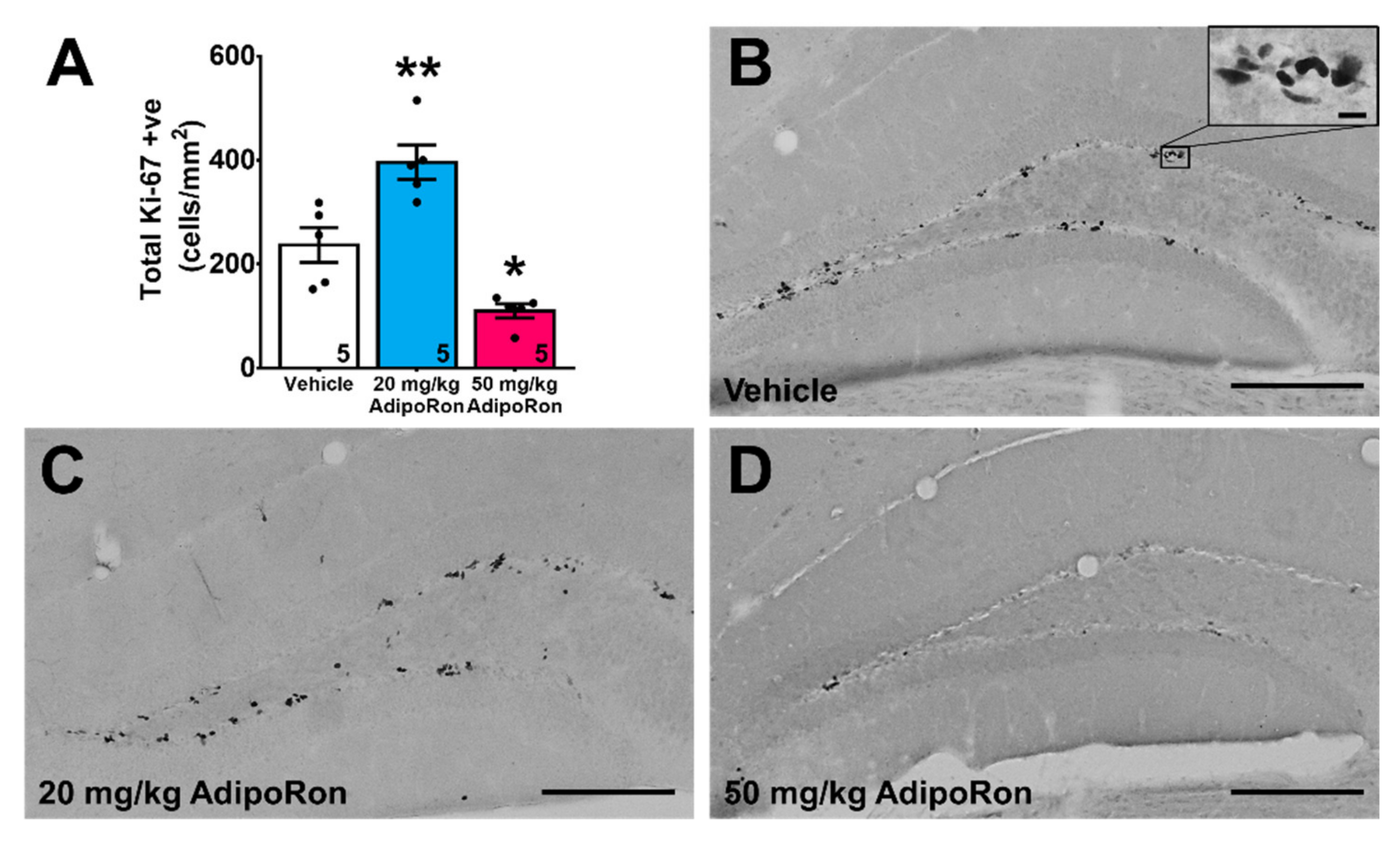
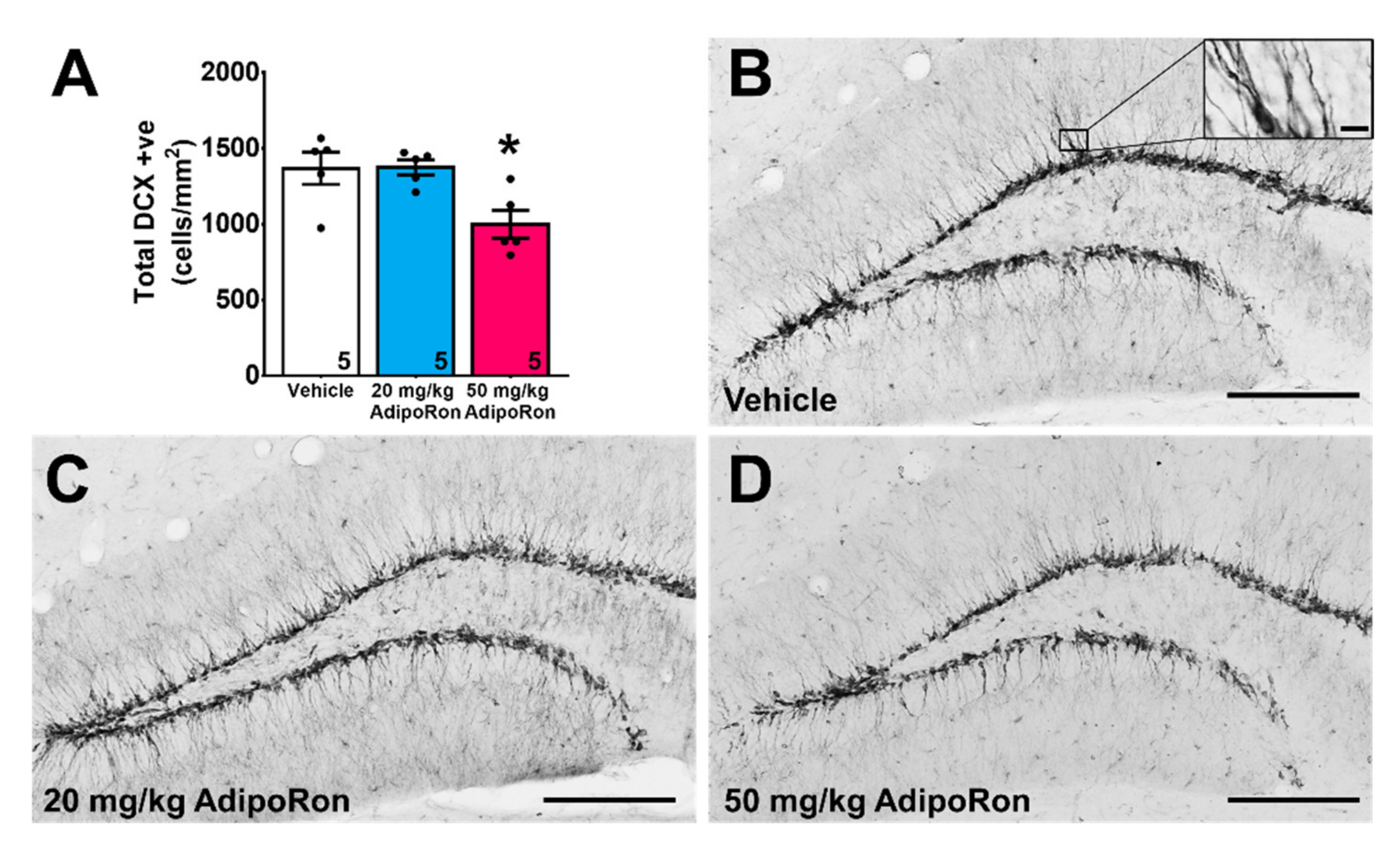
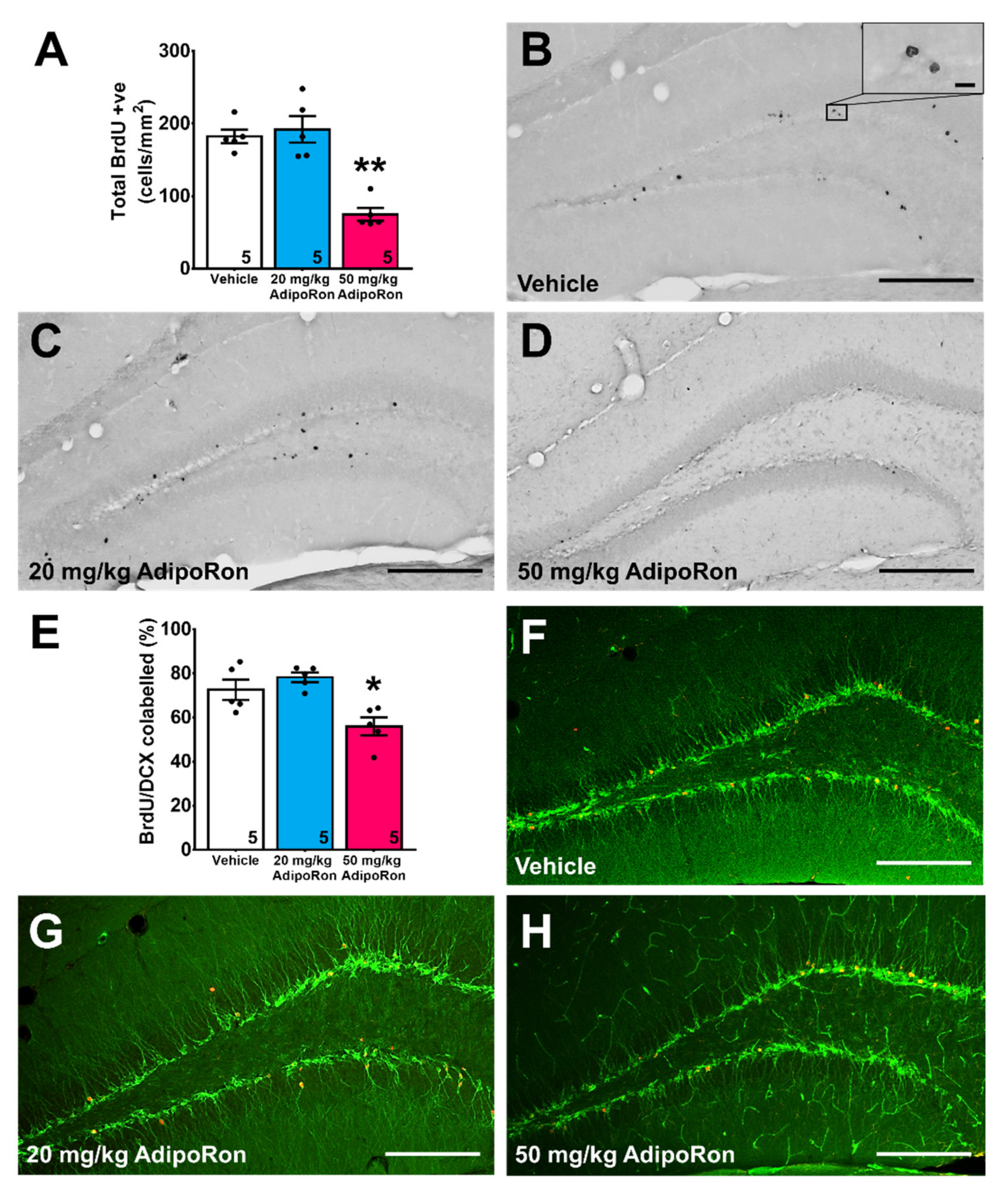
2.2. Treatment with 20 mg/kg AdipoRon Promoted Cell Proliferation, but 50 mg/kg AdipoRon Treatment Impaired Hippocampal Neurogenesis in the Dentate Gyrus

Hippocampal neurogenesis is involved in spatial learning and memory [41]. One-way ANOVA showed a significant effect of AdipoRon treatment on hippocampal cell proliferation (Figure 2A; F2,12 = 25.49, p < 0.0001). Post-hoc analysis showed that 20 mg/kg AdipoRon treatment significantly increased the number of proliferating cells (Figure 2A–C; p < 0.005 vs. Vehicle), whereas 50 mg/kg AdipoRon treatment suppressed cell proliferation (Figure 2A,D; p < 0.05 vs. Vehicle), highlighting differential effects of chronic treatment AdipoRon on cell proliferation.
AdipoRon treatment showed significant effects on the number of immature neurons labeled with doublecortin (DCX) in the dentate region (Figure 3A–D; F2,12 = 6.235, p < 0.05). Treatment with 20 mg/kg AdipoRon did not affect the number of immature neurons (Figure 3A; p > 0.05 vs. Vehicle), but 50 mg/kg treatment significantly reduced the number of immature neurons (Figure 3A; p < 0.05 vs. Vehicle).
AdipoRon treatment also significantly modulated cell survival in new-born cells labeled with BrdU (Figure 4A–D; F2,12 = 25.36, p < 0.0001). Post-hoc analysis revealed that 20 mg/kg AdipoRon treatment did not affect new-born cell survival (Figure 4A; p > 0.05 vs. Vehicle), whereas 50 mg/kg AdipoRon treatment significantly reduced cell survival, as evidenced by a significant decrease in BrdU positive cells (Figure 4A; p < 0.005 vs. Vehicle).
Lastly, quantification of BrdU/DCX co-labeled cells showed that AdipoRon treatment significantly affected neuronal differentiation (Figure 4E–H; F2,12 = 9.56, p < 0.005). Post-hoc test revealed that 20 mg/kg AdipoRon, but not 50 mg/kg AdipoRon treatment, significantly suppressed neuronal differentiation (Figure 4E; p < 0.05 vs. Vehicle).

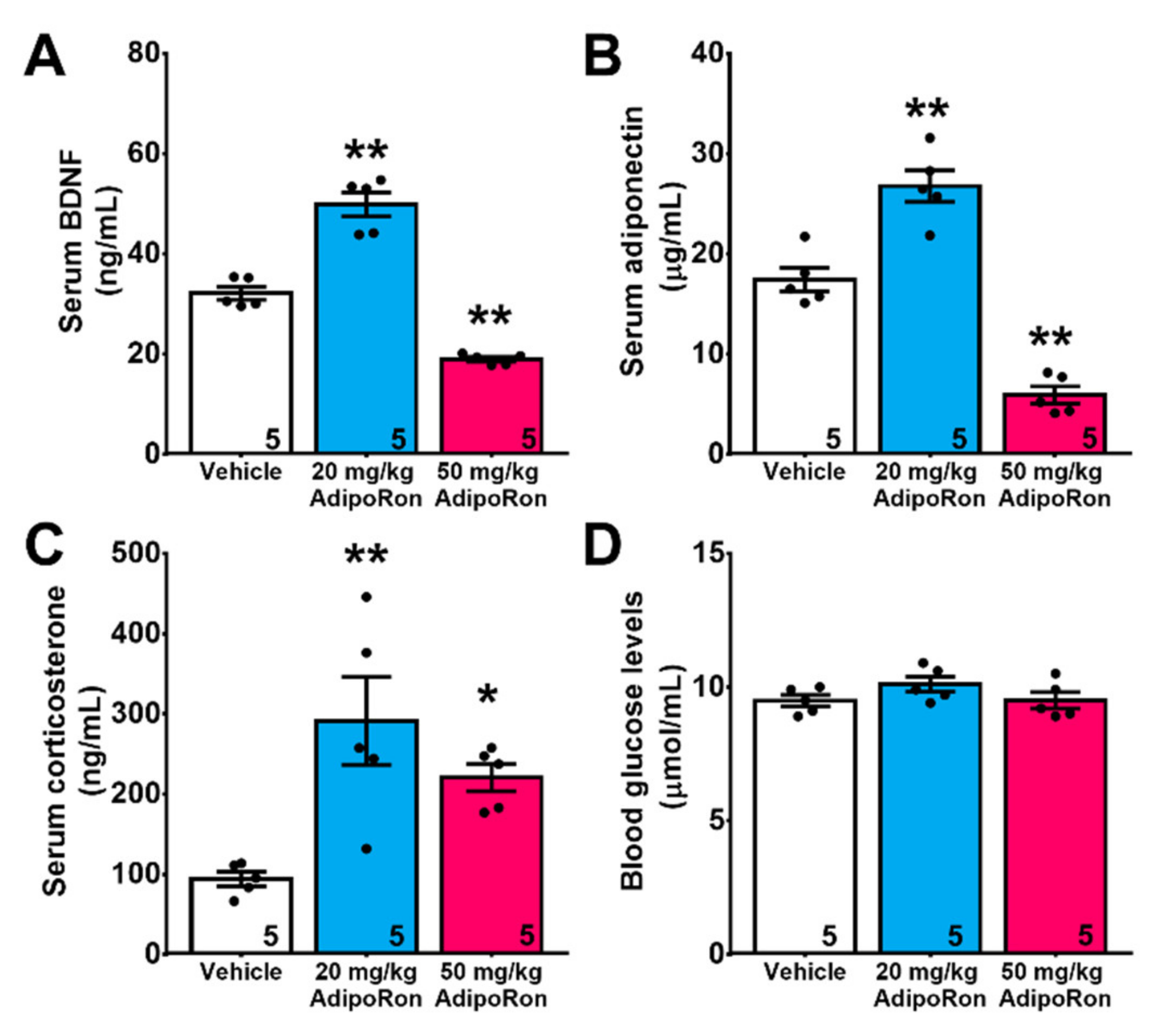
2.3. Chronic Treatment with AdipoRon Affects Serum Levels of BDNF, Adiponectin, and Corticosterone

Chronic treatment with AdipoRon significantly modulated serum levels of BDNF (Figure 5A; F2,12 = 93.83, p < 0.0001). Treatment with 20 mg/kg AdipoRon significantly increased serum BDNF levels (p < 0.005 vs. Vehicle), which was reduced by 50 mg/kg AdipoRon (Figure 5A; p < 0.005 vs. Vehicle). Similar effect was also observed in serum adiponectin (Figure 5B; F2,12 = 70.25, p < 0.0001). Treatment with 20 mg/kg AdipoRon significantly increased serum adiponectin levels (Figure 5B; p < 0.005 vs. Vehicle), whereas 50 mg/kg AdipoRon treatment reduced it (Figure 5B; p < 0.005 vs. Vehicle). Glucocorticoids suppresses neurogenesis [42], interestingly, the results showed that treatment with AdipoRon increased serum corticosterone levels regardless of the treatment dosages (Figure 5C; F2,12 = 8.909, p < 0.005). Of note, blood glucose levels were unaffected by AdipoRon treatment in all groups (Figure 5D; F2,12 = 1.705, p > 0.05 vs. Vehicle).

3. Discussion

The present study showed that chronic AdipoRon treatment at a low dose (20 mg/kg) and a high dose (50 mg/kg) have differential effects on hippocampal-dependent spatial learning, hippocampal neurogenesis, and the levels of serum biomarkers that can modulate neurogenesis. A low-dose AdipoRon treatment promoted cell proliferation in the hippocampal dentate gyrus with increased serum adiponectin and BDNF levels. Conversely, a high-dose treatment impaired spatial memory and suppressed hippocampal neurogenesis with reduced serum adiponectin and BDNF levels. The findings collectively suggested that chronic AdipoRon treatment at a high dose could be detrimental to hippocampal functions by impairing hippocampal neurogenesis (Figure 6).
Cell quantification showed that a low-dose (20 mg/kg) AdipoRon treatment promoted hippocampal cell proliferation with increased serum levels of adiponectin and BDNF. With the ability to cross the blood-brain barrier [5], adiponectin may mediate the AdipoRon action on hippocampal cell proliferation. The direct action of adiponectin on hippocampal cell proliferation is evidenced in vivo [14] and in vitro [5,43]. Besides, BDNF mediates hippocampal cell proliferation through TrkB [44]. Circulating and brain BDNF levels are positively correlated in humans and rodents [45]. The elevated serum BDNF level echoes our latest finding, illustrating that a low-dose AdipoRon treatment raises BDNF level in the dentate subregion (Lee et al., unpublished data). While the action of BDNF encompasses all stages of adult neurogenesis [46], we did not observe any neurotrophic effects of AdipoRon on neuronal differentiation and survival at a low dose. We speculate that BDNF could play a more critical role in forming dendritic arbors and spines [47]. A recent study shows that AdipoRon administration promotes spine formation in the apical dendrites of CA1 neurons in both 5 × FAD and 5 × FAD, Adipo-knockout mice [8], while we also observed an increased spine density in DG granule neurons in mice receiving low-dose AdipoRon treatment (Lee et al., unpublished data). The ability to cross BBB facilitates the direct action of AdipoRon on the hippocampus [8,38], where adiponectin receptors are expressed [12]. Intracerebral ventricular infusion of AdipoRon promotes cell proliferation in DG of APP/PS1 mice, and AdipoRon also restores Aβ-impaired neural stem cell proliferation [48]. Collectively, this study presents the potential direct and indirect actions of AdipoRon on hippocampal cell proliferation.
Meanwhile, the specific roles of AdipoR1 and AdipoR2 in adult hippocampal neurogenesis remain elusive. Full-length and dodecameric (high molecular weight, HMW) adiponectin have higher affinities for AdipoR2, while trimeric and globular adiponectin have higher affinities for AdipoR1 [49]. Trimeric adiponectin can be found in human CSF [2]. Our team has reported that trimeric adiponectin promotes neural progenitor cell proliferation in vitro in an AdipoR1-dependent manner by siRNA knockdown. Another study shows that both globular and full-length adiponectin promote neural progenitor cell proliferation in vitro [43]. The direct effect of full-length adiponectin suggests the potential involvement of AdipoR2 in modulating cell proliferation. Moreover, AdipoR2 may play a critical role in dendritic spine formation and granule neuron excitability in the dentate gyrus. Electrophysiological study reveals that AdipoR2 and adiponectin knockout induces hyperexcitability of the dentate granule neurons [17]. Chronic neuronal hyperexcitability could contribute to dendritic spine degeneration [50,51,52,53,54,55,56,57]. Notably, the spine density of dentate granule neurons is reduced in adiponectin deficiency [14]. Activating the adiponectin signaling by AdipoRon and adiponectin can restore hyperexcitability in dentate granule neurons [17] and promote dendritic spine formation [14], respectively. These findings warrant further investigation to examine whether AdipoR2 mediates dendritic spine formation in the dentate granule neurons.
In this study, AdipoRon treatment began in adolescence from six weeks of age. We cannot rule out the possibility that this development stage may play a role in affecting the proliferative effect of AdipoRon. The hippocampal cell proliferation rate is higher in adolescence than the adult and aged mice [58]. Besides, hippocampal cell proliferation is more responsive to drug treatment, like fluoxetine, in juvenile than in young adult brains [59].
In contrast, 50 mg/kg AdipoRon treatment is regarded as a high-dose treatment since serum adiponectin level was significantly reduced, implicating a homeostatic change by reducing adiponectin synthesis from the adipose tissues [60]. This adaptive response in adipose tissue is reported when adipocyte differentiation is suppressed by prolonged AdipoRon treatment (20 μM, 8 days) in C3H10T1/2 mouse embryonic mesenchymal stem cells [60]. The anti-adipogenic effect of AdipoRon is associated with reduced transcript expressions of adipocyte-specific markers, including adiponectin, but increased AMPK and ACC phosphorylation. Thus, a high-dose AdipoRon treatment may initiate negative feedback by reducing adiponectin secretion and suppressing adipogenesis to overcome hyperactivation of adiponectin receptor-mediated cascades.
AMPK is a downstream target of adiponectin [61] and AdipoRon [33]. AMPK is required to facilitate AdipoRon-induced hippocampal cell proliferation in APP/PS1 mice and in vitro, whereas Compound C, an AMPK antagonist, blunts the AdipoRon action [48]. We speculate the high-dose treatment may hyperactivate the AMPK signaling in the hippocampus. Overactivation of AMPK by AICAR infusion or AMPK overexpression induces AMPKThr172 phosphorylation but reduces BDNF expression in the hippocampal CA1 [62]. Memory deficit is subsequently presented in contextual fear conditioning after AMPK activation [62]. The abovementioned investigation only represents the scenario of AMPK hyperactivation. It is noteworthy that such pro-cognitive treatments as voluntary wheel running [5] also promotes AMPK activation in the hippocampus. The role of AMPK activation serves as an adaptive response of metabolic reprogramming [63]. Likewise, low-dose AdipoRon treatment increased AMPKThr172 phosphorylation in the dentate subregion (Lee et al., unpublished data).
AMPK activation can inhibit mTOR [64], whereas activating mTOR pathway promotes BDNF expression in the hippocampus [65]. Reduced BDNF levels impair neuronal differentiation and survival in the hippocampus [66], while depleting BDNF in the hippocampus impairs hippocampal-dependent spatial learning tasks [67]. Consistently, our results showed that a high-dose AdipoRon treatment abrogated hippocampal neurogenesis in concurrent with reductions in serum BDNF levels. Significant impairment in adult hippocampal neurogenesis may contribute to learning and memory deficits [68]. Collectively, the high-dose AdipoRon treatment may hyper-activate AMPK signaling, which in turn, inhibits mTOR activity and reduces BDNF levels, and consequently impairs hippocampal neurogenesis under physiological condition.
Lastly, AdipoRon increases circulating corticosterone levels upon treatment. The underlying reason could be explained by the activation of the adiponectin receptors in the adrenal gland. Adiponectin treatment increases corticosterone secretion in rat adrenocortical cells in vitro in a dose-dependent manner [69]. Caloric restriction [70] and physical exercise [71] are mild stressors that increase corticosterone levels, but they are effective in increasing BDNF levels and promote adult hippocampal neurogenesis [72]. Physical exercise could promote brain BDNF in rodents [73] and circulating BDNF in humans [74]. Caloric restriction [75] and physical exercise [76] can also enhance adiponectin levels. Hence, the increased BDNF and adiponectin levels may be sufficient to overcome the suppressive effects of corticosterone on hippocampal neurogenesis as physical exercise does [77].

4. Materials and Methods

4.1. Animals

Five-week-old male wild-type mice with C57BL/6J genetic background were housed in the Centralized Animal Facilities at The Hong Kong Polytechnic University. The animals were fed with standard chow and water ad libitum and kept under a 12-h:12-h light-dark cycle. All experimental procedures were approved and followed the Animal Subjects Ethics Sub-Committee’s guidelines, The Hong Kong Polytechnic University.

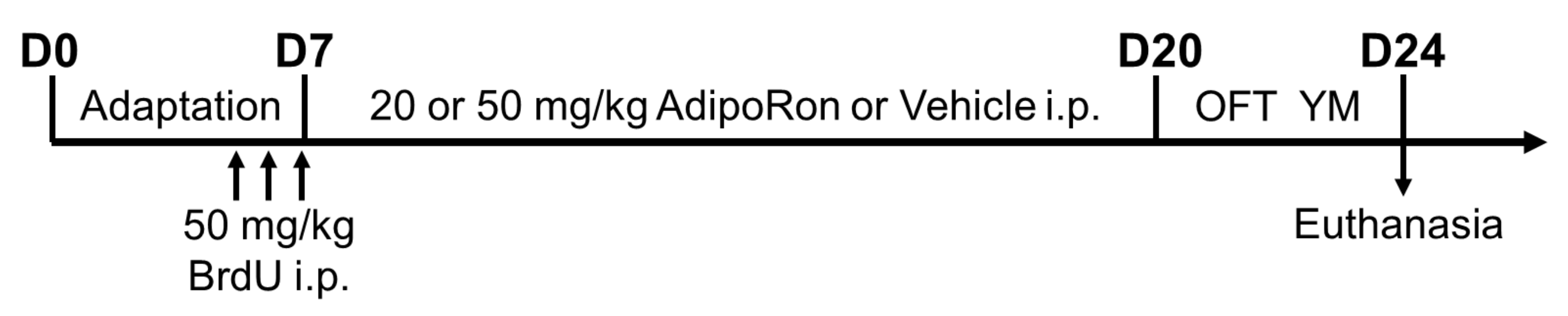
4.2. Drug Preparation and Treatment

AdipoRon (Sigma-Aldrich, MO, USA) was first dissolved in DMSO at 40 mg/mL, then suspended in 0.5% carboxymethylcellulose salt (Sigma-Aldrich, St. Louis, MO, USA) [35,78]. Animals received the AdipoRon treatment (20 or 50 mg/kg i.p.) or the vehicle as control treatment continuously for 14 days. Mice were injected with bromodeoxyuridine i.p. (BrdU: 50 mg/kg dissolved in 0.9% saline) before AdipoRon treatment to study newborn cell survival. The day after the last treatment, mice were subjected to an open field test and Y-maze task, as illustrated in Figure 7.

4.3. Open Field Test

Mice were first brought to the testing room for 2-hr adaptation. Each mouse was allowed to explore the open field (L × W × H: 40 × 40 × 30 cm3) for 10 min. Between trials, open field was cleaned by 70% ethanol wipe and two rounds of water wipes. The test box was dried before the next trial. Anxiety-like behavior and locomotor activity were analyzed using ANYMAZE software (Stoelting Co., Wood Dale, IL, USA). Locomotor activity was calculated as the total distance traveled over 10 min. Thigmotaxis is linked to anxiety-like behaviors [79,80,81]. Wall-hugging is counted when the animal performed vertical rear against the wall supported by two hind paws [82]. Wall sniffing behavior was excluded. Defecation is another indicator of anxiety [83]. The number of fecal boli was counted.

4.4. Y-Maze Task

Spatial recognition memory was assessed by the Y-maze task [84]. Three identical arms (L × W × H: 10 × 6 × 8 cm3) in Y-maze were pre-designated as starting arm, familiar arm, and novel arm, respectively. Three arms were counterbalanced to avoid recognition bias. In the 10-min habituation session, a mouse was released in the designated starting arm and was allowed to explore the maze with a blocked novel arm. After a 4-hr intermission time, the animal re-explored the maze with the unblocked novel arm for 5 min. Time spent in each arm was hand-scored by the trained researcher in a sample blinded manner. The exploration index was calculated as (N − F)/(N + F) and presented on a −1 to 1 scale.

4.5. Tissue Preparation

Mice were deeply anaesthetized with isoflurane (Zoetis, Surrey, UK). Blood was transcardially collected by a 25-G needle with a 1 mL syringe. Samples were clotted for 30 min at room temperature. Blood sera were then collected by centrifugation at 1000× g for 20 min at 4 °C and then stored at −80 °C until ELISA measurements. Animals were rapidly perfused with 0.9% saline, followed by fixation with 4% paraformaldehyde (PFA) in 0.01 M phosphate-buffered saline (PBS). The isolated brains were post-fixed in 4% PFA at 4 °C overnight. The brains were then transferred to 30% sucrose until they sank. Coronal sections (1-in-6 series, 30-μm thickness) were obtained using a vibratome (Leica Biosystems, Heidelberg, Germany). The slices were temporarily stored in a cryoprotectant composed of 30% glycerol and 30% ethylene glycol at 4 °C until immunostaining.

4.6. Immunohistochemistry and Immunofluorescent Staining

Immunostaining was performed by the free-floating method, as previously performed [85]. For BrdU staining, the antigens were retrieved in the citric acid buffer (pH 6.0) at 95 °C for 15 min, then denatured in 2 N HCl for 30 min and neutralized by 0.1 M borate buffer (pH 8.5) for 15 min at room temperature. The sections were incubated overnight with mouse anti-BrdU antibody (1:1000; Roche Life Science, Upper Bavaria, Germany), and then incubated with the biotinylated goat anti-mouse IgG (1:200; Vector Laboratories, Burlingame, CA, USA) for 2 h at room temperature. BrdU staining was visualized with the peroxidase method using the VECTASTAIN® ABC kit (HRP) (Vector Laboratories, Burlingame, CA, USA) and the DAB peroxidase substrate kit (Vector Laboratories, Burlingame, CA, USA). For doublecortin (DCX) and Ki-67 staining, sections were incubated with mouse anti-DCX (1:200; Santa Cruz Biotechnology, Dallas, TX, USA) or rabbit anti-Ki67 (1:1000; abcam, Cambridge, UK) antibody, then secondary antibodies, the biotinylated goat anti-mouse, or goat anti-rabbit IgG (1:200; Vector Laboratories, Burlingame, CA, USA), and visualized using the peroxidase method.
Immunofluorescent co-labeling of BrdU and DCX was performed as previously described [5,86]. After antigen retrieval, sections were incubated with primary antibodies overnight and secondary antibodies for 2 h, including goat anti-rabbit IgG Alexa Fluor-488 and goat anti-mouse IgG Alexa Fluor-568. The mounted sections were coverslipped with the fluorescent mounting medium (Dako, Carpinteria, CA, USA).

4.7. Quantification of BrdU, Ki67, and DCX Immunopositive Cells

BrdU-, Ki67-, and DCX-positive cells were counted in the 1-in-6 series of nine sections (from bregma −1.34 to −3.80 mm), using an optical fractionator system (grid size: 55 μm × 55 μm, counting frame: 35 μm × 35 μm) of StereoInvestigator (MicroBrightfield Inc., Williston, VT, USA) as previously described [5]. Cells located in the DG sub-granular zone and granular cell layer were counted, whereas those located in the uppermost focal plane were excluded. Quantification was performed in a sample-blinded manner.

4.8. Quantification of DCX/BrdU Co-Labeled Cells

Images of six sections from each animal were captured using LSM 800 confocal laser scanning microscope (Carl Zeiss Microscopy, White, Plains, NY, USA). Fifty BrdU positive cells were randomly selected to confirm the BrdU/DCX co-labeled ratio as an indicator for neuronal differentiation. Quantification of the co-labeling was performed in a sample blinded manner.

4.9. Measurement of Serum Glucose Levels, Corticosterone, Adiponectin and Brain-Derived Neurotrophic Factor (BDNF) Levels

Blood glucose from tail blood was measured using the Accu-Chek© Performa glucometer (Roche, Sydney, Australia) before sacrificing the animals. Serum levels of adiponectin, BDNF, and corticosterone were determined by commercial ELISA kits, including corticosterone ELISA kit (Assay Designs, Enzo Life Sciences, Lausen, Switzerland), mouse adiponectin ELISA kit (Immunodiagnostics, Hong Kong, China), and the total BDNF Quantikine ELISA kit (R & D system, Minneapolis, MN, USA) according to the manufacturer’s instructions.

4.10. Statistical Analyses

One-way ANOVA with Tukey’s posthoc test was performed using GraphPad Prism version 8.0.0 for Windows (GraphPad Software, San Diego, CA, USA). A probability (P) value of less than 0.05 is considered statistically significant. Data were shown as mean ± SEM.

5. Conclusions

In summary, our data demonstrated that a low-dose AdipoRon treatment promoted hippocampal cell proliferation and increased serum adiponectin and BDNF levels. In contrast, a high-dose of AdipoRon treatment was deleterious to hippocampal function, as evidenced by learning and memory deficits in hippocampus-dependent tasks. The ability to cross the BBB may allow AdipoRon to promote cell proliferation directly, though a low-dose AdipoRon treatment increased BDNF levels to elicit its neurotrophic effect indirectly. On the other hand, reduced neurotrophic factors and aberrant hippocampal neurogenesis might contribute to spatial memory with a high-dose AdipoRon treatment. The present study provides evidence of the beneficial effects of AdipoRon treatment on inducing hippocampal cell proliferation and the potential side effects of a high dose AdipoRon treatment on hippocampal neurogenesis impairment with learning and memory deficits.

Author Contributions

Conceptualization, T.H.L., B.R.C., K.L., P.M.-F.S., A.X., K.-F.S. and S.-y.Y.; methodology, T.H.L., K.L., P.M.-F.S. and S.-y.Y.; formal analysis, T.H.L.; investigation, T.H.L. and S.-y.Y.; resources, S.-y.Y.; data curation, T.H.L.; writing—original draft preparation, T.H.L.; writing—review and editing, T.H.L., B.RC., H.v.P., K.L., P.M.-F.S., A.X., K.-F.S. and S.-y.Y.; visualization, T.H.L.; supervision, S.-y.Y.; project administration, S.-y.Y.; funding acquisition, B.RC., H.v.P., A.X., K.-F.S., and S.-y.Y. All authors have read and agreed to the published version of the manuscript.

Funding

This research is funded by Hong Kong General Research Fund (GRF) (Early career scheme): 25100217, and GRF: 15100018, National Natural Science Foundation of China (Young Investigator Scheme): 81801346.

Institutional Review Board Statement

The study was conducted according to the guidelines of the Department of Health HKSAR, and approved by the Animal Subjects Ethics Sub-Committee of The Hong Kong Polytechnic University (ASESC: 16-17/10-RS-R-GRF).

Informed Consent Statement

Not applicable.

Data Availability Statement

The data that support the findings of this study are available from the corresponding author upon reasonable request.

Acknowledgments

The authors would like to thank the University Research Facility in Behavioral and Systems Neuroscience, The Hong Kong Polytechnic University, for providing research facilities and their technical support.

Conflicts of Interest

The authors declare no conflict of interest.

References

  1. Turer, A.T.; Scherer, P.E. Adiponectin: Mechanistic insights and clinical implications. Diabetologia 2012, 55, 2319–2326. [Google Scholar] [CrossRef] [Green Version]
  2. Kusminski, C.M.; McTernan, P.G.; Schraw, T.; Kos, K.; O’Hare, J.P.; Ahima, R.; Kumar, S.; Scherer, P.E. Adiponectin complexes in human cerebrospinal fluid: Distinct complex distribution from serum. Diabetologia 2007, 50, 634–642. [Google Scholar] [CrossRef]
  3. Schön, M.; Kovaničová, Z.; Košutzká, Z.; Nemec, M.; Tomková, M.; Jacková, L.; Máderová, D.; Slobodová, L.; Valkovič, P.; Ukropec, J.; et al. Effects of running on adiponectin, insulin and cytokines in cerebrospinal fluid in healthy young individuals. Sci. Rep. 2019, 9, 1959. [Google Scholar] [CrossRef] [Green Version]
  4. Kubota, N.; Yano, W.; Kubota, T.; Yamauchi, T.; Itoh, S.; Kumagai, H.; Kozono, H.; Takamoto, I.; Okamoto, S.; Shiuchi, T.; et al. Adiponectin Stimulates AMP-Activated Protein Kinase in the Hypothalamus and Increases Food Intake. Cell Metab. 2007, 6, 55–68. [Google Scholar] [CrossRef] [PubMed] [Green Version]
  5. Yau, S.Y.; Li, A.; Hoo, R.L.C.; Ching, Y.P.; Christie, B.R.; Lee, T.M.C.; Xu, A.; So, K.-F. Physical exercise-induced hippocampal neurogenesis and antidepressant effects are mediated by the adipocyte hormone adiponectin. Proc. Natl. Acad. Sci. USA 2014, 111, 15810–15815. [Google Scholar] [CrossRef] [PubMed] [Green Version]
  6. Cezaretto, A.; Suemoto, C.K.; Bensenor, I.; Lotufo, P.A.; de Almeida-Pititto, B.; Ferreira, S.R.G.; Aquino, E.M.L. Association of adiponectin with cognitive function precedes overt diabetes in the Brazilian Longitudinal Study of Adult Health: ELSA. Diabetol. Metab. Syndr. 2018, 10, 54. [Google Scholar] [CrossRef] [PubMed]
  7. Benavente, K.S.K.; Palmer, R.F.; Royall, D.R. Serum Adiponectin is Related to Dementia. J. Gerontol. Ser. A Boil. Sci. Med. Sci. 2019, 75, 779–783. [Google Scholar] [CrossRef] [PubMed]
  8. Ng, R.C.; Jian, M.; Ma, O.K.; Bunting, M.; Kwan, J.S.; Zhou, G.J.; Senthilkumar, K.; Iyaswamy, A.; Chan, P.K.; Li, M.; et al. Chronic oral administration of adipoRon reverses cognitive impairments and ameliorates neuropathology in an Alzheimer’s disease mouse model. Mol. Psychiatry 2020, 1–21. [Google Scholar] [CrossRef] [PubMed]
  9. Diniz, B.S.; Teixeira, A.L.; Campos, A.C.; Miranda, A.S.; Rocha, N.P.; Talib, L.L.; Gattaz, W.F.; Forlenza, O.V. Reduced serum levels of adiponectin in elderly patients with major depression. J. Psychiatr. Res. 2012, 46, 1081–1085. [Google Scholar] [CrossRef]
  10. Wang, X.-Q.; Li, W.-H.; Tang, Y.-H.; Wu, L.-F.; Zeng, G.-R.; Wang, Y.-H.; Cheng, Z.; Jiang, D.-J. The correlation between adiponectin and FGF9 in depression disorder. Brain Res. 2020, 1729, 146596. [Google Scholar] [CrossRef]
  11. Yamauchi, T.; Iwabu, M.; Okada-Iwabu, M.; Kadowaki, T. Adiponectin receptors: A review of their structure, function and how they work. Best Pr. Res. Clin. Endocrinol. Metab. 2014, 28, 15–23. [Google Scholar] [CrossRef]
  12. Thundyil, J.; Pavlovski, D.; Sobey, C.G.; Arumugam, T.V. Adiponectin receptor signalling in the brain. Br. J. Pharmacol. 2011, 165, 313–327. [Google Scholar] [CrossRef] [PubMed] [Green Version]
  13. Liu, J.; Guo, M.; Zhang, D.; Cheng, S.-Y.; Liu, M.; Ding, J.; Scherer, P.E.; Liu, F.; Lu, X.-Y. Adiponectin is critical in determining susceptibility to depressive behaviors and has antidepressant-like activity. Proc. Natl. Acad. Sci. USA 2012, 109, 12248–12253. [Google Scholar] [CrossRef] [Green Version]
  14. Zhang, D.; Wang, X.; Lu, X.-Y. Adiponectin Exerts Neurotrophic Effects on Dendritic Arborization, Spinogenesis, and Neurogenesis of the Dentate Gyrus of Male Mice. Endocrinology 2016, 157, 2853–2869. [Google Scholar] [CrossRef] [Green Version]
  15. Wang, M.; Jo, J.; Song, J. Adiponectin improves long-term potentiation in the 5XFAD mouse brain. Sci. Rep. 2019, 9, 1–8. [Google Scholar] [CrossRef] [Green Version]
  16. Bloemer, J.; Pinky, P.D.; Smith, W.D.; Bhattacharya, D.; Chauhan, A.; Govindarajulu, M.; Hong, H.; Dhanasekaran, M.; Judd, R.; Amin, R.H.; et al. Adiponectin Knockout Mice Display Cognitive and Synaptic Deficits. Front. Endocrinol. 2019, 10, 819. [Google Scholar] [CrossRef] [PubMed] [Green Version]
  17. Zhang, D.; Wang, X.; Wang, B.; Garza, J.C.; Fang, X.; Wang, J.; Scherer, P.E.; Brenner, R.; Zhang, W.; Lu, X.-Y. Adiponectin regulates contextual fear extinction and intrinsic excitability of dentate gyrus granule neurons through AdipoR2 receptors. Mol. Psychiatry 2016, 22, 1044–1055. [Google Scholar] [CrossRef] [PubMed] [Green Version]
  18. Ng, R.C.-L.; Cheng, O.-Y.; Jian, M.; Kwan, J.S.-C.; Ho, P.W.-L.; Cheng, K.K.-Y.; Yeung, P.K.K.; Zhou, L.L.; Hoo, R.L.-C.; Chung, S.K.; et al. Chronic adiponectin deficiency leads to Alzheimer’s disease-like cognitive impairments and pathologies through AMPK inactivation and cerebral insulin resistance in aged mice. Mol. Neurodegener. 2016, 11, 1–16. [Google Scholar] [CrossRef] [PubMed] [Green Version]
  19. Kim, M.W.; Abid, N.B.; Jo, M.H.; Jo, M.G.; Yoon, G.H.; Kim, M.O. Suppression of adiponectin receptor 1 promotes memory dysfunction and Alzheimer’s disease-like pathologies. Sci. Rep. 2017, 7, 12435. [Google Scholar] [CrossRef]
  20. Lee, T.H.-Y.; Cheng, K.K.Y.; Hoo, R.L.-C.; Siu, P.M.; Yau, S.-Y. The Novel Perspectives of Adipokines on Brain Health. Int. J. Mol. Sci. 2019, 20, 5638. [Google Scholar] [CrossRef] [Green Version]
  21. Lee, T.H.-Y.; Yau, S.-Y. From Obesity to Hippocampal Neurodegeneration: Pathogenesis and Non-Pharmacological Interventions. Int. J. Mol. Sci. 2020, 22, 201. [Google Scholar] [CrossRef] [PubMed]
  22. Knittle, J.L.; Timmers, K.; Ginsberg-Fellner, F.; Brown, R.E.; Katz, D.P. The growth of adipose tissue in children and adolescents. Cross-sectional and longitudinal studies of adipose cell number and size. J. Clin. Investig. 1979, 63, 239–246. [Google Scholar] [CrossRef] [Green Version]
  23. Spalding, K.L.; Arner, E.; Westermark, P.O.; Bernard, S.; Buchholz, B.A.; Bergmann, O.; Blomqvist, L.; Hoffstedt, J.; Näslund, E.; Britton, T.; et al. Dynamics of fat cell turnover in humans. Nat. Cell Biol. 2008, 453, 783–787. [Google Scholar] [CrossRef]
  24. Boitard, C.; Etchamendy, N.; Sauvant, J.; Aubert, A.; Tronel, S.; Marighetto, A.; Layé, S.; Ferreira, G. Juvenile, but not adult exposure to high-fat diet impairs relational memory and hippocampal neurogenesis in mice. Hippocampus 2012, 22, 2095–2100. [Google Scholar] [CrossRef]
  25. Boitard, C.; Parkes, S.L.; Cavaroc, A.; Tantot, F.; Castanon, N.; Layé, S.; Tronel, S.; Pacheco-Lopez, G.; Coutureau, E.; Ferreira, G. Switching Adolescent High-Fat Diet to Adult Control Diet Restores Neurocognitive Alterations. Front. Behav. Neurosci. 2016, 10, 225. [Google Scholar] [CrossRef] [PubMed] [Green Version]
  26. O’Leary, J.D.; Hoban, A.E.; Murphy, A.; O’Leary, O.F.; Cryan, J.F.; Nolan, Y.M. Differential effects of adolescent and adult-initiated exercise on cognition and hippocampal neurogenesis. Hippocampus 2019, 29, 352–365. [Google Scholar] [CrossRef] [PubMed]
  27. O’Leary, J.D.; Hoban, A.E.; Cryan, J.F.; O’Leary, O.F.; Nolan, Y.M. Differential effects of adolescent and adult-initiated voluntary exercise on context and cued fear conditioning. Neuropharmacology 2019, 145, 49–58. [Google Scholar] [CrossRef]
  28. Hotta, K.; Funahashi, T.; Arita, Y.; Takahashi, M.; Matsuda, M.; Okamoto, Y.; Iwahashi, H.; Kuriyama, H.; Ouchi, N.; Maeda, K.; et al. Plasma Concentrations of a Novel, Adipose-Specific Protein, Adiponectin, in Type 2 Diabetic Patients. Arter. Thromb. Vasc. Biol. 2000, 20, 1595–1599. [Google Scholar] [CrossRef] [PubMed] [Green Version]
  29. Berg, A.H.; Combs, T.P.; Du, X.; Brownlee, M.; Scherer, P.E. The adipocyte-secreted protein Acrp30 enhances hepatic insulin action. Nat. Med. 2001, 7, 947–953. [Google Scholar] [CrossRef]
  30. Fruebis, J.; Tsao, T.S.; Javorschi, S.; Ebbets-Reed, D.; Erickson, M.R.; Yen, F.T.; Bihain, B.E.; Lodish, H.F. Proteolytic cleavage product of 30-kDa adipocyte complement-related protein increases fatty acid oxidation in muscle and causes weight loss in mice. Proc. Natl. Acad. Sci. USA 2001, 98, 2005–2010. [Google Scholar] [CrossRef]
  31. Yamauchi, T.; Kamon, J.; Waki, H.; Terauchi, Y.; Kubota, N.; Hara, K.; Mori, Y.; Ide, T.; Murakami, K.; Tsuboyamakasaoka, N.; et al. The fat-derived hormone adiponectin reverses insulin resistance associated with both lipoatrophy and obesity. Nat. Med. 2001, 7, 941–946. [Google Scholar] [CrossRef]
  32. Yamauchi, T.; Nio, Y.; Maki, T.; Kobayashi, M.; Takazawa, T.; Iwabu, M.; Okada-Iwabu, M.; Kawamoto, S.; Kubota, N.; Kubota, T.; et al. Targeted disruption of AdipoR1 and AdipoR2 causes abrogation of adiponectin binding and metabolic actions. Nat. Med. 2007, 13, 332–339. [Google Scholar] [CrossRef] [PubMed]
  33. Okada-Iwabu, M.; Yamauchi, T.; Iwabu, M.; Honma, T.; Hamagami, K.-I.; Matsuda, K.; Yamaguchi, M.; Tanabe, H.; Kimura-Someya, T.; Shirouzu, M.; et al. A small-molecule AdipoR agonist for type 2 diabetes and short life in obesity. Nat. Cell Biol. 2013, 503, 493–499. [Google Scholar] [CrossRef]
  34. Yamashita, T.; Lakota, K.; Taniguchi, T.; Yoshizaki, A.; Sato, S.; Hong, W.; Zhou, X.; Sodin-Semrl, S.; Fang, F.; Asano, Y.; et al. An orally-active adiponectin receptor agonist mitigates cutaneous fibrosis, inflammation and microvascular pathology in a murine model of systemic sclerosis. Sci. Rep. 2018, 8, 1–14. [Google Scholar] [CrossRef] [Green Version]
  35. Fairaq, A.; Shawky, N.M.; Osman, I.; Pichavaram, P.; Segar, L. AdipoRon, an adiponectin receptor agonist, attenuates PDGF-induced VSMC proliferation through inhibition of mTOR signaling independent of AMPK: Implications toward suppression of neointimal hyperplasia. Pharmacol. Res. 2017, 119, 289–302. [Google Scholar] [CrossRef] [PubMed] [Green Version]
  36. Xiao, W.-Z.; Zhang, L. Adiponectin receptor agonist AdipoRon relieves endotoxin-induced acute hepatitis in mice. Chin. Med. J. 2019, 132, 2438–2445. [Google Scholar] [CrossRef]
  37. Zhang, Y.; Zhao, J.; Li, R.; Lau, W.B.; Yuan, Y.-X.; Liang, B.; Li, R.; Gao, E.-H.; Koch, W.J.; Ma, X.-L.; et al. AdipoRon, the first orally active adiponectin receptor activator, attenuates postischemic myocardial apoptosis through both AMPK-mediated and AMPK-independent signalings. Am. J. Physiol. Metab. 2015, 309, E275–E282. [Google Scholar] [CrossRef] [PubMed] [Green Version]
  38. Nicolas, S.; Debayle, D.; Béchade, C.; Maroteaux, L.; Gay, A.-S.; Bayer, P.; Heurteaux, C.; Guyon, A.; Chabry, J. Adiporon, an adiponectin receptor agonist acts as an antidepressant and metabolic regulator in a mouse model of depression. Transl. Psychiatry 2018, 8, 1–11. [Google Scholar] [CrossRef]
  39. Yau, S.-Y.; Lee, T.H.-Y.; Li, A.; Xu, A.; So, K.-F. Adiponectin Mediates Running-Restored Hippocampal Neurogenesis in Streptozotocin-Induced Type 1 Diabetes in Mice. Front. Neurosci. 2018, 12, 679. [Google Scholar] [CrossRef] [Green Version]
  40. Wang, P.; Liang, Y.; Chen, K.; Yau, S.-Y.; Sun, X.; Cheng, K.K.-Y.; Xu, A.; So, K.-F.; Li, A. Potential Involvement of Adiponectin Signaling in Regulating Physical Exercise-Elicited Hippocampal Neurogenesis and Dendritic Morphology in Stressed Mice. Front. Cell. Neurosci. 2020, 14, 189. [Google Scholar] [CrossRef]
  41. Deng, W.; Aimone, J.B.; Gage, F.H. New neurons and new memories: How does adult hippocampal neurogenesis affect learning and memory? Nat. Rev. Neurosci. 2010, 11, 339–350. [Google Scholar] [CrossRef] [PubMed]
  42. Saaltink, D.-J.; Vreugdenhil, E. Stress, glucocorticoid receptors, and adult neurogenesis: A balance between excitation and inhibition? Cell. Mol. Life Sci. 2014, 71, 2499–2515. [Google Scholar] [CrossRef] [PubMed]
  43. Zhang, D.; Guo, M.; Zhang, W.; Lu, X.Y. Adiponectin stimulates proliferation of adult hippocampal neural stem/progenitor cells through activation of p38 mitogen-activated protein kinase (p38MAPK)/glycogen synthase kinase 3beta (GSK-3beta)/beta-catenin signaling cascade. J. Biol. Chem. 2011, 286, 44913–44920. [Google Scholar] [CrossRef] [Green Version]
  44. Islam, O.; Loo, T.X.; Heese, K. Brain-Derived Neurotrophic Factor (BDNF) has Proliferative Effects on Neural Stem Cells through the Truncated TRK-B Receptor, MAP Kinase, AKT, and STAT-3 Signaling Pathways. Curr. Neurovascular Res. 2009, 6, 42–53. [Google Scholar] [CrossRef]
  45. Lang, U.E.; Hellweg, R.; Seifert, F.; Schubert, F.; Gallinat, J. Correlation Between Serum Brain-Derived Neurotrophic Factor Level and An In Vivo Marker of Cortical Integrity. Biol. Psychiatry 2007, 62, 530–535. [Google Scholar] [CrossRef]
  46. Pan, W.; Banks, W.A.; Fasold, M.B.; Bluth, J.; Kastin, A.J. Transport of brain-derived neurotrophic factor across the blood-brain barrier. Neuropharmacology 1998, 37, 1553–1561. [Google Scholar] [CrossRef]
  47. Bergami, M.; Rimondini, R.; Santi, S.; Blum, R.; Götz, M.; Canossa, M. Deletion of TrkB in adult progenitors alters newborn neuron integration into hippocampal circuits and increases anxiety-like behavior. Proc. Natl. Acad. Sci. USA 2008, 105, 15570–15575. [Google Scholar] [CrossRef] [PubMed] [Green Version]
  48. Liu, B.; Liu, J.; Wang, J.-G.; Liu, C.-L.; Yan, H. AdipoRon improves cognitive dysfunction of Alzheimer’s disease and rescues impaired neural stem cell proliferation through AdipoR1/AMPK pathway. Exp. Neurol. 2020, 327, 113249. [Google Scholar] [CrossRef] [PubMed]
  49. Yamauchi, T.; Kamon, J.; Ito, Y.; Tsuchida, A.; Yokomizo, T.; Kita, S.; Sugiyama, T.; Miyagishi, M.; Hara, K.; Tsunoda, M.; et al. Cloning of adiponectin receptors that mediate antidiabetic metabolic effects. Nat. Cell Biol. 2003, 423, 762–769. [Google Scholar] [CrossRef]
  50. Šišková, Z.; Justus, D.; Kaneko, H.; Friedrichs, D.; Henneberg, N.; Beutel, T.; Pitsch, J.; Schoch, S.; Becker, A.; von der Kammer, H.; et al. Dendritic Structural Degeneration Is Functionally Linked to Cellular Hyperexcitability in a Mouse Model of Alzheimer’s Disease. Neuron 2014, 84, 1023–1033. [Google Scholar] [CrossRef] [Green Version]
  51. Jiang, M.; Lee, C.L.; Smith, K.L.; Swann, J.W. Spine Loss and Other Persistent Alterations of Hippocampal Pyramidal Cell Dendrites in a Model of Early-Onset Epilepsy. J. Neurosci. 1998, 18, 8356–8368. [Google Scholar] [CrossRef] [PubMed] [Green Version]
  52. Musto, A.E.; Rosencrans, R.F.; Walker, C.P.; Bhattacharjee, S.; Raulji, C.M.; Belayev, L.; Fang, Z.; Gordon, W.C.; Bazan, N.G. Dysfunctional epileptic neuronal circuits and dysmorphic dendritic spines are mitigated by platelet-activating factor receptor antagonism. Sci Rep. 2016, 6, 30298. [Google Scholar] [CrossRef] [PubMed] [Green Version]
  53. Musto, A.E.; Gjorstrup, P.; Bazan, N.G. The omega-3 fatty acid-derived neuroprotectin D1 limits hippocampal hyperexcitability and seizure susceptibility in kindling epileptogenesis. Epilepsia 2011, 52, 1601–1608. [Google Scholar] [CrossRef]
  54. Palop., J.; Chin, J.; Roberson, E.D.; Wang, J.; Thwin, M.T.; Bien-Ly, N.; Yoo, J.; Ho, K.O.; Yu, G.Q.; Kreitzer, A.; et al. Aberrant excitatory neuronal activity and compensatory remodeling of inhibitory hippocampal circuits in mouse models of Alzheimer’s disease. Neuron 2007, 55, 697–711. [Google Scholar] [CrossRef] [Green Version]
  55. Minkeviciene, R.; Rheims, S.; Dobszay, M.B.; Zilberter, M.; Hartikainen, J.; Fulop, L.; Penke, B.; Zilberter, Y.; Harkany, T.; Pitkanen, A.; et al. Amyloid beta-induced neuronal hyperexcitability triggers progressive epilepsy. J. Neurosci. 2009, 29, 3453–3462. [Google Scholar] [CrossRef]
  56. Busche, M.A.; Eichhoff, G.; Adelsberger, H.; Abramowski, D.; Wiederhold, K.H.; Haass, C.; Staufenbiel, M.; Konnerth, A.; Garaschuk, O. Clusters of hyperactive neurons near amyloid plaques in a mouse model of Alzheimer’s disease. Science 2008, 321, 1686–1689. [Google Scholar] [CrossRef] [Green Version]
  57. Busche, M.A.; Chen, X.; Henning, H.A.; Reichwald, J.; Staufenbiel, M.; Sakmann, B.; Konnerth, A. Critical role of soluble amyloid-beta for early hippocampal hyperactivity in a mouse model of Alzheimer’s disease. Proc. Natl. Acad. Sci. USA 2012, 109, 8740–8745. [Google Scholar] [CrossRef] [PubMed] [Green Version]
  58. Seki, T.; Arai, Y. Age-related production of new granule cells in the adult dentate gyrus. NeuroReport 1995, 6, 2479–2482. [Google Scholar] [CrossRef]
  59. Hodes, G.E.; Yang, L.; van Kooy, J.; Santollo, J.; Shors, T.J. Prozac during puberty: Distinctive effects on neurogenesis as a function of age and sex. Neuroscience 2009, 163, 609–617. [Google Scholar] [CrossRef] [Green Version]
  60. Wang, S.J.; Lu, W.Y.; Liu, K.Y. Adiponectin receptor agonist AdipoRon suppresses adipogenesis in C3H10T1/2 cells through the adenosine monophosphateactivated protein kinase signaling pathway. Mol. Med. Rep. 2017, 16, 7163–7169. [Google Scholar] [CrossRef]
  61. Yamauchi, T.; Kamon, J.; Minokoshi, Y.; Ito, Y.; Waki, H.; Uchida, S.; Yamashita, S.; Noda, M.; Kita, S.; Ueki, K.; et al. Adiponectin stimulates glucose utilisation and fatty-acid oxidation by activating AMP-activated protein kinase. Nat. Med. 2002, 8, 1288–1295. [Google Scholar] [CrossRef]
  62. Domise, M.; Sauvé, F.; Didier, S.; Caillerez, R.; Bégard, S.; Carrier, S.; Colin, M.; Marinangeli, C.; Buée, L.; Vingtdeux, V. Neuronal AMP-activated protein kinase hyper-activation induces synaptic loss by an autophagy-mediated process. Cell Death Dis. 2019, 10, 1–15. [Google Scholar] [CrossRef] [Green Version]
  63. Wu, S.-B.; Wu, Y.-T.; Wu, T.-P.; Wei, Y.-H. Role of AMPK-mediated adaptive responses in human cells with mitochondrial dysfunction to oxidative stress. Biochim. Biophys. Acta Gen. Subj. 2014, 1840, 1331–1344. [Google Scholar] [CrossRef] [PubMed]
  64. Arsikin, K.; Kravic-Stevovic, T.; Jovanovic, M.; Ristic, B.; Tovilovic, G.; Zogovic, N.; Bumbasirevic, V.; Trajkovic, V.; Harhaji-Trajkovic, L. Autophagy-dependent and -independent involvement of AMP-activated protein kinase in 6-hydroxydopamine toxicity to SH-SY5Y neuroblastoma cells. Biochim. Biophys. Acta Mol. Basis Dis. 2012, 1822, 1826–1836. [Google Scholar] [CrossRef] [PubMed] [Green Version]
  65. Autry, A.E.; Adachi, M.; Nosyreva, E.D.; Na, E.S.; Los, M.F.; Cheng, P.-F.; Kavalali, E.T.; Monteggia, L.M. NMDA receptor blockade at rest triggers rapid behavioural antidepressant responses. Nat. Cell Biol. 2011, 475, 91–95. [Google Scholar] [CrossRef] [Green Version]
  66. Sairanen, M.; Lucas, G.; Ernfors, P.; Castrén, M.L.; Castrén, E. Brain-Derived Neurotrophic Factor and Antidepressant Drugs Have Different but Coordinated Effects on Neuronal Turnover, Proliferation, and Survival in the Adult Dentate Gyrus. J. Neurosci. 2005, 25, 1089–1094. [Google Scholar] [CrossRef] [PubMed] [Green Version]
  67. Heldt, S.A.; Stanek, L.; Chhatwal, J.P.; Ressler, K.J. Hippocampus-specific deletion of BDNF in adult mice impairs spatial memory and extinction of aversive memories. Mol. Psychiatry 2007, 12, 656–670. [Google Scholar] [CrossRef] [Green Version]
  68. Garthe, A.; Behr, J.; Kempermann, G. Adult-Generated Hippocampal Neurons Allow the Flexible Use of Spatially Precise Learning Strategies. PLoS ONE 2009, 4, e5464. [Google Scholar] [CrossRef] [Green Version]
  69. Paschke, L.; Zemleduch, T.; Rucinski, M.; Ziolkowska, A.; Szyszka, M.; Malendowicz, L.K. Adiponectin and adiponectin receptor system in the rat adrenal gland: Ontogenetic and physiologic regulation, and its involvement in regulating adrenocortical growth and steroidogenesis. Peptides 2010, 31, 1715–1724. [Google Scholar] [CrossRef] [PubMed]
  70. Perry, R.J.; Resch, J.M.; Douglass, A.M.; Madara, J.C.; Rabin-Court, A.; Kucukdereli, H.; Wu, C.; Song, J.D.; Lowell, B.B.; Shulman, G.I. Leptin’s hunger-suppressing effects are mediated by the hypothalamic-pituitary-adrenocortical axis in rodents. Proc. Natl. Acad. Sci. USA 2019, 116, 13670–13679. [Google Scholar] [CrossRef] [Green Version]
  71. Hare, B.D.; Beierle, J.A.; Toufexis, D.J.; Hammack, S.E.; Falls, W.A. Exercise-Associated Changes in the Corticosterone Response to Acute Restraint Stress: Evidence for Increased Adrenal Sensitivity and Reduced Corticosterone Response Duration. Neuropsychopharmacology 2014, 39, 1262–1269. [Google Scholar] [CrossRef] [Green Version]
  72. Liu, P.Z.; Nusslock, R. Exercise-Mediated Neurogenesis in the Hippocampus via BDNF. Front. Neurosci. 2018, 12, 52. [Google Scholar] [CrossRef] [Green Version]
  73. Neeper, S.A.; Góauctemez-Pinilla, F.; Choi, J.; Cotman, C. Exercise and brain neurotrophins. Nat. Cell Biol. 1995, 373, 109. [Google Scholar] [CrossRef]
  74. Szuhany, K.L.; Bugatti, M.; Otto, M.W. A meta-analytic review of the effects of exercise on brain-derived neurotrophic factor. J. Psychiatr. Res. 2015, 60, 56–64. [Google Scholar] [CrossRef] [PubMed] [Green Version]
  75. Miller, K.N.; Burhans, M.S.; Clark, J.P.; Howell, P.R.; Polewski, M.A.; de Muth, T.M.; Eliceiri, K.W.; Lindstrom, M.J.; Ntambi, J.M.; Anderson, R.M. Aging and caloric restriction impact adipose tissue, adiponectin, and circulating lipids. Aging Cell 2017, 16, 497–507. [Google Scholar] [CrossRef]
  76. Martinez-Huenchullan, S.F.; Maharjan, B.R.; Williams, P.F.; Tam, C.S.; McLennan, S.V.; Twigg, S.M. Differential metabolic effects of constant moderate versus high intensity interval training in high-fat fed mice: Possible role of muscle adiponectin. Physiol. Rep. 2018, 6, e13599. [Google Scholar] [CrossRef] [PubMed] [Green Version]
  77. Duman, R.S. Neurotrophic factors and regulation of mood: Role of exercise, diet and metabolism. Neurobiol. Aging 2005, 26, 88–93. [Google Scholar] [CrossRef]
  78. Zhuo, Y.; Hua, L.; Feng, B.; Jiang, X.; Li, J.; Jiang, D.; Huang, X.; Zhu, Y.; Li, Z.; Yan, L.; et al. Fibroblast growth factor 21 coordinates adiponectin to mediate the beneficial effects of low-protein diet on primordial follicle reserve. EBioMedicine 2019, 41, 623–635. [Google Scholar] [CrossRef] [PubMed] [Green Version]
  79. Costall, B.; Jones, B.; Kelly, M.; Naylor, R.; Tomkins, D. Exploration of mice in a black and white test box: Validation as a model of anxiety. Pharmacol. Biochem. Behav. 1989, 32, 777–785. [Google Scholar] [CrossRef]
  80. Borta, A.; Schwarting, R.K. Inhibitory avoidance, pain reactivity, and plus-maze behavior in Wistar rats with high versus low rearing activity. Physiol. Behav. 2005, 84, 387–396. [Google Scholar] [CrossRef] [PubMed]
  81. Bouwknecht, J.A.; Paylor, R. Pitfalls in the interpretation of genetic and pharmacological effects on anxiety-like behaviour in rodents. Behav. Pharmacol. 2008, 19, 385–402. [Google Scholar] [CrossRef]
  82. Sturman, O.; Germain, P.-L.; Bohacek, J. Exploratory rearing: A context- and stress-sensitive behavior recorded in the open-field test. Stress 2018, 21, 443–452. [Google Scholar] [CrossRef] [PubMed]
  83. Ramos, A. Animal models of anxiety: Do I need multiple tests? Trends Pharmacol. Sci. 2008, 29, 493–498. [Google Scholar] [CrossRef]
  84. Kraeuter, A.-K.; Guest, P.C.; Sarnyai, Z. The Y-Maze for Assessment of Spatial Working and Reference Memory in Mice. Methods Mol. Biol. 2019, 1916, 105–111. [Google Scholar] [CrossRef] [PubMed]
  85. Yau, S.-Y.; Lee, T.H.-Y.; Formolo, D.A.; Lee, W.-L.; Li, L.C.-K.; Siu, P.M.; Chan, C.C.H. Effects of Maternal Voluntary Wheel Running During Pregnancy on Adult Hippocampal Neurogenesis, Temporal Order Memory, and Depression-Like Behavior in Adult Female and Male Offspring. Front. Neurosci. 2019, 13, 470. [Google Scholar] [CrossRef] [PubMed]
  86. Yau, S.-Y.; Lau, B.W.-M.; Tong, J.-B.; Wong, R.; Ching, Y.-P.; Qiu, G.; Tang, S.-W.; Lee, T.M.; So, K.-F. Hippocampal Neurogenesis and Dendritic Plasticity Support Running-Improved Spatial Learning and Depression-Like Behaviour in Stressed Rats. PLoS ONE 2011, 6, e24263. [Google Scholar] [CrossRef] [PubMed] [Green Version]
Figure 1. Effects of AdipoRon on rodent behaviors. Administration of AdipoRon at a low dose (20 mg/kg) and high dose (50 mg/kg) for two weeks did not affect (A) locomotor activity and (B) mean speed traveled in the open field. Both dosages did not affect anxiety-like behaviors as indicated by (C) the percentage time spent at the center, (D) the number of vertical rears, and (E) the number of fecal boli in the open field. However, treatment with 50 mg/kg AdipoRon (high dose) impaired spatial recognition memory, as indicated by (F) reduced exploration index in the Y-maze task. The results were expressed as the mean ± SEM. n = 7 per group, ** p < 0.005 compared to the vehicle treated control.
Ijms 22 02068 g001
Figure 2. Effects of AdipoRon treatment on neural progenitor cell density. (A) Treatment with 20 mg/kg AdipoRon increased Ki67 positive cells. A total of eight sections were quantified in each sample. The results for the data were expressed as the mean ± SEM. n = 5 per group, * p < 0.05, ** p < 0.005 compared to the vehicle treated control. Representative image of Ki67 positive cells in the (B) control, (C) 20 mg/kg AdipoRon, and (D) 50 mg/kg AdipoRon treatment groups (Scale bars, 200 μm in 100×, 10 μm in 400×).
Ijms 22 02068 g002
Figure 3. Effects of AdipoRon treatment on the immature neuron density. (A) Treatment with 50 mg/kg AdipoRon significantly decreased DCX positive cells. A total of eight sections were quantified in each sample. The results were expressed as the mean ± SEM. n = 5 per group, * p < 0.05 compared to the control. Representative images of DCX-positive cells in (B) control animal, mice receiving (C) 20 mg/kg, and (D) 50 mg/kg AdipoRon treatments (Scale bars, 200 μm in 100×, 10 μm in 400×).
Ijms 22 02068 g003
Figure 4. Effects of AdipoRon treatment on newborn cell density and neuronal differentiation. (A) Treatment with 50 mg/kg AdipoRon significantly decreased cell survival. A total of eight sections were quantified in each sample. The results were expressed as the mean ± SEM. n = 5 per group, ** p < 0.005 compared to the vehicle-treated control. Representative image of BrdU positive cells in (B) control animal, (C) 20 mg/kg, and (D) 50 mg/kg AdipoRon treatment groups. (E) Treatment with 50 mg/kg AdipoRon significantly decreased neuronal differentiation in the dentate as estimated by the co-labeling ratio of BrdU and DCX positive cells. A total of 50 BrdU positive were selected from six sections. The results for the data were expressed as the mean ±SEM. n = 5 per group, * p < 0.05 compared to the vehicle-treated control. Representative image of BrdU/DCX-labeled cells in (F) control animal, (G) 20 mg/kg, and (H) 50 mg/kg AdipoRon treatment groups. * p < 0.05, ** p < 0.005 compared to the vehicle-treated control. (Scale bars, 200 μm in 100×, 10 μm in 400×).
Ijms 22 02068 g004
Figure 5. Effect of AdipoRon on blood levels of adiponectin, BDNF, corticosterone, and blood glucose levels. Treatment with 20 mg/kg AdipoRon levels significantly increased blood levels of (A) BDNF and (B) adiponectin levels, whereas treatment with 50 mg/kg AdipoRon showed opposite effects. (C) AdipoRon treatment increased serum levels of corticosterone but showed no effect on (D) blood glucose levels. The results were expressed as the mean ± SEM. n = 5 per group, * p < 0.05, ** p < 0.005 compared to the vehicle-treated control.
Ijms 22 02068 g005
Figure 6. Summary of AdipoRon actions on hippocampal function. A low-dose AdipoRon treatment significantly promoted hippocampal cell proliferation and increased serum levels of adiponectin and BDNF. Conversely, a high-dose AdipoRon treatment suppressed hippocampal neurogenesis, reduced serum levels of adiponectin and BDNF, leading to hippocampal-dependent memory deficits. Created with biorender.com.
Ijms 22 02068 g006
Figure 7. Timeline of AdipoRon treatment and behavioral tests. Before AdipoRon treatment, mice received three doses of BrdU injections (50 mg/kg i.p.) to study newborn cell survival. Mice have received AdipoRon treatment (20 or 50 mg/kg i.p.) or vehicle treatment continuously for 14 days. The day after, mice were subjected to the open field test and the Y-maze task on two consecutive days. The day after the Y-maze test, mice were sacrificed to collect blood samples for ELISA and fixed brain tissues for immunostaining.
Ijms 22 02068 g007
Publisher’s Note: MDPI stays neutral with regard to jurisdictional claims in published maps and institutional affiliations.

