Wearable Health Technology to Quantify the Functional Impact of Peripheral Neuropathy on Mobility in Parkinson’s Disease: A Systematic Review

 ,
,
Abstract
1. Introduction
2. Materials and Methods
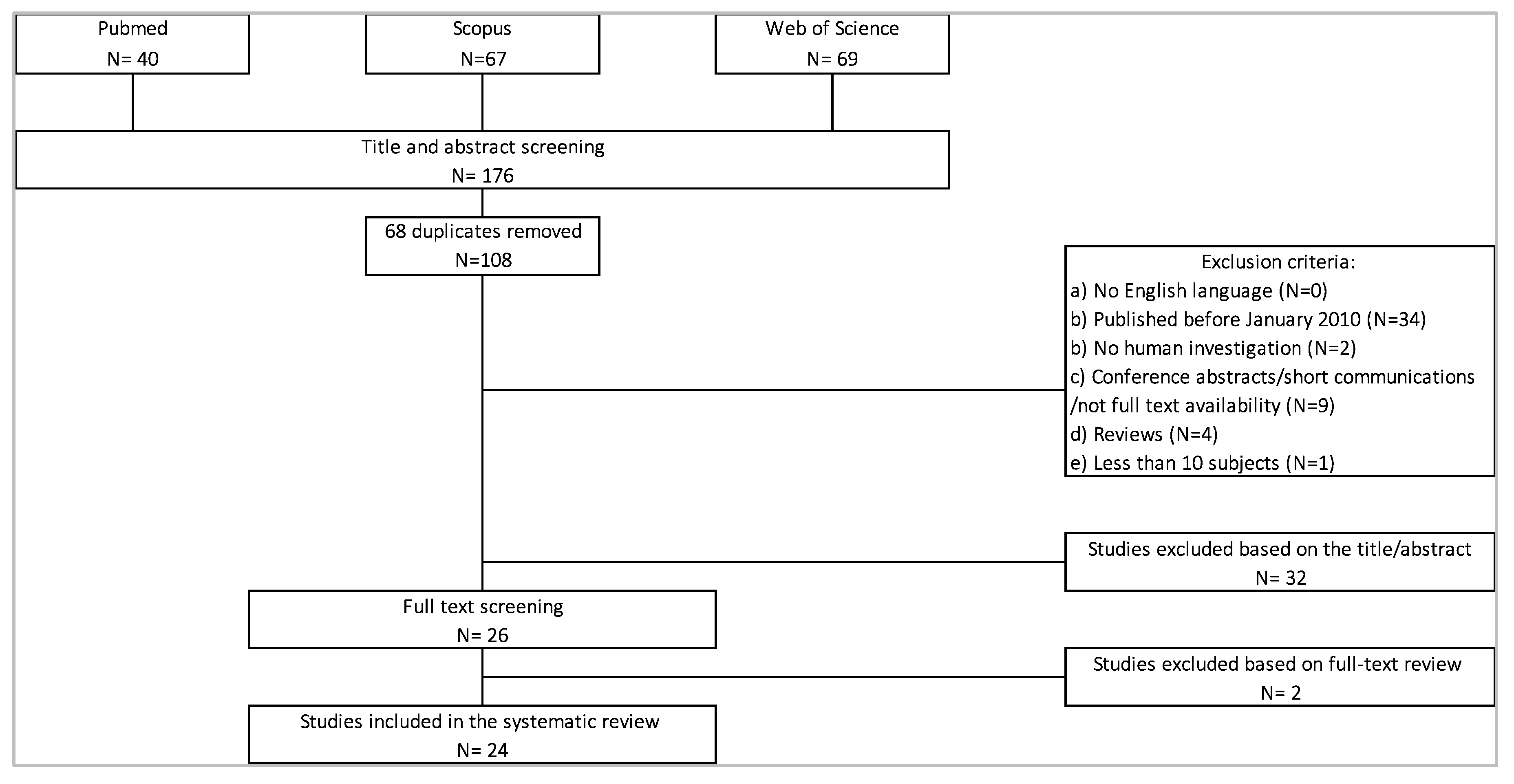
2.1. Search Strategy
- (1)
- To investigate the main characteristics and the most relevant gait and balance features for studying PNP with wearable technology, the following keywords were used: “peripheral neuropathy” OR “polyneuropathy” OR “small fiber neuropathy” AND “wearable sensor” OR “wearable” OR “mobile health technology” OR “technology assessment” OR “body-worn sensors” OR “inertial sensor” OR “inertial measurement unit” OR “acceleromet*” OR “gyroscope” AND “mobility” OR “gait” OR “balance” OR “postural balance” OR “postural stability” OR “postural strategies”.
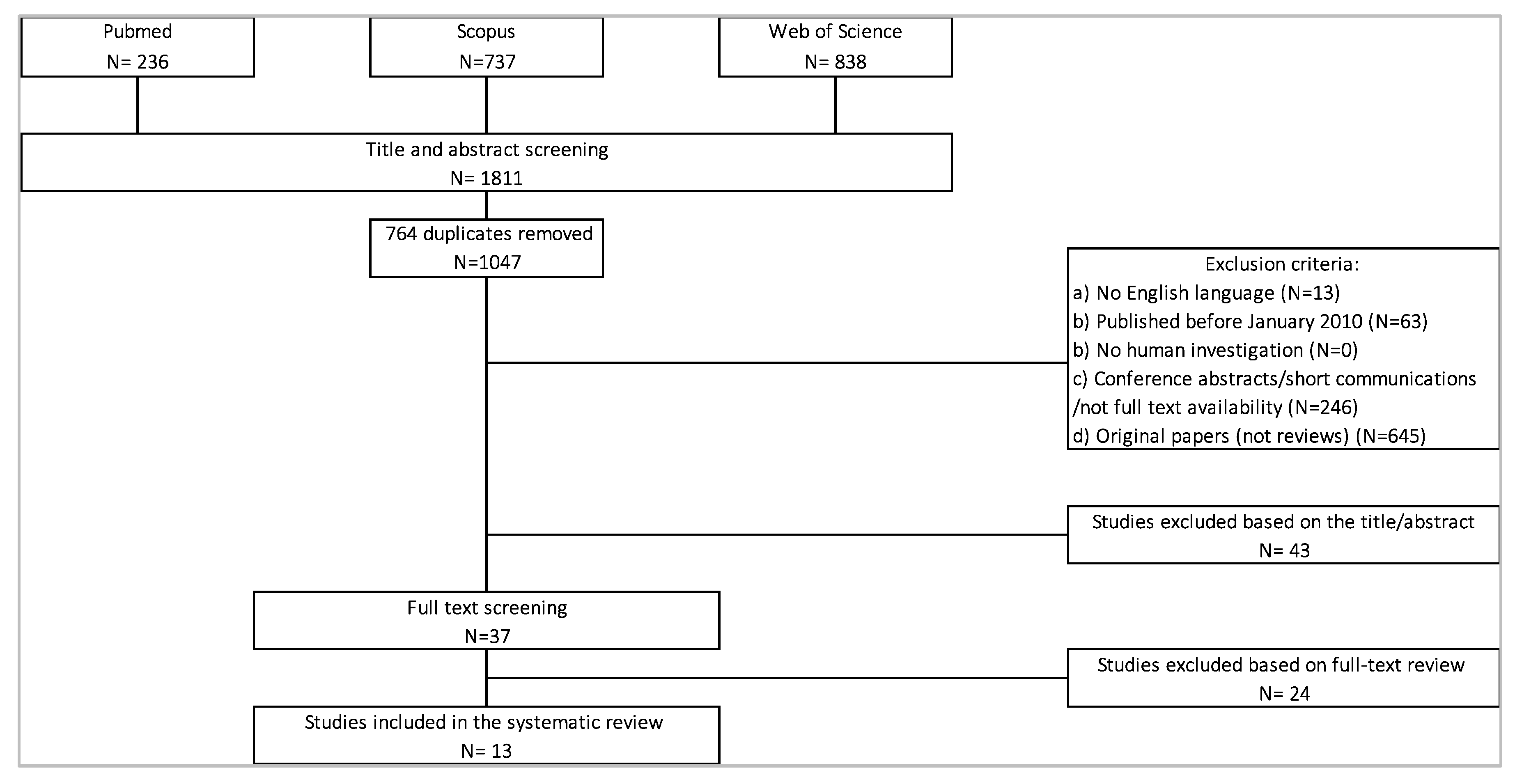
- (2)
- To investigate the main characteristics of wearable sensor assessments, and the most relevant gait and balance features in PD, the following keywords were used: “Parkinson” AND “wearable sensor” OR “wearable” OR “mobile health technology” OR “technology assessment” OR “inertial sensor” OR “inertial measurement unit” OR “acceleromet*” OR “gyroscope” AND “mobility” OR “gait” OR “balance” OR “postural balance”.
2.2. Selection Criteria
2.3. Data Extraction
3. Results
3.1. Sample Population Characteristics
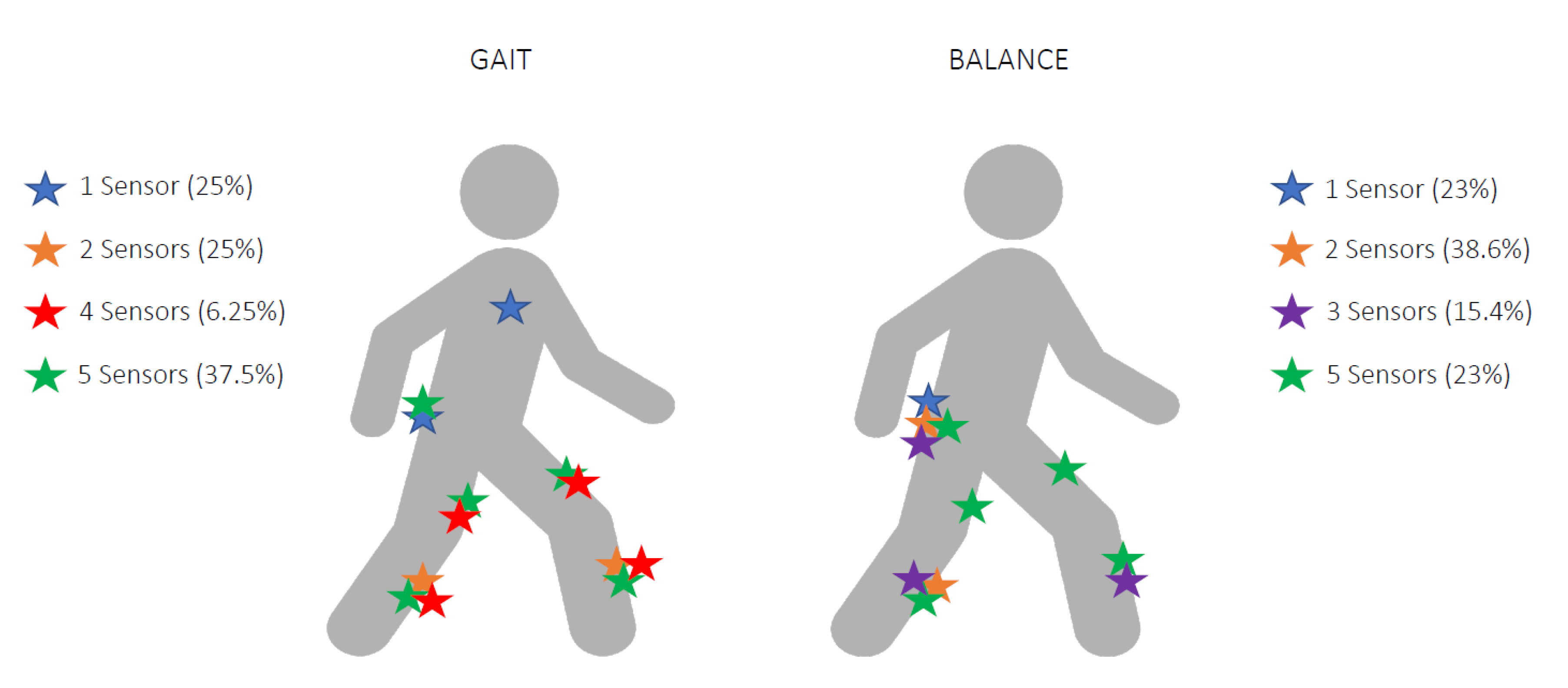
3.2. Sensor Type and Placement
3.2.1. PNP
3.2.2. PD
3.3. Parameters and Main Outcomes
3.3.1. PNP
- (1)
- Position of feet: Standing balance was assessed with feet together in eight (61.5%) studies, feet apart (spaced shoulder width) in two studies (15.3%), and both feet positions in one paper (7.6%), while two papers (15.3%) did not specify the position of the feet. In two studies patients were also asked to perform a semi-tandem position [33,43], while one other study introduced a detailed balance test protocol with single leg stance [24].
- (2)
- Open and closed eyes: Twelve studies (92.3%) analyzed balance with both open and closed eyes, and one study only used eyes-open condition [44].
- (3)
3.3.2. PD
4. Discussion
4.1. Gait and Walking Stability
4.2. Balance and Postural Stability
4.3. PNP Motor Assessment with Other Tools than Wearables
4.3.1. Gait Assessment
4.3.2. Balance and Postural Stability
5. Conclusions
- A combination of at least two sensors (one on lower back and one on at least one lower limb) may help gathering both PNP- and PD-specific features during gait and balance testing.
- Concerning parameters to analyze, particular attention should be given to gait speed, stride length, and gait variability. Gait variability may be particularly relevant for PNP-induced gait changes. Dual tasking assessments and irregular trajectories may unveil PNP-related gait deficits that are not visible during nonchallenging, single tasking walking conditions.
- Functional mobility tests (TUG test, functional reach test) can provide a comprehensive overview of function and mobility in PD patients with and without PNP.
- Balance tasks should include double leg stance with open- and with closed-eyes conditions.
- Total sway amplitude and AP and ML sway directions may be the most promising balance parameters to differentiate between PD and PD-PNP.
Author Contributions
Funding
Conflicts of Interest
Abbreviations
| ACC | Accelerometer |
| AP | Anterior-posterior |
| BBS | Berg Balance Scale |
| CFF | Cross-correlation function |
| CIDN | Chronic inflammatory demyelinating polyneuropathy |
| CIPN | Chemotherapy-induced peripheral neuropathy |
| COG | Center of gravity |
| COM | Center of mass |
| COP | Center of pressure |
| CV | Coefficient of variation |
| DFU | Diabetic foot ulcer |
| DM | Diabetes mellitus |
| DPN | Diabetic peripheral neuropathy (DPN) |
| EC | Closed eyes |
| EO | Open eyes |
| FAB | Fullerton Advance Balance test |
| Freq | Sample frequency |
| GYR | Gyroscop |
| HC | Healthy controls |
| iTUG | Instrumented, timed up-and-go test |
| MAG | Magnetometer |
| MeSH | Medical Subject Headings |
| ML | Medio-lateral |
| NeP-DPN | Neuropathic pain diabetic neuropathy |
| PD | Parkinson ’s Ddisease |
| PNP | Peripheral neuropathy |
| PNP-LL | Peripheral neuropathy of the lower limbs |
| PNP-PD | Peripheral neuropathy in Parkison disease |
| PNS | Peripheral nervous system |
| PRISMA | Preferred Reporting Items for Systematic Reviews and Meta-Analyses |
| RMS | Root mean square |
| SOT | Sensory organization test |
| TBS | Tinetti Balance scale |
| TUG | Timed up-and-go test |
Appendix A. Search Query
- (1)
- Pubmed
- -
- PN+wearable with MeSH
- -
- PD+wearable with MeSH terms
- (2)
- Scopus database
- -
- PN+wearables
- -
- PD+wearables
- (3)
- Web of Science database
- -
- PN+wearables
- -
- PD+wearables
References
- Poewe, W.; Seppi, K.; Tanner, C.M.; Halliday, G.M.; Brundin, P.; Volkmann, J.; Schrag, A.E.; Lang, A.E. Parkinson disease. Nat. Rev. Dis. Primers 2017, 3, 17013. [Google Scholar] [CrossRef] [PubMed]
- Spillantini, M.G.; Schmidt, M.L.; Lee, V.M.; Trojanowski, J.Q.; Jakes, R.; Goedert, M. Alpha-synuclein in Lewy bodies. Nature 1997, 388, 839–840. [Google Scholar] [CrossRef]
- Dennison, A.C.; Noorigian, J.V.; Robinson, K.M.; Fisman, D.N.; Cianci, H.J.; Moberg, P.; Bunting-Perry, L.; Martine, R.; Duda, J.; Stern, M.B. Falling in Parkinson Disease: Identifying and prioritizing risk factors in recurrent fallers. Am. J. Phys. Med. Rehabil. 2007, 86, 621–632. [Google Scholar] [CrossRef] [PubMed]
- Hammarlund, C.S.; Westergren, A.; Åström, I.; Edberg, A.-K.; Hagell, P. The Impact of Living with Parkinson’s Disease: Balancing within a Web of Needs and Demands. Parkinson Dis. 2018, 2018, 4598651. [Google Scholar] [CrossRef]
- Rodríguez-Leyva, I.; Calderón-Garcidueñas, A.L.; Jiménez-Capdeville, M.E.; Rentería-Palomo, A.A.; Hernandez-Rodriguez, H.G.; Valdés-Rodríguez, R.; Fuentes-Ahumada, C.; Torres-Álvarez, B.; Sepúlveda-Saavedra, J.; Soto-Domínguez, A.; et al. α-Synuclein inclusions in the skin of Parkinson’s disease and parkinsonism. Ann. Clin. Transl. Neurol. 2014, 1, 471–478. [Google Scholar] [CrossRef] [PubMed]
- Doppler, K.; Ebert, S.; Üçeyler, N.; Trenkwalder, C.; Ebentheuer, J.; Volkmann, J.; Sommer, C. Cutaneous neuropathy in Parkinson’s disease: A window into brain pathology. Acta Neuropathol. 2014, 128, 99–109. [Google Scholar] [CrossRef]
- Zis, P.; Sarrigiannis, P.G.; Rao, D.; Hewamadduma, C.; Hadjivassiliou, M. Chronic idiopathic axonal polyneuropathy: A systematic review. J. Neurol. 2016, 263, 1903–1910. [Google Scholar] [CrossRef]
- Karceski, S. Patient page. Parkinson disease and polyneuropathy. About Parkinson disease. Neurology 2011, 77, e132–e134. [Google Scholar] [CrossRef]
- Zis, P.; Grünewald, R.A.; Chaudhuri, R.K.; Hadjivassiliou, M. Peripheral neuropathy in idiopathic Parkinson’s disease: A systematic review. J. Neurol. Sci. 2017, 378, 204–209. [Google Scholar] [CrossRef]
- Toth, C.; Breithaupt, K.; Ge, S.; Duan, Y.; Terris, J.M.; Thiessen, A.; Wiebe, S.; Zochodne, D.W.; Suchowersky, O. Levodopa, methylmalonic acid, and neuropathy in idiopathic Parkinson disease. Ann. Neurol. 2010, 68, 28–36. [Google Scholar] [CrossRef]
- Toth, C.; Brown, M.S.; Furtado, S.; Suchowersky, O.; Zochodne, D. Neuropathy as a potential complication of levodopa use in Parkinson’s disease. Mov. Disord. 2008, 23, 1850–1859. [Google Scholar] [CrossRef] [PubMed]
- Ceravolo, R.; Cossu, G.; Bandettini di Poggio, M.; Santoro, L.; Barone, P.; Zibetti, M.; Frosini, D.; Nicoletti, V.; Manganelli, F.; Iodice, R.; et al. Neuropathy and levodopa in Parkinson’s disease: Evidence from a multicenter study. Mov. Disord. 2013, 28, 1391–1397. [Google Scholar] [CrossRef] [PubMed]
- DeMott, T.K.; Richardson, J.K.; Thies, S.B.; Ashton-Miller, J.A. Falls and Gait Characteristics Among Older Persons with Peripheral Neuropathy. Am. J. Phys. Med. Rehabil. 2007, 86, 125–132. [Google Scholar] [CrossRef]
- Beaulieu, M.L.; Müller, M.; Bohnen, N.I. Peripheral neuropathy is associated with more frequent falls in Parkinson’s disease. Parkinsonism Relat. Disord. 2018, 54, 46–50. [Google Scholar] [CrossRef]
- Warmerdam, E.; Hausdorff, J.M.; Atrsaei, A.; Zhou, Y.; Mirelman, A.; Aminian, K.; Espay, A.J.; Hansen, C.; Evers, L.J.W.; Keller, A.; et al. Long-term unsupervised mobility assessment in movement disorders. Lancet Neurol. 2020, 19, 462–470. [Google Scholar] [CrossRef]
- Godinho, C.; Domingos, J.; Cunha, G.V.; Santos, A.T.; Fernandes, R.M.; Abreu, D.; Gonçalves, N.; Matthews, H.; Isaacs, T.; Duffen, J.; et al. A systematic review of the characteristics and validity of monitoring technologies to assess Parkinson’s disease. J. Neuroeng. Rehabil. 2016, 13, 24. [Google Scholar] [CrossRef]
- Esser, P.; Collett, J.; Maynard, K.; Steins, D.; Hillier, A.; Buckingham, J.; Tan, G.D.; King, L.; Dawes, H. Single Sensor Gait Analysis to Detect Diabetic Peripheral Neuropathy: A Proof of Principle Study. Diabetes Metab. J. 2018, 42, 82–86. [Google Scholar] [CrossRef] [PubMed]
- Kang, G.E.; Najafi, B. Sensor-Based Daily Physical Activity: Towards Prediction of the Level of Concern about Falling in Peripheral Neuropathy. Sensors 2020, 20, 505. [Google Scholar] [CrossRef]
- Loprinzi, P.D.; Crush, E. Sensory Impairment, Functional Balance and Physical Activity with All-Cause Mortality. J. Phys. Act. Health 2016, 13, 980–987. [Google Scholar] [CrossRef]
- Moher, D.; PRISMA-P Group; Shamseer, L.; Clarke, M.; Ghersi, D.; Liberati, A.; Petticrew, M.; Shekelle, P.; Stewart, L.A. Preferred reporting items for systematic review and meta-analysis protocols (PRISMA-P) 2015 statement. Syst. Rev. 2015, 4, 1. [Google Scholar] [CrossRef]
- Morgan, C.; Rolinski, M.; McNaney, R.; Jones, B.; Rochester, L.; Maetzler, W.; Craddock, I.; Whone, A.L. Systematic Review Looking at the Use of Technology to Measure Free-Living Symptom and Activity Outcomes in Parkinson’s Disease in the Home or a Home-like Environment. J. Parkinson Dis. 2020, 10, 429–454. [Google Scholar] [CrossRef] [PubMed]
- Rovini, E.; Maremmani, C.; Cavallo, F. Automated Systems Based on Wearable Sensors for the Management of Parkinson’s Disease at Home: A Systematic Review. Telemed. J. E Health 2018, 25, 167–183. [Google Scholar] [CrossRef] [PubMed]
- Del Din, S.; Godfrey, A.; Mazzà, C.; Lord, S.; Rochester, L. Free-living monitoring of Parkinson’s disease: Lessons from the field. Mov. Disord. 2016, 31, 1293–1313. [Google Scholar] [CrossRef] [PubMed]
- Findling, O.; Van Der Logt, R.; Nedeltchev, K.; Achtnichts, L.; Allum, J.H.J. A comparison of balance control during stance and gait in patients with inflammatory and non-inflammatory polyneuropathy. PLoS ONE 2018, 13, e0191957. [Google Scholar] [CrossRef]
- Vienne-Jumeau, A.; Barrois, R.P.; Buffat, S.; Ricard, D.; Vidal, P.-P. Inertial Sensors to Assess Gait Quality in Patients with Neurological Disorders: A Systematic Review of Technical and Analytical Challenges. Front. Psychol. 2017, 8, 817. [Google Scholar] [CrossRef]
- Maetzler, W.; Domingos, J.; Srulijes, K.; Ferreira, J.J.; Bloem, B.R. Quantitative wearable sensors for objective assessment of Parkinson’s disease. Mov. Disord. 2013, 28, 1628–1637. [Google Scholar] [CrossRef]
- Ghislieri, M.; Gastaldi, L.; Pastorelli, S.; Tadano, S.; Agostini, V. Wearable Inertial Sensors to Assess Standing Balance: A Systematic Review. Sensors 2019, 19, 4075. [Google Scholar] [CrossRef]
- Hubble, R.P.; Naughton, G.A.; Silburn, P.A.; Cole, M.H. Wearable Sensor Use for Assessing Standing Balance and Walking Stability in People with Parkinson’s Disease: A Systematic Review. PLoS ONE 2015, 10, e0123705. [Google Scholar] [CrossRef]
- Karmakar, S.; Rashidian, H.; Chan, C.; Liu, C.; Toth, C. Investigating the role of neuropathic pain relief in decreasing gait variability in diabetes mellitus patients with neuropathic pain: A randomized, double-blind crossover trial. J. Neuroeng. Rehabil. 2014, 11, 125. [Google Scholar] [CrossRef]
- Lalli, P.; Chan, A.; Garven, A.; Midha, N.; Chan, C.; Brady, S.; Block, E.; Hu, B.; Toth, C. Increased gait variability in diabetes mellitus patients with neuropathic pain. J. Diabetes Complicat. 2013, 27, 248–254. [Google Scholar] [CrossRef]
- Ling, E.; Lepow, B.; Zhou, H.; Enriquez, A.; Mullen, A.; Najafi, B. The impact of diabetic foot ulcers and unilateral offloading footwear on gait in people with diabetes. Clin. Biomech. 2020, 73, 157–161. [Google Scholar] [CrossRef] [PubMed]
- Kang, G.E.; Yang, J.; Najafi, B. Does the Presence of Cognitive Impairment Exacerbate the Risk of Falls in People with Peripheral Neuropathy? An Application of Body-Worn Inertial Sensors to Measure Gait Variability. Sensors 2020, 20, 1328. [Google Scholar] [CrossRef]
- Schwenk, M.; Grewal, G.S.; Holloway, D.T.; Muchna, A.; Garland, L.; Najafi, B. Interactive Sensor-Based Balance Training in Older Cancer Patients with Chemotherapy-Induced Peripheral Neuropathy: A Randomized Controlled Trial. Gerontology 2016, 62, 553–563. [Google Scholar] [CrossRef] [PubMed]
- Najafi, B.; Khan, T.; Fleischer, A.E.; Wrobel, J. The Impact of Footwear and Walking Distance on Gait Stability in Diabetic Patients with Peripheral Neuropathy. J. Am. Podiatr. Med. Assoc. 2013, 103, 165–173. [Google Scholar] [CrossRef] [PubMed]
- Kelly, C.; Fleischer, A.E.; Yalla, S.; Grewal, G.S.; Albright, R.; Berns, D.; Crews, R.T.; Najafi, B. Fear of Falling Is Prevalent in Older Adults with Diabetes Mellitus But Is Unrelated to Level of Neuropathy. J. Am. Podiatr. Med. Assoc. 2013, 103, 480–488. [Google Scholar] [CrossRef]
- Grewal, G.; Sayeed, R.; Schwenk, M.; Bharara, M.; Menzies, R.; Talal, T.K.; Armstrong, D.G.; Najafi, B. Balance rehabilitation: Promoting the role of virtual reality in patients with diabetic peripheral neuropathy. J. Am. Podiatr. Med. Assoc. 2013, 103, 498–507. [Google Scholar] [CrossRef]
- Kang, G.E.; Zhou, H.; Varghese, V.; Najafi, B. Characteristics of the gait initiation phase in older adults with diabetic peripheral neuropathy compared to control older adults. Clin. Biomech. 2020, 72, 155–160. [Google Scholar] [CrossRef]
- Grewal, G.S.; Bharara, M.; Menzies, R.; Talal, T.K.; Armstrong, D.; Najafi, B. Diabetic Peripheral Neuropathy and Gait: Does Footwear Modify This Association? J. Diabetes Sci. Technol. 2013, 7, 1138–1146. [Google Scholar] [CrossRef]
- Najafi, B.; Talal, T.K.; Grewal, G.S.; Menzies, R.; Armstrong, D.G.; Lavery, L.A. Using Plantar Electrical Stimulation to Improve Postural Balance and Plantar Sensation Among Patients with Diabetic Peripheral Neuropathy: A Randomized Double Blinded Study. J. Diabetes Sci. Technol. 2017, 11, 693–701. [Google Scholar] [CrossRef]
- Caronni, A.; Picardi, M.; Pintavalle, G.; Aristidou, E.; Redaelli, V.; Antoniotti, P.; Sterpi, I.; Tropea, P.; Corbo, M. Responsiveness to rehabilitation of balance and gait impairment in elderly with peripheral neuropathy. J. Biomech. 2019, 94, 31–38. [Google Scholar] [CrossRef]
- De Bruin, E.D.; Hubli, M.; Hofer, P.; Wolf, P.; Murer, K.; Zijlstra, W. Validity and Reliability of Accelerometer-Based Gait Assessment in Patients with Diabetes on Challenging Surfaces. J. Aging Res. 2012, 2012, 954378. [Google Scholar] [CrossRef]
- Kang, G.E.; Zahiri, M.; Lepow, B.; Saleem, N.; Najafi, B. The Effect of Daily Use of Plantar Mechanical Stimulation Through Micro-Mobile Foot Compression Device Installed in Shoe Insoles on Vibration Perception, Gait, and Balance in People with Diabetic Peripheral Neuropathy. J. Diabetes Sci. Technol. 2019, 13, 847–856. [Google Scholar] [CrossRef]
- Najafi, B.; Horn, D.; Marclay, S.; Crews, R.T.; Wu, S.; Wrobel, J.S. Assessing Postural Control and Postural Control Strategy in Diabetes Patients Using Innovative and Wearable Technology. J. Diabetes Sci. Technol. 2010, 4, 780–791. [Google Scholar] [CrossRef] [PubMed]
- Fino, P.C.; Horak, F.B.; El-Gohary, M.; Guidarelli, C.; Medysky, M.E.; Nagle, S.J.; Winters-Stone, K.M. Postural sway, falls, and self-reported neuropathy in aging female cancer survivors. Gait Posture 2019, 69, 136–142. [Google Scholar] [CrossRef]
- Yalla, S.V.; Crews, R.T.; Fleischer, A.E.; Grewal, G.; Ortiz, J.; Najafi, B. An immediate effect of custom-made ankle foot orthoses on postural stability in older adults. Clin. Biomech. 2014, 29, 1081–1088. [Google Scholar] [CrossRef] [PubMed][Green Version]
- Toosizadeh, N.; Mohler, J.; Armstrong, D.G.; Talal, T.K.; Najafi, B. The Influence of Diabetic Peripheral Neuropathy on Local Postural Muscle and Central Sensory Feedback Balance Control. PLoS ONE 2015, 10, e0135255. [Google Scholar] [CrossRef] [PubMed]
- Turcot, K.; Allet, L.; Golay, A.; Hoffmeyer, P.; Armand, S. Postural Strategies in Diabetes Patients with Peripheral Neuropathy Determined Using Cross-Correlation Functions. Diabetes Technol. Ther. 2012, 14, 403–410. [Google Scholar] [CrossRef]
- Grewal, G.; Schwenk, M.; Lee-Eng, J.; Parvaneh, S.; Bharara, M.; Menzies, R.A.; Talal, T.K.; Armstrong, D.G.; Najafi, B. Sensor-Based Interactive Balance Training with Visual Joint Movement Feedback for Improving Postural Stability in Diabetics with Peripheral Neuropathy: A Randomized Controlled Trial. Gerontology 2015, 61, 567–574. [Google Scholar] [CrossRef]
- Horak, F.B.; Mancini, M. Objective biomarkers of balance and gait for Parkinson’s disease using body-worn sensors. Mov. Disord. 2013, 28, 1544–1551. [Google Scholar] [CrossRef]
- Oung, Q.W.; Hariharan, M.; Lee, H.L.; Basah, S.N.; Yaacob, S.; Sarillee, M.; Lee, C.H. Technologies for Assessment of Motor Disorders in Parkinson’s Disease: A Review. Sensors 2015, 15, 21710–21745. [Google Scholar] [CrossRef]
- Steins, D.; Dawes, H.; Esser, P.; Collett, J. Wearable accelerometry-based technology capable of assessing functional activities in neurological populations in community settings: A systematic review. J. Neuroeng. Rehabil. 2014, 11, 36. [Google Scholar] [CrossRef]
- Merola, A.; Sturchio, A.; Hacker, S.; Serna, S.; Vizcarra, J.A.; Marsili, L.; Fasano, A.; Espay, A.J. Technology-based assessment of motor and nonmotor phenomena in Parkinson disease. Expert Rev. Neurother. 2018, 18, 825–845. [Google Scholar] [CrossRef]
- Rovini, E.; Maremmani, C.; Cavallo, F. How Wearable Sensors Can Support Parkinson’s Disease Diagnosis and Treatment: A Systematic Review. Front. Neurosci. 2017, 11, 555. [Google Scholar] [CrossRef]
- Micó-Amigo, M.E.; Kingma, I.; Heinzel, S.; Rispens, S.M.; Heger, T.; Nussbaum, S.; Van Lummel, R.C.; Berg, D.; Maetzler, W.; Van Dieen, J.H. Potential Markers of Progression in Idiopathic Parkinson’s Disease Derived From Assessment of Circular Gait With a Single Body-Fixed-Sensor: A 5 Year Longitudinal Study. Front. Hum. Neurosci. 2019, 13, 59. [Google Scholar] [CrossRef]
- Hausdorff, J.M. Gait dynamics in Parkinson’s disease: Common and distinct behavior among stride length, gait variability, and fractal-like scaling. Chaos 2009, 19, 026113. [Google Scholar] [CrossRef] [PubMed]
- Zahiri, M.; Chen, K.M.; Zhou, H.; Nguyen, H.; Workeneh, B.T.; Yellapragada, S.V.; Sada, Y.H.; Schwenk, M.; Najafi, B. Using wearables to screen motor performance deterioration because of cancer and chemotherapy-induced peripheral neuropathy (CIPN) in adults—Toward an early diagnosis of CIPN. J. Geriatr. Oncol. 2019, 10, 960–967. [Google Scholar] [CrossRef] [PubMed]
- Jáuregui-Renaud, K.; Kanbayashi, Y.; Hosokawa, T.; Ta, L.E.; Ramirez, E.; Windebank, A.; Loprinzi, C.; Maiore, S.; Palazzo, E.; Lopez-Mendoza, J.; et al. Peripheral Neuropathy—A New Insight into Mechanism, Evaluation and Management of a Complex Disorder; InTech: Rijeka, Croatia, 2013. [Google Scholar]
- Brognara, L.; Palumbo, P.; Grimm, B.; Palmerini, L. Assessing Gait in Parkinson’s Disease Using Wearable Motion Sensors: A Systematic Review. Diseases 2019, 7, 18. [Google Scholar] [CrossRef] [PubMed]
- Del Din, S.; Hickey, A.; Ladha, C.; Stuart, S.; Bourke, A.K.; Esser, P.; Rochester, L.; Godfrey, A.; Stuart, S. Instrumented gait assessment with a single wearable: An introductory tutorial [version 1; peer review: 1 approved, 1 approved with reservations]. F1000Research 2016, 5, 2323. [Google Scholar] [CrossRef]
- Del Din, S.; Elshehabi, M.; Galna, B.; Hobert, M.A.; Warmerdam, E.; Suenkel, U.; Brockmann, K.; Metzger, F.; Hansen, C.; Berg, D.; et al. Gait analysis with wearables predicts conversion to Parkinson disease. Ann. Neurol. 2019, 86, 357–367. [Google Scholar] [CrossRef] [PubMed]
- Reinfelder, S.; Hauer, R.; Barth, J.; Klucken, J.; Eskofier, B.M. Timed Up-and-Go phase segmentation in Parkinson’s disease patients using unobtrusive inertial sensors. In Proceedings of the 37th Annual International Conference of the IEEE Engineering in Medicine and Biology Society (EMBC), Milan, Italy, 25–29 August 2015; pp. 5171–5174. [Google Scholar]
- Ducic, I.; Short, K.W.; Dellon, A.L. Relationship Between Loss of Pedal Sensibility, Balance, and Falls in Patients with Peripheral Neuropathy. Ann. Plast. Surg. 2004, 52, 535–540. [Google Scholar] [CrossRef]
- Allet, L.; Armand, S.; Golay, A.; Monnin, D.; De Bie, R.A.; De Bruin, E.D. Gait characteristics of diabetic patients: A systematic review. Diabetes Metab. Res. Rev. 2008, 24, 173–191. [Google Scholar] [CrossRef]
- Zampieri, C.; Salarian, A.; Carlson-Kuhta, P.; Aminian, K.; Nutt, J.G.; Horak, F.B. The instrumented timed up and go test: Potential outcome measure for disease modifying therapies in Parkinson’s disease. J. Neurol. Neurosurg. Psychiatry 2010, 81, 171–176. [Google Scholar] [CrossRef]
- Silva de Lima, A.L.; Smits, T.; Darweesh, S.K.L.; Valenti, G.; Milosevic, M.; Pijl, M.; Baldus, H.; de Vries, N.M.; Meinders, M.J.; Bloem, B.R. Home-based monitoring of falls using wearable sensors in Parkinson’s disease. Mov. Disord. 2020, 35, 109–115. [Google Scholar] [CrossRef]
- McIlroy, W.E.; Maki, B.E. Preferred placement of the feet during quiet stance: Development of a standardized foot placement for balance testing. Clin. Biomech. 1997, 12, 66–70. [Google Scholar] [CrossRef]
- Volmer-Thole, M.; Lobmann, R. Neuropathy and Diabetic Foot Syndrome. Int. J. Mol. Sci. 2016, 17, 917. [Google Scholar] [CrossRef]
- Yang, C.-C.; Hsu, Y.-L. A Review of Accelerometry-Based Wearable Motion Detectors for Physical Activity Monitoring. Sensors 2010, 10, 7772–7788. [Google Scholar] [CrossRef] [PubMed]
- Simmons, R.W.; Richardson, C.; Pozos, R. Postural stability of diabetic patients with and without cutaneous sensory deficit in the foot. Diabetes Res. Clin. Pract. 1997, 36, 153–160. [Google Scholar] [CrossRef]
- Horak, F.B.; Nashner, L.M.; Diener, H.C. Postural strategies associated with somatosensory and vestibular loss. Exp. Brain Res. 1990, 82, 167–177. [Google Scholar] [CrossRef] [PubMed]
- Melzer, I.; Benjuya, N.; Kaplanski, J. Postural stability in the elderly: A comparison between fallers and non-fallers. Age Ageing 2004, 33, 602–607. [Google Scholar] [CrossRef] [PubMed]
- Koehler-McNicholas, S.R.; Danzl, L.; Cataldo, A.Y.; Oddsson, L.I.E. Neuromodulation to improve gait and balance function using a sensory neuroprosthesis in people who report insensate feet—A randomized control cross-over study. PLoS ONE 2019, 14, e0216212. [Google Scholar] [CrossRef] [PubMed]
- Zivi, I.; Maffia, S.; Ferrari, V.; Zarucchi, A.; Molatore, K.; Maestri, R.; Frazzitta, G. Effectiveness of aquatic versus land physiotherapy in the treatment of peripheral neuropathies: A randomized controlled trial. Clin. Rehabil. 2017, 32, 663–670. [Google Scholar] [CrossRef] [PubMed]
- Ridao-Fernández, C.; Pinero-Pinto, E.; Chamorro-Moriana, G. Observational Gait Assessment Scales in Patients with Walking Disorders: Systematic Review. BioMed Res. Int. 2019, 2019, 2085039. [Google Scholar] [CrossRef] [PubMed]
- Lamkin-Kennard, K.A.; Popovic, M.B. Sensors: Natural and Synthetic Sensors. In Biomechatronics; Academic Press: Cambridge, MA, USA, 2019; pp. 81–107. [Google Scholar]
- Lamola, G.; Venturi, M.; Martelli, D.; Iacopi, E.; Fanciullacci, C.; Coppelli, A.; Rossi, B.; Piaggesi, A.; Chisari, C. Quantitative assessment of early biomechanical modifications in diabetic foot patients: The role of foot kinematics and step width. J. Neuroeng. Rehabil. 2015, 12, 98. [Google Scholar] [CrossRef] [PubMed]
- Gomes, A.A.; Ackermann, M.; Ferreira, J.P.; Orselli, M.I.V.; Sacco, I.C.N. Muscle force distribution of the lower limbs during walking in diabetic individuals with and without polyneuropathy. J. Neuroeng. Rehabil. 2017, 14, 111. [Google Scholar] [CrossRef] [PubMed]
- Hsu, W.-C.; Liu, M.-W.; Lu, T.-W. Biomechanical risk factors for tripping during obstacle—Crossing with the trailing limb in patients with type II diabetes mellitus. Gait Posture 2016, 45, 103–109. [Google Scholar] [CrossRef]
- Sacco, I.C.; Picon, A.P.; Macedo, D.O.; Butugan, M.K.; Watari, R.; Sartor, C.D. Alterations in the Lower Limb Joint Moments Precede the Peripheral Neuropathy Diagnosis in Diabetes Patients. Diabetes Technol. Ther. 2015, 17, 405–412. [Google Scholar] [CrossRef]
- Höhne, A.; Ali, S.; Stark, C.; Brüggemann, G.P. Reduced plantar cutaneous sensation modifies gait dynamics, lower-limb kinematics and muscle activity during walking. Eur. J. Appl. Physiol. 2012, 112, 3829–3838. [Google Scholar] [CrossRef]
- Zurales, K.; DeMott, T.K.; Kim, H.; Allet, L.; Ashton-Miller, J.A.; Richardson, J.K. Gait Efficiency on an Uneven Surface Is Associated with Falls and Injury in Older Subjects with a Spectrum of Lower Limb Neuromuscular Function: A Prospective Study. Am. J. Phys. Med. Rehabil. 2016, 95, 83–90. [Google Scholar] [CrossRef]
- Suda, E.Y.; Matias, A.B.; Bus, S.A.; Sacco, I.C.N. Impact of diabetic neuropathy severity on foot clearance complexity and variability during walking. Gait Posture 2019, 74, 194–199. [Google Scholar] [CrossRef]
- Portnoy, S.; Maayan, C.; Tsenter, J.; Ofran, Y.; Goldman, V.; Hiller, N.; Karniel, N.; Schwartz, I. Characteristics of ataxic gait in familial dysautonomia patients. PLoS ONE 2018, 13, e0196599. [Google Scholar] [CrossRef]
- Raspovic, A. Gait characteristics of people with diabetes-related peripheral neuropathy, with and without a history of ulceration. Gait Posture 2013, 38, 723–728. [Google Scholar] [CrossRef] [PubMed]
- Guillebastre, B.; Calmels, P.; Rougier, P. Effects of muscular deficiency on postural and gait capacities in patients with Charcot-Marie-Tooth disease. J. Rehabil. Med. 2013, 45, 314–317. [Google Scholar] [CrossRef]
- Hazari, A.; Maiya, A.G.; Shivashankara, K.N. Foot Kinetic and Kinematic Profile in Type 2 Diabetes Mellitus with Peripheral Neuropathy (A Hospital-Based Study from South India). J. Am. Podiatr. Med. Assoc. 2019, 109, 36–49. [Google Scholar] [CrossRef] [PubMed]
- Youdas, J.W.; Hollman, J.H.; Aalbers, M.J.; Ahrenholz, H.N.; Aten, R.A.; Cremers, J.J. Agreement Between the GAITRite Walkway System and a Stopwatch–Footfall Count Method for Measurement of Temporal and Spatial Gait Parameters. Arch. Phys. Med. Rehabil. 2006, 87, 1648–1652. [Google Scholar] [CrossRef]
- Vo, M.L.; Chin, R.L.; Miranda, C.; Latov, N. Changes in spatiotemporal gait parameters following intravenous immunoglobulin treatment for chronic inflammatory demyelinating polyneuropathy. Muscle Nerve 2017, 56, 732–736. [Google Scholar] [CrossRef] [PubMed]
- Maksimovic, A.; Hanewinckel, R.; Verlinden, V.J.A.; Ligthart, S.; Hofman, A.; Franco, O.H.; Van Doorn, P.A.; Tiemeier, H.; Dehghan, A.; Ikram, M.A. Gait characteristics in older adults with diabetes and impaired fasting glucose: The Rotterdam Study. J. Diabetes Complicat. 2016, 30, 61–66. [Google Scholar] [CrossRef] [PubMed]
- Hanewinckel, R.; Drenthen, J.; Verlinden, V.J.; Darweesh, S.K.; Van Der Geest, J.N.; Hofman, A.; Van Doorn, P.A.; Ikram, M.A. Polyneuropathy relates to impairment in daily activities, worse gait, and fall-related injuries. Neurology 2017, 89, 76–83. [Google Scholar] [CrossRef]
- De Mettelinge, T.R.; Delbaere, K.; Calders, P.; Gysel, T.; Noortgate, N.V.D.; Cambier, D. The Impact of Peripheral Neuropathy and Cognitive Decrements on Gait in Older Adults with Type 2 Diabetes Mellitus. Arch. Phys. Med. Rehabil. 2013, 94, 1074–1079. [Google Scholar] [CrossRef]
- Wuehr, M.; Schniepp, R.; Schlick, C.; Huth, S.; Pradhan, C.; Dieterich, M.; Brandt, T.; Jahn, K. Sensory loss and walking speed related factors for gait alterations in patients with peripheral neuropathy. Gait Posture 2014, 39, 852–858. [Google Scholar] [CrossRef]
- Falzone, Y.M.; Campagnolo, M.; Bianco, M.; Dacci, P.; Martinelli, D.; Ruiz, M.; Bocci, S.; Cerri, F.; Quattrini, A.; Comi, G.; et al. Functioning and quality of life in patients with neuropathy associated with anti-MAG antibodies. J. Neurol. 2018, 265, 2927–2933. [Google Scholar] [CrossRef]
- Rojhani-Shirazi, Z.; Barzintaj, F.; Salimifard, M.R. Comparison the effects of two types of therapeutic exercises Frenkele vs. Swiss ball on the clinical balance measures in patients with type II diabetic neuropathy. Diabetes Metab. Syndr. 2017, 11 (Suppl. S1), S29–S32. [Google Scholar] [CrossRef] [PubMed]
- Fernandes, J.; Kumar, S. Effect of lower limb closed kinematic chain exercises on balance in patients with chemotherapy-induced peripheral neuropathy. Int. J. Rehabil. Res. 2016, 39, 368–371. [Google Scholar] [CrossRef] [PubMed]
- Cammisuli, S.; Cavazzi, E.; Baldissarro, E.; Leandri, M. Rehabilitation of balance disturbances due to chemotherapy induced peripheral neuropathy: A pilot study. Eur. J. Phys. Rehabil. Med. 2016, 52, 479–488. [Google Scholar]
- De França Costa, I.M.P.; Nunes, P.S.; Neves, E.; Barreto, L.C.L.S.; Garcez, C.A.; Souza, C.C.; Oliveira, P.M.P.; Ferreira, L.A.S.; Lima, V.N.B.; Araújo, A.A.D.S. Evaluation of muscle strength, balance and functionality of individuals with type 2 Charcot-Marie-Tooth Disease. Gait Posture 2018, 62, 463–467. [Google Scholar] [CrossRef] [PubMed]
- Missaoui, B.; Thoumie, P. Balance training in ataxic neuropathies. Effects on balance and gait parameters. Gait Posture 2013, 38, 471–476. [Google Scholar] [CrossRef]
- Bragadin, M.M.; Francini, L.; Bellone, E.; Grandis, M.; Reni, L.; Canneva, S.; Gemelli, C.; Ursino, G.; Maggi, G.; Mori, L.; et al. Tinetti and Berg balance scales correlate with disability in hereditary peripheral neuropathies: A preliminary study. Eur. J. Phys. Rehabil. Med. 2014, 51, 423–427. [Google Scholar]
- Schlenstedt, C.; Brombacher, S.; Hartwigsen, G.; Weisser, B.; Möller, B.; Deuschl, G. Comparing the Fullerton Advanced Balance Scale With the Mini-BESTest and Berg Balance Scale to Assess Postural Control in Patients With Parkinson Disease. Arch. Phys. Med. Rehabil. 2015, 96, 218–225. [Google Scholar] [CrossRef]
- Miaskowski, C.; Mastick, J.; Paul, S.M.; Topp, K.; Smoot, B.; Abrams, G.; Chen, L.-M.; Kober, K.M.; Conley, Y.P.; Chesney, M.; et al. Chemotherapy-Induced Neuropathy in Cancer Survivors. J. Pain Symptom Manag. 2017, 54, 204–218. [Google Scholar] [CrossRef]
- Kober, K.M.; Mazor, M.; Abrams, G.; Olshen, A.; Conley, Y.P.; Hammer, M.; Schumacher, M.; Chesney, M.; Smoot, B.; Mastick, J.; et al. Phenotypic Characterization of Paclitaxel-Induced Peripheral Neuropathy In Cancer Survivors. J. Pain Symptom Manag. 2018, 56, 908–919. [Google Scholar] [CrossRef]
- Vollmers, P.L.; Mundhenke, C.; Maass, N.; Bauerschlag, D.; Kratzenstein, S.; Röcken, C.; Schmidt, T. Evaluation of the effects of sensorimotor exercise on physical and psychological parameters in breast cancer patients undergoing neurotoxic chemotherapy. J. Cancer Res. Clin. Oncol. 2018, 144, 1785–1792. [Google Scholar] [CrossRef]
- Rose, D.J.; Lucchese, N.; Wiersma, L.D. Development of a Multidimensional Balance Scale for Use with Functionally Independent Older Adults. Arch. Phys. Med. Rehabil. 2006, 87, 1478–1485. [Google Scholar] [CrossRef] [PubMed]
- Wilson, S.J.; Garner, J.C.; Loprinzi, P.D. The influence of multiple sensory impairments on functional balance and difficulty with falls among U.S. adults. Prev. Med. 2016, 87, 41–46. [Google Scholar] [CrossRef] [PubMed]
- Bronstein, A.M.; Pavlou, M. Chapter 16—Balance. In Handbook of Clinical Neurology; Barnes, M.P., Good, D.C., Eds.; Elsevier: Amsterdam, The Netherlands, 2013; pp. 189–208. [Google Scholar]
- Ghomian, B.; Kamyab, M.; Jafari, H.; Khamseh, M.; Healy, A. Rocker outsole shoe is not a threat to postural stability in patients with diabetic neuropathy. Prosthet. Orthot. Int. 2014, 40, 224–230. [Google Scholar] [CrossRef] [PubMed]
- Paton, J.; Glasser, S.; Collings, R.; Msc, J.F.M. Getting the right balance: Insole design alters the static balance of people with diabetes and neuropathy. J. Foot Ankle Res. 2016, 9, 40. [Google Scholar] [CrossRef] [PubMed]
- Manor, B.; Lipsitz, L.; Wayne, P.; Peng, C.-K.; Li, L. Complexity-based measures inform tai chi’s impact on standing postural control in older adults with peripheral neuropathy. BMC Complement. Altern. Med. 2013, 13, 87. [Google Scholar] [CrossRef] [PubMed]
- Alsubiheen, A.; Petrofsky, J.S.; Daher, N.; Lohman, E.; Balbas, E. Effect of Tai Chi Exercise Combined with Mental Imagery Theory in Improving Balance in a Diabetic and Elderly Population. Med. Sci. Monit. 2015, 21, 3054–3061. [Google Scholar] [CrossRef] [PubMed]
- Schmitt, A.C.; Repka, C.P.; Heise, G.D.; Challis, J.H.; Smith, J.D. Comparison of posture and balance in cancer survivors and age-matched controls. Clin. Biomech. 2017, 50, 1–6. [Google Scholar] [CrossRef]
- Ahmad, I.; Noohu, M.M.; Verma, S.; Singla, D.; Hussain, M.E. Effect of sensorimotor training on balance measures and proprioception among middle and older age adults with diabetic peripheral neuropathy. Gait Posture 2019, 74, 114–120. [Google Scholar] [CrossRef]
- Kneis, S.; Wehrle, A.; Müller, J.; Maurer, C.; Ihorst, G.; Gollhofer, A.; Bertz, H. It’s never too late—Balance and endurance training improves functional performance, quality of life, and alleviates neuropathic symptoms in cancer survivors suffering from chemotherapy-induced peripheral neuropathy: Results of a randomized controlled trial. BMC Cancer 2019, 19, 414. [Google Scholar] [CrossRef]
- Nardone, A.; Corna, S.; Turcato, A.M.; Schieppati, M. Afferent control of walking: Are there distinct deficits associated to loss of fibres of different diameter? Clin. Neurophysiol. 2014, 125, 327–335. [Google Scholar] [CrossRef]
- De Mettelinge, T.R.; Calders, P.; Palmans, T.; Bossche, L.V.; Noortgate, N.V.D.; Cambier, D. Vibration perception threshold in relation to postural control and fall risk assessment in elderly. Disabil. Rehabil. 2013, 35, 1712–1717. [Google Scholar] [CrossRef] [PubMed]
- Dixit, S.; Maiya, A.; Shasthry, B.; Kumaran, D.S.; Guddattu, V. Postural sway in diabetic peripheral neuropathy among Indian elderly. Indian J. Med. Res. 2015, 142, 713–720. [Google Scholar] [CrossRef] [PubMed]
- Paiva, A.F.G.; Thoumie, P.; Missaoui, B. How far do stabilometric and clinical parameters correlate in peripheral neuropathies? Gait Posture 2017, 52, 11–14. [Google Scholar] [CrossRef]
- McCrary, J.M.; Goldstein, D.; Sandler, C.X.; Barry, B.K.; Marthick, M.; Timmins, H.C.; Li, T.; Horvath, L.; Grimison, P.; Park, S.B. Exercise-based rehabilitation for cancer survivors with chemotherapy-induced peripheral neuropathy. Support. Care Cancer 2019, 27, 3849–3857. [Google Scholar] [CrossRef] [PubMed]
- Rinalduzzi, S.; Serafini, M.; Capozza, M.; Accornero, N.; Missori, P.; Trompetto, C.; Fattapposta, F.; Currà, A. Stance Postural Strategies in Patients with Chronic Inflammatory Demyelinating Polyradiculoneuropathy. PLoS ONE 2016, 11, e0151629. [Google Scholar] [CrossRef] [PubMed]
- Van Maurik, J.F.M.; Ter Horst, B.; Van Hal, M.; Kon, M.; Peters, E.J. Effect of surgical decompression of nerves in the lower extremity in patients with painful diabetic polyneuropathy on stability: A randomized controlled trial. Clin. Rehabil. 2014, 29, 994–1001. [Google Scholar] [CrossRef]
- Tozza, S.; Aceto, M.G.; Pisciotta, C.; Bruzzese, D.; Iodice, R.; Santoro, L.; Manganelli, F. Postural instability in Charcot-Marie-Tooth 1A disease. Gait Posture 2016, 49, 353–357. [Google Scholar] [CrossRef]
- Monfort, S.M.; Pan, X.; Patrick, R.; Ramaswamy, B.; Wesolowski, R.; Naughton, M.J.; Loprinzi, C.L.; Chaudhari, A.M.W.; Lustberg, M.B. Gait, balance, and patient-reported outcomes during taxane-based chemotherapy in early-stage breast cancer patients. Breast Cancer Res. Treat. 2017, 164, 69–77. [Google Scholar] [CrossRef]
- Varedi, M.; Lu, L.; Howell, C.R.; Partin, R.E.; Hudson, M.M.; Pui, C.-H.; Krull, K.R.; Robison, L.L.; Ness, K.K.; McKenna, R.F. Peripheral Neuropathy, Sensory Processing, and Balance in Survivors of Acute Lymphoblastic Leukemia. J. Clin. Oncol. 2018, 36, 2315–2322. [Google Scholar] [CrossRef]
- Provost, C.; Tasseel-Ponche, S.; Lozeron, P.; Piccinini, G.; Quintaine, V.; Arnulf, B.; Kubis, N.; Yelnik, A. Standing postural reaction to visual and proprioceptive stimulation in chronic acquired demyelinating polyneuropathy. J. Rehabil. Med. 2018, 50, 278–284. [Google Scholar] [CrossRef]
- Fulk, G.D.; Robinson, C.J.; Mondal, S.; Storey, C.M.; Hollister, A. The effects of diabetes and/or peripheral neuropathy in detecting short postural perturbations in mature adults. J. Neuroeng. Rehabil. 2010, 7, 44. [Google Scholar] [CrossRef] [PubMed]
- Razzak, R.A.; Hussein, W. Postural visual dependence in asymptomatic type 2 diabetic patients without peripheral neuropathy during a postural challenging task. J. Diabetes Complicat. 2016, 30, 501–506. [Google Scholar] [CrossRef] [PubMed]
- Rao, N.; Aruin, A.S. Auxiliary Sensory Cues Improve Automatic Postural Responses in Individuals with Diabetic Neuropathy. Neurorehabilit. Neural Repair 2010, 25, 110–117. [Google Scholar] [CrossRef] [PubMed]


| REFERENCE | POPULATION (Mean Age ± SD) | SENSORS (Number and Type) | SENSOR PLACEMENT | ASSESSMENT PROTOCOL | PARAMETERS EXTRACTED/INVESTIGATED/OUTCOMES | MAIN FINDINGS |
|---|---|---|---|---|---|---|
| Ling et al., 2020 [31] |
| 5 Inertial sensors (ACC, GYR and MAG) (LegSys™, BioSensics) Freq: 100 Hz |
| Straight walking test at preferred speed for 10 m on a flat floor |
| People with DPN and DFUs wearing offloading devices have poorer gait function compared to controls. DFUs and offloading devices further deteriorate gait beyond DPN, specifically for performance in gait speed, stride length and gait cycle time. Compared to controls, DPN showed 10% decreased in gait speed and increased stride length of 48%. |
| Kang et al., 2020 [37] |
| 5 Inertial sensors (ACC, GYR and MAG) (LegSys™, BioSensics) Freq: 100 Hz |
| Straight walking test at preferred speed for 12 m on a flat floor at two conditions: during single and dual (cognitive) task |
| For both single-task and dual-task gait conditions, number of steps, distance, and mediolateral body sway were significantly greater for the DPN group than for the CON group. Gait initiation steps and dynamic balance may be more sensitive than gait speed for detecting gait deterioration due to DPN. |
| Kang et al., 2020 [32] | 44 DPN + CIPN:
| 2 Inertial sensors (ACC, GYR and MAG) (LegSys™, BioSensics) Freq: 100 Hz |
| Straight walking test at preferred speed for 12 m on a flat floor at two conditions: during single and dual (cognitive) task |
| During dual-task walking, between-group differences were significant for gait variability for gait speed and stride length (51.4% and 71.1%, respectively; p = 0.014 and 0.011, respectively). The presence of cognitive impairment exacerbates the risk of falls in people with PN. |
| Kang and Najafi, 2020 [18] |
| 1 accelerometer (ACC) (PAMSys™, BioSensics LLC) Freq: 50 Hz |
| 48-h period recording |
| People with PN and low concern about falling tended to have more activity, but people with PN and high concern about falling tended to have less activity. Furthermore, the duration and amount of being active (i.e., walking bout and total step counts) may predict the level of concern about falling, and thus may be used as eHealth targets and strategies for fall risk assessment among people with PN. |
| Zahiri et al., 2019 [56] |
| 5 Inertial sensors (ACC, GYR and MAG) (LEGSys™and BalanSens™; Biosensics LLC) Freq: not reported |
|
|
| The deterioration in gait parameters was more pronounced in the CIPN+ than the CIPN- subgroup, when compared to the control group. CIPN+ on average had 8% and 18% slower stride velocity compared to the CIPN- and control groups, respectively. Stride velocity was also on average 11% slower in CIPN, when compared to control. Similar trends were observed for other gait parameters of interest. Results also suggest high visual dependency in the CIPN+ subgroup. The negative impact of CIPN on motor-performance is confirmed with the largest effects on ankle stability and stride time. Vibration perception threshold (VPT) is a predictor of motor deterioration and may be used to determine the severity of CIPN symptom |
| Kang et al., 2019 [42] |
|
| Gait assessment:
|
|
| Daily use of plantar mechanical stimulation through a micro-mobile foot compression device installed in a shoe insole is effective for improving vibration perception, which likely results in improvements in some balance outcomes and gait parameters. Key findings were improvements in plantar sensation in the foot, CoM sway in the ML direction during quiet standing and stride velocity and other spatiotemporal gait parameters in dual task condition after using the wearable foot compression device for four weeks. |
| Fino et al., 2019 [44] |
| 1 Inertial sensor (ACC and GYR) (Opal v1, APDM) Freq: 128 Hz. |
| Double leg stance test with eyes open for 30 feet apart | AP-sway, ML-sway, or resultant sway | Cancer survivors had worse sway than healthy control subjects in components related to sway magnitude and mediolateral frequency of sway, but no difference in the component related to resultant/AP sway jerk and frequency. Cancer survivors who reported neuropathy were more likely to have higher resultant/AP sway frequencies and jerk than asymptomatic survivors, while survivors who reported a fall were more likely to have lower frequencies of mediolateral sway than non-fallers. Neuropathy influenced the associations between specific characteristics of sway and falls, which may have implications for fall prevention interventions. |
| Caronni et al., 2019 [40] |
| 1 Inertial sensor (ACC and GYR) (mHT, mHealth Technologies) Freq: 100 Hz |
|
|
| After rehabilitation, patients with PN-LL consistently improved straight walking, walking along curved trajectories and transfers, with no apparent modification of static balance. Four gait measures (i.e., gait speed, angular velocities during TUG) and the TTD showed a large improvement after rehabilitation. The improvement was medium for the walking phases of the TUG test (i.e., W1, T1 and W2) and TUG transfers (i.e., STS and TAS). |
| Findling et al., 2018 [24] |
| 1 gyroscope SwayStar device (GYR) (Balance International Innovations GmbH) Freq: 100 Hz |
| 12 stance tasks:
| Global balance control index (BCI); trunk sway and trunk velocity | CIDP patients have reduced ability to decrease trunk sway with lower gait speed. A similar effect was noted for pitch velocity walking eyes closed. This is possibly associated with an increased risk of falls |
| Esser et al., 2018 [17] |
| 1 inertial sensor (ACC and GYR). Freq: 100 HZ |
|
| Step time, cadence, stride length, walking speed | A single IMU used in clinical setting has the potential to discriminate patients with DPN compared to healthy controls. Walking speed was the most sensitive parameter, while no significant differences were found in stride length compared to controls. |
| Najafi et al., 2017 [39] |
|
| Gait assessment:
|
|
| No differences were observed between the groups for baseline characteristics or for motor performance including postural sway and spatiotemporal parameters of gait. However, the majorities of measurable metrics were improved post-treatment in the intervention group with no significant changes in the control group. This study suggests that daily home use of plantar electrical-stimulation may be a practical means to enhance motor-performance and plantar-sensation in people with DPN. |
| Schwenk et al., 2016 [33] |
|
| Gait assessment:
|
|
| ML CoM sway, hip sway, and ankle sway were reduced in the intervention group compared to control group during balance assessment with feet close together and EO. Significant reductions in postural sway parameters were also found during the more challenging semi-tandem position, except for ankle sway. Older cancer patients with CIPN can significantly improve their postural balance with specifically tailored, sensor-based exercise training. |
| Toosizadeh et al., 2015 [46] |
| 2 Inertial sensors (ACC and GYR) (BalanSens™, Biosensics) Freq: not reported |
| 2 Romberg balance trials (with open and closed eyes) for 15 s | Center of gravity (COG) sway (total sway) and COG (AP) sway, COG (ML) sway; local- (in short time-intervals) and central- (in long time intervals) control balance strategies. | The rate of sway within local-control was significantly higher in the DPN group by 49%, which suggests a compromised local-control balance behavior in DPN patients. Unlike local-control, the rate of sway within central-control was 60% smaller in the DPN group, which suggests an adaptation mechanism to reduce the overall body sway in DPN patients. In the lack of sensory feedback cueing, DPN participants were highly unstable compared to controls. However, as soon as they perceived the magnitude of sway using sensory feedback, they chose a high rigid postural control strategy, probably due to high concerns for fall, which may increase the energy cost during extended period of standing. |
| Grewal et al., 2015 [48] |
| 5 Inertial sensors (ACC, GYR and MAG) (LEGSys™, Biosensics LLC) Freq: 100 HZ |
| Double leg stance for 30 s with open and closed eyes and feet together | COM sway, COM AP, COM ML sway, Hip sway. | On average, the CoM sway area for the intervention group (IG) was reduced significantly by 58.31% compared to a reduction of 7.8% in the control group (CG). The IG showed a significant reduction in the ML CoM sway; similarly, significant reductions were observed for the hip and ankle sway in the IG compared to the CG. During balance assessment with closed eyes, the IG achieved a reduction in CoM sway of 62.68%; however, none of the sway components (AP, ML or CoM area) reached significance. People with DPN can significantly improve their postural balance with diabetes specific, tailored, sensor-based exercise training |
| Yalla et al., 2014 [45] |
| 5 Inertial sensors (ACC and GYR) (BalanSens™, BioSensics LLC) Freq:100 Hz |
|
| Ankle, hip, and COM sway | The orthoses reduced center of mass sway on average by 49.0% and 40.7% during eyes-open balance trials. The reduction was amplified during the eyes-closed trials with average reductions of 65.9% and 47.8%, compared to barefoot and ‘shoes alone’ conditions. Ankle foot orthoses reduced postural sway and improved lower extremity coordination in the elderly participants without limiting their ability to perform a standard activity of daily living. |
| Karmakar et al., 2014 [29] |
| 2 Inertial sensors (ACC and GYR) (GaitMeter™) Freq: not reported |
| Straight walking test at preferred speed for 50 m on a flat floor and a 90° turn without rest time. | Step length, step velocity, gait variability | DPN subjects with neuropathic pain receiving pregabalin treatment had increasing variance for both step length and step velocity. No significant differences in durations of time required to walk, step length and step velocity measures were found between timepoints and interventions. The degree of variability in both step length and step velocity significantly increased for subjects receiving pregabalin for comparison of baseline and final visits. The potential relief of NeP using pharmacotherapy may not improve gait dysfunction. |
| Najafi et al., 2013 [34] |
| 5 Inertial sensors (ACC and GYR) (LEGSys™, Biosensics LLC) Freq: not reported |
| Straight walking test at preferred speed for 7 m (short distance) and 20 m (long distance) at two conditions: barefoot and with regular shoes. | Gait initiation velocity, stride velocity, gait variability, average range of motion of ML- and AP- CoM during each stride, double support time, stride time, stride length, number of steps. | Most gait parameters showed alterations in patients with DPN during the barefoot and shoe conditions compared with those in the control group. However, the effect size was usually larger in the long walking distance trials, and none of the observed differences were statistically significant in the short walking distance trials. Gait speed during the gait initiation and gait steady state phases was reduced on average by 15%. Variability was 84% higher in the DPN group. Double support time was more than 20% during the barefoot and shod conditions in those with DPN, suggesting a more altered gait while walking barefoot. The benefit of footwear was significant only during the long walking distance trials. |
| Lalli et al., 2013 [30] |
| 2 Inertial sensors (ACC and GYR) (GaitMeter™) Freq: not reported |
| Straight walking test at preferred speed for 50 m on a flat floor and a 90° turn without rest time. | Gait variability, cadence, step length, step velocity and total duration of walk | No differences were observed among groups in the total duration of walk, step length and step velocity. The degree of variability in both step length and velocity were both significant in participants with NeP-DPN compared to DPN. Participants with NeP-DPN had greater variance in gait when compared to DPN and controls. Also, patients with DPN or DM only were not significantly different from controls with respect to most gait measures utilized. NeP contributes to gait variability, potentially contributing to the risk of falling in DM patients. |
| Kelly et al., 2013 [35] |
| 5 Inertial sensors (ACC, GYR and MAG) (LEGSys™, Biosensics LLC) Freq: not reported |
| Straight walking test at preferred speed for 20 m on a flat floor |
| Gait performance was relatively worse in participants with DPN compared with DM individuals. However, only steps taken during gait initiation and double-support percentage achieved statistical significance. The DPN and non-DPN groups had almost the same level of concern about falling, suggesting a prevalence in older adults with DM but not a relation with DPN. |
| Grewal et al., 2013 [36] |
| 2 Inertial sensors (ACC and GYR) (BalanSens™, BioSensics LLC) Freq: 100 Hz |
| Double stance position for 30 s at open and closed eyes (width not specified) | COM sway (AP and ML) and sway area. Postural coordination between the upper and lower body (in the mediolateral and anteroposterior directions) | Significant reduction in center of mass sway after training. A higher postural stability deficit (high body sway) at baseline was associated with higher training gains in postural balance (reduction in center of mass sway). In addition, significant improvement was observed in postural coordination between the ankle and hip joints. |
| Grewal et al., 2013 [38] |
| A set of Inertial sensors (LEGSys™, Biosensics LLC) (ACC and GYR) Freq: not reported | Not reported | Not reported | Stride velocity, stride length, gait cycle time, double support time, AP- and ML- COM sway area, knee range of motion, gait variability, number of steps and total distance required to achieve gait steady state | During gait initiation, number of steps, knee range of motion and CV stride velocity revealed significant differences among groups. The presence of PNP increases the number of steps required to reach steady state gait by nealry 90% compared to healthy individuals. During steady state gait, double support, COM sway area anf CV stride velocity were significantly different between groups. The reuslts demonstrates that neuropathy deteriorates gait, but the presence of foot ulcers does not alter gait parameters further than neuropathy. In addition, patients with foot ulcers demonstrated a better gait compared with DPN patients without ulcers. |
| Turcot et al., 2012 [47] |
| 3 Inertial sensors (ACC and GYR) (Physilog®, BioAGM). Freq: 200 Hz |
| Double leg stance for 30 s with open and closed eyes (width not specified) | Angular velocity at trunk and ankle levels in two terms: RMS and with cross-correlation function (CCF), to investigate the coordination of human movements in motor control. CFF was calculated between trunk and right ankle, trunk and left ankle, right and left ankle. | The analyses of anterior-posterior angular velocities between the trunk and both ankles showed positive CCFs in the eyes open condition in 23/25 patients and in all patients in the eyes closed condition. It has been demonstrated that the level of PNP was linked to postural strategies and instability during different standing tasks. RMS of the angular velocities at the trunk and ankle levels increases as the task complexity increases. These results highlighted the relation of the level of PNP with postural strategies and instability. |
| de Bruin et al., 2012 [41] |
| 1 accelerometer (DynaPort Mini-Mod, McRoberts BV) (ACC) Freq: not reported |
| Walking at preferred velocity under two conditions. Single task: walking on the walkway; dual task: walking on the walkway with a counting task. The walkway contained a paved trajectory, cobble stones, and gravel rocks | Step time, step length, velocity, cadence | Significant differences between single versus dual task walking at baseline were identified for all gait parameters. Gait speed, step length, and cadence were significantly decreased under dual tasking, and step duration was significantly increased compared to normal walking. Gait speed, cadence, step duration, and step length under more challenging conditions can be reliably measured in adults with diabetes |
| Najafi et al., 2010 [43] |
| 2 Inertial sensors (ACC, GYR and MAG) (BalanSens™, Biosensics) Freq: not reported |
| Double leg stance for 30 s with open (EO) and closed eyes (EC) and feet together, with firm and foam surfaces. | COM sway area, hip and ankle motions | DPN individuals exhibit significantly greater COM sway than healthy subjects during both EO and EC conditions. Sway area was significantly higher than healthy subjects on average by 98%. No significant difference was observed for both ankle and hip sways during EO. At EC, both ankle and hip sways were significantly higher in DPN subjects. Results suggest that postural compensatory strategies during EO condition is significantly better in healthy subjects compared to DPN subjects. During EC condition, although postural control strategy was better in healthy subjects, the observed difference was not significant. It has been shown that PNP significantly affects postural compensantory strategies. |
| REFERENCE | REVIEW CHARACTERISTICS | NUMBER OF STUDIES INVESTIGATING PD | SAMPLE SIZE (H&Y Stage) | SENSORS (Number and Type) | EXTRACTED PARAMETERS |
|---|---|---|---|---|---|
| Morgan et al., 2020 [21] | Analysis of gait during home assessment | 65 papers | Almost half of the studies used between 10 and 49 PD participants. 12 studies used fewer than 10 and 8 more than 100 participants. | 45.5% of the studies used 1 sensor at the lower back; 2 studies used 3 sensors at lower back and feet; 1 paper used 1 sensor on the chest, 1 used 1 sensor on the wrist. 2 papers do not discribe the position | Features not specified. |
| Ghislieri et al., 2019 [27] | Analysis of standing balance | 14 papers | From 10 to 58 PD patients (and one study with 104 patients) | The 93% of studies used 1 sensors on the lower back. 1 study used 3 sensors: 1 on the lower back and 2 on lower limbs | Jerk index, sway amplitude, range of acceleration signals, frequency dispersion and centroidal frequency. |
| Rovini et al., 2018 [22] | Analysis of gait during home assessment | 30 papers | Ranging from 1 to 75 PD patients | 6 papers (28.2%) used 1 sensor: 4 on the waist and 2 on the lower back. 10 (33.3%) papers used 2 sensors: 5 on the wrists, 1 on the feet, 3 on the ankles, one on ankle and dominant leg. 6 studies used 3 sensors on the waist and feet. 2 papers used 5 sensors (on wrists, ankles and trunk; on shanks, wrists and sternum). The last 3 papers used more than 6 sensors. | Average time and distance walked, cadence, gait speed, step length, swing time, double support time; stride time and stride time variability. Inter-trial variability, inter-subject variability; inter-task variability. Number of turns per hour, turn angle amplitude, turn duration, turn mean velocity, number of steps per turn, hourly frequency of turning, duration of each turn, number of steps per turn, peak and average rotational turning rate, jerk, variability of these measures throughout the day and week. |
| Merola et al., 2018 [52] | Analysis of gait and balance | 6 papers | From 6 to 40 (and 2 studies with 190 and 139 PD patients) | Not reported | Gait: temporal (reaction time, gait cycle duration), spatial (step length, step height) and biomechanical (ankle torque, vertical landing force) variables, and gait strategies (i.e., number of steps, single versus multiple step response). Balance and postural instability: trajectory of the center of pressure (COP) and center of mass (COM) misplacement, trunk acceleration and postural sway |
| Vienne et al., 2017 [25] | General analysis of gait | 16 papers | Not reported | 11 studies (68.7%) described the assessment of PD with 1 sensor at the lower back. one paper used one sensor at one ankle, one at one shank and one at one foot. One paper used 2 sensors (upper and lower back), and one paper utilized 3 sensors at lower back and shanks | Features not specified. |
| Rovini et al., 2017 [53] | Analysis of wearable sensors on support of PD treatment and diagnosis | 80 papers | From 5 to 47 (and 1 study of 75 PD patients) | Not reported | Statistical (e.g., mean, variance, skewness, kurtosis), frequency (e.g., energy, power spectral density, fundamental frequency), and spatiotemporal/kinematic (e.g., stride length, TUG time, stride velocity) features; step or stride segmentation. |
| Godinho et al., 2016 [16] | Mobile health technology characteristics | 76 papers | Not reported | Not reported | ISway measures (jerk, RMS amplitude and mean velocity from the time-domain measures, and centroidal frequency); gait parameters with a high degree of accuracy; total number of walking bouts, the percent of time spent walking, the total number of steps, median walking bout duration, median number of steps, and median cadence per bout. Quality-related sensor derived measures included: frequency measures, regularity measures and the harmonic ratio. |
| Del Din et al., 2016 [23] | Analysis of gait during home assessment | 19 papers | From 2 to 169 PD participants (and one study of 467 patients) | 9 studies (47.3%) used 1 sensor on lower back; 3 used 2 sensors on thighs; 2 papers used 2 sensors on feet; 1 on both shanks and 1 used 1 sensor on the chest; the other papers used more than 4 sensors. | Number of walking bouts, walking duration, total number of steps, median number of steps per bout, bout duration, cadence, step and stride regularity, frequency domain measures (harmonic ratio, amplitude, slope and width of dominant frequency), step duration, step symmetry, acceleration range and dynamic stability |
| Oung et al., 2015 [50] | Assessment of motor disorders in PD | Not reported | Not reported | Not reported | Step frequency, stride length, entropy and arm swing |
| Hubble et al., 2015 [28] | Analysis of standing balance and walking stability | 26 papers | From 5 to 67 PD patients | 20 studies (76.9%) used 1 sensor on the lower back (sacrum/L3/L4/L5); 2 studies used 2 sensors on the shanks; 2 studies used 1 sensor on sternum/chest; 1 study utilized one sensor on the wrist; and another one on the lateral side of the pelvis. | Sway velocity (23% of studies), RMS accelerations (19% of studies) and jerk (19% of studies). Harmonic ratio (31% of studies) and stride time variability (27% of studies). |
| Steins et al., 2014 [51] | Assessment of functional activities with wearable devices | 6 papers | Not reported | Not reported | Stride length, stride velocity, cadence, and turning velocity |
| Maetzler et al., 2013 [26] | Quantitative objective assessment of gait and balance | 16 papers | Not reported | Gait: 4 papers used one sensor on the lower back (44.4%). 2 papers utilized 1 sensor on the shank and 2 papers 2 sensors on both feet. 1 paper used 1 sensor on the forearm and two studies used more than 5 sensors. Balance: 5 papers used 1 sensor on lower back (100%). | Gait: Phase coordination index of gait cycle; stride length; frequency-based measures of gait (harmonic ratio, amplitude, slope and width of dominant frequency); cadence, step time variability; peak trunk horizontal velocity, turning duration, turn-to-sit duration; time- and amplitude-based measures of sit-to-stand and stand-to-sit; peak trunk rotation velocity and rotation range of motion, turning velocity; Walk peak roll velocity, total turning duration, turn peak yaw and roll velocity. Balance: Velocity, jerk, acceleration, frequency-based measures; displacement, velocity; Peak trunk acceleration during anticipatory postural adjustments towards the stance leg; Hilbert-Huang transformation of postural parameters |
| Horak et al., 2013 [49] | Biomarkers of gait and balance | Not reported | Not reported | Not reported | Gait: Stride Time Variability, double support time, peak arm velocity, trunk rotation, gait velocity, cadence, stride length. Balance: Postural sway (area, velocity, frequency) and jerk. |
Publisher’s Note: MDPI stays neutral with regard to jurisdictional claims in published maps and institutional affiliations. |
© 2020 by the authors. Licensee MDPI, Basel, Switzerland. This article is an open access article distributed under the terms and conditions of the Creative Commons Attribution (CC BY) license (http://creativecommons.org/licenses/by/4.0/).
Share and Cite
Corrà, M.F.; Warmerdam, E.; Vila-Chã, N.; Maetzler, W.; Maia, L. Wearable Health Technology to Quantify the Functional Impact of Peripheral Neuropathy on Mobility in Parkinson’s Disease: A Systematic Review. Sensors 2020, 20, 6627. https://doi.org/10.3390/s20226627
Corrà MF, Warmerdam E, Vila-Chã N, Maetzler W, Maia L. Wearable Health Technology to Quantify the Functional Impact of Peripheral Neuropathy on Mobility in Parkinson’s Disease: A Systematic Review. Sensors. 2020; 20(22):6627. https://doi.org/10.3390/s20226627
Chicago/Turabian StyleCorrà, Marta Francisca, Elke Warmerdam, Nuno Vila-Chã, Walter Maetzler, and Luís Maia. 2020. "Wearable Health Technology to Quantify the Functional Impact of Peripheral Neuropathy on Mobility in Parkinson’s Disease: A Systematic Review" Sensors 20, no. 22: 6627. https://doi.org/10.3390/s20226627
APA StyleCorrà, M. F., Warmerdam, E., Vila-Chã, N., Maetzler, W., & Maia, L. (2020). Wearable Health Technology to Quantify the Functional Impact of Peripheral Neuropathy on Mobility in Parkinson’s Disease: A Systematic Review. Sensors, 20(22), 6627. https://doi.org/10.3390/s20226627