Wireless Motion Sensors—Useful in Assessing the Effectiveness of Physiotherapeutic Methods Used in Patients with Knee Osteoarthritis—Preliminary Report

 , , , , , , , , ,
Abstract
1. Introduction
2. Materials and Methods
2.1. Participants
- advanced osteoarthritis of the knee (grade III or IV according to the Kellgren-Lawrence classification system),
- neurological diseases,
- rheumatic diseases (rheumatoid arthritis, ankylosing spondylitis, psoriatic arthritis),
- sciatica,
- assisted gait,
- any congenital or acquired lower limb deformity,
- prior surgery that could affect knee joint function (anterior cruciate ligament reconstruction, meniscectomy, total knee replacement, total hip replacement, osteotomy, and arthrodesis of the lower limb).
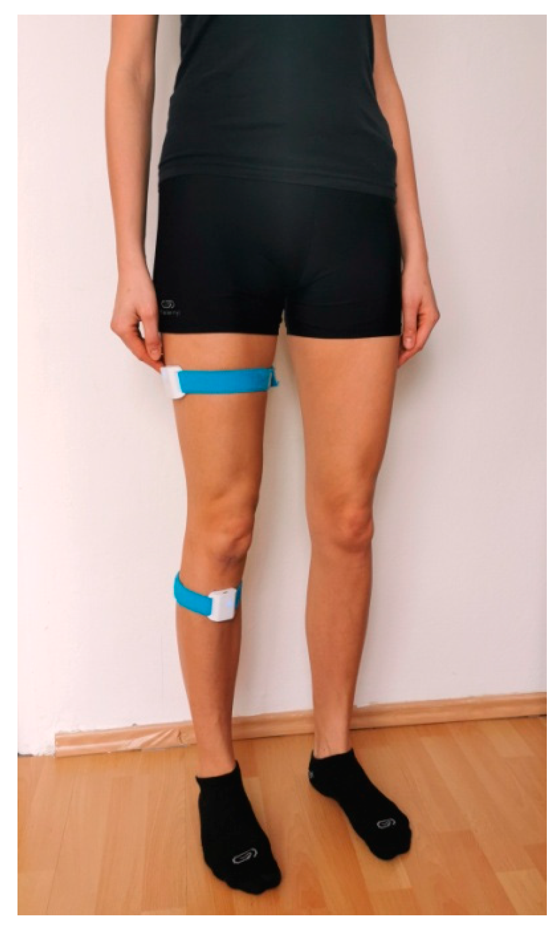
2.2. Sensors and Application
2.3. Experimental Procedures and Instruments
2.3.1. Proprioception Tests (Orthyo® system)
2.3.2. WOMAC
2.3.3. VAS
2.4. Testing and Intervention Procedures
2.4.1. Testing
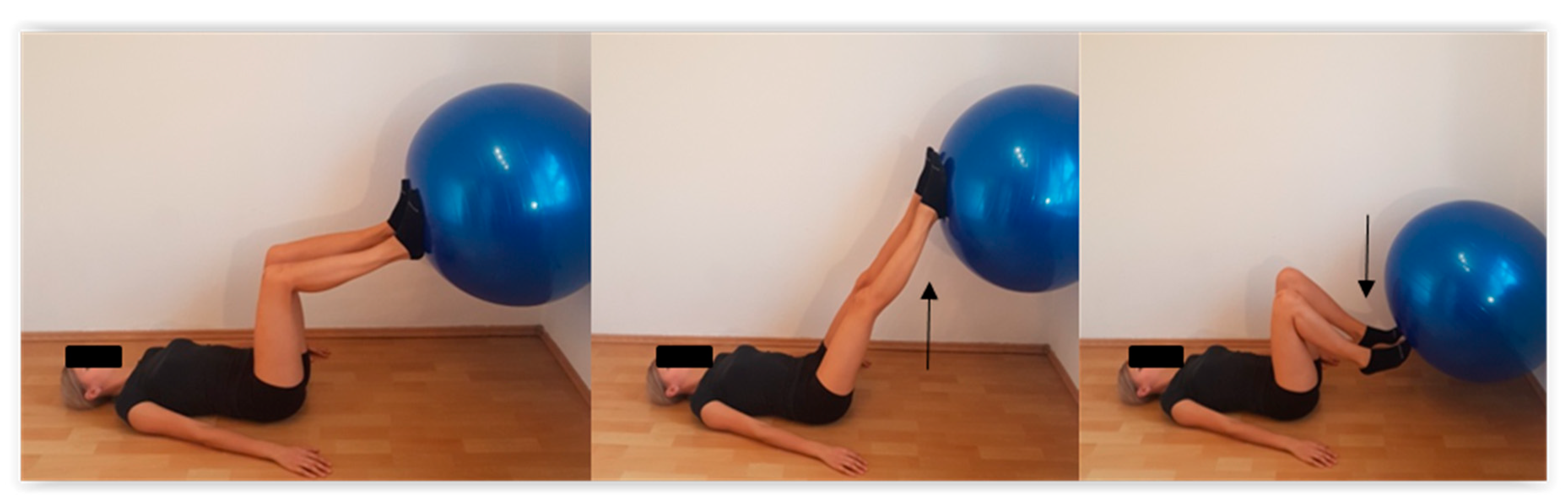
2.4.2. Intervention
2.5. Statistical Analysis
3. Results
3.1. Proprioception Measurements
3.2. WOMAC
3.3. VAS
4. Discussion
5. Conclusions
- The Orthyo® system used in conjunction with a mobile device fitted with the Android 5.0 Lollipop operating system allowed for a quick and accurate assessment of the knee joint position sense, which made it a great verification tool for evaluating the effectiveness of selected rehabilitation programs.
- A 10-day rehabilitation regimen did not significantly affect the knee joint position sense, regardless of the type of therapeutic intervention used.
- Even a relatively short (10-day) period of rehabilitation produced significant functional improvement and pain reduction, regardless of the type of therapeutic intervention used.
- For a complete functional evaluation of the patient with OAK, muscle activity measurements and gait analysis should be added.
Limitations
Author Contributions
Funding
Conflicts of Interest
References
- Mills, K.; Hettinga, B.A.; Pohl, M.B.; Ferber, R. Between-limb kinematic asymmetry during gait in unilateral and bilateral mild to moderate knee osteoarthritis. Arch. Phys. Med. Rehabil. 2013, 94, 2241–2247. [Google Scholar] [CrossRef] [PubMed]
- Ogunbode, A.M.; Adebusoye, L.A.; Olowookere, O.O.; Alonge, T.O. Physical functionality and self-rated health status of adult patients with knee osteoarthritis presenting in a primary care clinic. Ethiop. J. Health Sci. 2014, 24, 319–328. [Google Scholar] [CrossRef] [PubMed]
- Abbott, J.H.; Usiskin, I.M.; Wilson, R.; Hansen, P.; Losina, E. The quality-of-life burden of knee osteoarthritis in New Zealand adults: A model-based evaluation. PLoS ONE 2017, 12, e0185676. [Google Scholar] [CrossRef] [PubMed]
- Mahmoudian, A.; van Dieen, J.H.; Baert, I.A.C.; Jonkers, I.; Bruijn, S.M.; Luyten, F.P.; Faber, G.S.; Verschueren, S.M.P. Changes in proprioceptive weighting during quiet standing in women with early and established knee osteoarthritis compared to healthy controls. Gait Posture 2016, 44, 184–188. [Google Scholar] [CrossRef]
- Tasci Bozbas, G.; Sendur, O.F.; Aydemir, A.H. Primary knee osteoarthritis increases the risk of falling. J. Back Musculoskelet. Rehabil. 2017, 30, 785–789. [Google Scholar] [CrossRef]
- Smith, T.O.; Higson, E.; Pearson, M.; Mansfield, M. Is there an increased risk of falls and fractures in people with early diagnosed hip and knee osteoarthritis? Data from the Osteoarthritis Initiative. Int. J. Rheum. Dis. 2018, 21, 1193–1201. [Google Scholar] [CrossRef]
- Knoop, J.; Steultjens, M.P.M.; van der Leeden, M.; van der Esch, M.; Thorstensson, C.A.; Roorda, L.D.; Lems, W.F.; Dekker, J. Proprioception in knee osteoarthritis: A narrative review. Osteoarthr. Cartil. 2011, 19, 381–388. [Google Scholar] [CrossRef]
- Cammarata, M.L.; Schnitzer, T.J.; Dhaher, Y.Y. Does knee osteoarthritis differentially modulate proprioceptive acuity in the frontal and sagittal planes of the knee? Arthritis Rheum. 2011, 63, 2681–2689. [Google Scholar] [CrossRef]
- Baert, I.A.C.; Mahmoudian, A.; Nieuwenhuys, A.; Jonkers, I.; Staes, F.; Luyten, F.P.; Truijen, S.; Verschueren, S.M.P. Proprioceptive accuracy in women with early and established knee osteoarthritis and its relation to functional ability, postural control, and muscle strength. Clin. Rheumatol. 2013, 32, 1365–1374. [Google Scholar] [CrossRef]
- Van der Esch, M.; Knoop, J.; Hunter, D.J.; Klein, J.-P.; van der Leeden, M.; Knol, D.L.; Reiding, D.; Voorneman, R.E.; Gerritsen, M.; Roorda, L.D.; et al. The association between reduced knee joint proprioception and medial meniscal abnormalities using MRI in knee osteoarthritis: Results from the Amsterdam osteoarthritis cohort. Osteoarthr. Cartil. 2013, 21, 676–681. [Google Scholar] [CrossRef][Green Version]
- Chen, Y.; Yu, Y.; He, C. Correlations Between Joint Proprioception, Muscle Strength, and Functional Ability in Patients with Knee Osteoarthritis. Sichuan Da Xue Xue Bao Yi Xue Ban 2015, 46, 880–884. [Google Scholar] [PubMed]
- Chang, A.H.; Lee, S.J.; Zhao, H.; Ren, Y.; Zhang, L.-Q. Impaired varus-valgus proprioception and neuromuscular stabilization in medial knee osteoarthritis. J. Biomech. 2014, 47, 360–366. [Google Scholar] [CrossRef] [PubMed]
- Rahlf, A.L.; Petersen, E.; Rehwinkel, D.; Zech, A.; Hamacher, D. Validity and Reliability of an Inertial Sensor-Based Knee Proprioception Test in Younger vs. Older Adults. Front. Sports Act. Living 2019, 1, 27. [Google Scholar] [CrossRef]
- Négyesi, J.; Galamb, K.; Szilágyi, B.; Nagatomi, R.; Hortobágyi, T.; Tihanyi, J. Age-specific modifications in healthy adults’ knee joint position sense. Somatosens. Mot. Res. 2019, 36, 262–269. [Google Scholar] [CrossRef]
- Noor, R.; Olyaei, G.; Hadian, M.R.; Talebian, S.; Bashir, M.S. A reliable and accurate system of joint position sense measurement. Biomed. Res. 2018, 29. [Google Scholar] [CrossRef]
- Clark, N.C.; Akins, J.S.; Heebner, N.R.; Sell, T.C.; Abt, J.P.; Lovalekar, M.; Lephart, S.M. Reliability and measurement precision of concentric-to-isometric and eccentric-to-isometric knee active joint position sense tests in uninjured physically active adults. Phys. Ther. Sport 2016, 18, 38–45. [Google Scholar] [CrossRef]
- Irving, F.; Russell, J.; Smith, T. Reliability of knee joint position sense measurement: A comparison between goniometry and image capture methods. Eur. J. Physiother. 2016, 18, 95–102. [Google Scholar] [CrossRef][Green Version]
- Arvin, M.; Hoozemans, M.; Burger, B.; Verschueren, S.; Van Dieen, J.; Pijnappels, M. Reproducibility of a knee and hip proprioception test in healthy older adults. Aging Clin. Exp. Res. 2014, 27, 171–177. [Google Scholar] [CrossRef]
- Sanchez-Ramirez, D.C.; van der Leeden, M.; Knol, D.L.; van der Esch, M.; Roorda, L.D.; Verschueren, S.; van Dieën, J.; Lems, W.F.; Dekker, J. Association of postural control with muscle strength, proprioception, self-reported knee instability and activity limitations in patients with knee osteoarthritis. J. Rehabil. Med. 2013, 45, 192–197. [Google Scholar] [CrossRef]
- Collins, A.T.; Blackburn, J.T.; Olcott, C.W.; Miles, J.; Jordan, J.; Dirschl, D.R.; Weinhold, P.S. Stochastic resonance electrical stimulation to improve proprioception in knee osteoarthritis. Knee 2011, 18, 317–322. [Google Scholar] [CrossRef]
- Clausen, B.; Holsgaard-Larsen, A.; Søndergaard, J.; Christensen, R.; Andriacchi, T.P.; Roos, E.M. The effect on knee-joint load of instruction in analgesic use compared with neuromuscular exercise in patients with knee osteoarthritis: Study protocol for a randomized, single-blind, controlled trial (the EXERPHARMA trial). Trials 2014, 15, 444. [Google Scholar] [CrossRef] [PubMed]
- Smith, T.O.; King, J.J.; Hing, C.B. The effectiveness of proprioceptive-based exercise for osteoarthritis of the knee: A systematic review and meta-analysis. Rheumatol. Int. 2012, 32, 3339–3351. [Google Scholar] [CrossRef] [PubMed]
- Mohd Sharif, N.A.; Usman, J.; Wan Safwani, W.K.Z.; Siew Li, G.; Abdul Karim, S.; Mohamed, N.A.; Khan, S.S.; Khan, S.J. Effects of simple knee sleeves on pain and knee adduction moment in early unilateral knee osteoarthritis. Proc. Inst. Mech. Eng. Part H J. Eng. Med. 2019, 233, 1132–1140. [Google Scholar] [CrossRef] [PubMed]
- Hu, X.; Lai, Z.; Wang, L. Effects of Taichi exercise on knee and ankle proprioception among individuals with knee osteoarthritis. Res. Sports Med. 2019, 1–11. [Google Scholar] [CrossRef] [PubMed]
- Lai, Z.; Zhang, Y.; Lee, S.; Wang, L. Effects of strength exercise on the knee and ankle proprioception of individuals with knee osteoarthritis. Res. Sports Med. 2018, 26, 138–146. [Google Scholar] [CrossRef] [PubMed]
- Yang, J.; Lee, B.; Kim, C. Changes in proprioception and pain in patients with neck pain after upper thoracic manipulation. J. Phys. Ther. Sci. 2015, 27, 795–798. [Google Scholar] [CrossRef]
- Jeong, H.S.; Lee, S.-C.; Jee, H.; Song, J.B.; Chang, H.S.; Lee, S.Y. Proprioceptive Training and Outcomes of Patients With Knee Osteoarthritis: A Meta-Analysis of Randomized Controlled Trials. J. Athl. Train. 2019, 54, 418–428. [Google Scholar] [CrossRef]
- Wellsandt, E.; Golightly, Y. Exercise in the management of knee and hip osteoarthritis. Curr. Opin. Rheumatol. 2018, 30, 151–159. [Google Scholar] [CrossRef]
- Legha, A.; Burke, D.L.; Foster, N.E.; van der Windt, D.A.; Quicke, J.G.; Healey, E.L.; Runhaar, J.; Holden, M.A. Do comorbidities predict pain and function in knee osteoarthritis following an exercise intervention, and do they moderate the effect of exercise? Analyses of data from three randomized controlled trials. Musculoskelet. Care 2019, 18, 3–11. [Google Scholar] [CrossRef]
- Gezginaslan, Ö.; Öztürk, E.A.; Cengiz, M.; Mirzaoğlu, T.; Çakcı, F.A. Effects of isokinetic muscle strengthening on balance, proprioception, and physical function in bilateral knee osteoarthritis patients with moderate fall risk. Turk. J. Phys. Med. Rehabil. 2018, 64, 353–361. [Google Scholar] [CrossRef]
- Park, H.-R.; Im, S.; Kim, H.; Jung, S.-Y.; Kim, D.; Jang, E.J.; Sung, Y.-K.; Cho, S.-K. Validation of algorithms to identify knee osteoarthritis patients in the claims database. Int. J. Rheum. Dis. 2019, 22, 890–896. [Google Scholar] [CrossRef] [PubMed]
- Lisiński, P.; Wareńczak, A.; Hejdysz, K.; Sip, P.; Gośliński, J.; Owczarek, P.; Jonak, J.; Goślińska, J. Mobile Applications in Evaluations of Knee Joint Kinematics: A Pilot Study. Sensors 2019, 19, 3675. [Google Scholar] [CrossRef] [PubMed]
- Jogi, P.; Overend, T.J.; Spaulding, S.J.; Zecevic, A.; Kramer, J.F. Effectiveness of balance exercises in the acute post-operative phase following total hip and knee arthroplasty: A randomized clinical trial. SAGE Open Med. 2015, 3, 2050312115570769. [Google Scholar] [CrossRef] [PubMed]
- Brokelman, R.B.G.; Haverkamp, D.; van Loon, C.; Hol, A.; van Kampen, A.; Veth, R. The validation of the visual analogue scale for patient satisfaction after total hip arthroplasty. Eur. Orthop. Traumatol. 2012, 3, 101–105. [Google Scholar] [CrossRef] [PubMed]
- Lin, D.-H.; Lin, Y.-F.; Chai, H.-M.; Chai, H.-M.; Han, Y.-C.; Jan, M.-H. Comparison of proprioceptive functions between computerized proprioception facilitation exercise and closed kinetic chain exercise in patients with knee osteoarthritis. Clin. Rheumatol. 2007, 26, 520–528. [Google Scholar] [CrossRef] [PubMed]
- Kumar, S.; Kumar, A.; Kumar, R. Proprioceptive training as an adjunct in osteoarthritis of knee. J. Musculoskelet. Res. 2013, 16, 1350002. [Google Scholar] [CrossRef]
- Sekir, U.; Gür, H. A multi-station proprioceptive exercise program in patients with bilateral knee osteoarthrosis: Functional capacity, pain and sensoriomotor function. A randomized controlled trial. J. Sports Sci. Med. 2005, 4, 590–603. [Google Scholar]
- Abdelazeem, F.; Nambi, G.; Elnegamy, T. Comparative study on Virtual Reality Training (VRT) over Sensory Motor Training (SMT) in Unilateral Chronic Osteoarthritis—A Randomized Control Trial. Int. J. Med. Res. Health Sci. 2016, 5, 7–16. [Google Scholar]
- Hancock, G.E.; Hepworth, T.; Wembridge, K. Accuracy and reliability of knee goniometry methods. J. Exp. Orthop. 2018, 5, 46. [Google Scholar] [CrossRef]
- Cho, Y.R.; Hong, B.Y.; Lim, S.H.; Kim, H.W.; Ko, Y.J.; Im, S.A.; Lee, J.I. Effects of joint effusion on proprioception in patients with knee osteoarthritis: A single-blind, randomized controlled clinical trial. Osteoarthr. Cartil. 2011, 19, 22–28. [Google Scholar] [CrossRef]
- Olagbegi, O.M.; Adegoke, B.O.; Odole, A.C. Effectiveness of three modes of kinetic-chain exercises on quadriceps muscle strength and thigh girth among individuals with knee osteoarthritis. Arch. Physiother. 2017, 7, 9. [Google Scholar] [CrossRef] [PubMed]
- AdemolaGbiri, C. Comparative Efficacy of Open-chain and Close-chain Kinematics on Proprioception, Muscles’ Strength and Functional Performances in Individual with Knee Osteoarthritis. Occup. Med. Health Aff. 2013, 1, 1–5. [Google Scholar] [CrossRef]
- Cho, S.-H.; Bae, C.-H.; Gak, H.-B. Effects of closed kinetic chain exercises on proprioception and functional scores of the knee after anterior cruciate ligament reconstruction. J. Phys. Ther. Sci. 2013, 25, 1239–1241. [Google Scholar] [CrossRef] [PubMed]
- Kachanathu, S.J.; Kaur, H.; Natho, M.; Nuhmani, S. The effect of open and closed kinematics chain exercises in the management of meniscal injuries. JSIR 2013, 2, 927–931. [Google Scholar]
- Baratta, R.; Solomonow, M.; Zhou, B.H.; Letson, D.; Chuinard, R.; D’Ambrosia, R. Muscular coactivation. The role of the antagonist musculature in maintaining knee stability. Am. J. Sports Med. 1988, 16, 113–122. [Google Scholar] [CrossRef] [PubMed]
- Mondam, S.; Babu, S.; Kumar, R.; Prakash, J. A Comparative Study of Proprioceptive Exercises versus Conventional Training Program on Osteoarthritis of Knee. RJRS 2012, 1, 2277–2502. [Google Scholar]
- Jan, M.-H.; Lin, C.-H.; Lin, Y.-F.; Lin, J.-J.; Lin, D.-H. Effects of weight-bearing versus nonweight-bearing exercise on function, walking speed, and position sense in participants with knee osteoarthritis: A randomized controlled trial. Arch. Phys. Med. Rehabil. 2009, 90, 897–904. [Google Scholar] [CrossRef]
- Salaheldin, G.; Hassanien, W.F.H. Closed versus open kinetic chain exercises in treatment of patients with hemophilic arthritis. AAMJ 2014, 12, 287–310. [Google Scholar]
- Aslan, H.; Buddhadev, H.H.; Suprak, D.N.; San Juan, J.G. Acute effects of two hip flexor stretching techniques on knee joint position sense and balance. Int. J. Sports Phys. Ther. 2018, 13, 846–859. [Google Scholar] [CrossRef]
- Tsauo, J.-Y.; Cheng, P.-F.; Yang, R.-S. The effects of sensorimotor training on knee proprioception and function for patients with knee osteoarthritis: A preliminary report. Clin. Rehabil. 2008, 22, 448–457. [Google Scholar] [CrossRef]
- Kaya Mutlu, E.; Ercin, E.; Razak Ozdıncler, A.; Ones, N. A comparison of two manual physical therapy approaches and electrotherapy modalities for patients with knee osteoarthritis: A randomized three arm clinical trial. Physiother. Theory Pract. 2018, 34, 600–612. [Google Scholar] [CrossRef]
- Ko, T.; Lee, S.; Lee, D. Manual Therapy and Exercise for OA Knee: Effects on Muscle Strength, Proprioception, and Functional Performance. J. Phys. Ther. Sci. 2009, 21, 293–299. [Google Scholar] [CrossRef]
- Nor Azlin, M.N.; Su Lyn, K. Effects of passive joint mobilization on patients with knee osteoarthritis. Sains Malays. 2011, 40, 1461–1465. [Google Scholar]
- Ling, S.M.; Conwit, R.A.; Talbot, L.; Shermack, M.; Wood, J.E.; Dredge, E.M.; Weeks, M.J.; Abernethy, D.R.; Metter, E.J. Electromyographic Patterns Suggest Changes in Motor Unit Physiology Associated with Early Osteoarthritis of the Knee. Osteoarthr. Cartil. 2007, 15, 1134–1140. [Google Scholar] [CrossRef] [PubMed]
- Costello, K.E.; Astephen Wilson, J.L.; Stanish, W.D.; Urquhart, N.; Hubley-Kozey, C.L. Differences in Baseline Joint Moments and Muscle Activation Patterns Associated With Knee Osteoarthritis Progression When Defined Using a Clinical Versus a Structural Outcome. J. Appl. Biomech. 2020, 36, 39–51. [Google Scholar] [CrossRef]
- Rutherford, D.J.; Hubley-Kozey, C.L.; Stanish, W.D. Changes in knee joint muscle activation patterns during walking associated with increased structural severity in knee osteoarthritis. J. Electromyogr. Kinesiol. 2013, 23, 704–711. [Google Scholar] [CrossRef]
- Coudeyre, E.; Jegu, A.G.; Giustanini, M.; Marrel, J.P.; Edouard, P.; Pereira, B. Isokinetic muscle strengthening for knee osteoarthritis: A systematic review of randomized controlled trials with meta-analysis. Ann. Phys. Rehabil. Med. 2016, 59, 207–215. [Google Scholar] [CrossRef]
- Vårbakken, K.; Lorås, H.; Nilsson, K.G.; Engdal, M.; Stensdotter, A.K. Relative difference in muscle strength between patients with knee osteoarthritis and healthy controls when tested bilaterally and joint-inclusive: An exploratory cross-sectional study. BMC Musculoskelet. Disord. 2019, 20, 593. [Google Scholar] [CrossRef]
- Tadano, S.; Takeda, R.; Sasaki, K.; Fujisawa, T.; Tohyama, H. Gait characterization for osteoarthritis patients using wearable gait sensors (H-Gait systems). J. Biomech. 2016, 49, 684–690. [Google Scholar] [CrossRef]
- Agostini, V.; Gastaldi, L.; Rosso, V.; Knaflitz, M.; Tadano, S. A Wearable Magneto-Inertial System for Gait Analysis (H-Gait): Validation on Normal Weight and Overweight/Obese Young Healthy Adults. Sensors 2017, 17, 2406. [Google Scholar] [CrossRef]
- Seeger, J.B.; Schikschneit, J.P.; Schuld, C.; Rupp, R.; Rickert, M.; Jahnke, A.; Maier, G.S.; Clarius, M. Instrumented gait analysis in patients with medial osteoarthritis of the knee after mobile-bearing unicompartmental knee arthroplasty. Knee 2018, 25, 392–397. [Google Scholar] [CrossRef] [PubMed]
- Dawe, R.J.; Yu, L.; Leurgans, S.E.; Truty, T.; Curran, T.; Hausdorff, J.M.; Wimmer, M.A.; Block, J.A.; Bennett, D.A.; Buchman, A.S. Expanding instrumented gait testing in the community setting: A portable, depth-sensing camera captures joint motion in older adults. PLoS ONE 2019, 14, e0215995. [Google Scholar] [CrossRef] [PubMed]


| Variable | Group | No. | Mean ± SD | Median | Min–Max | p-Value |
|---|---|---|---|---|---|---|
| age (years) | E | 27 | 65.0 ± 7.4 | 64.0 | 55.0–78.0 | 0.10 |
| MT | 27 | 66.1 ± 4.7 | 65.0 | 58.0–76.0 | ||
| C | 27 | 63.0 ± 6.6 | 60.0 | 53.0–76.0 | ||
| weight (kg) | E | 27 | 76.1 ± 11.3 | 75.0 | 58.0–100.0 | 0.52 |
| MT | 27 | 76.2 ± 11.5 | 75.0 | 58.0–105.0 | ||
| C | 27 | 73.5 ± 12.3 | 68.0 | 56.0–90.0 | ||
| height (m) | E | 27 | 1.67 ± 0.07 | 1.66 | 1.53–1.82 | 0.69 |
| MT | 27 | 1.66 ± 0.07 | 1.66 | 1.55–1.82 | ||
| C | 27 | 1.68 ± 0.07 | 1.67 | 1.56–1.80 | ||
| BMI (kg/m2) | E | 27 | 27.3 ± 3.8 | 26.8 | 20.4–36.1 | 0.07 |
| MT | 27 | 27.6 ± 4.0 | 26.9 | 20.9–38.3 | ||
| C | 27 | 25.9 ± 3.8 | 24.5 | 20.5–34.3 |
| Variable | Group | Before | After | p-Value3 | ||||||
|---|---|---|---|---|---|---|---|---|---|---|
| Mean ± SD | Median | Range | p-Value1 | Mean ± SD | Median | Range | p-Value2 | |||
| End angle (°) Left | E | 67.0 ± 9.3 | 66.6 | 51.6–87.0 | 0.31 | 67.4 ± 7.5 a | 68.1 | 52.9–86.4 | 0.04 | 0.84 |
| MT. | 67.0 ± 9.1 | 66.7 | 46.3–85.8 | 72.7 ± 9.5 a | 71.4 | 48.2–101.7 | 0.02 * | |||
| C | 69.9 ± 6.5 | 72.3 | 54.9–77.7 | 69.5 ± 10.7 | 669.0 | 50.9–98.0 | 0.65 * | |||
| End angle (°) Right | E | 68.4 ± 9.3 | 67.7 | 46.6–85.6 | 0.99 | 68.3 ± 8.4 | 69.1 | 44.2–86.7 | 0.88 | 0.63 * |
| MT. | 70.4 ± 12.9 | 68.4 | 53.0–107.6 | 68.8 ± 11.2 | 66.6 | 47.6–98.5 | 0.53 | |||
| C | 69.1 ± 8.1 | 68.4 | 59.1–87.8 | 67.4 ± 7.0 | 70.5 | 51.5–76.6 | 0.29 | |||
Left | E | 11.0 ± 13.6 | 4.0 | 0–40.8 | 0.38 | 6.8 ± 5.4 | 6.7 | 0–17.0 | 0.96 | 0.33 * |
| MT. | 8.4 ± 15.1 | 4.1 | 0.4–79.4 | 10.3 ± 16.5 | 3.6 | 0.3–78.2 | 0.27 * | |||
| C | 6.0 ± 8.7 | 3.1 | 0.1–32.3 | 9.7 ± 11.0 | 5.0 | 0.1–32.2 | 0.26 * | |||
Right | E | 6.6 ± 8.7 | 2.9 | 0.1–33.9 | 0.41 | 5.1 ± 7.1 | 3.3 | 0.1–36.4 | 0.47 | 0.39 * |
| MT. | 5.2 ± 5.5 | 4.0 | 0–27.1 | 5.2 ± 4.9 | 3.8 | 0.1–15.9 | 0.77 * | |||
| C | 9.6 ± 9.7 | 7.5 | 0.1–32.8 | 7.3 ± 7.0 | 6.2 | 5.90–25.8 | 0.22 | |||
| Variable | Group | Before | After | p-Value3 | ||||||
|---|---|---|---|---|---|---|---|---|---|---|
| Mean ± SD | Median | Range | p-Value1 | Mean ± SD | Median | Range | p-Value2 | |||
| WOMAC | E | 45.9 ± 13.7 a | 42.0 | 19.0–65.0 | <0.01 | 39.7 ± 12.8 a | 45.0 | 12.0–58.0 | <0.01 | <0.01 |
| MT. | 46.3 ± 19.0 b | 44.0 | 2.0–81.0 | 40.1 ± 21.7 b | 33.0 | 1.0–84.0 | 0.01 | |||
| C | 19.3 ± 17.6 a,b | 13.0 | 2.0–78.0 | 18.4 ± 18.6 a,b | 12.0 | 1.0–75.0 | 0.11 * | |||
| Variable | Group | Before | After | p-Value3 | ||||||
|---|---|---|---|---|---|---|---|---|---|---|
| Mean ± SD | Median | Range | p-Value1 | Mean ± SD | Median | Range | p-Value2 | |||
| VAS left | E | 5.2 ± 2.7 a | 6.0 | 0–10.0 | 0.99 | 3.4 ± 2.1 a | 3.0 | 0–9.0 | 0.72 | <0.01 * |
| MT. | 5.2 ± 2.4 b | 6.0 | 0–8.0 | 3.2 ± 2.7 b | 3.0 | 0–8.0 | <0.01 * | |||
| VAS right | E | 5.0 ± 2.7 a | 5.0 | 0–10.0 | 0.89 | 3.4 ± 2.2 a | 3.0 | 0–9.0 | 0.56 | <0.01 * |
| MT. | 4.9 ± 2.7 b | 5.0 | 0–9.0 | 3.0 ± 2.6 | 2.0 | 0–8.0 | <0.01 | |||
© 2020 by the authors. Licensee MDPI, Basel, Switzerland. This article is an open access article distributed under the terms and conditions of the Creative Commons Attribution (CC BY) license (http://creativecommons.org/licenses/by/4.0/).
Share and Cite
Goślińska, J.; Wareńczak, A.; Miedzyblocki, M.; Hejdysz, K.; Adamczyk, E.; Sip, P.; Chlebuś, E.; Gośliński, J.; Owczarek, P.; Woźniak, A.; et al. Wireless Motion Sensors—Useful in Assessing the Effectiveness of Physiotherapeutic Methods Used in Patients with Knee Osteoarthritis—Preliminary Report. Sensors 2020, 20, 2268. https://doi.org/10.3390/s20082268
Goślińska J, Wareńczak A, Miedzyblocki M, Hejdysz K, Adamczyk E, Sip P, Chlebuś E, Gośliński J, Owczarek P, Woźniak A, et al. Wireless Motion Sensors—Useful in Assessing the Effectiveness of Physiotherapeutic Methods Used in Patients with Knee Osteoarthritis—Preliminary Report. Sensors. 2020; 20(8):2268. https://doi.org/10.3390/s20082268
Chicago/Turabian StyleGoślińska, Jagoda, Agnieszka Wareńczak, Margaret Miedzyblocki, Krystyna Hejdysz, Ewa Adamczyk, Paweł Sip, Ewa Chlebuś, Jarosław Gośliński, Piotr Owczarek, Adam Woźniak, and et al. 2020. "Wireless Motion Sensors—Useful in Assessing the Effectiveness of Physiotherapeutic Methods Used in Patients with Knee Osteoarthritis—Preliminary Report" Sensors 20, no. 8: 2268. https://doi.org/10.3390/s20082268
APA StyleGoślińska, J., Wareńczak, A., Miedzyblocki, M., Hejdysz, K., Adamczyk, E., Sip, P., Chlebuś, E., Gośliński, J., Owczarek, P., Woźniak, A., & Lisiński, P. (2020). Wireless Motion Sensors—Useful in Assessing the Effectiveness of Physiotherapeutic Methods Used in Patients with Knee Osteoarthritis—Preliminary Report. Sensors, 20(8), 2268. https://doi.org/10.3390/s20082268